Primary lacrimal gland adenocarcinoma of the third eyelid in a horse
←
→
Page content transcription
If your browser does not render page correctly, please read the page content below
Veterinary Ophthalmology (2011) 14, 1, 48–54
CASE REPORT
Primary lacrimal gland adenocarcinoma of the
third eyelid in a horse
Rachel L. Mathes,* Karen Paige Carmichael,† John Peroni‡ and Phillip Anthony Moore*
*Department of Small Animal Medicine and Surgery, College of Veterinary Medicine, University of Georgia, Athens, GA 30602, USA; †Department of
Pathology, College of Veterinary Medicine, University of Georgia, Athens, GA 30602, USA; and ‡Department of Large Animal Medicine and Surgery,
College of Veterinary Medicine, University of Georgia, Athens, GA 30602, USA
Address communications to: Abstract
R. L. Mathes A 5-year-old Draft Horse gelding presented for evaluation of a large, fleshy, ulcerated
Tel.: (706) 206-7948
third eyelid mass OD of 3 weeks duration. Complete ophthalmic examination, ocular
Fax: (706) 542-6460 ultrasound and skull radiographs revealed a large soft-tissue mass involving the entire
e-mail: rmathes@uga.edu third eyelid OD and extending into the ventral right orbit to the level of the globe
equator. No other abnormalities were noted on physical or ophthalmic examination.
Surgical removal via exenteration was performed 3 months after initial presentation.
A lacrimal adenocarcinoma of the third eyelid was diagnosed based on histopathology.
Concurrent asymptomatic intra-ductal and intra-acinar Demodex caballi parasites were
found in the eyelid sebaceous glands, likely as an incidental finding. No tumor
recurrence or metastasis has occurred 12 months after excision. To the author’s
knowledge, this case is the first reported primary lacrimal adenocarcinoma in a horse.
Complete surgical excision was curative.
Key Words: adnexa, demodex, equine ophthalmology, exenteration, lacrimal
adenocarcinoma, third eyelid neoplasia
after initial presentation.15 The tissue of origin is the large
INTRODUCTION
lacrimal gland of the third eyelid located at the base of the
Primary neoplasia of the third eyelid, or nictitans mem- third eyelid cartilage. Adenomas of the accessory lacrimal
brane, in horses is well described with the most common gland, located in the dorsal orbit, have been reported in
tumor being squamous cell carcinoma.1–3 These tumors are dogs.17
epithelial in origin and are thought to be primarily UV- Demodex spp. are intrafollicular mites that are found on
mediated tumors.4–6 Other tumors that reportedly may humans18 and many species of animals including dogs,19
affect the third eyelid in horses include lymphangioma,7 ruminants,20–25 cats,26 sea lions,27 bears,28 hamsters,29
lymphangiosarcoma,8 papilloma,3,9 hemangiosarcoma,2,10 ferrets,30 voles,31 mice31 and many others. These mites are
basal cell tumor,11 solid carcinoma12 and sebaceous adeno- species specific and may be found in the nonpathologic state
carcinoma.13 Conjunctival pseudotumors have been or may be causative in certain types of skin disease. Blephari-
described arising from the third eyelid, however, these tis caused by Demodex spp. has been reported in humans.32 A
lesions are proliferative inflammatory masses and do not case of palpebral demodicosis has been described in cattle33
represent true neoplasia.14 and these mites have been found in cattle sebaceous
In dogs and cats, primary adenocarcinoma of the gland of glands.34 In horses, Demodex spp. are divided into Demodex
the third eyelid has been described.15,16 Primary third equi, a species found in the hair follicles, and Demodex caballi,
eyelid tumors in dogs and cats are rare.15,16 This tumor is found in the sebaceous glands.35 Although there are ana-
locally invasive and local recurrence is high with surgical tomic differences in these species, the location in which they
excision.15,16 Reportedly, complete excision of the third are found is distinctive, allowing differentiation between
eyelid is the treatment of choice for canine adenocarcino- these Demodex spp.35 Demodex caballi are found as an uncom-
mas arising in this location.16 The solitary feline case mon incidental finding in horses, not reported to cause
report described diffuse metastasis and a survival of 4 weeks blepharitis.35
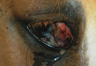
! 2011 American College of Veterinary OphthalmologistsLACRIMAL GLAND ADENOCARCINOMA IN A HORSE 49 The case report herein describes a primary adenocarci- noma of the lacrimal gland of the third eyelid in a horse trea- ted with surgical excision. Concurrent asymptomatic sebaceous gland Demodex equi was found on histopathology and is discussed. To the author’s knowledge, this is the first report of a primary lacrimal adenocarcinoma of the third eyelid in a horse diagnosed histologically. This tumor was successfully treated surgically and was locally expansive without distant metastasis. CASE REPORT History, physical examination, initial ophthalmic examination and diagnostics A 5-year-old Grade Draft Horse gelding presented to the Figure 1. A photograph of the right eye in a 5-year-old Draft Horse University of Georgia Veterinary Teaching Hospital for gelding at initial presentation is depicted here. There is a large, pink, evaluation of a mass on the right third eyelid with a duration firm mass originating from the third eyelid with multifocal ulcerations of approximately 3 weeks. The mass had progressed in size and a moderate amount of surface mucopurulent discharge and hemor- rapidly during this time and the horse was not on any prior rhage. The visible mass measured approximately 3.4 cm horizontally by medications. The horse was on pasture and fed grass hay 2.8 cm vertically and caused moderate enophthalmia. from round bales in the pasture as well as one-and-one-half scoops of a high fat, high protein balanced feed (Omolene Schirmer tear test (STT) and intraocular pressure (IOP) OD 300, Land O’ Lakes Purina Mills, Gray Summit, MO, USA) were not possible due to the large mass and enophthalmia. once per day. Vaccinations for equine encephalomyelitis The left eye was normal on complete ophthalmic exami- eastern and western, tetanus (Cephalovac EWT, Boehringer nation with a STT value of 26 mm/min (Schirmer Tear Ingelheim Vetmedica, Inc., St. Joseph, MO, USA), rabies Test, Merck and Company, Inc., Whitehouse Station, NJ, (Imrab 3, Merial, Duluth, GA, USA) and west Nile virus USA) and an IOP of 18 mmHg using applanation tonometry (West Nile Recombitek, Merial, Duluth, GA, USA) were (Tonopen XL; Reichert Instruments, Depew, NY, USA). given annually in the spring and were up to date at the time Differential diagnoses for the mass included squamous of presentation. Six weeks prior to presentation, the horse cell carcinoma,1 hemangiosarcoma,2 papilloma,3 basal cell was dewormed with ivermectin (Eqvalan, Merial, Duluth, tumor11 and pseudotumor14. Impression smears were evalu- GA, USA). The Coggins test was performed 1 month prior ated for cytology and showed moderate numbers of neu- to presentation and the results were negative. trophils, bacterial cocci and necrotic cell debris. An ocular Upon physical examination, the horse was overweight ultrasound was performed and showed a large echogenic (BCS 7/9) with a normal temperature (38.2 "C), respiration mass of the third eyelid with extension ventrally and caudally (28 bpm) and pulse (48 bpm). All other findings on the phys- to the level of or caudal to the globe equator. The mass mea- ical examination were unremarkable. The horse was light in sured 5.2 cm rostral to caudal and 3.4 cm lateral to medial color; however, the palpebrae OD were pigmented. A com- on ultrasonographic evaluation. (Fig. 2) The echogenicity plete ophthalmic examination was performed with slit-lamp and echotexture of the orbital portion of the mass were simi- biomicroscopy and indirect ophthalmoscopy. On examina- lar to that of the visible extra-ocular mass and continuous tion, there was a large, fleshy, pink to red, ulcerated, lobu- with it. The mass was routinely biopsied by taking several lated mass extending from the OD third eyelid with surface tissue sections for histopathologic evaluation. Rec- complete involvement of the third eyelid and loss of normal ommendations for exenteration of the right orbit were given architecture. (Fig. 1) The mass was firm on palpation with to the owner at the initial presentation and evaluation. The purulent mucoid discharge and hemorrhage on the surface. horse was placed on triple antibiotic ointment (Neomycin The visible mass measured approximately 3.4 cm horizon- and polymyxin B and bacitracin ophthalmic ointment, tally by 2.8 cm vertically. The mass extended to the dorsona- Bausch and Lomb, Rochester, NY, USA) every 8 h in the sal bulbar conjunctiva and could be seen extending into the right eye, flunixin meglumine (0.5 mg/kg PO Q12 h; ventral fornix. The third eyelid was markedly elevated with Banamine, Schering Plough, Union, NJ, USA) and trimeth- pressure noted on the globe and subsequent enophthalmia. aprim sulfamethozale (25 mg/kg PO Q12 h; Amneal There was moderate chemosis (characterized as 2 + on a Pharmaceuticals, Hauppauge, NY, USA) for 10 days and 0–4 + sequential grading scale) and moderate conjunctival discharged pending biopsy results. hyperemia (2 + ) OD. A small superficial corneal ulcer was diagnosed adjacent to the area of corneal contact with the Biopsy mass nasally. A full intraocular examination was precluded by Histopathologic examination of the initial biopsies from the the large mass, however, no significant findings were noted. mass revealed disorganized clusters of amphophilic to baso- ! 2011 American College of Veterinary Ophthalmologists, Veterinary Ophthalmology, 14, 48–54
50 M A T H E S   ET AL.

Figure 2. A photograph is shown depicting an 8–5 m Hz ocular ultra-
sound of the right globe and third eyelid mass in a sagittal plane with
the ventral orbital contents positioned to the right in the photograph
and the cornea positioned at the top. A mixed echogenic soft tissue mass
is noted rostral and ventral to the globe, expanding the vental orbital
space and extending into the ventral orbit to the level of or past the
globe equator. The mass measures 5.2 cm rostral to caudal, depicted by     Figure 3. A partial skull radiograph of the right globe and surround-
the dotted line. The echogenicity and echotexture of the intraorbital      ing bone is shown. There is a large soft tissue mass that extends rostral
portion of the mass is similar to the visible extraocular portion and      to the globe, causing moderate enophthalmia. The surrounding orbital
continuous with it.                                                        bone is normal (arrow), indicating no radiographic evidence of osseous
                                                                           involvement.
philic neoplastic cells. The cells had marked anisocytosis
and anisokaryosis, intracellular vacuolation and a mitotic                 large soft tissue mass ventral to and overlying the right globe
index of 10/10 high powered fields. There were frequent                    with normal ventral and dorsal orbital bone. (Fig. 3) Com-
bizarre mitotic figures. Intersecting the neoplastic cells were            plete blood count and blood chemistry were unremarkable.
disorganized connective tissue elements with severe lym-
phoplasmacytic infiltrates. There was severe multifocal                    Surgical procedure
necrosis and severe lymphoplasmacytic inflammation within                  The horse was premedicated prior to general anesthesia with
the neoplastic cell population. Moderate hemorrhage was                    potassium penicillin (22,000 U/kg IV; Pfizer, New York,
noted diffusely. Definitive diagnosis was difficult due to the             NY, USA), gentamicin (6.6 mg/kg IV; Butler Animal Health
large amount of necrosis. Presumptive diagnosis of squa-                   and Supply, Dublin, OH, USA), phenylbutazone (2.2 mg/kg
mous cell carcinoma was made with recommendations for                      PO; First Priority, Inc., Elgin, IL, USA), acepromazine
histopathologic submission of the entire mass. The owner                   (0.03 mg/kg IM; Fort Dodge, Fort Dodge, IO, USA), xyla-
was contacted and recommendations for exenteration and                     zine (0.25 mg/kg IV; Lloyd Laboratories, Shenandoah, IO,
histolopathologic evaluation were given.                                   USA), and butorphanol (0.02 mg/kg IV; Akorn, Inc., Deca-
                                                                           tur, IL, USA). Anesthesia induction was performed using
Follow-up examination                                                      diazepam (0.05 mg/kg IV; Hospira, Lake Forest, IL, USA)
The horse returned for follow up examination 3 months                      and ketamine (2.3 mg/kg IV; Fort Dodge, Fort Dodge, IO,
after initial examination. The mass had significantly                      USA). General anesthesia was maintained with isoflurane
improved and regressed with the medications given; how-                    1.5% (Abbott Animal Health, North Chicago, IL, USA).
ever, approximately 2 weeks before the second presentation,                The horse was placed in left lateral recumbancy and a trans-
had started to grow again. Therefore, the owner elected to                 palpebral exenteration was performed routinely as previously
have an exenteration performed on the right orbit. On pre-                 described.36 The excised tissue was submitted for histopath-
sentation, the appearance of the mass was similar to that at               ologic evaluation. Small sections of ventral periosteal bone
initial presentation, however, it was slightly smaller. There              and soft tissue were removed and submitted separately for
was complete involvement of the third eyelid and the mass                  histopathologic evaluation. Recovery was routine and the
extended into the ventral fornix, however, the dorsonasal                  patient was discharged the following day with a bandage and
conjunctiva appeared normal grossly. The left eye was unre-                oral phenylbutazone 2.2 mg/kg every 12 h for 2 days.
markable. There were no changes to the physical examina-
tion and no lymphadenomegaly was noted.                                    Histopathologic evaluation
   Routine skull radiographs were performed prior to sur-                  Grossly, the third eyelid was markedly expanded by a firm
gery to evaluate the orbital bone. The radiographs revealed a              soft tissue irregular mass extending ventral to the globe with

                                                   ! 2011 American College of Veterinary Ophthalmologists, Veterinary Ophthalmology, 14, 48–54
LACRIMAL GLAND ADENOCARCINOMA IN A HORSE 51
no normal third eyelid architecture recognized. The globe index was 24/10 hpf with frequent bizarre mitotic figures
and surrounding adnexa were unremarkable on gross exami- noted. There was multifocal moderate central necrosis
nation. Histopathology revealed clusters of haphazardly within the neoplastic cell populations and connective tissue
arranged basophilic neoplastic glandular epithelial cells elements. The neoplastic cells and interspersed connective
forming disorganized acinar structures diffusely and severely tissue elements were diffusely and severely infiltrated by
expanding the nictitans. (Fig. 4) The neoplastic cells had large numbers of lymphocytes and neutrophils with moder-
marked anisocytosis, anisokaryosis, prominent nucleoli and ate numbers of plasma cells, macrophages and a few eosin-
intracytoplasmic secretory material. (Figs 5,6) The mitotic ophils. (Figs 4,5) There was multifocal fibrosis present
(scirrhous reaction). Neoplastic epithelial cells did not
extend to the deep margins of the ventral orbit. Paraffin
embedded sections were stained with mucicarmine and peri-
odic acid Schiff-hematoxylin (PASH) stains routinely with
positive and negative controls. The neoplastic cells had vari-
Figure 4. A photomicrograph at 20· magnification of the lacrimal
gland adenocarcinoma depicts disorganized clusters of haphazardly
arranged basophilic neoplastic glandular epithelial cells forming disor-
ganized acinar structures (arrows). The neoplastic cells are intersected
by scant connective tissue elements with inflammatory cell infiltrates
(arrowheads). Intralesional necrosis was noted multifocally throughout
the neoplastic cell clusters and connective tissue elements. Figure 6. A photomicrograph at 100· magnification of the lacrimal
gland adenocarcinoma after staining with periodic acid Schiff-hematox-
ylin (PASH) depicts neoplastic glandular epithelial cells with PASH
positive intracytoplasmic secretory material (arrows). This, along with
the negative staining for mucicarmine, indicates that the secretory
material is serous and is consistent with lacrimal gland secretions.
Figure 5. A photomicrograph at 40· magnification of the lacrimal
gland adenocarcinoma depicts neoplastic glandular epithelial cells in
disorganized acini (arrows) with marked anisocytosis, anisokaryosis,
prominent nucleoli and intracytoplasmic basophilic to amphophilic
secretory material (arrowheads). Frequent bizarre mitotic figures were
noted. Admixed within the neoplastic cell populations and intersecting Figure 7. A photomicrograph at 20· magnification depicts intra-aci-
connective tissue elements are moderate to severe infiltrates of nar and intra-ductal cross sections of parasites multifocally within the
lymphocytes and neutrophils with moderate numbers of plasma cells, Meibomian glands (arrows). There is mild inter-acinar multifocal lym-
macrophages and a few eosinophils. phoplasmacytic inflammation.
! 2011 American College of Veterinary Ophthalmologists, Veterinary Ophthalmology, 14, 48–5452 M A T H E S ET AL.
able intracytoplasmic PASH positive secretory granules cous glandular origin of the neoplastic cell population. This
present. (Fig. 6) The neoplastic cells were negative for muci- combined with the conversion of lacrimal gland tissue into
carmine staining, indicating that the intracytoplasmic secre- neoplastic cells lead the authors’ to diagnose an undifferenti-
tory material was not mucoid. This along with the PASH ated solid carcinoma of the lacrimal gland, although a tumor
positive staining indicates that the secretory material is ser- of salivary gland origin could not be definitely ruled out.12
ous, as would be expected in a tumor of lacrimal gland ori- In the case reported here, an adenocarcinoma of the third
gin. This staining is identical to that of the normal equine eyelid lacrimal gland was easily diagnosed based on light
lacrimal gland control. The sebaceous glands were noted to microscopy as the neoplastic cells retained characteristics of
have intra-acinar and intra-ductal cross sections of parasites the lacrimal gland cells of origin such as acinar formation
multifocally with mild inter-acinar multifocal lymphoplas- and intra-cytoplasmic secretory material. The tumor
macytic inflammation. (Fig. 7) reported here was present for approximately 4 months prior
to surgical intervention. There was no clinical evidence of
Follow up metastasis and 16 months after initial tumor formation, the
The surgery site healed well after exenteration. Fifteen patient remains disease-free with no evidence of distant
months after initial presentation, the patient remains clini- metastasis or local recurrence. The histopathologic charac-
cally normal with no evidence of recurrence or metastasis as teristics and apparent different biological behavior of the
determined by full physical and ophthalmic examination primary lacrimal gland adenocarcinoma reported here likely
with right orbital palpation and oral examination. represents a different, previously unreported type of tumor.
While it is difficult to definitively state that this tumor repre-
sents a unique previously undiagnosed tumor instead of a
DISCUSSION
more biologically benign variant of the previously reported
To the author’s knowledge, the case herein describes the solid carcinoma, the marked histomorphological differences
first report of lacrimal adenocarcinoma of the third eyelid in and clinical presentation would seem to indicate that this is
a horse diagnosed histologically. This tumor was treated indeed a previously unrecognized tumor type in horses. The
successfully with surgical removal. The reported ‘regression’ lacrimal adenocarcinoma reported here is histopathologi-
of the tumor after initial presentation and medical therapy cally very similar to the previously reported tumors in dogs
was likely due to the anti-inflammatory effects of flunixin and cats, further supporting this assertion.
meglumine.37,38 Both histopathologic sections showed mod- Based on these reports in dogs, cats and horses, it is clear
erate to severe intralesional inflammation characterized by that adenocarcinomas of the third eyelid may metastasize. It
primarily lymphoplasmacytic inflammation. Flunixin meg- is difficult to predict how quickly this will occur, based on
lumine, a nonsteroidal anti-inflammatory, inhibits prosta- the limited case numbers. As with any malignant tumor,
glandin release in inflammation37 and has specific ability to early-intervention surgery with complete margins is recom-
inhibit activation of proinflammatory factors.39 Although mended. In this case, exenteration or removal of the orbital
the owner reports rapid recurrence of the tumor following contents resulted in complete excision of the tumor.
apparent regression, the biological behavior of the tumor Removal of the third eyelid was not recommended, in this
based on this description is difficult to ascertain. In dogs, case, at there was ultrasonographic evidence of deep ventral
surgical excision of third eyelid adenocarcinomas resulted in orbital invasion. The tumor did not invade the surrounding
regrowth, however, this appeared to be slow.16 One of the bony orbit. The other descriptions of this tumor in dogs and
seven dogs with reported third eyelid gland adenocarcinoma cats would also suggest that ability to invade bony structures
had evidence of metastasis to the regional lymph node.16 is limited or nonexistent, as this has not been reported.
The reported case in a cat described diffuse distant metasta- The initial surface biopsies of the tumor reported here
sis 4 weeks after diagnosis, however, the tumor had report- showed neoplastic epithelial cells in clusters with marked
edly been present for 5 months prior to presentation.15 inflammation and necrosis. Based on these findings, a pre-
There is a solitary case report in a horse describing an sumptive diagnosis of squamous cell carcinoma was made.
undifferentiated solid carcinoma thought to originate from In horses, squamous cell carcinomas are the most common
the third eyelid lacrimal gland.12 This tumor was character- tumors of the third eyelid and certain breeds, such as Drafts,
ized by solid sheets of neoplastic epithelial cells and irregular Appaloosas, Paints and ‘white-faced’ horses, are predis-
tubular structures. The tumor had been present for posed.2,5,40 Exposure to ultraviolet (UV) radiation is a major
3 months prior to presentation and, at presentation, had risk factor for development of these epithelial tumors and
extensive metastasis to the lymph nodes, ventral eyelid, UV-induced mutations of the p53 tumor suppressor gene
esophagus and salivary glands.12 Unlike the case reported have been suggested.4 In addition, UV-mediated changes to
here, the tumor cells described in the solid carcinoma of the the subconjunctival tissues, such as solar elastosis, may be
lacrimal gland were undifferentiated and did not have light- found in the surrounding connective tissues when this tumor
microscopic characteristics of lacrimal glandular cells. is present.6,41 Adenocarcinoma formation is not linked to
Secretion granules were identified on electron microscopy UV radiation and no breed predilections have been
and were speculated to indicate serous, mucous or seromu- reported. In our case, the horse was a Draft horse that was
! 2011 American College of Veterinary Ophthalmologists, Veterinary Ophthalmology, 14, 48–54LACRIMAL GLAND ADENOCARCINOMA IN A HORSE 53
light in color, although the palpebrae were pigmented. This 2. Gearhart PM, Steficek BA, Peteresen-Jones SM. Hemangiosarco-
finding is likely coincidental. In addition, there was no ma and squamous cell carcinoma in the third eyelid of a horse.
evidence of UV radiation damage to the surrounding Vet Ophthalmol 2007; 10: 121–126.
3. Lavach JD, Severin GA. Neoplasia of the equine eye, adnexa,
conjunctival tissue, although surrounding conjunctival tis-
and orbit: a review of 68 cases. Journal of the American Veterinary
sue was not present on the initial biopsy sections examined. Medical Association 1977; 170: 202–203.
Histopathologically, the neoplastic adenocarcinoma tumor 4. Sironi G, Riccaboni P, Mertel L et al. p53 protein expression
cells did not exhibit intracellular bridging, dyskeratosis, ker- in conjunctival squamous cell carcinomas of domestic animals.
atosis or keratin pearl formation, all frequent findings in Vet Ophthalmol 1999; 2: 227–231.
squamous cell carcinomas.6,42 Both adenocarcinomas and 5. Mosunic CB, Moore PA, Carmicheal KP et al. Effects of treat-
squamous cell carcinomas may have associated lymphoplas- ment with and without adjuvant radiation therapy on recurrence
of ocular and adnexal squamous cell carcinoma in horses: 157
macytic inflammation and necrosis. After the complete mass
cases (1985-2002). Journal of the American Veterinary Medical Asso-
was available for histopathologic examination, the diagnosis ciation 2004; 225: 1733–1738.
was clear, underscoring the need for the clinician to obtain 6. Kafarnik C, Rawlings M, Dubielzig RR. Corneal stromal invasive
large diagnostic samples in cases of third eyelid neoplasia. squamous cell carcinoma: a retrospective morphological descrip-
On histopathology of the eyelids, concurrent sebaceous tion in 10 horses. Vet Ophthalmol 2009; 12: 6–12.
gland Demodex caballi was found. The surrounding connec- 7. Gehlen H, Wohlsein P. Cutaneous lymphangioma in a young
Standardbred mare. Equine Veterinary Journal 2000; 32: 86–88.
tive tissue contained mild numbers of lymphocytes and
8. Puff C, Herder V, Philipp A et al. Lymphangiosarcoma in the
plasma cells, suggestive of a mild inflammatory reaction sec-
nictitating membrane of a horse. Journal of Veterinary Diagnostic
ondary to the intrafollicular mite presence. There were no Investigation 2008; 20: 108–110.
other pathologic changes to the eyelids or haired skin on 9. Junge RE, Sundberg JP, Lancaster WD. Papillomas and squa-
physical examination and the owner did not report hair loss, mous cell carcinomas of horses. Journal of the American Veterinary
pruritis or excoriations of the right palpebrae. After ques- Medical Association 1984; 185: 656–659.
tioning, the owner also did not report any skin pathology 10. Sansom J, Donaldson D, Smith K et al. Haemangiosarcoma
involving the third eyelid in the horse: a case series. Equine Veteri-
anywhere else on the body or any history of skin disease.
nary Journal 2006; 38: 277–282.
Demodex caballi is an uncommon incidental finding in
11. Baril C. Basal cell tumour of third eyelid in a horse. Canadian
horses.35 Demodex spp. may be present in a clinically normal Veterinary Journal 1973; 14: 66–67.
state and may be an incidental finding in many species.18,43 12. van der Linde-Sipman JS, van der Gaag I, van der Velden MA.
Concurrent neoplasia and demodicosis has been reported. Solid carcinoma of the glandula superficialis palpebrae tertiae in a
In a cat, cutaneous xanthomas with clinical demodicosis horse. Zentralbl Veterinarmed A 1986; 33: 208–211.
resolved with oral milbemycin and low-fat dietary 13. Kunze DJ SG, Tvedten HW. Sebaceous adenocarcinoma of the
third eyelid of a horse. J Equine Med and Surg 1979; 3:
changes.44 The demodicosis was suspected to be due to
452–455.
immunosuppressive therapy, in this patient, and not directly
14. Moore CP, Grevan VL, Champagne ES et al. Equine conjunctival
linked to the xanthomas. In humans, eyelid basal cell carci- pseudotumors. Vet Ophthalmol 2000; 3: 57–63.
nomas45 and sebaceous adenomas46 have been linked to eye- 15. Komaromy AM, Ramsey DT, Render JA et al. Primary adenocar-
lid demodicosis. It has been suggested that the Demodex spp. cinoma of the gland of the nictitating membrane in a cat. Journal
may cause irritation leading to tumor formation. Alterna- of the American Animal Hospital Association 1997; 33: 333–336.
tively, tumor formation may alter the local immunologic 16. Wilcock B, Peiffer R, Jr. Adenocarcinoma of the gland of the
third eyelid in seven dogs. Journal of the American Veterinary
environment, allowing demodicosis. In this case, the Demo-
Medical Association 1988; 193: 1549–1550.
dex caballi presence is likely coincidental as no parasitic
17. Barron CN. The comparative pathology of neoplasms of the eye-
organisms were found in the conjunctiva or adjacent to the lids and conjunctiva with special reference to those of epithelial
tumor. In addition, no overt pathology was noted in the origin. Acta Derm Venereol Suppl (Stockh) 1962; 42(Suppl 51):
otherwise histologically normal sebaceous glands, palpebral 1–100.
haired skin and adjacent conjunctiva. 18. Rufli T, Mumcuoglu Y. The hair follicle mites Demodex follicu-
This case describes the first reported equine lacrimal ade- lorum and Demodex brevis: biology and medical importance. A
review. Dermatologica 1981; 162: 1–11.
nocarcinoma of the third eyelid. This tumor was treated with
19. Greve JH, Gaafar SM. Natural transmission of Demodex canis in
complete surgical excision and 12-months after surgery, the
dogs. Journal of the American Veterinary Medical Association 1966;
patient remains disease-free with no recurrence or metasta- 148: 1043–1045.
sis. Complete surgical excision should be considered the 20. Fisher WF. Natural transmission of Demodex bovis Stiles in
treatment of choice for lacrimal adenocarcinomas in horses cattle. Journal of Parasitology 1973; 59: 223–224.
and early intervention may prevent distant metastasis. 21. Carpenter JW, Freeny JC, Patton CS. Occurrence of Demodex
Owen 1843 on a white-tailed deer from Oklahoma. Journal of
Wildlife Diseases 1972; 8: 112–114.
REFERENCES 22. Slingenbergh J, Mohammed AN, Bida SA. Studies on bovine
demodecosis in northern Nigeria Specification and host parasite
1. Sundberg JP, Burnstein T, Page EH et al. Neoplasms of Equidae.
relationships. Tijdschrift voor Diergeneeskunde 1980; 105(suppl 2):
Journal of the American Veterinary Medical Association 1977; 170:
90–94.
150–152.
! 2011 American College of Veterinary Ophthalmologists, Veterinary Ophthalmology, 14, 48–5454 M A T H E S ET AL.
23. Yeruham I, Rosen S, Hadani A. Sheep demodecosis (Demodex 36. Brooks DE. Orbit. In: Equine Surgery. 2nd edn. W. B. Saunders,
ovis Railliet, 1895) in Israel. Revue d’Elevage et de Medecine Veteri- Philadelphia, PA, 1992.
naire des Pays Tropicaux 1986; 39: 363–365. 37. Lees P, Higgins AJ. Flunixin inhibits prostaglandin E2 produc-
24. Fleischer P, Lukesova D, Skrivanek M et al. [First report of de- tion in equine inflammation. Research in Veterinary Science 1984;
modicosis in goats in the Czech Republic]. Vet Med (Praha) 1996; 37: 347–349.
41: 289–293. 38. Higgins AJ, Lees P, Taylor JB et al. Flunixin meglumine: quantita-
25. Matthes HF, Bukva V. Features of bovine demodecosis (Demodex tive determination in and effects on composition of equine
bovis Stiles, 1892) in Mongolia: preliminary observations. Folia inflammatory exudate. British Veterinary Journal 1986; 142: 163–
Parasitol (Praha) 1993; 40: 154–155. 169.
26. Willoughby K. Demodex mite in a cat. Veterinary Record 1989; 39. Bryant CE, Farnfield BA, Janicke HJ. Evaluation of the ability of
124: 152. carprofen and flunixin meglumine to inhibit activation of nuclear
27. Dailey MD, Nutting WB. Demodex zalophi sp. nov(Acari : Dem- factor kappa B. American Journal of Veterinary Research 2003; 64:
odicidae) from Zalophus californianus, the California sea lion. 211–215.
Acarologia 1980; 21: 423–428. 40. Elce YA, Orsini JA, Blikslager AT. Expression of cyclooxygenase-
28. Forrester DJ, Spalding MG, Wooding JB. Demodicosis in black 1 and -2 in naturally occurring squamous cell carcinomas in
bears (Ursus americanus) from Florida. Journal of Wildlife Diseases horses. American Journal of Veterinary Research 2007; 68: 76–80.
1993; 29: 136–138. 41. Campbell GA, Gross TL, Adams R. Solar elastosis with squa-
29. Owen D, Young C. The occurrence of Demodex aurati and mous cell carcinoma in two horses. Veterinary Pathology 1987; 24:
Demodex criceti in the Syrian hamster (Mesocricetus auratus) in 463–464.
the United Kingdom. Veterinary Record 1973; 92: 282–284. 42. McGavin MD, Zachary JF. Pathologic basis of veterinary disease. 4th
30. Noli C, van der Horst HH, Willemse T. Demodicosis in ferrets edn. Elsevier Mosby, St.Louis, 2007.
(Mustela putorius furo). Veterinary Quarterly 1996; 18: 28–31. 43. Nutting WB. Hair follicle mites (Demodex spp.) of medical and
31. Nutting WB, Emejuaiwe SO, Tisdel MO. Demodex gapperi sp. veterinary concern. Cornell Vet 1976; 66: 214–231.
n(Acari: Demodicidae) from the red-backed vole, Clethrionomys 44. Vogelnest LJ. Cutaneous xanthomas with concurrent demodicosis
gapperi. Journal of Parasitology 1971; 57: 660–665. and dermatophytosis in a cat. Australian Veterinary Journal 2001;
32. Martinaud C, Gaillard T, Pons S et al. [Chronic blepharitis: 79: 470–475.
which role for Demodex folliculorum? A case report] Ann Biol 45. Erbagci Z, Erbagci I, Erkilic S. High incidence of demodicidosis
Clin (Paris) 2009; 67: 701–704. in eyelid basal cell carcinomas. International Journal of Dermatology
33. Gearhart MS, Crissman JW, Georgi ME. Bilateral lower palpe- 2003; 42: 567–571.
bral demodicosis in a dairy cow. Cornell Vet 1981; 71: 305–310. 46. Dhingra KK, Saroha V, Gupta P et al. Demodex-associated der-
34. Baker KP. Demodex spp. in the meibomian glands of Irish cattle. matologic conditions–A coincidence or an etiological correlate.
Veterinary Record 1973; 92: 699–700. Review with a report of a rare case of sebaceous adenoma. Pathol-
35. Desch CE, Jr, Nutting WB. Redescription of Demodex caballi ogy, Research and Practice 2009; 205: 423–426.
(= D. folliculorum var. equi Railliet, 1895) from the horse, Equus
caballus. Acarologia 1979; 20: 235–240.
! 2011 American College of Veterinary Ophthalmologists, Veterinary Ophthalmology, 14, 48–54You can also read


