Development of an Improved Carotenoid Extraction Method and to Characterize the Carotenoid Composition under Oxidative Stress and Cold Temperature ...
←
→
Page content transcription
If your browser does not render page correctly, please read the page content below
Journal of
           Fungi
Article
Development of an Improved Carotenoid Extraction
Method and to Characterize the Carotenoid
Composition under Oxidative Stress and Cold
Temperature in the Rock Inhabiting Fungus Knufia
petricola A95
Kerstin Flieger 1 , Nicole Knabe 2 and Jörg Toepel 3, *
 1    Department of Plant Physiology, Institute of Biology, Leipzig University, Johannisallee 21-23, 04103 Leipzig,
      Germany; k.flieger@uni-leipzig.de
 2    Department of Materials & Environment, Bundesanstalt für Material-forschung und-prüfung, BAM, Unter
      den Eichen 87, 12205 Berlin, Germany; knanic@hotmail.com
 3    Department of Solar Materials, Applied Biocatalytics, Helmholtz Centre for Environmental Research,
      Permoser Strasse 15, 04318 Leipzig, Germany
 *    Correspondence: joerg.toepel@ufz.de; Tel.: +49-341-235-482282; Fax: +49-341-235-451286

 Received: 2 October 2018; Accepted: 8 November 2018; Published: 9 November 2018

 Abstract: Black yeasts are a highly specified group of fungi, which are characterized by a high
 resistance against stress factors. There are several factors enabling the cells to survive harsh
 environmental conditions. One aspect is the pigmentation, the melanin black yeasts often display
 a highly diverse carotenoid spectrum. Determination and characterization of carotenoids depend
 on an efficient extraction and separation, especially for black yeast, which is characterized by thick
 cell walls. Therefore, specific protocols are needed to ensure reliable analyses regarding stress
 responses in these fungi. Here we present both. First, we present a method to extract and analyze
 carotenoids and secondly we present the unusual carotenoid composition of the black yeast Knufia
 petricola A95. Mechanical treatment combined with an acetonitrile extraction gave us very good
 extraction rates with a high reproducibility. The presented extraction and elution protocol separates
 the main carotenoids (7) in K. petricola A95 and can be extended for the detection of additional
 carotenoids in other species. K. petricola A95 displays an unusual carotenoid composition, with
 mainly didehydrolycopene, torulene, and lycopene. The pigment composition varied in dependency
 to oxidative stress but remained relatively constant if the cells were cultivated under low temperature.
 Future experiments have to be carried out to determine if didehydrolycopene functions as a protective
 agent itself or if it serves as a precursor for antioxidative pigments like torulene and torularhodin,
 which could be produced after induction under stress conditions. Black yeasts are a promising source
 for carotenoid production and other substances. To unravel the potential of these fungi, new methods
 and studies are needed. The established protocol allows the determination of carotenoid composition
 in black yeasts.

 Keywords: Knufia petricola A95; HPLC analysis; carotenoids; black yeast; didehydrolycopene

1. Introduction
      A wide variety of natural pigments are produced by a wide spectrum of organisms, including
bacteria, plants, and fungi. Carotenoids are the main pigments besides the essential photosynthetic
chlorophyll pigments. The carotenoid synthesis pathways of most organisms share two steps, which
start with the synthesis of geranylgeranyl pyrophosphate (GGPP) from the head-to-tail condensation

J. Fungi 2018, 4, 124; doi:10.3390/jof4040124                                           www.mdpi.com/journal/jof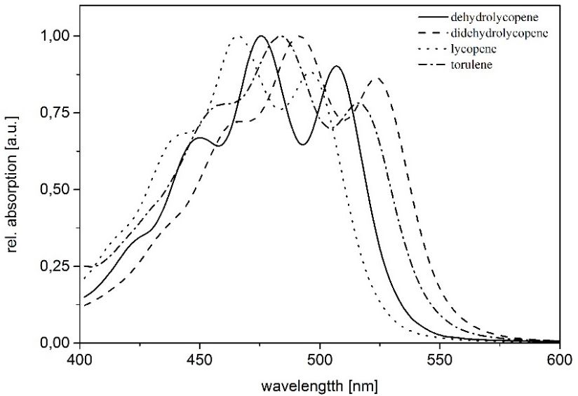
J. Fungi 2018, 4, 124                                                                                2 of 12

of four C5 isoprene units and the tail-to-tail condensation of two GGPP units to produce the colorless
precursor phytoene. These reactions are catalyzed by the enzymes GGPP synthase and phytoene
synthase respectively. The introduction of conjugated double bonds into the phytoene backbone
yields molecules able to absorb visible light and provides the characteristic yellow, orange to red
colors of the carotenoids. Four desaturation steps are necessary for the conversion of phytoene to
the maximally de-saturated red lycopene. These steps are carried out by a single enzyme [1,2], a
carotene cyclase encoded by Al-2 gene in Neurospora crassa. Similar genes were found also in Exophiala
strains [3]. Later reactions including cyclisation, isomerization, hydroxylation, and oxidation result in
the more than 600 different natural carotenoids known so far [1,4,5]. Those can be divided into a class
of hydrocarbons (carotenes) and their oxygenated derivatives (xanthophylls). In general, carotenoids
have several functions, ranging from supporting light harvesting during photosynthesis, to protecting
the cell from high light exposure, and the structural support of the photosynthetic apparatus [6]. They
are important for the cells to survive UV radiation, oxidative stress, and water or salt stress. These
properties make carotenoids interesting for industrial applications, for example as food colorants,
nutritional supplements, cosmetics, or health purposes. Especially in fungi, oxidative stress protection
seems to be the major function and is intensively investigated [6]. Furthermore, fungi and yeasts
were recently screened for commercial carotenoid production. Several Basidiomycetous yeast genera
(e.g., Rhodotorula or Phaffia) were found to be candidates for a commercial utilization [7–9]. Interestingly
it seems that extremophilic fungi produce higher amounts and a higher diversity of carotenoids [10–13].
One important group of extremophilic fungi are the so called black fungi or black yeasts, which can
cause human diseases. Such yeasts can be found in almost any environment and often form subaerial
biofilms (SABs) [14–17]. Among black fungi, several salt and cold tolerant fungi were found, such
species could be candidates for industrial carotenoid production [10,12]. The most obvious feature
of black fungi is their dark pigmentation, which results from inclusion of melanin into the cell wall.
Formation of melanin together with the production of secondary metabolites including carotenoids
and mycosporines, are passive physiological adaptations that help these fungi resisting environmental
stresses [18–22]. Secondly black fungi are characterized by a thick cell wall providing a high mechanical
protection especially under water stress [22].
      The rock inhabiting fungus K. petricola A95(syn. Sarcinomyces petricola), which was isolated from
a marble rock near the Philopappos monument on Musaios Hill, Athens (Greece) [23] belongs to an
ancestral lineage of the order Chaetothyriales [24] and possesses all characteristic features of black
fungi, including meristematic growth, melaninized cell walls and extensive secondary metabolite
production [22]. The desiccation resistant black yeast K. petricola A95 is recently under investigation
regarding stress response mechanisms and adaptation processes [25]. Since it produces melanin,
carotenoids and mycosporins [21], among other stress related substances, it is an ideal candidate
to investigate the role of the single components regarding stress response in black fungi. The high
variety of carotenoids in fungi and the growing fungal related biotechnology demand techniques and
protocols to investigate pigment composition and production under various environmental conditions.
Recent reviews show the growing interest of biotechnology industry to produce such pigments in yeast
cultures or other fungi and listed important factors (carbon source, aeration, light etc.) and limiting
steps (production rates, enzymatic activity etc.) in carotenoid production [7,8,26]. To analyze carotenoid
concentration, accumulation, and conversion a highly efficient, cost effective reliable system with a
low detection limit is needed. HPLC is one of the standard methods used for such analyses. However,
sample preparation, especially the difficulties of extracting specific carotenoids (low concentration or
derivatives), requires a specific sample treatment (cell disruption), extraction medium, and solvent
mixture. Several studies discussed separation of xanthophylls with HPLC, but they reported difficulties
for some carotenoids. Most studies propose a C18 column in combination with a wide variety of
solvents; e.g., studies used water-acetone mixtures [27,28], acetonitrile or hexane as extraction medium
or solvent. All of the extraction solutions or solvents face the same problem: that they are not equally
suitable for all pigments. Additionally, a comparison of carotenoids by spectroscopic measurements is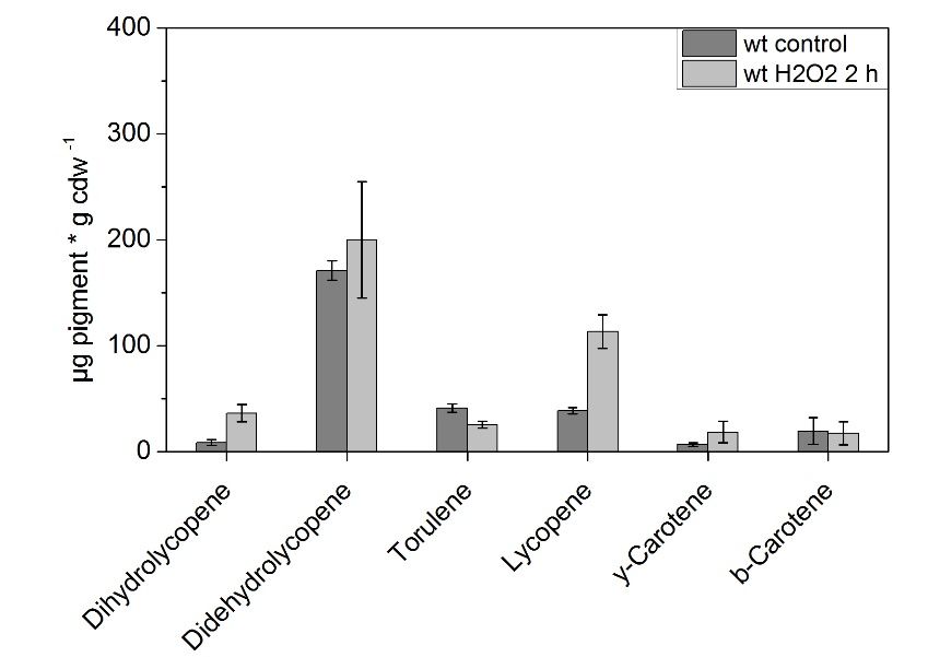
J. Fungi 2018, 4, 124                                                                               3 of 12

difficult, since the absorption spectra depend on the solvent used [29] and the solvents can chemically
react with pigments and such reactions can result in pigment derivatives [30].
      Several methods were tested to increase extraction efficiency, mechanical or chemical treatment,
and the use of specific solvents. The enhancement of pigment extraction was demonstrated by using
mechanical or chemical treatment [31,32], ultra-sonication for cell disruption, and by using different
extraction media [33]. Cell consumption is critical for quantitative pigment analysis. As [34,35]
demonstrated the extraction efficiency depends on two parameters: an efficient cell disruption and a
proper extraction medium. Mechanical treatment seems to be the preferred method for cells with thick
cell walls [31,32]. Other parameters that influence pigment stability during extraction and therefore
should be considered are pH-values, oxygen concentration, light and temperature.
      The main fungal pigments are β-carotene, astaxanthin, lycopene, and neurosporene. Recently
the production of torulene and torularhodin in fungi has gained some interest, especially as stress
response to UV radiation [10,36,37]. The exact function of carotenoids in fungi is still under discussion,
since mutants lacking carotenoids display the same growth rates as wild type strains. In the cases
investigated, a lack of carotenoids has no apparent phenotypic consequences on growth or morphology
in laboratory cultures. However, secondary effects of carotenoids, especially in regard to membrane
integrity, are under discussion [6]. It is known in fungi that carotenoid formation is activated by blue
light and H2 O2 , which generates oxidative stress [38]. The rock inhabiting fungus K. petricola A95
produces: β-carotene, γ-carotene, phytoene, torulene, and torularhodin and desiccation/rehydration
stresses affect the formation of the colorless phytone as well as the other carotenoids [22]. A new
pigment extraction protocol was used to investigate the carotenoid composition of the rock inhabiting
yeast K. petricola A95 under control conditions but also under temperature and oxidative stress. This
new, robust, and reproducible method allows quantitative analysis of carotenoids in black yeasts to
be used to determine the adaptation responses to extreme environmental conditions in high stress
resistant fungi.

2. Material and Methods
     Cell cultures of K. petricola A95 were grown on MEA (malt extract agar) plates at 25 ◦ C and
4 ◦ C.Cell colonies were harvested after 7 days and freeze dried. Oxidative stress related pigment
accumulation was tested with cell cultures grown (equal cell number) in liquid BG11 (cyanobacteria
media,) media with 2% malt extract and 0.2% glucose and with subsequent addition of 20 mM H2 O2 for
2 h. K. petricola A95 was cultivated in BG11 media to investigate biofilm formation and interaction with
cyanobacteria as described in [39], especially since such biofilms are exposed to harsh environmental
conditions, leading to adaptations in pigment compositions in both organisms.
     All cells were freeze dried after harvesting (Labconco FreeZone 2.5), dry weights of the cells were
determined and the cells were homogenized. A mill for glass beads with integrated CO2 cooling was
used for the extraction of the pigments. The freeze dried material was transferred into a glass vessel
with ground glass joint (Sartorius). The glass beads used had a mixing ratio of 1:3 (w/w), including
1 part of cells with a diameter of 1 mm and 3 parts of beads with an average diameter of 0.25 mm
(Carl ROTH GmbH). Simultaneously the samples were disrupted with 100% HPLC grade acetonitrile
(Sigma-Aldrich) for 3 min. To reduce the amount of fine particles in the supernatant, the samples were
centrifuged at 20,800 g (Centrifuge 5417 C, Eppendorf) twice for 1 min after the disrupting procedure.
The remaining pellets were tested for extraction efficiency by applying extraction solvents afterwards,
to make sure, the pigment extraction was complete.
     The determination of the different carotenoids was realized via HPLC (DIONEX, Ultimate 3000)
A reversed phase C18 column (300-5, C18-250 × 4 mm, from CS Chromatography Service) was used.
A two eluent protocol was used with following gradient (Table 1)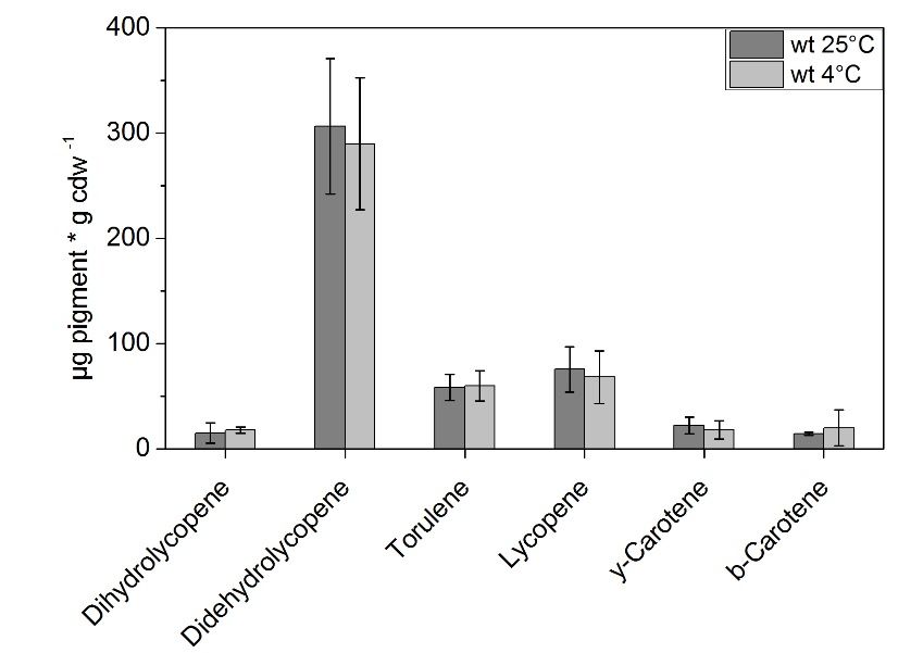
J. Fungi
J. Fungi 2018,
2018, 4,
4, 124
x FOR PEER REVIEW 44 of
of 12
12
Table 1. Eluent protocol used for carotenoid separation.
Table 1. Eluent protocol used for carotenoid separation.
Time [min] Eluent A Eluent B
Time [min] 0 60%A
Eluent 40%Eluent B
7 50% 50%
0 60% 40%
7 17 40%
50% 60% 50%
17 21 30%
40% 70% 60%
21 28.5 20%
30% 80% 70%
28.5 29.5 10%
20% 90% 80%
29.5
30.5 to 42 min 10%
60% 40% 90%
30.5 to 42 min 60% 40%
Eluent A was composed of 80% acetonitrile (HPLC grade; Sigma-Aldrich) and 20% aqua dest.
(v/v) Eluent
and EluentA wasB was 100% ethyl
composed of 80%acetate (HPLC grade;
acetonitrile (HPLCSigma-Aldrich). The totaland
grade; Sigma-Aldrich) protocol is 30 dest.
20% aqua min
long and
(v/v) andwas carried
Eluent B wasout withethyl
100% a flow rate of
acetate 0.8 mL/min.
(HPLC grade; The used solvents
Sigma-Aldrich). were
The degassed
total protocolfor 15 min
is 30
by ultrasonication
long and was carried prior
outuse.
with a flow rate of 0.8 mL/min. The used solvents were degassed for 15 min
Pigment identification
by ultrasonication prior use. and quantification was established for lycopene, didehydrolycopene,
dehydrolycopene, β-carotene,
Pigment identification γ-carotene,
and torulene,
quantification was and as well forfor
established torularhodin.
lycopene, Authentic standards
didehydrolycopene,
were obtained for β-carotene
dehydrolycopene, β-carotene,and lycopenetorulene,
γ-carotene, from Sigma-Aldrich.
and as well for The quantification
torularhodin. was based
Authentic on the
standards
β-carotene
were obtained calibration curve, only,
for β-carotene because lycopene
and lycopene is not solubleThe
from Sigma-Aldrich. in the extraction solvent.
quantification was basedLycopene
on the
was dissolved
β-carotene in 100%curve,
calibration hexane (HPLC
only, grade;
because Sigma-Aldrich).
lycopene Calculation
is not soluble of pigment
in the extraction concentrations
solvent. Lycopene
weredissolved
was based on in β-carotene calibration
100% hexane (HPLCand drySigma-Aldrich).
grade; weight as reference values. Therefore,
Calculation of pigmentpeak areas were
concentrations
related
were to β-carotene
based concentration
on β-carotene calibrationandand then divided
dry weight as by the specific
reference values.extinction
Therefore, coefficients
peak areasofwere the
pigments
related to in acetonitrile
β-carotene [40,41]. The pigments
concentration and then were detected
divided by theatspecific
440 nm extinction
for β-carotene, at 460 nm
coefficients for
of the
of γ-carotene,
pigments lycopene and
in acetonitrile dehydrolycopene
[40,41]. The pigments were at 470detected
nm, torulene
at 440 atnm480fornm and torularhodin
β-carotene, at 460 nmwas for
detected
of at 497lycopene
γ-carotene, nm. Theanddrydehydrolycopene
weight was subsequently at 470 nm, included.
toruleneDuring the and
at 480 nm HPLC measurements,
torularhodin was
absorption
detected at spectra
497 nm.were
The ascertained
dry weight by wasthesubsequently
HPLC-detector itself, supported
included. During the byHPLC
a tungsten lamp. The
measurements,
absorption spectra
spectra were
weredetermined
ascertainedfrom by the402HPLC-detector
to 762 nm. Furthermore, concentrations
itself, supported of purchased
by a tungsten lamp.
standards (β-carotene and lycopene) were verified by photometric measurements
The absorption spectra were determined from 402 to 762 nm. Furthermore, concentrations of purchased (Carl Zeiss Specord
M500), to prevent
standards overloading
(β-carotene of thewere
and lycopene) column. In all
verified bydetermined
photometricconditions three (Carl
measurements different
Zeissbiological
Specord
replicates
M500), with a dry
to prevent weight of of
overloading about 0.1 g were
the column. Inanalyzed. Pigment
all determined concentrations
conditions were subjected
three different biologicalto
the one-way
replicates with analysis of variance
a dry weight (ANOVA)
of about 0.1 g were test in orderPigment
analyzed. to establish comparisons
concentrations between
were subjecteddifferent
to the
stress treatments.
one-way analysis of All datas generated
variance (ANOVA)or analyzed
test in orderduring this study
to establish are included
comparisons between in different
this published
stress
article.
treatments. All datas generated or analyzed during this study are included in this published article.
3. Results
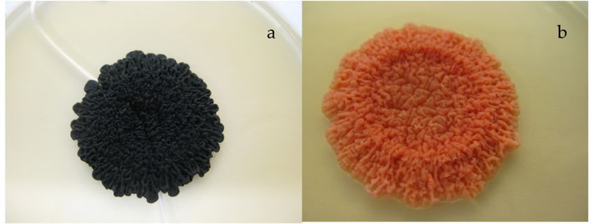
Pigment extraction was carried out with K. petricola A95 WT in comparison to a spontaneous
spontaneous
melanin deficient mutant of K. petricola
petricola A95
A95 (hereafter
(hereafter mdK)
mdK) (Figure
(Figure 1).
1).
Figure 1. Colonies of K. petricola
petricola A95
A95 (a)
(a) WT
WT and
and (b)
(b) the
the spontaneous
spontaneous melanin
melanin deficient
deficient mutant
mutant (mdK)
(mdK)
grown on MEA plates for 14 days at 25 ◦°C.
at 25 0.5 cm.
C. Colony size approximately 0.5 cm.
The
The mutant
mutant allowed
allowed us to determine
us to determine the
the quality
quality of
of the
the carotenoid
carotenoid extraction
extraction since
since the
the containing
containing
carotenoids were visible
carotenoids were visible in
in this
this mutant
mutant and
and not
not masked
masked by by melanin
melanin as
as in
in the
the WT
WT of K. petricola
of K. petricola A95.
A95.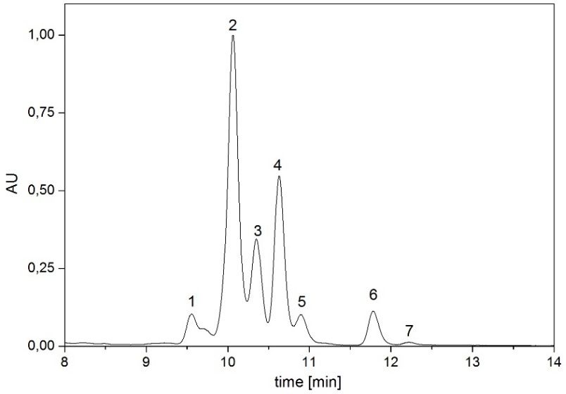
J. Fungi 2018, 4, 124                                                                               5 of 12

Pigment extraction was carried out for freeze dried cell pellets with a minimum weight of 15 mg.
Torularhodin was the pigment with the lowest concentration and minimum weight of the used material
was adjusted to determine the specific amount of torularhodin accordingly. The thick cell wall of
K. petricola A95 caused the use of relative long cell disruption times, and the shorter extraction lead to
a higher variation in extraction efficiency. Therefore to ensure quantitative pigment extraction, cell
pellets were tested for the remaining pigments in a second extraction step with 100% acetonitrile and
100% hexane. No significant amounts of pigments were detectable via HPLC analysis (
J. Fungi
J. Fungi 2018,
2018, 4, x FOR PEER REVIEW
4, 124 6 of6 12
of 12
J. Fungi 2018, 4, x FOR PEER REVIEW 6 of 12
Figure 2. Representative HPLC chromatogram of carotenoids in K. petricola A95 detected in our
experiments.
Figure
Figure The peaks were
2. 2.Representative
Representative HPLC
HPLCdetermined via reference
chromatogram
chromatogram substances
of carotenoids
of carotenoids ininand identified
K.K.petricola as dehyrolycopene
petricolaA95
A95 detected
detected in in
ourour
(1), didehydrolycopene
experiments.
experiments. The
The peaks
peaks (2),
were
were torulene
determined
determined (3),
via
via lycopene (4), γ-carotene
referencesubstances
reference substancesand (5) and
andidentified β-carotene (6) and(1),
identifiedasasdehyrolycopene
dehyrolycopene
torularhodin
(1), (7). (2), torulene
didehydrolycopene
didehydrolycopene (2), torulene (3), (4),
(3), lycopene lycopene (4), γ-carotene
γ-carotene (5) and(6)β-carotene
(5) and β-carotene (6) and(7).
and torularhodin
torularhodin (7).
Figure
Figure 3. 3. Absorption
Absorption spectra
spectra ofofmain
maincarotenoids
carotenoidsininK.K.petricola
petricolaA95
A95(determined
(determined via
via HPLC
HPLC detector
detector in
in according
according eluent
Figure 3.eluent mixture).
mixture).
Absorption The
The of
spectra maxima
maxima of each pigment
of each pigment
main carotenoids was determined:
was determined:
in K. petricola didehydrolycopene
didehydrolycopene
A95 (determined (463
(463
via HPLC detector nm,
nm,
492innm,492 nm,
according
523 523 nm);
eluent
nm); torulene
mixture).
torulene (454 (454
The nm,
484484
maxima
nm, ofnm,
nm, 515
each
515 nm);dehydrolycopene
pigment
nm); dehydrolycopene
was determined:(448 nm,
nm,476
476nm, 507
didehydrolycopene
(448 nm, 507nm);
(463
nm);
lycopene
nm,
lycopene (440
492(440
nm, nm,
523
nm, 465nm,
nm);
465 nm,496
496nm).
torulene nm).nm, 484 nm, 515 nm); dehydrolycopene (448 nm, 476 nm, 507 nm);
(454
lycopene (440 nm, 465 nm, 496 nm).
Table 2. Pigments
Table 2. Pigmentsabsorption maxima
absorption andand
maxima concentrations of the
concentrations of black
the black Knufia
yeastyeast petricola
Knufia A95 grown
petricola A95
onTable
MEA plates
grown 2. MEAunder
onPigments standard
plates growth
maximaconditions.
under standard
absorption growth conditions. of the black yeast Knufia petricola A95
and concentrations
grown on MEA plates under standard
Pigment growth conditions.
Absorption
Pigment AbsorptionMaxima
Maxima[nm][nm]Concentration [µg/g [µg/g
Concentration dry weight]
dry weight]
dihydrolycopene
Pigment 448,
Absorption 476, 507
Maxima [nm] Concentration30.74
[µg/g dry weight]
dihydrolycopene 448, 476, 507 30.74
didehydrolycopene
dihydrolycopene 463, 476,
448, 492, 523 383.12
didehydrolycopene 463, 492,507
523 30.74383.12
torulene
didehydrolycopene 454, 492,
463, 484, 515 85.39
torulene 454, 484,523
515 383.12 85.39
lycopene
torulene 440,
454, 465, 496 100.30
lycopene 440,484,
465,515
496 85.39100.30
γ-carotene
lycopene
γ-carotene 460,
440, 489
465,
460, 496
489 23.88 23.88
100.30
β-carotene
γ-carotene
β-carotene 457,
460, 483
457,489
483 7.53 7.53
23.88
torularhodin
torularhodin
β-carotene 469,
469,497,
483525
497,
457, 525 5.84 5.84
7.53
torularhodin 469, 497, 525 5.84
Short-term incubation (2 h) of WT K. petricola A95 under oxidative stress (treatment with 20 mM
H2Short-term
O2Short-term
) showed incubation
anincubation
increase in(2(2concentration
h)h)ofofWT
WTK. for several
K. petricola
petricola A95carotenoids
A95 under compared
underoxidative
oxidative stressto growth under
(treatment
stress (treatment with normal
with2020
mM mM
conditions
H2HO22O) 2)showed for an
showed theincrease
same time
increase inin period (Figure
concentration
concentration for4).
forThe
several concentrations
carotenoids
several of lycopene
compared
carotenoids (38.7
to growth
compared to 113.2
tounder
growth µg
normal g
under
cdw −1), γ-carotene (6.6 to 18.2 µg g cdw−1) and dihydrolycopene (8.5 to 36.2 µg g cdw−1) increased
conditions for the same time period (Figure 4). The concentrations of lycopene
normal conditions for the same time period (Figure 4). The concentrations of lycopene (38.7 to (38.7 to 113.2 µg g
strongly
cdw
113.2 µg under
g cdw the
−1 ),
−1), γ-carotene applied
(6.6 to 18.2
γ-carotene conditions
µgto
(6.6 (differences
g 18.2
cdw µg g cdw−significant
−1) and dihydrolycopene for (8.5
p = 0.05).
to 36.2
1 ) and dihydrolycopene The
µg concentrations
(8.5gtocdw of β-−1 )
−1) increased
36.2 µg g cdw
stronglystrongly
increased under the applied
under conditions
the applied (differences
conditions significant
(differences for p = 0.05).
significant for p =The concentrations
0.05). of β-
The concentrationsJ. Fungi 2018,4,4,124
2018,
J. Fungi x FOR PEER REVIEW 7 of 712of 12
J. Fungi 2018, 4, x FOR PEER REVIEW 7 of 12
carotene and torulene did not increase during the incubation and for didehydrolycopene an increase
ofwas and and
β-carotene
carotene
detectable, torulene
torulene did
did not
however, the not increase
increase
high during during
variation the the
thebetween
incubationincubation
and and
did for
notdidehydrolycopene
for didehydrolycopene
samples allow usanto draw an
increase
increase
was was detectable,
detectable, however,
however, the the
high high variation
variation between
between the the
samplessamples
did did
not not allow
allow
conclusions. Didehydrolycopene showed in general the highest variation in pigment concentration us us
to to draw
draw
conclusions.
conclusions.
in Didehydrolycopeneshowed
Didehydrolycopene
all samples. showedin ingeneral
general the
the highest
highest variation
variation in pigment
pigment concentration
concentration in
allinsamples.
all samples.
Figure 4. Carotenoid composition of WT K. petricola A95 incubated with and without 20 mM H2O2 for
Figure
2Figure 4.4.Carotenoid
Carotenoid
h, determined composition
analysisof
composition
via HPLC (nWT
of petricolaA95
= 3).K.Significant
petricola A95 incubated
incubated
differences pwith
at with and
and
< 0.05 without
without
with 2020
respect mM
to mM 2H
O22 O
theHcontrol 2 for
for
2 are
h,
2 h,determined
determined via
viaHPLC
HPLC analysis
analysis (n
(n
marked by asterisks (one-way ANOVA). = 3).
3). Significant
Significant differences
differences atat
p p
< < 0.05
0.05 with
with respect
respect to to
the the control
control
areare markedby
marked byasterisks
asterisks(one-way
(one-way ANOVA).
ANOVA).
Incubation under low temperature did not influence the pigment composition (Figure 5),
Incubation
Incubation
compared under
undergrown
to cultures low
low temperature
temperature
under normal did
did not influence
not influence
temperatures thepigment
(thethe
ANOVA pigmentTest composition
composition
showed (Figure
no (Figure 5), 5),
significant
compared
compared toto cultures
cultures grown
grown under
under normal
normal temperatures
temperatures (the
(theANOVA
ANOVA Test showed
Test
differences between the control samples and temperature stressed cells for all pigments). However, showed no significant
no significant
adifferences
differences betweenin
between
strong increase the
the control
control
the samples and
samples and temperature
didehydrolycopene temperature
concentrationstressed
stressed
(up to cells
300for
cells for
µgall
allgpigments).
pigments).
cdw−1) under However,
However,
both a
a strong
strong increase
increase in the in the didehydrolycopene
didehydrolycopene concentration
concentration (up to(up
300 to
µg 300
g cdw µg−1g) under
cdw −1) under both
both conditions
conditions was visible after long term incubation. Therefore, the total measured pigment
wasconditions
concentration was
visible after visible
long
increasedterm toafter long
incubation.
~500 µg term incubation.
Therefore,
g cdw totalTherefore,
the results
−1. Identical measured the
pigment
for all experimentstotal concentration
measured
are exhibitedpigment
increased
for the
concentration
tospontaneous
~500 µg g cdw increased
−
mdK of1 to
. Identical~500 µg g
K. petricolaresults cdw
A95. The
−1. Identical results for all experiments are exhibited for the
forspontaneous
all experiments mutantaredisplays
exhibited for theamount
a similar spontaneous mdK of
of pigments
spontaneous
K.per dry weight
petricola A95. mdK
andofthe
The K. carotenoid
petricola A95.
spontaneous The spontaneous
concentrations
mutant displaysdid mutant
not displays
change
a similar ofa pigments
similartoamount
as a response
amount of pigments
temperature
per dry stressand
weight
per
(datadry weight
not shown). and the carotenoid concentrations did not change as a response
the carotenoid concentrations did not change as a response to temperature stress (data not shown). to temperature stress
(data not shown).
Figure5.5.Pigment
Figure Pigment composition
composition of K. petricola
of K. petricolaA95
A95(WT)
(WT)determined
determined viavia HPLC
HPLC after
after growth
growth under
under
Figure
normal 5. Pigment
conditions (25 ◦ C) and under
composition of K. petricola
low A95 (WT)
temperature (4 ◦ C) for 7 days
determined via (nHPLC
= 3). after growth
Significant under
differences
normal conditions (25 °C) and low temperature (4 °C) for 7 days (n = 3). Significant differences
normal conditions
p p<J. Fungi 2018, 4, 124 8 of 12
4. Discussion
For an efficient pigment analysis, two steps are important: First, a solvent for extraction should
be selected that allows the quantitative determination of all relevant pigments. In specific cases and
with unknown samples, a selection and/or a mixture of media should be used, like acetonitrile, ethyl
acetate, isopropanol, and hexane. The most suitable solvents seemed to be isopropanol and hexane,
which were used to verify the extraction efficiency (re-extraction of the pellet), but caused a baseline
shift in the HPLC. It is noteworthy to state that DMSO is to our opinion not a suitable extraction
solvent; the difficulties to remove DMSO from the system limit the application in an adequate eluent
system. After centrifugation and removal, the supernatant residues of DMSO still remain in the sample,
which is problematic for HPLC analyses. Additionally, the proposed DMSO based method of pigment
extraction [37,44] should be tested accordingly, depending on the systems used, to ensure optimal
pigment analysis results. Thus, DMSO extraction is always a time consuming method and the results
are hardly comparable with other methods. Secondly an efficient cell rupture method under conditions
that ensure the pigment stability should be selected. Black yeasts are characterized by a thick cell
wall with high melanin concentration. In general pigment extraction should be performed under low
oxygen, and if possible under oxygen free conditions. The major challenge in pigment analysis is to
recover pigments with minimum risk of damage but with high efficiency. A mechanical treatment is
needed in the most cases but the method applied should be tested carefully due to the high sensitivity
of the molecules. Especially for black yeasts and other fungi with a thick cell wall, a mechanical cell
disruption seems necessary and provides better results as a chemical treatment [31,32].
K. petricola A95 displayed an uncommon pigment composition; especially in the high
concentrations of dihydrolycopene, didehydrolycopene and torulene, which are not so often
described in fungi. A normally high abundance of carotene derivatives and lycopene can be
observed in pigmented fungi [45,46]. Several yeasts showed a high torulene concentration but no
dihydrolycopene was detected in yeast so far [46,47]. The abundance of didehydrolycopene and
torulene indicates a similar production pathway as described for N. crassa, leading in this fungus to
neurosporaxanthin [2,48], a carotenoid that was not detectable in K. petricola A95 under the applied
conditions. It is noteworthy to mention that the determination of the abundance of astaxanthin
and neurosporaxanthin is technically possible with the described protocol. The only pigment left
in the pellet was melanin in the WT. Hence comparing the results with methods developed for
pigments in algae or higher plants [26,27,31,49–51] is not advisable due to the thick cultured cell wall
of the supervised fungi. Although [49] compared different methods using maize seeds, they are not
characterized by a thick cell wall like black yeast. Similar to our study, the decision regarding efficacy
was made with respect to the color of the pellet. In general, these studies provide a wide method spectra
and were useful regarding extraction, elution, and calibration of the authentic standards, see also [31,51].
Fungi pigments were mainly analyzed, focusing on certain valuable pigments only. Knowledge
regarding pigment composition in fungi is needed for species description, however, such studies used
standard methods and did not compare different methods [5–7,10,33,34,52]. Methodological overviews
for fungi pigment extraction are still missing [38]. Therefore, the established method was compared
to [19]. The combination of extraction media and the eluent system of [19] resulted in a strong injection
peak in the presented HPLC system and was adjusted afterwards, concerning the different tested
method combinations shown.
The detected carotenoids are consistent with [22], they found also carotene derivatives, torulene,
and torularhodin. Our extraction method, especially the extraction medium and the mechanical
treatment, gave us additionally the possibility to quantify lycopene and didehydrolycopene, unusual
pigments in fungi and not often found in yeast strains.
To our opinion didehydrolycopene functions as a synthesis interface, comparable to β-carotene in
photosynthetic organisms. Such a pool of didehydrolycopene can be converted fast to stress response
pigments and helps cells to respond quickly to environmental stresses. The two main pigments in
K. petricola A95, lycopene and didehydrolycopene, display much higher concentrations, as in otherJ. Fungi 2018, 4, 124 9 of 12
fungi detected [53] and β-carotene concentrations are lower compared to other fungi and yeasts.
The detected lycopene derivatives are the major carotenoids in K. petricola A95. Future experiments
must be carried out to find out if these pigments have a photoprotective role or if they function as
a precursor for other pigments. In N. crassa the conversion of didehydrolycopene to torulene by
cyclisation was described [54]. The already described torularhodin and torulene could be involved in
photoprotection [6,13,37,55]. The antioxidant properties of torulene are attributed to its conjugated
double bond system; in fact, torulene has more antioxidant efficiency than β-carotene, which presents
less of a double bond on its chemical structure than torulene [56]. However, several other carotenoids
were also described to have an antioxidant activity.
Another explanation for the stable carotenoid concentrations in K. petricola A95 would be that
the mechanism of carotenoid action is more likely to consist of shielding sensitive molecules or
organelles than of the neutralization of harmful oxidants [52]. Therefore, carotenoids do not play a
major physiological role in fungal cells, but they may have beneficial effects under specific conditions.
Several studies showed an increase in carotenoid concentration under stress [45,57]. The potential
photoprotective pigment torulene [9,58] showed low concentrations in K. petricola A95, which would
imply a less important role in K. petricola A95, but an inducible production under a stress situation
out of the more stable dehydrolycopene seems possible. However, we could not detect an increase in
torulene concentrations if the cells were exposed to oxidative or temperature stress. Such an induction
was described for a black yeast under oxidative stress [59], in red yeast under salt stress [58], and
a light dependent increase of the overall carotenoid concentration was described for N. crassa [38]
incubated with H2 O2 . Such an inducible protective system would enable K. petricola A95 to react fast
to stress situations. A similar system was described for the red yeast Dioszegia with the xanthophyll
plectaniaxanthin [60]. The photoprotective carotenoid torularhodin was induced in K. petricola A95
under cold temperatures, but very low amounts and other stresses or longer incubation times have to
be investigated to unravel the function of this pigment in K. petricola A95.
The major changes under the applied conditions were detected for lycopene and derivatives,
which should therefore be considered as the most important pigments in K. petricola A95. The high
concentration of the unusual carotenoid didehydrolycopene in K. petricola A95 should be investigated
in detail, especially since the photoprotective role and the biotechnological potential of lycopene
derivatives was described before [49]. Screening other black yeasts regarding their pigmentation
could result in a wide diversity of pigments with multiple promising applications. Black fungi are
remarkable in their stress resistance and we showed that their carotenoid pigmentation is complex
and future studies have to be performed to determine the specific function of the specific pigments.
The protocol presented will allow the quantitative analysis of black fungi, characterized by thick cell
walls and melanin pigmentation, regarding stress response and adaptation to extreme environmental
conditions. Future experiments have to unravel the function of melanin, carotenoids, and the cell wall
structure regarding the stress responses in detail.
5. Conclusions
Extremotolerant and extremophile black yeasts are a promising source of pigments and other
chemicals. New protocols and studies are needed to determine the capacity for production of these high
stress resistance fungi. The established protocol allows the determination of carotenoid composition in
black yeasts. Oxidative stress results in an adaptation in pigment composition. Future experiments
have to be carried out to determine if didehydrolycopene functions as a protective agent itself or if
it serves as a precursor for antioxidative pigments like torulene and torularhodin, which could be
produced after induction under stress conditions.
Author Contributions: J.T. and N.K. performed the growth experiments and drafted this manuscript. K.F.
developed the HPLC methods and participated in the conception of the study, analysis of data and revision of the
manuscript. J.T. and N.K. participated in the conception of the study, supervised the experiments, and revised the
manuscript. All authors read and approved the final manuscript.J. Fungi 2018, 4, 124 10 of 12
Funding: This work was supported financially by internal funds of the BAM.
Acknowledgments: We thank Anna Gorbushina (BAM Berlin) and Christian Wilhelm (University of Leipzig) for
their support.
Conflicts of Interest: The authors declare no conflict of interest.
References
1. Diaz-Sanchez, V.; Estrada, A.F.; Trautmann, D.; Limon, M.C.; Al-Babili, S.; Avalos, J. Analysis of al-2 mutations
in Neurospora. PLoS ONE 2011, 6. [CrossRef] [PubMed]
2. Hausmann, A.; Sandmann, G. A single five-step desaturase is involved in the carotenoid biosynthesis
pathway to β-carotene and torulene in Neurospora crassa. Fungal Genet. Biol. 2000, 30, 147–153. [CrossRef]
[PubMed]
3. Tafer, H.; Lopandic, K.; Blasi, B.; Poyntner, C.; Sterflinger, K. Draft genome sequence of Exophiala mesophila, a
black yeast with high bioremediation potential. Genome Announc. 2015, 3. [CrossRef] [PubMed]
4. Sandmann, G. Carotenoid biosynthesis in microorganisms and plants. Eur. J. Biochem. 1994, 223, 7–24.
[CrossRef] [PubMed]
5. Armstrong, G.A.; Hearst, J.E. Carotenoids 2: Genetics and molecular biology of carotenoid pigment
biosynthesis. FASEB J. 1996, 10, 228–237. [CrossRef] [PubMed]
6. Avalos, J.; Carmen Limon, M. Biological roles of fungal carotenoids. Curr. Genet. 2014, 61, 309–324. [CrossRef]
[PubMed]
7. Frengova, G.I.; Beshkova, D.M. Carotenoids from Rhodotorula and Phaffia: Yeasts of biotechnological
importance. J. Ind. Microbiol. Biotechnol. 2009, 36, 163–180. [CrossRef] [PubMed]
8. Mata-Gomez, L.C.; Montanez, J.C.; Mendez-Zavala, A.; Aguilar, C.N. Biotechnological production of
carotenoids by yeasts: An overview. Microb. Cell Fact. 2014, 13, 12. [CrossRef] [PubMed]
9. Moline, M.; Libkind, D.; van Broock, M. Production of torularhodin, torulene, and β-carotene by Rhodotorula
yeasts. Methods Mol. Biol. 2012, 898, 275–283. [PubMed]
10. Amaretti, A.; Simone, M.; Quartieri, A.; Masino, F.; Raimondi, S.; Leonardi, A.; Rossi, M. Isolation of
carotenoid-producing yeasts from an alpine glacier. Chem. Eng. Trans. 2014, 38, 217–222.
11. Arcangeli, C.; Cannistraro, S. In situ raman microspectroscopic identification and localization of carotenoids:
Approach to monitoring of UV-B irradiation stress on antarctic fungus. Biopolymers 2000, 57, 179–186. [CrossRef]
12. Libkind, D.; Moline, M.; Sampaio, J.P.; van Broock, M. Yeasts from high-altitude lakes: Influence of UV
radiation. FEMS Microbiol. Ecol. 2009, 69, 353–362. [CrossRef] [PubMed]
13. Moline, M.; Libkind, D.; Dieguez Mdel, C.; van Broock, M. Photoprotective role of carotenoids in yeasts:
Response to UV-B of pigmented and naturally-occurring albino strains. J. Photochem. Photobiol. B 2009, 95,
156–161. [CrossRef] [PubMed]
14. Teixeira, M.M.; Moreno, L.F.; Stielow, B.J.; Muszewska, A.; Hainaut, M.; Gonzaga, L.; Abouelleil, A.;
Patane, J.S.; Priest, M.; Souza, R.; et al. Exploring the genomic diversity of black yeasts and relatives
(Chaetothyriales, Ascomycota). Stud. Mycol. 2017, 86, 1–28. [CrossRef] [PubMed]
15. De Hoog, G.S.; Vicente, V.A.; Gorbushina, A.A. The bright future of darkness—the rising power of black
fungi: Black yeasts, microcolonial fungi, and their relatives. Mycopathologia 2013, 175, 365–368. [CrossRef]
[PubMed]
16. Gunde-Cimerman, N.; Grube, M.; de Hoog, G.S. The emerging potential of melanized fungi: Black yeast
between beauty and the beast. Fungal Biol. 2011, 115, 935–936. [CrossRef] [PubMed]
17. Gorbushina, A.A.; Broughton, W.J. Microbiology of the atmosphere-rock interface: How biological
interactions and physical stresses modulate a sophisticated microbial ecosystem. Annu. Rev. Microbiol. 2009,
63, 431–450. [CrossRef] [PubMed]
18. Sterflinger, K. Temperature and NaCl-tolerance of rock-inhabiting meristematic fungi. Antonie Van
Leeuwenhoek 1998, 74, 271–281. [CrossRef] [PubMed]
19. Kogej, T.; Stein, M.; Volkmann, M.; Gorbushina, A.A.; Galinski, E.A.; Gunde-Cimerman, N. Osmotic
adaptation of the halophilic fungus hortaea werneckii: Role of osmolytes and melanization. Microbiol-Sgm
2007, 153, 4261–4273. [CrossRef] [PubMed]J. Fungi 2018, 4, 124 11 of 12
20. Kogej, T.; Wheeler, M.H.; Rizner, T.L.; Gunde-Cimerman, N. Evidence for 1,8-dihydroxynaphthalene melanin
in three halophilic black yeasts grown under saline and non-saline conditions. FEMS Microbiol. Lett. 2004,
232, 203–209. [CrossRef]
21. Volkmann, M.; Gorbushina, A.A. A broadly applicable method for extraction and characterization of
mycosporines and mycosporine-like amino acids of terrestrial, marine and freshwater origin. FEMS Microbiol.
Lett. 2006, 255, 286–295. [CrossRef] [PubMed]
22. Gorbushina, A.A.; Kotlova, E.R.; Sherstneva, O.A. Cellular responses of microcolonial rock fungi to long-term
desiccation and subsequent rehydration. Stud. Mycol. 2008, 61, 91–97. [CrossRef] [PubMed]
23. Wollenzien, U.; de Hoog, G.S.; Krumbein, W.; Uijthof, J.M. Sarcinomyces petricola, a new microcolonial fungus
from marble in the mediterranean basin. Antonie Van Leeuwenhoek 1997, 71, 281–288. [CrossRef] [PubMed]
24. Gueidan, C.; Villasenor, C.R.; de Hoog, G.S.; Gorbushina, A.A.; Untereiner, W.A.; Lutzoni, F. A rock-
inhabiting ancestor for mutualistic and pathogen-rich fungal lineages. Stud. Mycol. 2008, 61, 111–119.
[CrossRef] [PubMed]
25. Nai, C.; Wong, H.Y.; Pannenbecker, A.; Broughton, W.J.; Benoit, I.; de Vries, R.P.; Gueidan, C.;
Gorbushina, A.A. Nutritional physiology of a rock-inhabiting, model microcolonial fungus from an ancestral
lineage of the Chaetothyriales (Ascomycetes). Fungal Genet. Biol. 2013, 56, 54–66. [CrossRef] [PubMed]
26. Sandmann, G. Carotenoids of biotechnological importance. Adv. Biochem. Eng. Biotechnol. 2015, 148, 449–467.
[PubMed]
27. Weber, R.W.; Anke, H.; Davoli, P. Simple method for the extraction and reversed-phase high-performance
liquid chromatographic analysis of carotenoid pigments from red yeasts (Basidiomycota, fungi). J. Chromatogr.
A 2007, 1145, 118–122. [CrossRef] [PubMed]
28. Bona-Lovasz, J.; Bona, A.; Ederer, M.; Sawodny, O.; Ghosh, R. A rapid method for the extraction and analysis
of carotenoids and other hydrophobic substances suitable for systems biology studies with photosynthetic
bacteria. Metabolites 2013, 3, 912–930. [CrossRef] [PubMed]
29. Zang, L.Y.; Sommerburg, O.; VanKuijk, F.J.G.M. Absorbance changes of carotenoids in different solvents.
Free Radic. Biol. Med. 1997, 23, 1086–1089. [CrossRef]
30. Takaichi, S. Characterization of carotenes in a combination of a C (18) HPLC column with isocratic elution
and absorption spectra with a photodiode-array detector. Photosynthesis Res. 2000, 65, 93–99. [CrossRef]
[PubMed]
31. Geciova, J.; Bury, D.; Jelen, P. Methods for disruption of microbial cells for potential use in the dairy
industry—A review. Int. Dairy J. 2002, 12, 541–553. [CrossRef]
32. Gu, Z.; Deming, C.; Yongbin, H.; Zhigang, C.; Feirong, G. Optimization of carotenoids extraction from
Rhodobacter sphaeroides. LWT-Food Sci. Technol. 2008, 41, 1082–1088. [CrossRef]
33. Papaioannou, E.; Roukas, T.; Liakopoulou-Kyriakides, M. Effect of biomass pre-treatment and solvent
extraction on β-carotene and lycopene recovery from Blakeslea trispora cells. Prep. Biochem. Biotechnol. 2008,
38, 246–256. [CrossRef] [PubMed]
34. Craft, N.E.; Soares, J.H. Relative solubility, stability, and absorptivity of lutein and β-carotene in
organic-solvents. J. Agric. Food Chem. 1992, 40, 431–434. [CrossRef]
35. Su, Q.; Rowley, K.G.; Balazs, N.D.H. Carotenoids: Separation methods applicable to biological samples.
J. Chromatogr. B 2002, 781, 393–418. [CrossRef]
36. Sperstad, S.; Lutnaes, B.F.; Stormo, S.K.; Liaaen-Jensen, S.; Landfald, B. Torularhodin and torulene are the
major contributors to the carotenoid pool of marine Rhodosporidium babjevae (golubev). J. Ind. Microbiol.
Biotechnol. 2006, 33, 269–273. [CrossRef] [PubMed]
37. Moline, M.; Flores, M.R.; Libkind, D.; Dieguez Mdel, C.; Farias, M.E.; van Broock, M. Photoprotection by
carotenoid pigments in the yeast rhodotorula mucilaginosa: The role of torularhodin. Photochem. Photobiol.
Sci. 2010, 9, 1145–1151. [CrossRef] [PubMed]
38. Iigusa, H.; Yoshida, Y.; Hasunuma, K. Oxygen and hydrogen peroxide enhance light-induced carotenoid
synthesis in Neurospora crassa. FEBS Lett. 2005, 579, 4012–4016. [CrossRef] [PubMed]
39. Seiffert, F.; Bandow, N.; Bouchez, J.; von Blanckenburg, F.; Gorbushina, A.A. Microbial colonization of
bare rocks: Laboratory biofilm enhances mineral weathering. Procedia Earth Planet. Sci. 2014, 10, 123–129.
[CrossRef]
40. Britton, G.; Liaaen-Jensen, S.; Pfander, H. Carotenoids Handbook; Birkhäuser Verlag: Basel, Switzerland, 2004;
ISBN 3764361808.J. Fungi 2018, 4, 124 12 of 12
41. Foppen, F.H. Tables for the identification of carotenoid pigments. Chromatogr. Rev. 1971, 14, 133–298. [CrossRef]
42. Choi, S.K.; Kim, J.H.; Park, Y.S.; Kim, Y.J.; Chang, H.I. An efficient method for the extraction of astaxanthin
from the red yeast Xanthophyllomyces dendrorhous. J. Microbiol. Biotechnol. 2007, 17, 847–852. [PubMed]
43. Sedmak, J.J.; Weerasinghe, D.; Jolly, S. Extraction and quantitation of astaxanthin from Phaffia rhodozyma.
Biotechnol. Technol. 1990, 4, 107–112. [CrossRef]
44. Libkind, D.; Perez, P.; Sommaruga, R.; Dieguez, M.D.; Ferraro, M.; Brizzio, S.; Zagarese, H.; van Broock, M.
Constitutive and UV-inducible synthesis of photoprotective compounds (carotenoids and mycosporines) by
freshwater yeasts. Photochem. Photobiol. Sci. 2004, 3, 281–286. [CrossRef] [PubMed]
45. Gmoser, R.; Ferreira, J.A.; Lennartsson, P.R.; Taherzadeh, M.J. Filamentous ascomycetes fungi as a source of
natural pigments. Fungal Biol. Biotechnol. 2017, 4, 4. [CrossRef] [PubMed]
46. Zoz, L.; Carvalho, J.C.; Soccol, V.T.; Casagrande, T.C.; Cardoso, L. Torularhodin and torulene: Bioproduction,
properties and prospective applications in food and cosmetics—A review. Braz. Arch. Biol. Technol. 2015, 58,
278–288. [CrossRef]
47. Buzzini, P.; Innocenti, M.; Turchetti, B.; Libkind, D.; van Broock, M.; Mulinacci, N. Carotenoid profiles
of yeasts belonging to the genera Rhodotorula, Rhodosporidium, Sporobolomyces, and Sporidiobolus. Can. J.
Microbiol. 2007, 53, 1024–1031. [CrossRef] [PubMed]
48. Avalos, J.; Prado-Cabrero, A.; Estrada, A.F. Neurosporaxanthin production by Neurospora and Fusarium.
Methods Mol. Biol. 2012, 898, 263–274. [PubMed]
49. Albrecht, M.; Takaichi, S.; Steiger, S.; Wang, Z.-Y.; Sandmann, G. Novel hydroxycarotenoids with improved
antioxidative properties produced by gene combination in Escherichia coli. Nat. Biotechnol. 2000, 18, 843.
[CrossRef] [PubMed]
50. Olives Barba, A.I.; Camara Hurtado, M.; Sanchez Mata, M.C.; Fernadez Ruiz, V.; Lopez Saenz de Tejada, M.
Application of a UV–vis detection-HPLC method for a rapid determination of lycopene and β-carotene in
vegetables. Food Chem. 2006, 95, 328–336. [CrossRef]
51. Busch, M.; Seuter, A.; Hain, R. Functional analysis of the early steps of carotenoid biosynthesis in tobacco.
Plant Physiol. 2012, 128, 439–453. [CrossRef] [PubMed]
52. Schnitzler, N.; Peltroche-Llacsahuanga, H.; Bestier, N.; Zundorf, J.; Lutticken, R.; Haase, G. Effect of melanin
and carotenoids of Exophiala (Wangiella) dermatitidis on phagocytosis, oxidative burst, and killing by human
neutrophils. Infect. Immun. 1999, 67, 94–101. [PubMed]
53. Zhang, Z.; Zhang, X.; Tan, T. Lipid and carotenoid production by Rhodotorula glutinis under
irradiation/high-temperature and dark/low-temperature cultivation. Bioresour. Technol. 2014, 157, 149–153.
[CrossRef] [PubMed]
54. Arrach, N.; Schmidhauser, T.J.; Avalos, J. Mutants of the carotene cyclase domain of al-2 from Neurospora
crassa. Mol. Genet. Genom. 2002, 266, 914–921.
55. Du, C.; Li, Y.; Guo, Y.; Han, M.; Zhang, W.; Qian, H. The suppression of torulene and torularhodin treatment
on the growth of PC-3 xenograft prostate tumors. Biochem. Biophys. Res. Commun. 2016, 469, 1146–1152.
[CrossRef] [PubMed]
56. Sakaki, H.; Nochide, H.; Komemushi, S.; Miki, W. Effect of active oxygen species on the productivity of
torularhodin by Rhodotorula glutinis No. 21. J. Biosci. Bioeng. 2002, 93, 338–340. [CrossRef]
57. Nanou, K.; Roukas, T.; Papadakis, E.; Kotzekidou, P. Carotene production from waste cooking oil by Blakeslea
trispora in a bubble column reactor: The role of oxidative stress. Eng. Life Sci. 2017, 17, 775–780. [CrossRef]
58. Li, C.; Zhang, N.; Li, B.; Xu, Q.; Song, J.; Wei, N.; Wang, W.; Zou, H. Increased torulene accumulation in red
yeast Sporidiobolus pararoseus NGR as stress response to high salt conditions. Food Chem. 2017, 237, 1041–1047.
[CrossRef] [PubMed]
59. Geis, P.A.; Szaniszlo, P.J. Carotenoid-pigments of the dematiaceous fungus Wangiella-dermatitidis. Mycologia
1984, 76, 268–273. [CrossRef]
60. Madhour, A.; Anke, H.; Mucci, A.; Davoli, P.; Weber, R.W.S. Biosynthesis of the xanthophyll plectaniaxanthin
as a stress response in the red yeast Dioszegia (Tremellales, Heterobasidiomycetes, fungi). Phytochemistry 2005,
66, 2617–2626. [CrossRef] [PubMed]
© 2018 by the authors. Licensee MDPI, Basel, Switzerland. This article is an open access
article distributed under the terms and conditions of the Creative Commons Attribution
(CC BY) license (http://creativecommons.org/licenses/by/4.0/).You can also read