Gut-Liver Axis: How Do Gut Bacteria Influence the Liver? - MDPI
←
→
Page content transcription
If your browser does not render page correctly, please read the page content below
medical
           sciences
Review
Gut–Liver Axis: How Do Gut Bacteria Influence
the Liver?
Peter Christopher Konturek 1, *, Igor Alexander Harsch 1 , Kathrin Konturek 1 , Monic Schink 2 ,
Thomas Konturek 3 , Markus F. Neurath 2 and Yurdaguel Zopf 2
 1   Department of Internal Medicine 2nd, Thuringia-Clinic Saalfeld, Teaching Hospital of the University of Jena,
     68, D-07318 Jena, Germany; iharsch@thueringen-kliniken.de (I.A.H.); kathrin.konturek@web.de (K.K.)
 2   1st Department of Internal Medicine, University Erlangen-Nuremberg, 91054 Erlangen, Germany;
     Monic.Schink@uk-erlangen.de (M.S.); Markus.Neurath@uk-erlangen.de (M.F.N.);
     Yurdaguel.Zopf@uk-erlangen.de (Y.Z.)
 3   Department of Medicine, St. Elizabeth’s Medical Center, Tufts University School of Medicine,
     Boston, MA 02135, USA; konturek@me.com
 *   Correspondence: pkonturek@web.de; Tel.: +49-367-154-1400

 Received: 17 July 2018; Accepted: 10 September 2018; Published: 17 September 2018

 Abstract: Chronic liver diseases are a major cause of morbidity and mortality worldwide.
 Recently, gut dysbiosis was identified as an important factor in the pathogenesis of liver diseases.
 The relationship between gut microbiota and the liver is still not well understood; however,
 dysfunction of the gut mucosal barrier (“leaky gut”) and increased bacterial translocation into the
 liver via the gut–liver axis probably play crucial roles in liver disease development and progression.
 The liver is an important immunological organ, and, after exposure to gut-derived bacteria via portal
 circulation, it responds with activation of the innate and adaptive immune system, leading to hepatic
 injury. A better understanding of the pathophysiological links among gut dysbiosis, the integrity
 of the gut barrier, and the hepatic immune response to gut-derived factors is essential for the
 development of new therapies to treat chronic liver diseases.

 Keywords: gut microbiota; dysbiosis; chronic liver diseases

1. Introduction
      The gut microbiota forms a complex microbial community that has a major impact on human
health [1]. The more than 100 trillion microorganisms in the gut show high metabolic activity and are
in continuous dialogue with the host immune system [2]. Moreover, the gut microbiota is an important
source of metabolites, hormones, and neuro-mediators that directly regulate gut function and indirectly
modulate the function of extra-intestinal organs such as the liver, brain, and kidney [3].
      The impact of the gut microbiota on human health and the physiology of the gastrointestinal tract
is a rapidly evolving field of research. Multiple studies showed that a well-balanced intestinal flora is
essential for health [4], implying that human health strongly depends on the composition and function
of the gut microbiota. Interestingly, each human being has their individual characteristic composition
of gut microbiota termed “microbial fingerprint” [5].
      Multiple factors may influence microbiota composition and can increase the risk of dysbiosis,
including diet, exposure to stress, the broad use of antibiotics, aging, and comorbid conditions [6].
      Importantly, communication between the host and gut microbiota occurs bidirectionally.
This interaction between the gut microbiota and host is especially apparent in the immune system.
Gut microorganisms impact the development and function of the immune system. On the other hand,
the host immune system (both innate and adaptive) shapes the microbiota composition and diversity
in the gut.

Med. Sci. 2018, 6, 79; doi:10.3390/medsci6030079                                  www.mdpi.com/journal/medsci
Med. Sci. 2018, 6, 79 2 of 9
There is strong evidence that gut microorganisms have an important impact on the physiology
of the gastrointestinal tract itself and may also affect the function of extra-intestinal organs including
the liver, kidney, or brain, the cardiovascular system, and/or the bone system. The communication
with organs beyond the gastrointestinal tract (“extra-intestinal functions”) occurs directly via Toll-like
receptors (TLR) and indirectly via different bacterial metabolites and signaling molecules [7].
In recent years, it became clear that the intestinal flora plays a crucial role in human physiology
and is involved in the pathophysiology of chronic diseases within and outside the gut [8].
A well-balanced diet rich in fibers and unsaturated fats, like a “Mediterranean diet”, leads to
an increase in anti-inflammatory bacterial taxa such as Bifidobacteria or Akkermansia. These bacteria
provide important beneficial signals for the host immune system, and may be responsible for the
development of immune tolerance to food. Moreover, these bacteria strengthen the gut barrier,
and thus, prevent bacterial translocation through the gut wall and resulting endotoxemia. In contrast,
the “western diet” contains food rich in saturated fats and carbohydrates leading to a significant
increase in pro-inflammatory taxa such as Bacteroides. This switch in the gut microbiota composition
may have detrimental effects on the gut barrier resulting in low-grade inflammation of the intestinal
mucosa [9].
2. Gut–Liver Axis
From the physiological point of view, one of the most important links between gut microbiota
and extra-intestinal organs is the gut–liver axis. It represents a close functional and bidirectional
communication between the intestine and the liver.
Since many years, it is well known that the liver is continually exposed not only to products of
digestion and absorption, but also to gut-derived factors including bacteria and bacterial components
like lipopolysaccharide (LPS). The venous system of the portal circulation defines the gut–liver axis
and highlights the close anatomical and functional interaction of the gastrointestinal tract and the
liver [10]. Physiologically, this axis seems to be an important operative unit that protects the host
against potentially harmful and toxic substances from the gut, thereby maintaining the homeostasis of
the immune system [11].
The portal vein is the direct venous outflow from the intestine. When the intestinal barrier is
damaged and shows increased permeability, the liver is automatically exposed to numerous toxic
factors derived from the intestine, as well as to intestinal bacteria. An existing intestinal dysbiosis
would further amplify these processes [12].
A dysfunctional intestinal microbiota is associated with an increase in intestinal permeability,
and consequently, an exposure of the liver to bacterial components termed pathogen-associated
molecular patterns (PAMP) and damage-associated molecular patterns (DAMP); both can result in
hepatic injury. PAMPs may act directly on hepatocytes or cells of the hepatic innate immune system
such as Kupffer cells or stellate cells. The activated hepatic immune system induces pro-inflammatory
pathways and may also influence antiviral and anti-apoptotic pathways in hepatocytes. These effects
can be both detrimental (activation of immune response, release of pro-inflammatory cytokines) and
beneficial (cytoprotection and regeneration of hepatocytes). It is important to mention that the liver
not only receives microbial input, but conversely influences the intestinal microbes via bile acids and
immunoglobulin A (IgA) antibodies, thereby playing an important regulatory role in the control of
microbial populations [13].
3. Dysbiosis and Liver Diseases
Our understanding of liver diseases, especially liver cirrhosis, changed dramatically over the last
decade with the introduction of culture-independent microbiome analysis. Experimental studies in
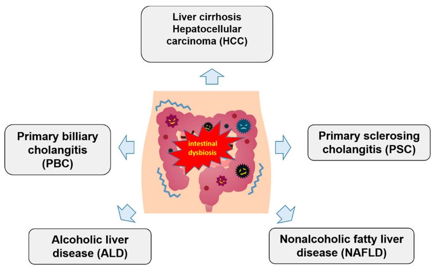
animals and humans showed that many liver diseases are linked to intestinal dysbiosis (Figure 1).
However, it is still not known if intestinal dysbiosis is a cause or a result of liver damage, the often
cited “chicken-egg” problem [14].Med. Sci. 2018, 6, 79 3 of 9
In recent years, a continuous increase in the prevalence and the mortality of liver diseases was
observed. The most common chronic liver diseases in western countries are alcoholic liver disease
(ALD) and nonalcoholic fatty liver disease (NAFLD) that can progress to liver cirrhosis and/or
hepatocellular carcinoma [15,16].
A better understanding of the pathophysiological connections among gut dysbiosis, the integrity
of the gut barrier, and the hepatic immune response to gut-derived factors is crucial for the development
of new therapies to treat chronic liver diseases or to at least prevent their progression and the
development of complications.
Figure 1. Schematic association between gut microbiota dysbiosis and chronic liver diseases.
4. Gut Microbiota and Alcoholic Liver Disease
Excessive alcohol consumption is a leading cause of chronic liver disease worldwide [17].
The stages of ALD are hepatic steatosis, steatohepatitis, and ultimately, liver cirrhosis and/or
hepatocellular carcinoma, often with an overlapping histology. It was suggested that not only the
quantity of alcohol, but also the interplay with genetic host and/or environmental factors may be
responsible for the development of hepatic injury in ALD [18].
Furthermore, experimental and clinical data imply that intestinal microbiota might also play
an important role in the pathogenesis of ALD [19]. This potential link between gut bacteria and hepatic
injury due to alcohol consumption was postulated more than 20 years ago. Adachi et al. showed for
the first time that antibiotics prevent liver injury in rats following long-term exposure to ethanol [20].
Casafont Morencos et al. demonstrated the presence of excessive or abnormal microbiota in the small
bowel (small intestinal bacterial overgrowth (SIBO)) in patients with alcoholic cirrhosis, and postulated
its role in the pathogenesis of ALD [21]. The recent meta-analysis by Shah et al. further supports this
observation showing higher SIBO prevalence in patients with chronic liver disease as compared to
a healthy control [22]. In addition, an imbalance between native Firmicutes and Bacteroidetes species
was described, with the former decreasing and the latter increasing [23].
The main characteristic of ALD is an increased gut permeability due to the direct toxic effect of
alcohol on the epithelial cells in the gastrointestinal tract and the decreased expression of tight-junction
proteins. This disruption of the intestinal barrier results in significant elevation of endotoxin plasma
levels that may cause hepatic injury (“autotoxic concept”). Furthermore, it was reported that individual
susceptibility to ALD may depend on the composition of the gut microbiota [24]. This hypothesis is
supported by animal studies showing the acceleration of alcohol-induced inflammation in germ-free
mice after transplantation of gut microbiota from alcoholic patients [25].Med. Sci. 2018, 6, 79 4 of 9
With the establishment of the dysfunctional intestinal microbiota as an important factor
responsible for the onset and maintenance of ALD, different strategies to modulate gut microbiota
were investigated in experimental and clinical studies. Most studies investigating the modulation
of gut microbiota in ALD focused on the role of different probiotics in the treatment of this
disease. The potential beneficial effects of probiotics on ALD include (1) quantitative and qualitative
improvement of gut microbiota composition (increase in Lactobacilli and Bifidobacteria, increase in
gut microbiota diversity); (2) improvement of liver function tests; (3) strengthening of gut-barrier
permeability; (4) decrease in pro-inflammatory cytokines like tumor necrosis factor α (TNFα) and
bacterial endotoxin levels in the blood; and (5) histologic improvement of liver steatosis and hepatic
inflammation [26,27]. Modulation of the gut microbiota appears to be a promising therapeutic strategy
in patients with ALD. Future therapy may employ engineered microbiota that decrease the permeability
of the gut barrier and reduce the release of pro-inflammatory cytokines in the gut. Further clinical
trials focusing on the role of gut microbiota in ALD are needed.
5. Gut Microbiota and Nonalcoholic Liver Disease
Nonalcoholic fatty liver disease represents the most common form of chronic liver disease that
is closely linked to metabolic syndrome and increased insulin resistance. In recent years, both the
incidence and prevalence of this disease substantially increased. NAFLD is recognized as a global
public health problem [28]. It is a very common disease, and interestingly, it especially affects patients
with obesity and diabetes type 2. NAFLD is characterized by the excessive accumulation of triglycerides
in hepatocytes in the absence of alcohol consumption (defined as less than 20 g and 30 g per day in
women and men, respectively). Similar to ALD, NAFLD may progress to steatohepatitis, liver cirrhosis,
and even hepatocellular carcinoma [29].
Multiple risk factors contribute to the pathogenesis of NAFLD, among them are genetic and dietary factors,
distribution of adipose tissue (in particular, the presence of visceral fat), and dysbiotic intestinal microbiota.
Recent studies indicate a strong involvement of gut microbiota in the pathophysiology of NAFLD [30].
An underlying intestinal dysbiosis can cause hepatic steatohepatitis via the following
pathophysiological events: (1) increase in hepatic inflammation leading to the development of
steatohepatitis (due to metabolic entotoxemia and TLR-mediated cytokine production); (2) increase in
insulin resistance; (3) hepatic de novo lipogenesis (steatosis); (4) change in bile-acid metabolism and
farnesoid X receptor (FXR) signaling; (5) change in gut-barrier permeability (“leaky gut”) and induction
of oxidative stress and inflammation by endogenous ethanol; and (6) decreased very-low-density
lipoprotein (VLDL) assembly and secretion due to changed choline metabolism in the dysbiotic gut.
Recent research indicates that compositional alterations of intestinal microbiota play a critical role in
the development of NAFLD. Typical compositional changes observed in NAFLD are an increase in
Bacteroidetes, a decrease in Firmucutes, and a rise in pro-inflammatory taxa such as Proteobacteria
and Enterobacteriaceae [31].
Loss of barrier integrity due to dysbiosis leads to a consequent increase in bacterial translocation
and to metabolic endotoxemia, which are key pathophysiological events for hepatic TLR system
activation, and thus, for a local hepatic and systemic inflammatory response.
Gut-microbiota-targeted therapies in NAFLD include the use of probiotics, a collection of bacteria
with beneficial effects on the host metabolism, and prebiotics, which are indigestible food ingredients
that selectively stimulate the growth of anti-inflammatory taxa and suppress that of pro-inflammatory
taxa. Recently, the effect of synbiotics, which are a combination of prebiotics and probiotics, on NAFLD
was investigated [32,33]. Studies with probiotic-based therapy in NAFLD were mainly performed in
animal models for this disease using high-fat diet (HFD)-induced fatty liver disease. Probiotic bacteria
like Lactobacillus or Bifidobacterium demonstrated some promising effects, e.g., attenuation of hepatic fat
accumulation, reduction of insulin resistance, limitation of oxidative and inflammatory liver damage,
and decrease in serum lipids [34]. Small human studies showed a beneficial effect of probiotics on liver
damage, along with a decrease in aminotransferases levels, and a reduction in total cholesterol andMed. Sci. 2018, 6, 79 5 of 9
low-density lipoprotein cholesterol (LDL-C) concentrations [35]. Similarly, some prebiotics limited
liver injury and reduced the levels of serum aminotrasfersases and insulin [36–38].
In the first randomized human study, Mofidi et al. showed positive effects of synbiotics on
fibrosis and the serum level of aminotransferases [39]. A recent meta-analysis by Khalesi et al. found
that probiotics and synbiotics given as supplements to nonalcoholic steatohepatitis (NASH) patients
improve the serum concentration of liver enzymes [40].
6. Gut Microbiota and Immune-Mediated Liver Diseases
There is accumulating evidence that dysfunctional gut microbiota might be implicated in the
pathogenesis of autoimmune diseases, primary billiary cirrhosis (PBC) and primary sclerosing
cholangitis (PSC). These two clinical entities represent chronic cholestatic liver diseases mediated
by the immune system. They are characterized by portal inflammation and slowly progress to liver
fibrosis and cirrhosis.
Since many years, it is postulated that these diseases may be triggered by unknown environmental
factors in genetically susceptible subjects. The gut microbiota could represent the missing link in
these pathogenetic events. Potential causes/trigger for the development of PSC/PBC are (1) intestinal
dysbiosis; (2) a change in bile-acid composition; (3) compositional alterations of billiary microbiota;
and (4) diverse unfavorable bacterial products (PAMPs) and metabolites [41].
In a recent publication, Tang et al. demonstrated reduced microbial species richness and a distinct
overall microbial diversity in PBC patients compared with healthy controls. PBC microbial dysbiosis
was characterized by altered abundances of 12 genera, and the dysbiosis was partially reversed during
ursodeoxycholic acid (UDCA) treatment [42].
7. Dysbiosis and Liver Cirrhosis
Liver cirrhosis is a severe liver disease characterized by loss of liver cells and irreversible scarring
of the liver. It represents an end-stage of all chronic liver diseases. Therefore, it is not surprising
that patients with liver cirrhosis have intestinal dysbiosis characterized by significant compositional
shifts toward pro-inflammatory bacterial taxa. Gut dysbiosis in liver cirrhosis is accompanied
by impaired gut-barrier function, pathological bacterial translocation and “immune exhaustion”.
Bacterial components and toxins (defined as endotoxemia) reaching the liver via a disrupted gut
barrier accelerate the already existing hepatic injury and increase the systemic inflammatory response.
These processes may then induce and promote portal hypertension and other complications of liver
cirrhosis like variceal bleeding or ascites [43].
Therapeutic strategies targeting the gut microbiota in liver cirrhosis comprise the use of antibiotics,
prebiotics, probiotics, synbiotics, and/or fecal microbiota transplantation [44]. The locally acting
antibiotic, rifaximine, was shown to reduce the incidence of hepatic encephalopathy (HE) and to
decrease the risk of variceal bleeding [45,46].
The role of probiotics in liver cirrhosis is controversially discussed. The evidence supporting a positive
effect of prebiotics and probiotics is rather weak due to small and/or heterogeneous studies. However,
it was demonstrated that some negative pathophysiologic aspects of liver cirrhosis can be reversed
by the use of probiotics. The following beneficial processes were observed: (1) reduction in arterial
ammonia concentration; (2) improvement in both overt and minimal hepatic encephalopathy; (3) decrease
in bacterial translocation and metabolic endotoxemia; (4) occurrence of anti-inflammatory effects and
reduction of pro-inflammatory cytokines such as TNFα; (5) reduction of systemic inflammatory reaction;
and (6) improvement in hemodynamic parameters in liver cirrhosis patients [47,48].
Probiotics show promising effects in patients with liver cirrhosis. However, studies investigating
their role in the treatment of liver cirrhosis have several limitations. The majority of studies used
probiotics for a short period of time (Med. Sci. 2018, 6, 79 6 of 9
well-designed randomized controlled trials are required to judge the exact role of probiotic-based
therapy in patient with liver cirrhosis.
Not long ago, researchers started investigating the role of fecal microbiota transplantation (FMT)
in liver cirrhosis. FMT is a procedure of transplantation of fecal bacteria from a healthy donor into
a patient’s gut for restoration of normal colonic flora. This method is gaining popularity because of its
high effectivity in the therapy of recurrent Clostridium difficile infection [49]. The first controlled study
with FMT (used as enema) in patients with liver cirrhosis demonstrated that this method significantly
improves cognitive functions of the patients and reduces their hospital stay due to a beneficial shift
in gut microbiota composition and an increased microbial diversity [50]. The FMT with antibiotic
pretreatment was well tolerated.
8. Targeting Gut Microbiota in Hepatocarcinogenesis
Hepatocellular carcinoma (HCC) is the fifth most common cancer and the third most common
cause of cancer-related mortality worldwide. There is emerging evidence that the gut microbiota may
have influence on the development and progression of this malignancy. The possible mechanisms
through which the gut microbiota is implicated in the pathogenesis of HCC involve increased
prevalence of pro-inflammatory bacteria due to the intestinal dysbiosis, increased barrier permeability
and bacterial translocation from the gut, direct damage of the liver cells by bacterial endotoxins,
and microbiota-mediated alterations in bile-acid metabolism.
The recent study by Ponziani et al. [51] showed significant changes in the gut microbiota profile
among patients with HCC. The stool of patients with HCC compared to healthy subjects showed
a significant decrease in alpha-diversity. The sequencing of 16S bacterial RNA showed in the stool of
HCC patients increased abundance of Bacteroides, Ruminococcus, Enterococcus, Phascolarctobacterium,
and Oscillospira and decreased abundance of Bifidobacteria and Blautia as compared with liver cirrhosis
without HCC. In particular, the deficiency of anti-inflammatory bacteria such as Bifidobacteria or Blautia
can enhance intestinal and liver inflammation and cause the progression of hepatocarcinogenesis.
All these observations indicate that modulation of the gut microbiota with probiotics in patients
with liver cirrhosis and at increased risk for HCC could decrease intestinal permeability and inhibit
microbiota-mediated process of carcinogenesis in the liver. In an animal model, Li et al. demonstrated
that the probiotic mixture, Prohep, composed of Lactobacillus rhamnosus GG (LGG), viable Escherichia coli
Nissle 1917 (EcN), and heat-inactivated VSL#3 (1:1:1), caused a significant reduction (almost 40%) of
HCC growth. The authors postulate that the reduction in the recruitment of T helper 17 (Th17) cells
from the gut to the tumor site and decreased plasma levels of pro-angiogenic interleukin 17 (IL-17) may
be consequences of the probiotic treatment and responsible for the inhibition of tumor growth [52].
In another animal model of carcinogen-induced hepatocarcinogenesis, the reduction of circulating
bacterial LPS using antibiotics prevented HCC growth [53].
These studies indicate that manipulation of the gut microbiota with anti-inflammatory bacteria
may prevent bacterial translocation with endotoxin absorption and development of hepatocellular
carcinoma in patients who are at risk of developing this malignancy (such as patients with liver
cirrhosis or nonalcoholic hepatic steatohepatitis). Further studies in humans should shed more light
on the role of probiotics in liver carcinogenesis.
9. Conclusions
In summary, intestinal dysbiosis is observed in many chronic liver diseases (e.g., NAFLD, ALD,
immune-mediated liver diseases, liver cirrhosis and hepatic carcinogenesis). There is increasing
evidence for an adverse role of intestinal dysbiosis in the pathogenesis and progression of these
diseases. Amelioration of the dysbiosis through the use of prebiotics, probiotics, and fecal microbiota
transplantation improves the gut-barrier function and appears to be a promising new approach to
managing chronic liver diseases.Med. Sci. 2018, 6, 79 7 of 9
Funding: This research received no external funding.
Conflicts of Interest: The authors declare no conflicts of interest.
References
1. Jandhyala, S.M.; Talukdar, R.; Subramanyam, C.; Vuyyuru, H.; Sasikala, M.; Reddy, D.N. Role of the normal
gut microbiota. World J. Gastroenterol. 2015, 21, 8787–8803. [CrossRef] [PubMed]
2. Round, J.L.; Mazmanian, S.K. The gut microbiota shapes intestinal immune responses during health and
disease. Nat. Rev. Immunol. 2009, 9, 313–323. [CrossRef] [PubMed]
3. Park, W. Gut microbiomes and their metabolites shape human and animal health. J. Microbial. 2018, 56, 151–153.
[CrossRef] [PubMed]
4. Feng, Q.; Chen, W.D.; Wang, Y.D. Gut Microbiota: An Integral Moderator in Health and Disease.
Front. Microbial. 2018, 9, 151. [CrossRef] [PubMed]
5. Bhattarai, Y.; Muniz Pedrogo, D.A.; Kashyap, P.C. Irritable bowel syndrome: A gut microbiota-related
disorder? Am. J. Physiol. Gastrointest. Liver Physiol. 2017, 312, G52–G62. [CrossRef] [PubMed]
6. Sommer, F.; Backhed, F. The gut microbiota—Masters of host development and physiology. Nat. Rev. Microbiol.
2013, 11, 227–238. [CrossRef] [PubMed]
7. Schroeder, B.O.; Backhed, F. Signals from the gut microbiota to distant organs in physiology and disease.
Nat. Med. 2016, 22, 1079–1089. [CrossRef] [PubMed]
8. Clemente, J.C.; Ursell, L.K.; Parfrey, L.W.; Knight, R. The impact of the gut microbiota on human health:
An integrative view. Cell 2012, 148, 1258–1270. [CrossRef] [PubMed]
9. Bretin, A.; Gewirtz, A.T.; Chassaing, B. Microbiota and Metabolism—What’s New in 2018. Am. J. Physiol.
Endocrinol. Metab. 2018. [CrossRef] [PubMed]
10. O’Hara, P.; Karlsen, T.H.; LaRusso, N.F. Cholangiocytes and the environment in primary sclerosing
cholangitis: Where is the link? Gut 2017, 66, 1873–1877. [CrossRef] [PubMed]
11. Ponziani, F.R.; Zocco, M.A.; Cerrito, L.; Gasbarrini, A.; Pompili, M. Bacterial translocation in patients with liver
cirrhosis: Physiology, clinical consequences, and practical implications. Expert Rev. Gastroenterol. Hepatol. 2018,
1–16. [CrossRef] [PubMed]
12. Carotti, S.; Guarino, M.P.L.; Vespasiani-Gentilucci, U.; Morini, S. Starring role of toll-like receptor-4 activation
in the gut-liver axis. World J. Gastrointest. Pathophysiol. 2015, 6, 99–109. [CrossRef] [PubMed]
13. Brandl, K.; Kumar, V.; Eckmann, L. Gut-liver axis at the frontier of host-microbial interactions. Am. J. Physiol.
Gastrointest. Liver Physiol. 2017, 312, G413–G419. [CrossRef] [PubMed]
14. Betrapally, N.S.; Gillevet, P.M.; Bajaj, J.S. Gut microbiome and liver disease. Transl. Res. J. Lab. Clin. Med.
2017, 179, 49–59. [CrossRef] [PubMed]
15. Mathurin, P.; Lucey, M.R. Alcohol, liver disease, and transplantation: Shifting attitudes and new
understanding leads to changes in practice. Curr. Opin. Organ Transplant. 2018, 23, 175–179. [CrossRef]
[PubMed]
16. Younossi, Z.; Stepanova, M.; Ong, J.P.; Jacobson, I.M.; Bugianesi, E.; Duseja, A.; Eguchi, Y.; Wong, V.W.;
Negro, F.; Yilmaz, Y.; et al. Non-alcoholic Steatohepatitis is the Fastest Growing Cause of Hepatocellular
Carcinoma in Liver Transplant Candidates. Clin. Gastroenterol. Hepatol. 2018. [CrossRef] [PubMed]
17. Pimpin, L.; Cortez-Pinto, H.; Negro, F.; Corbould, E.; Lazarus, J.V.; Webber, L.; Sheron, N. Burden of liver
disease in Europe: Epidemiology and analysis of risk factors to identify prevention policies. J. Hepatol. 2018.
[CrossRef] [PubMed]
18. Parker, R.; Kim, S.J.; Gao, B. Alcohol, adipose tissue and liver disease: Mechanistic links and clinical
considerations. Nat. Rev. Gastroenterol. Hepatol. 2018, 15, 50–59. [CrossRef] [PubMed]
19. Cassard, A.M.; Ciocan, D. Microbiota, a key player in alcoholic liver disease. Clin. Mol. Hepatol. 2018, 24, 100–107.
[CrossRef] [PubMed]
20. Adachi, Y.; Moore, L.E.; Bradford, B.U.; Gao, W.; Thurman, R.G. Antibiotics prevent liver injury in rats
following long-term exposure to ethanol. Gastroenterology 1995, 108, 218–224. [CrossRef]
21. Morencos, F.C.; De las Heras Castano, G.; Ramos, L.M.; Arias, M.J.L.; Ledesma, F.; Romero, F.P. Small bowel
bacterial overgrowth in patients with alcoholic cirrhosis. Dig. Dis. Sci. 1996, 41, 552–556. [CrossRef]Med. Sci. 2018, 6, 79 8 of 9
22. Shah, A.; Shanahan, E.; Macdonald, G.A.; Fletcher, L.; Ghasemi, P.; Morrison, M.; Jones, M.; Holtmann, G.
Systematic Review and Meta-Analysis: Prevalence of Small Intestinal Bacterial Overgrowth in Chronic Liver
Disease. Semin. Liver Dis. 2017, 37, 388–400. [CrossRef] [PubMed]
23. Quigley, E.M.; Stanton, C.; Murphy, E.F. The gut microbiota and the liver. Pathophysiological and clinical
implications. J. Hepatol. 2013, 58, 1020–1027. [CrossRef] [PubMed]
24. Sanduzzi Zamparelli, M.; Rocco, A.; Compare, D.; Nardone, G. The gut microbiota: A new potential driving
force in liver cirrhosis and hepatocellular carcinoma. United Eur. Gastroenterol. J. 2017, 5, 944–953. [CrossRef]
[PubMed]
25. Lopis, M.; Cassard, A.M.; Wrzosek, L.; Boschat, L.; Bruneau, A.; Ferrere, G.; Puchois, V.; Martin, J.C.;
Lepage, P.; Le Roy, T.; et al. Intestinal microbiota contributes to individual susceptibility to alcoholic liver
disease. Gut 2016, 65, 830–839. [CrossRef] [PubMed]
26. Hong, M.; Han, D.H.; Hong, J.; Kim, D.J.; Suk, K.T. Are Probiotics Effective in Targeting Alcoholic Liver
Diseases? Probiotics Antimicrob. Proteins 2018. [CrossRef] [PubMed]
27. Li, F.; Duan, K.; Wang, C.; McClain, C.; Feng, W. Probiotics and Alcoholic Liver Disease: Treatment and
Potential Mechanisms. Gastroenterol. Res. Pract. 2016, 5491465. [CrossRef] [PubMed]
28. Konturek, P.C. Therapy of nonalcoholic steatohepatitis: The proven and the new. MMW Fortschritte der
Medizin 2018, 160, 50–54. [CrossRef] [PubMed]
29. Tilg, H.; Moschen, A.R. Evolution of inflammation in nonalcoholic fatty liver disease: The multiple parallel
hits hypothesis. Hepatology 2010, 52, 1836–1846. [CrossRef] [PubMed]
30. Doulberis, M.; Kotronis, G.; Gialamprinou, D. Non-alcoholic fatty liver disease: An update with special
focus on the role of gut microbiota. Metab. Clin. Exp. 2017, 71, 182–197. [CrossRef] [PubMed]
31. De Faria Ghetti, F.; Oliveira, D.G.; de Oliveira, J.M.; de Castro, L.E.V.V.; Cesar, D.E.; Moreira, A.P.B. Influence of
gut microbiota on the development and progression of nonalcoholic steatohepatitis. Eur. J. Nutr. 2018, 57, 861–876.
[CrossRef] [PubMed]
32. Ma, J.; Zhou, Q.; Li, H. Gut Microbiota and Nonalcoholic Fatty Liver Disease: Insights on Mechanisms and
Therapy. Nutrients 2017, 9, 1124. [CrossRef] [PubMed]
33. Paolella, G.; Mandato, C.; Pierri, L.; Poeta, M.; Di Stasi, M.; Vajro, P. Gut-liver axis and probiotics: Their role
in non-alcoholic fatty liver disease. World J. Gastroenterol. 2014, 20, 15518–15531. [CrossRef] [PubMed]
34. Xu, R.Y.; Wan, Y.P.; Fang, Q.Y.; Lu, W.; Cai, W. Supplementation with probiotics modifies gut flora and
attenuates liver fat accumulation in rat nonalcoholic fatty liver disease model. J. Clin. Biochem. Nutr. 2012,
50, 72–77. [CrossRef] [PubMed]
35. Wai-Sun Wong, V.; Wong, G.L.H.; Chim, A.M.L.; Chu, W.C.W.; Yeung, D.K.W.; Li, K.C.T.; Chan, H.L.Y. Treatment
of nonalcoholic steatohepatitis with probiotics. A proof-of-concept study. Ann. Hepatol. 2013, 12, 256–262.
36. Parnell, J.A.; Raman, M.; Rioux, K.P.; Reimer, R.A. The potential role of prebiotic fibre for treatment and
management of non-alcoholic fatty liver disease and associated obesity and insulin resistance. Liver Int. 2012,
32, 701–711. [CrossRef] [PubMed]
37. Bomhof, M.R.; Parnell, J.A.; Ramay, H.R.; Crotty, P.; Rioux, K.P.; Probert, C.S.; Jayakumar, S.; Raman, M.;
Reimer, R.A. Histological improvement of non-alcoholic steatohepatitis with a prebiotic: A pilot clinical trial.
Eur. J. Nutr. 2018. [CrossRef] [PubMed]
38. Daubioul, C.A.; Horsmans, Y.; Lambert, P.; Danse, E.; Delzenne, N.M. Effects of oligofructose on glucose and
lipid metabolism in patients with nonalcoholic steatohepatitis: Results of a pilot study. Eur. J. Clin. Nutr.
2005, 59, 723–726. [CrossRef] [PubMed]
39. Mofidi, F.; Poustchi, H.; Yari, Z.; Nourinayyer, B.; Merat, S.; Sharafkhah, M.; Malekzadeh, R.; Hekmatdoost, A.
Synbiotic supplementation in lean patients with non-alcoholic fatty liver disease: A pilot, randomised,
double-blind, placebo-controlled, clinical trial. Br. J. Nutr. 2017, 117, 662–668. [CrossRef] [PubMed]
40. Khalesi, S.; Johnson, D.W.; Campbell, K.; Williams, S.; Fenning, A.; Saluja, S.; Irwin, C. Effect of probiotics
and synbiotics consumption on serum concentrations of liver function test enzymes: A systematic review
and meta-analysis. Eur. J. Nutr. 2017. [CrossRef] [PubMed]
41. Mattner, J. Impact of Microbes on the Pathogenesis of Primary Biliary Cirrhosis (PBC) and Primary Sclerosing
Cholangitis (PSC). Int. J. Mol. Sci. 2016, 17, 1864. [CrossRef] [PubMed]
42. Tang, R.; Wei, Y.; Li, Y.; Chen, W.; Chen, H.; Wang, Q.; Yang, F.; Miao, Q.; Xiao, X.; Zhang, H.; et al.
Gut microbial profile is altered in primary biliary cholangitis and partially restored after UDCA therapy. Gut
2018, 67, 534–541. [CrossRef] [PubMed]Med. Sci. 2018, 6, 79 9 of 9
43. Acharya, C.; Sahingur, S.E.; Bajaj, J.S. Microbiota, cirrhosis, and the emerging oral-gut-liver axis. JCI Insight
2017, 2, e94416. [CrossRef] [PubMed]
44. Woodhouse, C.A.; Patel, V.C.; Singanayagam, A.; Shawcross, D.L. Review article: The gut microbiome as
a therapeutic target in the pathogenesis and treatment of chronic liver disease. Aliment. Pharmacol. Ther.
2018, 47, 192–202. [CrossRef] [PubMed]
45. Bass, N.M.; Mullen, K.D.; Sanyal, A.; Poordad, F.; Neff, G.; Leevy, C.B.; Sigal, S.; Sheikh, M.Y.; Beavers, K.;
Frederick, T.; et al. Rifaximin treatment in hepatic encephalopathy. N. Engl. J. Med. 2010, 362, 1071–1081.
[CrossRef] [PubMed]
46. Vlachogiannakos, J.; Viazis, N.; Vasianopoulou, P.; Vafiadis, I.; Karamanolis, D.G.; Ladas, S.D.
Long-term administration of rifaximin improves the prognosis of patients with decompensated alcoholic
cirrhosis. J. Gastroenterol. Hepatol. 2013, 28, 450–455. [CrossRef] [PubMed]
47. Dhiman, R.K.; Rana, B.; Agrawal, S.; Garg, A.; Chopra, M.; Thumburu, K.K.; Khattri, A.; Malhotra, S.;
Duseja, A.; Chawla, Y.K. Probiotic VSL#3 reduces liver disease severity and hospitalization in patients with
cirrhosis: A randomized, controlled trial. Gastroenterology 2014, 147, 1327–1337. [CrossRef] [PubMed]
48. Altamirano-Barrera, A.; Uribe, M.; Chávez-Tapia, N.C.; Nuño-Lámbarri, N. The role of the gut microbiota in
the pathology and prevention of liver disease. J. Nutr. Biochem. 2018, 60, 1–8. [CrossRef] [PubMed]
49. Konturek, P.C.; Haziri, D.; Brzozowski, T.; Hess, T.; Heyman, S.; Kwiecien, S.; Konturek, S.J.; Koziel, J.
Emerging role of fecal microbiota therapy in the treatment of gastrointestinal and extra-intestinal diseases.
J. Physiol. Pharmacol. 2015, 66, 483–491. [PubMed]
50. Bajaj, J.S.; Kassam, Z.; Fagan, A.; Gavis, E.A.; Liu, E.; Cox, I.J.; Kheradman, R.; Heuman, D.; Wang, J.;
Gurry, T.; et al. Fecal microbiota transplant from a rational stool donor improves hepatic encephalopathy:
A randomized clinical trial. Hepatology 2017, 66, 1727–1738. [CrossRef] [PubMed]
51. Ponziani, F.R.; Bhoori, S.; Castelli, C.; Putignani, L.; Rivoltini, L.; Del Chierico, F.; Sanguinetti, M.; Morelli, D.;
Paroni Sterbini, F.; Petito, V.; et al. Hepatocellular carcinoma is associated with gut microbiota profile and
inflammation in nonalcoholic fatty liver disease. Hepatology 2018. [CrossRef] [PubMed]
52. Li, J.; Sung, C.Y.J.; Lee, N.; Ni, Y.; Pihlajamäki, J.; Panagiotou, G.; El-Nezami, H. Probiotics modulated gut
microbiota suppresses hepatocellular carcinoma growth in mice. Proc. Natl. Acad Sci. USA 2016, 113, E1306–E1315.
[CrossRef] [PubMed]
53. Yu, L.X.; Yan, H.X.; Liu, Q.; Yang, W.; Wu, H.P.; Dong, W.; Tang, L.; Lin, Y.; He, Y.Q.; Zou, S.S.; et al.
Endotoxin accumulation prevents carcinogen-induced apoptosis and promotes liver tumorigenesis in
rodents. Hepatology 2010, 52, 1323–1333. [CrossRef] [PubMed]
© 2018 by the authors. Licensee MDPI, Basel, Switzerland. This article is an open access
article distributed under the terms and conditions of the Creative Commons Attribution
(CC BY) license (http://creativecommons.org/licenses/by/4.0/).You can also read