Fremanezumab-A Humanized Monoclonal Anti-CGRP Antibody-Inhibits Thinly Myelinated (A ) But Not Unmyelinated (C) Meningeal Nociceptors
←
→
Page content transcription
If your browser does not render page correctly, please read the page content below
The Journal of Neuroscience, November 1, 2017 • 37(44):10587–10596 • 10587

Neurobiology of Disease

Fremanezumab—A Humanized Monoclonal Anti-CGRP
Antibody—Inhibits Thinly Myelinated (A␦) But Not
Unmyelinated (C) Meningeal Nociceptors
Agustin Melo-Carrillo,1,2 Andrew M. Strassman,1,2 Rony-Reuven Nir,1,2 XAaron J. Schain,1,2 X Rodrigo Noseda,1,2
X Jennifer Stratton,3 and X Rami Burstein1,2
1Department of Anesthesia, Critical Care & Pain Medicine, Beth Israel Deaconess Medical Center, Boston, Massachusetts 02215, 2Harvard Medical School,
Boston, Massachusetts 02115, and 3TEVA Biologics, Redwood City, California 94061

Calcitonin gene-related peptide (CGRP), the most abundant neuropeptide in primary afferent sensory neurons, is strongly implicated in
the pathophysiology of migraine headache, but its role in migraine is still equivocal. As a new approach to migraine treatment, humanized
anti-CGRP monoclonal antibodies (CGRP-mAbs) were developed to reduce the availability of CGRP, and were found effective in reducing
the frequency of chronic and episodic migraine. We recently tested the effect of fremanezumab (TEV-48125), a CGRP-mAb, on the activity
of second-order trigeminovascular dorsal horn neurons that receive peripheral input from the cranial dura, and found a selective inhibition of
high-threshold but not wide-dynamic range class of neurons. To investigate the basis for this selective inhibitory effect, and further explore the
mechanism of action of CGRP-mAbs, we tested the effect of fremanezumab on the cortical spreading depression-evoked activation of mecha-
nosensitive primary afferent meningeal nociceptors that innervate the cranial dura, using single-unit recording in the trigeminal ganglion of
anesthetized male rats. Fremanezumab pretreatment selectively inhibited the responsiveness of A␦ neurons, but not C-fiber neurons, as re-
flected in a decrease in the percentage of neurons that showed activation by cortical spreading depression. These findings identify A␦ meningeal
nociceptors as a likely site of action of fremanezumab in the prevention of headache. The selectivity in its peripheral inhibitory action may partly
account for fremanezumab’s selective inhibition of high-threshold, as a result of a predominant A-␦ input to high-threshold neurons, but not
wide dynamic-range dorsal horn neurons, and why it may not be effective in all migraine patients.
Key words: calcitonin gene-related peptide; cortical spreading depression; headache; migraine; pain; trigeminovascular

      Significance Statement
      Recently, we reported that humanized CGRP monoclonal antibodies (CGRP-mAbs) prevent activation and sensitization of high-
      threshold (HT) but not wide-dynamic range trigeminovascular neurons by cortical spreading depression (CSD). In the current
      paper, we report that CGRP-mAbs prevent the activation of A␦ but not C-type meningeal nociceptors by CSD. This is the first
      identification of an anti-migraine drug that appears to be selective for A␦-fibers (peripherally) and HT neurons (centrally). As the
      main CGRP-mAb site of action appears to be situated outside the brain, we conclude that the initiation of the headache phase of
      migraine depends on activation of meningeal nociceptors, and that for selected patients, activation of the A␦-HT pain pathway
      may be sufficient for the generation of headache perception.

Introduction                                                                                                              dorsal root and trigeminal ganglia (O’Connor and van der Kooy,
Calcitonin gene-related peptide (CGRP) is the most abundant                                                               1988; McCarthy and Lawson, 1990; Price and Flores, 2007). A
neuropeptide present in primary afferent sensory neurons in                                                               large body of evidence in both man and animals supports an
                                                                                                                          important role for CGRP in the pathophysiology of migraine
                                                                                                                          headache (Hansen and Ashina, 2014; Karsan and Goadsby, 2015;
Received Aug. 4, 2017; revised Sept. 5, 2017; accepted Sept. 13, 2017.
   Author contributions: A.M.-C., A.M.S., A.J.S., J.S., and R.B. designed research; A.M.-C., A.M.S., and R.B. performed   Russo, 2015). The mechanism of CGRP’s involvement in mi-
research; A.M.-C., A.M.S., R.N., and R.B. contributed unpublished reagents/analytic tools; A.M.-C., A.M.S., R.-R.N.,
R.N., and R.B. analyzed data; A.M.-C., A.M.S., R.-R.N., and R.B. wrote the paper.
   This work was supported by a grant from TEVA Pharmaceutical Industries; NIH Grants R37-NS079678, RO1                      Correspondence should be addressed to Rami Burstein, Beth Israel Deaconess Medical Center, CLS-649, 3 Black-
NS069847, and RO1 NS094198 (R.B.); and a grant from R. Chemers Neustein (A.S.).                                           fan Circle, Boston, MA 02115. E-mail: rburstei@bidmc.harvard.edu.
   Commercial interest: TEVA Pharmaceutical Industries holds the patent for treating episodic and chronic migraine           DOI:10.1523/JNEUROSCI.2211-17.2017
with Fremanezumab and funded parts of the study. J.S. is an employee of TEVA and R.B. is a consultant to TEVA.            Copyright © 2017 the authors 0270-6474/17/3710587-10$15.00/0
10588 • J. Neurosci., November 1, 2017 • 37(44):10587–10596 Melo-Carrillo et al. • Inhibition of Meningeal Nociceptors by CGRP-mAb
graine is not well understood, but one line of research has focused to allow insertion of a glass micropipette for recording of electrocortico-
on its role in the physiology of neurons that supply the sensory gram activity. A second burr hole was drilled ⬃5 mm caudal to the first
innervation of the cranial meninges, a sensory pathway that has one (⬃5 mm rostral to the transverse sinus), for induction of cortical
been critically implicated in the generation of migraine headache spreading depression by pinprick (brief insertion of a metal microelec-
(Moskowitz, 1984; Strassman et al., 1996; Burstein et al., 2015). trode into the cortex). The dura exposed by these burr holes was not
CGRP is present in a substantial subpopulation of the meningeal removed. All of the exposed dura was kept moist using a modified syn-
thetic interstitial fluid (in mM: 135 NaCl, 5 KCl, 1 MgCl2, 5 CaCl2, 10
sensory neurons (O’Connor and van der Kooy, 1988; Tsai et al.,
glucose, and 10 HEPES, pH 7.2). For neural recording from the left
1988; Uddman et al., 1989; Keller and Marfurt, 1991; Messlinger
trigeminal ganglion, a craniotomy was made on the right (contralateral)
et al., 1993; Edvinsson et al., 2001), and can be released from both side to allow the microelectrodes to be advanced through the contralat-
the peripheral endings in the meninges (Zagami et al., 1990; Ku- eral cortex to reach the ganglion, using an angled approach. This was
rosawa et al., 1995; Eltorp et al., 2000) and the central endings in done to avoid damage to the ipsilateral cortex. The craniotomy was made
the medullary and upper cervical dorsal horn (Storer et al., 2004; to allow electrode insertions into the right cortex covering an area of
Fischer et al., 2005). Peripherally, CGRP produces meningeal va- 1–3 mm caudal to bregma, and 1.5–3 mm lateral. The dura covering this
sodilatation (McCulloch et al., 1986; Uddman et al., 1986; Edvin- area of cortex was removed to allow microelectrode insertion.
sson et al., 1987, 1998; Kurosawa et al., 1995; Williamson et al., Neural recording. A glass micropipette filled with 0.9% saline (⬃1 M⍀,
1997), and thus contributes to meningeal neurogenic inflamma- 7 m tip) was inserted through the rostral burr hole 500 m below the
tion. The actions of CGRP as a neuromodulator when released cortical surface for electrocorticogram recording. A platinum-coated
from the central terminals of meningeal sensory neurons has not tungsten microelectrode (impedance 150 –300 k⍀) was advanced into
yet been well elucidated, but there is evidence from studies at the trigeminal ganglion for single-unit recording. To reach the ganglion
spinal levels that CGRP contributes to sensitization of dorsal via a contralateral approach, the electrode was angled medially 21° and
horn neurons (Sun et al., 2004a,b; Han et al., 2005; Bird et al., was advanced through the contralateral cortex (see above for coordi-
2006; Adwanikar et al., 2007). Among the most well documented nates). Dural afferent neurons in the ganglion were identified by their
constant latency response to single shock stimulation delivered to the
effects of the anti-migraine agent sumatriptan is inhibition of
dura overlying the ipsilateral transverse sinus with a bipolar stimulating
neuropeptide release from both the peripheral and central termi-
electrode (0.5 ms pulse, 5 mA, 0.75 Hz). The response latency was used to
nals of meningeal sensory neurons (Messlinger et al., 1998; Dur- calculate conduction velocity (c.v.), based on a conduction distance to
ham and Russo, 1999; Eltorp et al., 2000). the trigeminal ganglion of 12 mm. Neurons were classified as either
As a new approach to migraine treatment, humanized CGRP C-units (c.v. ⱕ1.5 ms) or A␦-units (c.v. 1.5–5 m/s). Neurons with con-
monoclonal antibodies (CGRP-mAbs) have been developed as duction velocity ⬎5 m/s were not studied. Spike 2 software (CED) was
therapeutic agents, and were found to reduce the frequency of used for acquisition and waveform discrimination of the electrically
attacks in chronic migraine patients (Dodick et al., 2014a,b; Bigal evoked spikes, and for off-line analysis.
et al., 2015a,b; Sun et al., 2016). Recently, we examined the effect Mechanical receptive fields of dural afferents were initially identified
of fremanezumab, a CGRP-mAb, on the central neurons in the by probing the dura with blunt forceps and von Frey hairs. Only neurons
meningeal sensory pathway, and found a selective inhibition of for which a mechanical receptive field could be identified were selected
the high-threshold (HT) but not the wide-dynamic range (WDR) for study.
class of dorsal horn neurons (Melo-Carrillo et al., 2017). This Experimental paradigm. Ongoing discharge activity was recorded
selective action is presumed to be through a peripheral site of continuously throughout the experiment. After recording of baseline
action, since the antibodies do not cross the blood– brain barrier. spontaneous activity an intravenous infusion was made of either the
Such a selective inhibition of HT neurons has not been reported humanized monoclonal anti-CGRP antibody fremanezumab (TEVA
Pharmaceutical Industries) or the corresponding human IgG2 isotype
previously, and it is not known what type of peripheral action
control antibody (isotype-conAb), both were given at a dose of 30 mg/kg
could produce such a selective central inhibition. To investigate
in saline. Four hours after drug administration, a single wave of CSD was
this question, we examined the effect of fremanezumab on the induced by briefly inserting a metal microelectrode into the cortex
evoked activity of mechanosensitive primary afferent neurons in through the caudal burr hole, and spontaneous activity was recorded
the trigeminal ganglion that innervate the cranial dura. Neuronal continuously for another 60 min.
activation was evoked by cortical spreading depression (CSD), a Data analysis. The 30 min period before CSD induction (3.5 h after
cortical event that has been implicated in the initiation of mi- drug administration) was used as the baseline for determination of CSD
graine attacks (Bolay and Moskowitz, 2005) and that is effective responses. A neuron was considered to have a response to CSD if its firing
in activating both first- and second-order neurons in the menin- rate increased above baseline by 2 SD for a period at least 10 min. Calcu-
geal sensory pathway (Zhang et al., 2010, 2011). lation of response magnitudes were based on two 10 min periods where
mean firing rates were highest during the two 30 min periods before
Materials and Methods (baseline firing rate) and after occurrence of CSD (post-CSD firing rate).
The latency from CSD to the onset of response, as well as response dura-
Surgical preparation. Experiments were approved by the Beth Israel Dea-
coness Medical Center and Harvard Medical School standing commit- tion were calculated for each neuron.
tees on animal care, and were in accordance with the U.S. National Baseline firing rates were compared between (1) the isotype (control)
Institutes of Health Guide for the Care and Use of Laboratory Animals. and CGRP-mAb groups, and (2) A␦ and C fibers using the nonparamet-
Male Sprague-Dawley rats (225–325 g) were anesthetized with urethane ric Mann–Whitney analysis. Firing rates before and after CSD were com-
(1.5 g/kg, i.p.), and treated with atropine (0.4 ml, i.p.) to reduce intraoral pared using the nonparametric Wilcoxon analysis. Latency and duration
secretions. The femoral vein was cannulated for drug infusion, an endo- of CSD responses were compared based on Group (isotype/CGRP-mAb)
tracheal tube was implanted, and the rat was mounted in a stereotaxic and neuron type (A␦/C), comprising four subgroups, using the nonpara-
apparatus. Core temperature was maintained at 37°C using a feedback metric Kruskal–Wallis test. The association between responsiveness to
controlled heating pad, and pO2 was maintained ⬎98% by O2 inhalation CSD (yes/no) and Group (isotype/CGRP-mAb) per neuron type (A␦/C)
through the endotracheal tube. A craniotomy was made to expose the left were tested using 2 tests. All analyses were performed using Statistical
transverse sinus, to allow electrical and mechanical stimulation of dural Package for the Social Sciences (SPSS v22, IBM). Continuous variables
primary afferent nociceptors. A small burr hole was drilled in the calvar- are presented as median (interquartile range: 25th–75th percentile). Sta-
ium on the left side, at ⬃1 mm rostral to bregma and ⬃1.5–2 mm lateral, tistical significance was set at p ⬍ 0.05.Melo-Carrillo et al. • Inhibition of Meningeal Nociceptors by CGRP-mAb J. Neurosci., November 1, 2017 • 37(44):10587–10596 • 10589
Table 1. Summary of results
Neurons (N ⫽ 49)
Isotype (n ⫽ 25) CGRP-mAb (n ⫽ 24)
A␦ (n ⫽ 9) C (n ⫽ 16) A␦ (n ⫽ 10) C (n ⫽ 14)
Activation by CSD Yes (n ⫽ 6) No (n ⫽ 3) Yes (n ⫽ 7) No (n ⫽ 9) Yes (n ⫽ 2) No (n ⫽ 8) Yes (n ⫽ 8) No (n ⫽ 6)
Baseline, mean spikes/s 0.79 (1.4) 0.49 (0.8) 0.53 (0.6) 0.30 (0.5) 0.03 (0) 0.44 (1.0) 0.32 (1.0) 1.70 (1.9)
Post-CSD, mean spikes/s 1.29 (2.1)* 0.33 (2.0) 1.10 (2.0)* 0.29 (0.5) 0.35 (0.1) 0.44 (1.1) 1.07 (2.0)* 0.96 (1.8)
Latency, min 6 (7.7) NA 5 (4.0) NA 5, 8a NA 5 (3.7) NA
Duration, min 30 (26.2) NA 60 (30.0) NA 20, 60a NA 60 (7.5) NA
Data are represented as median (IQR). NA, Not applicable.
*Statistically significant difference compared to baseline ( p ⬍ 0.05).
a
Analyses were not carried out due to small sample (n ⫽ 2); results are displayed per neuron (n ⫽ 2).
Results group remained unchanged after the occurrence of CSD (base-
Single-unit recordings were obtained from 19 A␦- and 30 C-class line: 0.44 (1.0)[median (IQR)]; post-CSD: 0.44 (1.14) [median
meningeal nociceptors in the trigeminal ganglion that were iden- (IQR)]; Z ⫽ ⫺0.98, p ⫽ 0.326; Fig. 2E–G).
tified by their response to electrical and mechanical stimulation Isotype versus CGRP-mAb
of the dura overlying the ipsilateral transverse sinus. There was a significant association between responsiveness to
The effect of CSD on neuronal discharge was tested following CSD and Group such that of a total of eight CSD activated neu-
intravenous infusion of either CGRP-mAb (n ⫽ 10 A␦ and 14 rons, 6 (75%) were treated with isotype and only 2 (25%) were
C-fibers) or the corresponding isotype antibody (n ⫽ 9 A␦- and 16 treated with CGRP-mAb. Additionally, of 9 isotype-treated neu-
C-fibers). CSD was induced by pinprick 4 h after the drug infusion. rons, 6 (66.6%) were and 3 (33.3%) were not activated by CSD.
Before CSD, neurons displayed firing rates of 0.37 (1.47) [median As to the CGRP-mAb-treated group, of 10 neurons, 2 (20%) were
(IQR)] in CGRP-MAb-treated animals, and 0.45 (0.69) [median and 8 (80%) were not activated by CSD ((1)
2
⫽ 4.23, p ⫽ 0.040)].
(IQR)] in the isotype-treated animals (Z ⫽ ⫺0.13, p ⫽ 0.897). There
was no significant difference in the baseline firing rates between CSD effects on C-fibers
CGRP-MAb-treated A␦ neurons 0.08 (0.95) [median (IQR)] and Isotype-treated group
the isotype-treated A␦ neurons 0.79 (1.37) [median (IQR); Z ⫽ In the isotype-treated group (Fig. 3; Table 1), CSD activated 7/16
⫺1.72, p ⫽ 0.095]. Similarly, the CGRP-mAb-treated C-fibers (43%) C-type meningeal nociceptors; i.e., the firing rate of each
0.46 (1.64) [median (IQR)] were comparable to the isotype- of these neurons increased by ⬎2 SD after occurrence of CSD
treated group 0.37 (0.51) [median (IQR); Z ⫽ ⫺1.43, p ⫽ 0.154]. compared with their baseline firing (Fig. 3A–C). These neurons
Following CSD, according to the aforementioned criteria, an demonstrated a baseline firing rate of 0.53 (0.61)[median (IQR)]
increase in firing rate was observed in 10/24 (41%) neurons in that increased to 1.1 (2.0)[median (IQR); Z ⫽ ⫺2.36, p ⫽ 0.018]
CGRP-MAb-treated animals and 13/25 (52%) neurons in isotype- after occurrence of CSD (Fig. 3B). Their activation started 5.0
treated animals (Table 1). (4.0) [median (IQR)] min after occurrence of CSD (range: 3–25
min; Fig. 3D) and lasted for 60.0 (30.0) [median (IQR)] min
CSD effects on A␦-fibers (range:15– 60 min; Fig. 3E). No significant change was observed
Isotype-treated group in the activity level of the remaining nine neurons after the CSD
In the isotype-treated group (Fig. 1; Table 1), CSD activated 6/9 (baseline: 0.3 (0.56)[median (IQR)]; post-CSD: 0.29 (0.53)[me-
(66%) A␦-meningeal nociceptors; i.e., the firing rate of each of dian (IQR)]; Z ⫽ ⫺0.98, p ⫽ 0.327; (Fig. 3F–H ).
these neurons increased by ⬎2 SD after occurrence of CSD as CGRP-mAb-treated group
compared with their baseline firing (Fig. 1A–C). These neurons In the CGRP-mAb-treated group (Fig. 4; Table 1), CSD activated
demonstrated a baseline firing rate of 0.79 (1.42)[median (IQR)] 8/14 (57%) C-type meningeal nociceptors (Fig. 4A–C). These
that increased to 1.29 (2.16)[median (IQR); Z ⫽ ⫺2.20, p ⫽ neurons demonstrated a baseline firing rate of 0.32 (1.04)[me-
0.028] after occurrence of CSD (Fig. 1B). Their activation started dian (IQR)] that increased to 1.07 (2.07)[median (IQR); Z ⫽
6.0 (7.75) [median (IQR)] min after occurrence of CSD (range: ⫺2.52, p ⫽ 0.012] after occurrence of CSD (Fig. 4B). As in the
2–18 min; Fig. 1D) and lasted for 30.0 (26.25) [median (IQR)] isotype-treated group, their activation started 5.0 (3.75)[median
min (range: 15– 60 min; Fig. 1E). No significant change was ob- (IQR)] min after occurrence of CSD (range: 2–10 min; Fig. 4D)
served in the activity level of the remaining 3 neurons after CSD and lasted for 60 (7.5)[median (IQR)] min (range: 20 – 60 min;
occurrence (baseline: 0.49 (0.83)[median (IQR)]; post-CSD: 0.33 Fig. 4E). No significant change was observed in the activity level
(0.93) [median (IQR)]; Z ⫽ ⫺1.60, p ⫽ 0.109; Fig. 1F–H ). of the remaining six neurons by the CSD (baseline: 1.70
CGRP-mAb-treated group (1.98)[median (IQR)]; post-CSD 0.96 (1.82) [median (IQR)];
In contrast, in the CGRP-mAb-treated group (Fig. 2; Table 1), Z ⫽ ⫺1.57, p ⫽ 0.116; Fig. 4F–H ).
CSD activated only 2/10 (20%) A␦-meningeal nociceptors (Fig. Isotype versus CGRP-mAb
2 A, B). These neurons demonstrated a baseline firing rate of 0.03 No significant association was found between responsiveness to
(0.0)[median (IQR)] that increased to 0.35 (0.18)[median (IQR); CSD and Group. Namely, of 15 CSD-activated neurons, 7 (46.7%)
Z ⫽ ⫺1.34, p ⫽ 0.180] after occurrence of CSD. Similar to the were treated with isotype and 8 (53.3%) were treated with CGRP-
isotype-treated group, activation latencies (8 and 5 min) and mAb. Additionally, of a total of 16 isotype-treated neurons, 7
duration (20 and 60 min) for these two neurons were within the (43.8%) were and 9 (56.3%) were not activated by CSD. As to the
same range (Fig. 2C,D). The activity level of the majority of the CGRP-mAb-treated group, of a total of 14 neurons, 8 (57.1%) were
A␦-units (8/10) which were studied in this CGRP-mAb-treated and 6 (42.9%) were not activated by CSD ((1)
2
⫽ 0.54, p ⫽ 0.464).10590 • J. Neurosci., November 1, 2017 • 37(44):10587–10596 Melo-Carrillo et al. • Inhibition of Meningeal Nociceptors by CGRP-mAb
ISOTYPE
Activated by CSD
A CSD
Pinprick
25 mV
Mean spikes/sec
6
(1.97)
(1.02) (1.05)
0
0 30 60 90 120
Time (min)
B C D E
4
* 4 30 70
3 3
50
Mean spikes/sec
Mean spikes/sec
20
Minutes
Minutes
2 2
30
10
1 1
10
0 0 0 0
Baseline After CSD Baseline After CSD A delta A delta
Not activated by CSD
F
Pinprick CSD
25 mV
(0.33)
Mean spikes/sec
1.5 (0.49)
0
0 30 60 90 120
Time (min)
G H
2 3
Mean spikes/sec
Mean spikes/sec
2
1
1
0 0
Baseline After CSD Baseline After CSD
Figure 1. Effect of CSD on activity of A␦ meningeal primary afferent nociceptors in animals treated with isotype. A, CSD recording (top trace) and plot of firing rate (bottom trace) for one A␦
neuron that was activated by CSD. B, Here and in the next 3 figures, box plots of neuronal firing rate before and after CSD illustrate the median and interquartile range of 25th–75th percentile. C, Plot
of baseline and post-CSD firing rates (see Materials and Methods) for the isotype-treated A␦ neurons that were activated by CSD (n ⫽ 6). D, E, Plots of the latency to the onset of activation (D) and
the duration of activation (E) for the neurons shown in B. F, Example of one A␦-fiber that was not activated by CSD. G, Box plots of neuronal firing rate before and after CSD. H, Plot as in B, for the
isotype-treated A␦ neurons that were not activated by CSD (n ⫽ 3). Asterisks in Figs. 1B, 3B, and 4B indicate statistically significant difference ( p ⬍ 0.05).Melo-Carrillo et al. • Inhibition of Meningeal Nociceptors by CGRP-mAb J. Neurosci., November 1, 2017 • 37(44):10587–10596 • 10591
CGRP-mAb
Activated by CSD
A CSD
25 mV Pinprick
Mean spikes/sec
1.5
(0.03) (0.25)
0
0 30 60 90 120
Time (min)
B C D
0.50 30 70
50
Mean spikes/sec
20
Minutes
Minutes
0.25
30
10
10
0 0
0 A delta
Baseline After CSD A delta
Not activated by CSD
E CSD
Pinprick
25 mV
Mean spikes/sec
5
(0) (0)
0
0 30 60 90 120
Time (min)
F 3
G 3
Mean spikes/sec
Mean spikes/sec
2 2
1 1
0 0
Baseline After CSD Baseline After CSD
Figure 2. Effect of CSD on activity of A␦ meningeal primary afferent nociceptors in animals treated with CGRP-mAb. A, CSD recording (top trace) and plot of firing rate (bottom trace) for one A␦
neuron that was activated by CSD. B, Plot of baseline and post-CSD firing rates for the CGRP-mAb-treated A␦ neurons that were activated by CSD (n ⫽ 2). C, D, Plots of the latency to the onset of
activation (C) and the duration of activation (D) for the neurons shown in B. E, Example of one A␦ fiber that was not activated by CSD. F, Box plots of neuronal firing rate before and after CSD. G, Plot
as in B, for the mAb-treated A␦ neurons that were not activated by CSD (n ⫽ 8).10592 • J. Neurosci., November 1, 2017 • 37(44):10587–10596 Melo-Carrillo et al. • Inhibition of Meningeal Nociceptors by CGRP-mAb
ISOTYPE
Activated by CSD
A 25 mV Pinprick CSD
Mean spikes/sec
5
(0) (0.08)
0
0 30 60 90 120
Time (min)
B C D E
2.5
* 4 30 70
2.0
3
50
Mean spikes/sec
Mean spikes/sec
20
1.5
Minutes
Minutes
2
30
1.0
10
1
0.5
10
0 0 0 0
Baseline After CSD Baseline After CSD
Not activated by CSD
F
Pinprick CSD
25 mV
Mean spikes/sec
1.5
(0.16) (0.09)
0
0 30 60 90 120
Time (min)
G H
2.5 3.50
2.0
Mean spikes/sec
Mean spikes/sec
1.5
1.75
1.0
0.5
0 0
Baseline After CSD Baseline After CSD
Figure 3. Effect of CSD on activity of C-fiber meningeal primary afferent nociceptors in animals treated with isotype. A, CSD recording (top trace) and plot of firing rate (bottom trace) for one
C-fiber neuron that was activated by CSD. B, Box plots of neuronal firing rate before and after CSD. C, Plot of baseline and post-CSD firing rates for the isotype-treated C-fiber neurons that were
activated by CSD (n ⫽ 7). D, E, Plots of the latency to the onset of activation (D) and the duration of activation (E) for the neurons shown in B. F, Example of one C-fiber neuron that was not activated
by CSD. G, Box plots of neuronal firing rate before and after CSD. H, Plot as in B, for the isotype-treated C-fiber neurons that were not activated by CSD (n ⫽ 9).Melo-Carrillo et al. • Inhibition of Meningeal Nociceptors by CGRP-mAb J. Neurosci., November 1, 2017 • 37(44):10587–10596 • 10593
CGRP-mAb
Activated by CSD
A
Pinprick CSD
25 mV
Mean spikes/sec
6
(0.25) (0.18) (0.57)
0
0 30 60 90 120
Time (min)
B C D E
4
* 4 30 70
3 3
50
Mean spikes/sec
Mean spikes/sec
20
Minutes
Minutes
2 2
30
10
1 1
10
0 0 0 0
Baseline After CSD Baseline After CSD
Not activated by CSD
F
Pinprick CSD
25 mV
Mean spikes/sec
5
(1.56) (0.46)
0
0 30 60 90 120
Time (min)
G H
3 3.50
Mean spikes/sec
Mean spikes/sec
2
1.75
1
0 0
Baseline After CSD Baseline After CSD
Figure 4. Effect of CSD on activity of C-fiber meningeal primary afferent nociceptors in animals treated with CGRP-mAb. A, CSD recording (top trace) and plot of firing rate (bottom trace) for one
C-fiber neuron that was activated by CSD. B, Box plots of neuronal firing rate before and after CSD. C, Plot of baseline and post-CSD firing rates for the CGRP-mAb-treated C-fiber neurons that were
activated by CSD (n ⫽ 8). D, E, Plots of the latency to the onset of activation (D) and the duration of activation (E) for the neurons shown in B. F, Example of one A␦ fiber that was not activated by
CSD. G, Box plots of neuronal firing rate before and after CSD. H, Plot as in B, for the mAb-treated A␦ neurons that were not activated by CSD (n ⫽ 6).10594 • J. Neurosci., November 1, 2017 • 37(44):10587–10596 Melo-Carrillo et al. • Inhibition of Meningeal Nociceptors by CGRP-mAb
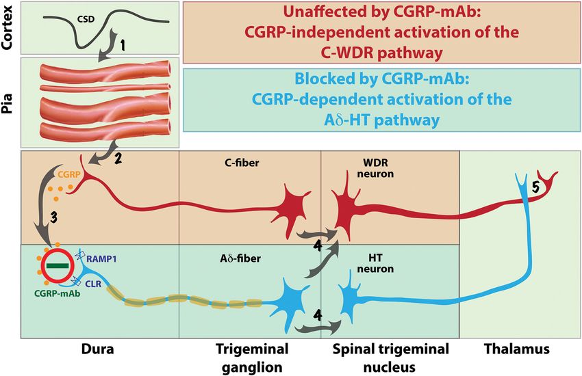
Figure 5. Proposed mechanisms for prevention of migraine by CGRP-mAbs. CSD induces brief constriction, brief dilatation, and prolonged constriction of pial arteries (Step 1), as well as
immediate and delayed activation of C-fiber meningeal nociceptors containing CGRP (Step 2). Upon their CGRP-independent activation, meningeal C-fibers release CGRP in the dura and by doing so;
mediate a CGRP-dependent activation of the nearby A␦-fibers (Step 3). Once activated, C-fiber meningeal nociceptors converge on and activate WDR neurons in the spinal trigeminal nucleus,
whereas A␦-fibers converge on and activate both WDR and HT neurons (Step 4) that eventually transmit the nociceptive signals from the dura to the thalamus (Step 5). The absence of CGRP receptors
from the meningeal C-fibers renders the activation of the C-WDR pathway CGRP-independent (red), and thus, unresponsive to the CGRP-mAb. In contrast, the presence of CGRP receptors on
meningeal A␦-fibers renders the activation of the A␦-HT pathway CGRP-dependent (blue), and thus, responsive to the CGRP-mAb.
Discussion (Strassman et al., 2004). According to this scenario, the A␦ acti-
This is the first study to test the effects of a CGRP-mAb on the vation by CSD involves increased CGRP release in the dura, most
response properties of peripheral trigeminovascular neurons. It likely by the CGRP-positive C-fibers, whereas the prevention of
is also the first study that identifies a molecule that prevents CSD their activation by fremanezumab is achieved by the sequestra-
from activating thinly myelinated A␦ but not unmyelinated C tion of the secreted CGRP molecules (Fig. 5, steps 2–3).
type meningeal nociceptors. This selectivity may be critical for Another explanation for the selective inhibition of the A␦-
the unique prophylactic profile of this class of drugs in migraine fibers by fremanezumab is that the mechanosensitivity of the A␦
therapy. Mechanistically, the findings define A␦ meningeal meningeal nociceptors is greater than the mechanosensitivity
nociceptors as a likely anti-nociceptive site of action of freman- of the C-fibers (Levy and Strassman, 2002), in which case, the
ezumab, which is critical for its ability to prevent the headache. CSD-induced cerebral vasodilatation should have a greater effect
The findings may also explain fremanezumab’s selective inhibi- on the activation of the A␦-fibers because the vasodilatation-
tion of HT but not WDR trigeminovascular neurons in the dorsal associated stretching of the wall of the blood vessels is a mechan-
horn (Melo-Carrillo et al., 2017). Clinically, fremanezumab’s se- ical stimulus. According to this scenario, the blockade of CGRP
lective inhibition of peripheral A␦ meningeal nociceptors and action by fremanezumab might have a greater effect on A␦-fibers
central HT trigeminovascular neurons may also explain how than C-fibers, because CGRP receptors are present on cerebral
CGRP-mAbs prevent the headaches, and equally important, why vascular smooth muscle cells (Eftekhari et al., 2013; Miller et al.,
it may not work for everybody (Bigal et al., 2015a,b). 2016), and CSD-induced cerebral vasodilatation (Bolay et al.,
By far, the most important finding of this study is that 2002; Ayata and Lauritzen, 2015) is partly dependent on CGRP
fremanezumab prevented the activation of A␦ but not C-type (Colonna et al., 1994; Reuter et al., 1998).
meningeal nociceptors by CSD. Mechanistically, the most obvi- Recently, we showed that fremanezumab prevents CSD-
ous explanation for this selectivity is that the calcitonin recep- induced activation and sensitization of HT but not WDR trigemi-
tor-like receptor (CLR) and/or the receptor activity-modifying novascular neurons, and proposed that in migraine patients
protein 1 (RAMP1) are expressed exclusively in the A␦- but not whose headaches are reduced by the CGRP-mAb, the headaches
C-fibers. In support of this explanation, Eftekhari et al. (2013) are initiated and/or maintained by the HT rather than the WDR
showed that in the rat and human dura, CGRP is expressed in C- neurons (Melo-Carrillo et al., 2017). When we wrote this paper,
but not A-fibers whereas CLR and RAMP1 are expressed in the A- but we did not know that fremanezumab prevents the activation of
not C-fibers. Our electrophysiological/functional findings suggest A␦- but not C-type meningeal nociceptors. The current study
that most dural CLR- and RAMP1-positive A-fibers are thinly suggests that the selective inhibition of (central) HT trigemino-
myelinated A␦-fibers; a conclusion supported by anatomical vascular neurons by fremanezumab is secondary to its selective
studies showing that almost all A-fibers in the dura are A␦ inhibition of the (peripheral) A␦ meningeal nociceptors (Fig. 5,Melo-Carrillo et al. • Inhibition of Meningeal Nociceptors by CGRP-mAb J. Neurosci., November 1, 2017 • 37(44):10587–10596 • 10595
step 4). The functional and anatomical relationship between A␦ mainly through their ability to prevent the activation of periph-
nociceptors and HT trigeminal neurons has been recognized for eral trigeminovascular neurons of the A␦ type by events that lead
quite some time. In the primate, HT neurons in the spinal trigeminal to cerebral release of CGRP, such as during migraine headache
nucleus, defined by their responses to noxious pinch and pinprick (Goadsby et al., 1990). Because CGRP-mAb therapy is effective in
but not to innocuous brush, exhibit exclusive input from A␦ fibers the prevention of new headache attacks, it is reasonable to con-
(Price et al., 1976). In humans, A␦ fibers, especially the mechanosen- clude that the activation of A␦-fibers in the dura, which leads to
sitive and mechanoheat-sensitive type I which are also capsaicin- activation of HT neurons in the spinal trigeminal nucleus, is re-
insensitive, are the only type of nociceptors that mediate the sponsible and sufficient for the initiation of the headache.
perception of pinprick (Magerl et al., 2001). In agreement with One caveat regarding the present findings is that the sample
these, it is now well established that central branches of A␦ but not included only male rats. Our prior study on fremanezumab ef-
C-type nociceptors terminate in the two laminae (I and V) where we fects on the central neurons of the trigeminovascular pathway in
recorded all of our HT neurons (Bráz and Basbaum, 2009). the dorsal horn did include both male and female rats, and found
The functional network and the physiological events that may no significant difference between males and females in the effects
explain fremanezumab’s selective inhibition of meningeal A␦- of fremanezumab on neuronal activity. Nonetheless, it remains
(but not C-) fibers and HT (but not WDR) trigeminovascular to be determined in future studies whether the drug has effects on
neurons’ (Melo-Carrillo et al., 2017) responses to CSD is sum- the meningeal primary afferent neurons in females, and whether
marized in Figure 5. Scientifically, these findings may be the first the effects in females are similar to those found in the males, as
to allow us to distinguish between HT and WDR neurons’ con- was the case for the central neurons.
tribution to the perception of pain at different times after their
activation (e.g., early vs late) and under different conditions References
(e.g., acute, chronic, neuropathic, inflammatory). Clinically, Adwanikar H, Ji G, Li W, Doods H, Willis WD, Neugebauer V (2007) Spinal
CGRP1 receptors contribute to supraspinally organized pain behavior
these findings force us to modify a notion that dominated the and pain-related sensitization of amygdala neurons. Pain 132:53– 66.
understanding of the different stages of allodynia, hyperalgesia, CrossRef Medline
and central sensitization, the role they play in the chronification Ayata C, Lauritzen M (2015) Spreading depression, spreading depolariza-
of pain and migraine, and their impact on treatment. Regarding tions, and the cerebral vasculature. Physiol Rev 95:953–993. CrossRef
the A␦-HT versus C-WDR pathways, the complete or nearly Medline
complete prevention of headache by fremanezumab suggests that Bigal ME, Dodick DW, Rapoport AM, Silberstein SD, Ma Y, Yang R, Loupe
PS, Burstein R, Newman LC, Lipton RB (2015a) Safety, tolerability, and
the headache perception of some patients is mediated by the
efficacy of TEV-48125 for preventive treatment of high-frequency episodic
neuronal A␦-HT pathway alone, and conversely, the complete or migraine: a multicentre, randomised, double-blind, placebo-controlled,
near complete lack of effect (i.e., no or insignificant reduction in phase 2b study. Lancet Neurol 14:1081–1090. CrossRef Medline
migraine days) suggests that the headache perception of some Bigal ME, Edvinsson L, Rapoport AM, Lipton RB, Spierings EL, Diener HC,
patients could be mediated by the neuronal C-WDR pathway Burstein R, Loupe PS, Ma Y, Yang R, Silberstein SD (2015b) Safety,
alone. Regarding allodynia, hyperalgesia and central sensitiza- tolerability, and efficacy of TEV-48125 for preventive treatment of chronic
migraine: a multicentre, randomised, double-blind, placebo-controlled,
tion, a synthesis of all currently available clinical data on the
phase 2b study. Lancet Neurol 14:1091–1100. CrossRef Medline
treatment outcome of the four CGRP-mAbs under development Bird GC, Han JS, Fu Y, Adwanikar H, Willis WD, Neugebauer V (2006)
indicates clearly that a class of drugs that is most likely to block Pain-related synaptic plasticity in spinal dorsal horn neurons: role of
the flow of nociceptive signals from the peripheral meninges to CGRP. Mol Pain 2:31. CrossRef Medline
the spinal trigeminal nucleus, is capable of reducing migraine in Bolay H, Moskowitz MA (2005) The emerging importance of cortical
patients whose attacks were frequent enough before treatment as spreading depression in migraine headache. Rev Neurol 161:655– 657.
to classify them chronic migraineurs. Until now, we thought that CrossRef Medline
Bolay H, Reuter U, Dunn AK, Huang Z, Boas DA, Moskowitz MA (2002)
the transition from acute to chronic migraine follows, at least in Intrinsic brain activity triggers trigeminal meningeal afferents in a mi-
part, the transition from early stage central sensitization that de- graine model. Nat Med 8:136 –142. CrossRef Medline
pends on pain signals that come from peripheral nociceptors to Bráz JM, Basbaum AI (2009) Triggering genetically-expressed transneuro-
late stage central sensitization that is independent of the pain nal tracers by peripheral axotomy reveals convergent and segregated
signals from the nociceptors, and accordingly, proposed that late- sensory neuron-spinal cord connectivity. Neuroscience 163:1220 –1232.
phase central sensitization and allodynia explain the inferior ef- CrossRef Medline
Burstein R, Jakubowski M (2004) Analgesic triptan action in an animal
ficacy of late treatment with drugs that lack the ability to reverse
model of intracranial pain: a race against the development of central
the sensitization(Burstein and Jakubowski, 2004; Burstein et al., sensitization. Ann Neurol 55:27–36. CrossRef Medline
2004; Levy et al., 2004). Our current and recent (Melo-Carrillo et Burstein R, Collins B, Jakubowski M (2004) Defeating migraine pain with
al., 2017) studies call for a revision of this concept. Fremanezum- triptans: a race against the development of cutaneous allodynia. Ann
ab’s ability to reduce migraine in patients (i.e., the responders) Neurol 55:19 –26. CrossRef Medline
who are likely to exhibit signs of late-phase, already established, Burstein R, Noseda R, Borsook D (2015) Migraine: multiple processes,
complex pathophysiology. J Neurosci 35:6619 – 6629. CrossRef Medline
allodynia and central sensitization (Lipton et al., 2017) suggests
Colonna DM, Meng W, Deal DD, Busija DW (1994) Calcitonin gene-
that their ongoing allodynia and central sensitization continue to related peptide promotes cerebrovascular dilation during cortical spread-
depend on the pain signals they receive from the meningeal ing depression in rabbits. Am J Physiol 266:H1095–H1102. Medline
nociceptors. Conversely, fremanezumab’s inability to reduce Dodick DW, Goadsby PJ, Spierings EL, Scherer JC, Sweeney SP, Grayzel DS
migraine in this patient population (i.e., the nonresponders) sug- (2014a) Safety and efficacy of LY2951742, a monoclonal antibody to
gests that their ongoing allodynia and central sensitization prog- calcitonin gene-related peptide, for the prevention of migraine: a
ress into the established phase whereby the activity of their central phase 2, randomised, double-blind, placebo-controlled study. Lancet
Neurol 13:885– 892. CrossRef Medline
trigeminovascular neurons is in fact independent of the pain sig- Dodick DW, Goadsby PJ, Silberstein SD, Lipton RB, Olesen J, Ashina M,
nals they receive from the meningeal nociceptors. Wilks K, Kudrow D, Kroll R, Kohrman B, Bargar R, Hirman J, Smith J; the
In summary, our findings provide direct evidence for the as- ALD403 study investigators (2014b) Safety and efficacy of ALD403, an
sertion that the prophylactic effect of CGRP-mAbs is achieved antibody to calcitonin gene-related peptide, for the prevention of frequent10596 • J. Neurosci., November 1, 2017 • 37(44):10587–10596 Melo-Carrillo et al. • Inhibition of Meningeal Nociceptors by CGRP-mAb
episodic migraine: a randomised, double-blind, placebo-controlled, explor- tive substance P in the brain stem upon stimulation of the cranial dura
atory phase 2 trial. Lancet Neurol 13:1100 –1107. CrossRef Medline mater with low pH: inhibition by the serotonin (5-HT1) receptor agonist
Durham PL, Russo AF (1999) Regulation of calcitonin gene-related peptide CP 93,129. Br J Pharmacol 125:1726 –1732. CrossRef Medline
secretion by a serotonergic antimigraine drug. J Neurosci 19:3423–3429. Miller S, Liu H, Warfvinge K, Shi L, Dovlatyan M, Xu C, Edvinsson L (2016)
Medline Immunohistochemical localization of the calcitonin gene-related peptide
Edvinsson L, Ekman R, Jansen I, McCulloch J, Uddman R (1987) Calcitonin binding site in the primate trigeminovascular system using functional
gene-related peptide and cerebral blood vessels: distribution and vasomo- antagonist antibodies. Neuroscience 328:165–183. CrossRef Medline
tor effects. J Cereb Blood Flow Metab 7:720 –728. CrossRef Medline Moskowitz MA (1984) The neurobiology of vascular head pain. Ann Neurol
Edvinsson L, Mulder H, Goadsby PJ, Uddman R (1998) Calcitonin gene- 16:157–168. CrossRef Medline
related peptide and nitric oxide in the trigeminal ganglion: cerebral vaso- O’Connor TP, van der Kooy D (1988) Enrichment of a vasoactive neuro-
dilatation from trigeminal nerve stimulation involves mainly calcitonin peptide (calcitonin gene related peptide) in the trigeminal sensory pro-
gene-related peptide. J Auton Nerv Syst 70:15–22. CrossRef Medline jection to the intracranial arteries. J Neurosci 8:2468 –2476. Medline
Edvinsson L, Elsås T, Suzuki N, Shimizu T, Lee TJ (2001) Origin and Price DD, Dubner R, Hu JW (1976) Trigeminothalamic neurons in nucleus
co-localization of nitric oxide synthase, CGRP, PACAP, and VIP in the caudalis responsive to tactile, thermal, and nociceptive stimulation of
cerebral circulation of the rat. Microsc Res Tech 53:221–228. CrossRef monkey’s face. J Neurophysiol 39:936 –953. Medline
Medline Price TJ, Flores CM (2007) Critical evaluation of the colocalization between
Eftekhari S, Warfvinge K, Blixt FW, Edvinsson L (2013) Differentiation of calcitonin gene-related peptide, substance p, transient receptor potential
nerve fibers storing CGRP and CGRP receptors in the peripheral trigemi- vanilloid subfamily type 1 immunoreactivities, and isolectin b(4) binding
novascular system. J Pain 14:1289 –1303. CrossRef Medline in primary afferent neurons of the rat and mouse. J Pain 8:263–272.
Eltorp CT, Jansen-Olesen I, Hansen AJ (2000) Release of calcitonin gene- CrossRef Medline
related peptide (CGRP) from guinea pig dura mater in vitro is inhibited Reuter U, Weber JR, Gold L, Arnold G, Wolf T, Dreier J, Lindauer U, Dirnagl
by sumatriptan but unaffected by nitric oxide. Cephalalgia 20:838 – 844. U (1998) Perivascular nerves contribute to cortical spreading depression-
CrossRef Medline associated hyperemia in rats. Am J Physiol 274:H1979 –H1987. Medline
Fischer MJ, Koulchitsky S, Messlinger K (2005) The nonpeptide calcitonin Russo AF (2015) Calcitonin gene-related peptide (CGRP): a new target for
gene-related peptide receptor antagonist BIBN4096BS lowers the activity migraine. Annu Rev Pharmacol Toxicol 55:533–552. CrossRef Medline
of neurons with meningeal input in the rat spinal trigeminal nucleus. Storer RJ, Akerman S, Goadsby PJ (2004) Calcitonin gene-related peptide
J Neurosci 25:5877–5883. CrossRef Medline (CGRP) modulates nociceptive trigeminovascular transmission in the
Goadsby PJ, Edvinsson L, Ekman R (1990) Vasoactive peptide release in the cat. Br J Pharmacol 142:1171–1181. CrossRef Medline
extracerebral circulation of humans during migraine headache. Ann Neu- Strassman AM, Raymond SA, Burstein R (1996) Sensitization of meningeal
rol 28:183–187. CrossRef Medline sensory neurons and the origin of headaches. Nature 384:560 –564. CrossRef
Han JS, Li W, Neugebauer V (2005) Critical role of calcitonin gene-related
Medline
peptide 1 receptors in the amygdala in synaptic plasticity and pain behav-
Strassman AM, Weissner W, Williams M, Ali S, Levy D (2004) Axon diam-
ior. J Neurosci 25:10717–10728. CrossRef Medline
eters and intradural trajectories of the dural innervation in the rat. J Comp
Hansen JM, Ashina M (2014) Calcitonin gene-related peptide and migraine
Neurol 473:364 –376. CrossRef Medline
with aura: a systematic review. Cephalalgia 34:695–707. CrossRef Medline
Sun H, Dodick DW, Silberstein S, Goadsby PJ, Reuter U, Ashina M, Saper J,
Karsan N, Goadsby PJ (2015) Calcitonin gene-related peptide and migraine.
Cady R, Chon Y, Dietrich J, Lenz R (2016) Safety and efficacy of AMG
Curr Opin Neurol 28:250 –254. CrossRef Medline
334 for prevention of episodic migraine: a randomised, double-blind,
Keller JT, Marfurt CF (1991) Peptidergic and serotoninergic innervation of
placebo-controlled, phase 2 trial. Lancet Neurol 15:382–390. CrossRef
the rat dura mater. J Comp Neurol 309:515–534. CrossRef Medline
Medline
Kurosawa M, Messlinger K, Pawlak M, Schmidt RF (1995) Increase of men-
Sun RQ, Lawand NB, Lin Q, Willis WD (2004a) Role of calcitonin gene-
ingeal blood flow after electrical stimulation of rat dura mater encephali:
related peptide in the sensitization of dorsal horn neurons to mechanical
mediation by calcitonin gene-related peptide. Br J Pharmacol 114:1397–
1402. CrossRef Medline stimulation after intradermal injection of capsaicin. J Neurophysiol 92:
Levy D, Strassman AM (2002) Mechanical response properties of A and C 320 –326. CrossRef Medline
primary afferent neurons innervating the rat intracranial dura. J Neuro- Sun RQ, Tu YJ, Lawand NB, Yan JY, Lin Q, Willis WD (2004b) Calcitonin
physiol 88:3021–3031. CrossRef Medline gene-related peptide receptor activation produces PKA- and PKC-dependent
Levy D, Jakubowski M, Burstein R (2004) Disruption of communication mechanical hyperalgesia and central sensitization. J Neurophysiol 92:2859 –
between peripheral and central trigeminovascular neurons mediates the 2866. CrossRef Medline
antimigraine action of 5HT 1B/1D receptor agonists. Proc Natl Acad Sci Tsai SH, Tew JM, McLean JH, Shipley MT (1988) Cerebral arterial innerva-
U S A 101:4274 – 4279. CrossRef Medline tion by nerve fibers containing calcitonin gene-related peptide (CGRP): I.
Lipton RB, Munjal S, Buse DC, Bennett A, Fanning KM, Burstein R, Reed ML Distribution and origin of CGRP perivascular innervation in the rat.
(2017) Allodynia is associated with initial and sustained response to J Comp Neurol 271:435– 444. CrossRef Medline
acute migraine treatment: results from the American migraine prevalence Uddman R, Edvinsson L, Ekblad E, Hakanson R, Sundler F (1986) Calci-
and prevention study. Headache 57:1026 –1040. CrossRef Medline tonin gene-related peptide (CGRP): perivascular distribution and vaso-
Magerl W, Fuchs PN, Meyer RA, Treede RD (2001) Roles of capsaicin- dilatory effects. Regul Pept 15:1–23. CrossRef Medline
insensitive nociceptors in cutaneous pain and secondary hyperalgesia. Uddman R, Hara H, Edvinsson L (1989) Neuronal pathways to the rat mid-
Brain 124:1754 –1764. CrossRef Medline dle meningeal artery revealed by retrograde tracing and immunocyto-
McCarthy PW, Lawson SN (1990) Cell type and conduction velocity of rat chemistry. J Auton Nerv Syst 26:69 –75. CrossRef Medline
primary sensory neurons with calcitonin gene-related peptide-like im- Williamson DJ, Hargreaves RJ, Hill RG, Shepheard SL (1997) Intravital
munoreactivity. Neuroscience 34:623– 632. CrossRef Medline microscope studies on the effects of neurokinin agonists and calcitonin
McCulloch J, Uddman R, Kingman TA, Edvinsson L (1986) Calcitonin gene-related peptide on dural vessel diameter in the anaesthetized rat.
gene-related peptide: functional role in cerebrovascular regulation. Proc Cephalalgia 17:518 –524. CrossRef Medline
Natl Acad Sci U S A 83:5731–5735. CrossRef Medline Zagami AS, Goadsby PJ, Edvinsson L (1990) Stimulation of the superior
Melo-Carrillo A, Noseda R, Nir R, Schain AJ, Stratton J, Strassman AM, sagittal sinus in the cat causes release of vasoactive peptides. Neuropep-
Burstein R (2017) Selective inhibition of trigeminovascular neurons by tides 16:69 –75. CrossRef Medline
fremanezumab: a humanized monoclonal anti-CGRP antibody. J Neuro- Zhang X, Levy D, Noseda R, Kainz V, Jakubowski M, Burstein R (2010)
sci 37:7149 –7163. CrossRef Medline Activation of meningeal nociceptors by cortical spreading depression:
Messlinger K, Hanesch U, Baumgärtel M, Trost B, Schmidt RF (1993) In- implications for migraine with aura. J Neurosci 30:8807– 8814. CrossRef
nervation of the dura mater encephali of cat and rat: ultrastructure and Medline
calcitonin gene-related peptide-like and substance P-like immunoreac- Zhang X, Levy D, Kainz V, Noseda R, Jakubowski M, Burstein R (2011)
tivity. Anat Embryol 188:219 –237. Medline Activation of central trigeminovascular neurons by cortical spreading
Messlinger K, Ebersberger A, Schaible HG (1998) Release of immunoreac- depression. Ann Neurol 69:855– 865. CrossRef MedlineYou can also read