Verification of neuroprotective effects of alpha-lipoic acid on chronic neuropathic pain in a chronic constriction injury rat model
←
→
Page content transcription
If your browser does not render page correctly, please read the page content below
Open Life Sciences 2021; 16: 222–228
Research Article
Junhao Wang#, Zhaohui Lou*#, Haiyang Xi, Zhi Li, Lepeng Li, Zhenzhen Li, Kai Zhang,
Tetsuya Asakawa*
Verification of neuroprotective effects of alpha-
lipoic acid on chronic neuropathic pain in a
chronic constriction injury rat model
https://doi.org/10.1515/biol-2021-0026 Keywords: neuropathic pain, peripheral nerve injury,
received June 05, 2020; accepted January 04, 2021 alpha-lipoic acid, satellite glial cells, dorsal root ganglia
Abstract: Treatment of neuropathic pain is far from satis-
factory. This study sought evidence of a neuroprotective
effect of alpha-lipoic acid (ALA) to treat neuropathic pain
in a chronic constriction injury (CCI) rat model. A total of 1 Introduction
48 rats were randomly divided into sham, CCI, or CCI +
ALA groups. Mechanical and thermal nociceptive thresh- Peripheral nerve injury (PNI) is commonly caused by
olds were evaluated as behavioral assessments. Dorsal crush injury or mechanical trauma, leading to pain char-
root ganglia cells were assessed morphologically with acterized by hyperalgesia and allodynia [1]. Spontaneous
hematoxylin and eosin staining and for apoptosis with pain is frequent and may be intolerable. Although this is
P53 immunohistochemical staining. Compared with the not a life-threatening condition, it seriously diminishes
sham group, the CCI group had a shorter paw withdrawal quality of life and is a frequent reason for doctor visits.
threshold and paw withdrawal latency, abnormal mor- Neuropathic pain is common in the general population.
phologic manifestations, and increased numbers of satel- Approximately 80% of adults at some time or other com-
lite glial cells and P53+ cells. These changes were significantly plain of lower back or leg pain, but current therapy
reversed by treatment with ALA. Our study indicates neu- cannot completely prevent pain or relieve the conse-
roprotective effects of ALA on chronic neuropathic pain in a quences of neurologic damage [2,3]. PNI is therefore an
CCI rat model. ALA is potentially considered to be devel- important and increasing public health concern.
oped as a treatment for neuropathic pain caused by periph- Mechanisms underlying PNI are complicated and not
eral nerve injury, which requires further verification. fully understood. Currently, there is increasing focus on
the pathogenesis of dorsal root ganglia (DRG) damage. A
recent study reported that peripheral neuropathic pain
# Equal contributors. is caused by abnormal spontaneous activity of both
damaged and undamaged DRG [4]. The DRG are sensitive
* Corresponding author: Zhaohui Lou, Department of Orthopedic to mechanical stimuli, with the adventitial coat being a
Surgery, First Affiliated Hospital of Zhengzhou University, very sensitive receptor for mechanical trauma. Under cer-
Zhengzhou, Henan 450052, China, tel: +86-371-6796611, tain pathologic conditions such as trauma or inflamma-
fax: +86-371-6796611, e-mail: louzhaohui@126.com
tion, DRG are stimulated and cause pain, as the ganglia
* Corresponding author: Tetsuya Asakawa, Department of
Neurosurgery, Hamamatsu University School of Medicine, have no effective blood-nerve barrier. The DRG neuroglia
Handayama, 1-20-1, Higashi-ku, Hamamatsu City, Shizuoka 431- are composed of satellite glial cells (SGCs) [5]. Neurons in
3192, Japan; Research Base of Traditional Chinese Medicine the sensory ganglia are surrounded by SGCs, which play
Syndrome, Fujian University of Traditional Chinese Medicine,
Fuzhou 350122, China, tel: +81-53-4352283, fax: +81-53-4352282,
crucial roles in the initiation and promotion of peripheral
e-mail: asakawat1971@gmail.com neuropathic pain [6,7]. Previous studies demonstrated
Junhao Wang, Haiyang Xi, Zhi Li, Lepeng Li, Kai Zhang: Department that SGCs can be triggered and activated by cytokines
of Orthopedic Surgery, First Affiliated Hospital of Zhengzhou
during the inflammatory response [8,9], which is often
University, Zhengzhou, Henan 450052, China
Zhenzhen Li: Institute of Clinical Medicine, First Affiliated Hospital closely associated with neuropathic pain. Activation of
of Zhengzhou University, Zhengzhou, Henan, China SGCs is therefore considered a hallmark of PNI.
Open Access. © 2021 Junhao Wang et al., published by De Gruyter. This work is licensed under the Creative Commons Attribution 4.0
International License.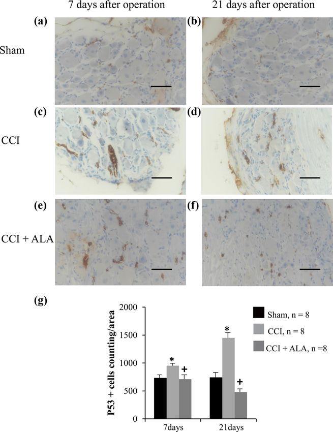
Neuroprotective effects of ALA in a chronic constriction injury 223
Alpha-lipoic acid (ALA) exhibits a strong neuropro- 2.2 Surgical procedures
tective effect. It is used to treat diseases caused by oxida-
tive stress (OS) such as diabetic neuropathy [10] and The CCI models were established according to the methods
multiple sclerosis [11] and to alleviate the inflammatory described in a previous study [16]. Briefly, the rats were
response [12]. It is also used as a modulator of various anesthetized with tiletamine/zolazepam (Zoletil WK001,
inflammatory signaling pathways [13]. As early in 2008, Virbac, France), 50 mg/kg intraperitoneally. The right
Melli et al. reported that ALA is neuroprotective in sen- sciatic nerve was exposed at the level of the mid-thigh,
sory neurons and prevents apoptosis of DRG cells [14]. A and the connective tissue around the nerve was cleared.
later study found that ALA reduces expression of oxides Four ligatures were then tied around the nerve at 1–2 mm
and increases antioxidants in sciatic nerve crush injury intervals using 4-0 chromic gut suture material.
rat models [15]. We thus wondered whether ALA would
have neuroprotective effects in PNI. In the present study,
we employed chronic constriction injury (CCI) in rats as a
model of chronic neuropathic pain [16] and P53 as a cel- 2.3 Behavioral assessment
lular apoptosis marker. The aim was to look for evidence
of neuroprotective effects of ALA in CCI rat models, To assess the pain, mechanical and thermal nociceptive
potentially leading to a novel treatment for neuropathic thresholds were examined as previously described [19].
pain associated with PNI. To examine the mechanical nociceptive threshold, ani-
mals were placed individually into a small plastic cage
with an open wire mesh bottom. Before testing, rats were
left in the test cages for 30 min until their grooming and
exploratory behaviors ceased and all four paws were
2 Materials and methods placed on the bottom. Von Frey filaments (North Coast
Medical Inc, Morgan Hill, CA, ranged from 1.08 to 40 g)
2.1 Animals were applied vertically to the planter surface of the paw
with an upward force just sufficient to bend the micro-
A total of 48 male Sprague-Dawley rats (240 ± 20 g, age 2 filament. One of the tactile-defensive behaviorals, namely,
months) were used. All rats were housed and fed at room brisk paw withdrawal was observed. We recorded the sti-
temperature (23°C) with a humidity of 55 ± 15% and a 12 h mulus intensity at the time of foot and leg reactions,
light/dark cycle (lights on at 7:00 a.m.). Food and water including foot reflexes, leg-stripping, and leg-turning, at
were provided ad libitum. The animals were randomly intervals of 10 s per stimulus. Filaments were used in
divided into three groups. The dose of ALA (50 mg/kg) ascending order. Each filament was used once prior to
was selected according to the previous rat studies advancing to the next filament. The smallest filament
[17,18]. In the sham group (n = 16), the right sciatic that conducted a paw withdrawal response was considered
nerve was exposed without any other treatment. The the threshold stimulus. The paw withdrawal threshold
CCI group (n = 16) underwent nerve injury as described (PWT) index was recorded and averaged over five measure-
below and had a sham injection (0.9% saline solution, ments. For thermal nociceptive thresholds, animals were
50 mg/kg) every day after surgery. The CCI + ALA group placed in an acrylic box with a transparent glass plate and
(n = 16) also underwent nerve injury, and the rats were irradiated with radiant heat on both hind paws. The leg lift
then treated with ALA (50 mg/kg) injections every day avoidance time (the time it took to respond to the thermal
after surgery immediately prior to conducting beha- stimulus) was counted as the paw withdrawal latency
vioral assessments. (PWL). A low power (40 mW/cm2) intensity, a 20-second
cutoff time, and a heating rate of 1°C/s were chosen
Ethical approval: The research related to animal use has according to the previous study [19]. The stimulus interval
been complied with all the relevant national regulations was 5 min, and the average of three measurements was
and institutional policies for the care and use of animals calculated.
and was approved and supervised by the Animal Care and The behavioral assessments were performed 1 day
Use Committee of the First Affiliated Hospital of Zhengzhou before surgery and 3, 7, 14, and 21 days postsurgery.
University. Animals were kept in the testing chambers for 30 minutes224 Junhao Wang et al.
before each measurement so as to ensure they were images included converting the image to black and white,
accustomed to the test environment. After the behavioral subtracting the background, elevating contrast, reducting
tests, all animals were submitted to the morphological the noise, and finally perfoming measurements. In this
experiments. study, we set all DRG areas (ipsilateral to injury) as the
regions of interest (ROI). A standard Erode and Dilate
filter was employed to reduce the background noise.
Dilate was used in the cell area to obtain a value compar-
2.4 Morphology able to the area before applying the Erode filter. Image
binarization was performed to correct filter operations.
DRG tissues were examined morphologically for evidence Areas of objects presenting in the ROI were included in
of nerve damage with hematoxylin and eosin (HE) staining a measurements unit [20], which were set as 20,000
and for cellular apoptosis with P53 immunohistochemical pixels, corresponding to 9,965 µm2 according to a pre-
staining. svious study [21]. Using these methods, the edematous
Rats in each group were anesthetized with tiletamine/ DRG neurons and surrounding SGCs were identified. All
zolazepam (50 mg/kg) either on day 7 (n = 8) or day 21 these processes were performed by the same experi-
(n = 8) after surgery. They were transcardially perfused enced researcher blinded to the treatments of groups.
with saline, followed by 250 mL of 4% paraformaldehyde
(PH0427, Phygene, Fujian, China) in phosphate-buffered
saline (PBS, 0.1 M, pH 7.4). DRG tissues from L4 to L6,
corresponding to sciatic afferent fibers, were removed
and fixed with 4% paraformaldehyde in PBS (pH 7.4)
for 4 h. After rinsing, the DRG tissues were processed
and embedded in paraffin. Subsequently, 5 µm thick sec-
tions were cut using a microtome with disaposable stain-
less knives (Yamato Kohki Industrial, Saitama, Japan).
Some tissue sections were examined after standard HE
staining. The remaining sections were submitted to P53
staining. To determine P53 immunoreactivity, anti-P53
mouse monoclonal antibodies (1:100, ZM-0408, ZSGB-
BIO, Beijing, China) were applied to the sections overnight
at 4°C. Secondary antibodies conjugated to horseradish
peroxidase (SPN-9001, anti-rabbit; SPN-9002, anti-mouse,
ZSGB-BIO, Beijing, China) were then applied for 15 min
at 37°C as per the manufacturer’s instructions. Diamino-
benzidine color reagent (SN640500, Celnovte Biotechnology,
Henan, China) was used to develop color.
The SGCs surrouding the neurons were identified
using the methods described by a previous study [20].
Briefly, all the sections were observed and quantitatively
analyzed using a slice-analyzing system including an
optical microscope (Nikon Ni-E, Japan) and the Image J
1.80 software package. DRG neurons were divided into
Figure 1: Changes in paw withdrawal thresholds (PWTs) and paw
large (>50 µm), medium (30–50 µm), and small (10–30 µm) withdrawal latency (PWL) induced by chronic constriction injury
with visible nucleoli. The numbers of surrounding SGCs (CCI) and its treatment with alpha-lipoic acid (ALA). (a) The PWTs in
and P53-immunopositive (P53+) cells in each of eight the CCI group were significantly lower than those in the sham group,
consecutive sections were calculated using the Scion whereas ALA treatment significantly enhanced PWTs, although not
Image 4.0.3 software package as following steps: All to the level of the sham group. (b) Similar to the PWTs, the PWLs of
the CCI group were significantly lower than those of the sham group.
areas of the DRG were snapped for the following image
Treatments with ALA significantly enhanced the PWLs, although not
analysis using at least sixty images in each group. After to the level of the sham group. ** means P < 0.01, *** means
acquisition of the images, they were transformed into P < 0.001, sham vs CCI; ++ means P < 0.01, +++ means P < 0.001,
TIFF format (1,080 × 800 pixels). The processes of the CCI vs CCI + ALA.Neuroprotective effects of ALA in a chronic constriction injury 225
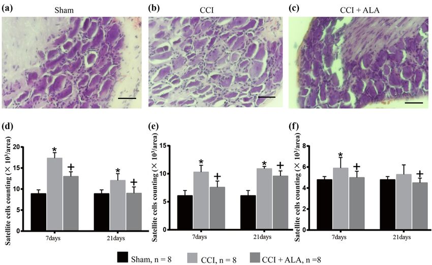
Figure 2: Morphology of dorsal root ganglia (DRG) and satellite glial cells (SGC) (hematoxylin and eosin). (a) Representative photo of DRG
cells in the sham group with normally shaped neuronal cells. (b) Representative photo of DRG cells in the CCI group. Edematous neurons
and the enhanced SCGs surrounding the neurons can be observed. (c) Representative photo of DRG cells in the CCI + ALA. Although there
are some edematous neurons and enhanced SGCs, they are fewer than in the CCI group. (d–f) SGC cells counts/area surrounding large
(>50 µm, d), medium (30–50 µm, e), and small (10–30 µm, f) DRG neurons. The results were analogous; cell numbers/area were signifi-
cantly higher in the CCI group (vs sham group) and could be reduced after ALA treatment (vs CCI + ALA group). * means P < 0.05, sham vs
CCI; + means P < 0.05, CCI vs CCI + ALA; scale bar = 50 μm; magnification: 40×; one area = 20,000 pixels = 9,965 µm2.
2.5 Statistic analysis The cytoplasm was concentrated, and the cell body
volume contracted and deformed. Occasionally, apop-
Data were analyzed using SPSS software (V 19.0, IBM, totic neuronal cells were observed, and the number of
USA). All data are reported as mean ± standard deviation. SGCs was obviously enhanced. Such changes indicated
All statistical results were tested on both sides. Normality an enhanced inflammatory reaction associated with CCI.
of distribution and homogeneity of variance tests were In the CCI + ALA group, there was less neuronal cell
performed first. Analysis of variance followed by body edema and fewer vacuolar changes than in the CCI
Bonferroni post hoc correction was then selected for mul- group, but ALA treatment did not completely restore the
tiple comparisons. P < 0.05 was considered to be statisti- tissue to a normal appearance (Figure 2c).
cally significant. Counting the numbers of SGCs/area yielded similar
results. Here, we set one area as 20,000 pixels, corre-
sponding to 9,965 µm2. The numbers of SGS/area in the
3 Results CCI group were significantly higher than those in the
sham group (Figure 2d, surrounding large DRG neurons
CCI treatment significantly shortened the PWT (Figure 1a) over 50 µm, at both 7 and 21 days after operation; Figure 2e,
and PWL (Figure 1b) as compared with the sham group. surrounding medium DRG neurons [30–50 µm], at both 7
These changes were partly ameliorated by ALA treat- and 21 days after operation; Figure 2f, surrounding small
ment, although not to the normal levels of the sham DRG neurons [10–30 µm], at 7 days after operation). ALA
group. The same differences between groups were pre- treatment partially relieved this effect, although without
sent at postoperative days 3, 7, 14, and 21 (Figure 1). restoring the DRG tissues to normal at both 7 and 21 days
In the sham group, DRG cells appeared morphologi- after operation. This suggests that ALA treatment partly
cally normal (Figure 2a). No edema or abnormal aggrega- relieves the inflammatory reaction induced by CCI
tion or proliferation of cells was observed. In the CCI (Figure 2d–f).
group, the cell bodies of DRG neurons were markedly Likewise, one area was set as 20,000 pixels, corre-
edematous with evident vacuolar-like changes (Figure 2b). sponding to 9,965 µm2. Compared with the sham group226 Junhao Wang et al.
(Figure 3a and b), the numbers of P53+ cells/area were pain [26,27]. A number of studies have demonstrated that
significantly higher in the CCI group (Figure 3c and d), a variety of injurious stimuli may trigger activation of
suggesting that apoptosis was induced by CCI. The find- SGCs [27–29]. SGC activation is regarded as a neurophy-
ings were similar at both 7 days (Figure 3c) and 21 days siologic reaction to neuronal stress induced by these sti-
(Figure 3d) postoperatively. Treatment with ALA signifi- muli [26]. Activated SGCs commonly have remarkable
cantly reduced the number of P53+ cells/area compared cellular proliferation [28]. HE staining in our CCI rat
with the CCI group (Figure 3), indicating that ALA can models showed proliferated and abnormally aggregated
relieve CCI-induced apoptosis at both 7 days (Figure 3e) SGCs. Along with other abnormal morphologic manifes-
and 21 days (Figure 3f) after the operation. tations in the DRG neurons, we confirmed that the CCI
procedure induces neurologic damage. ALA treatment
reduced these physiologic changes, by reducing the
4 Discussion
The present study used ALA to treat chronic neuropathic
pain in a CCI rat model. We found that ALA treatment
significantly shortened PWT and PWL, improved mor-
phologic changes in the DRG neurons, reduced the aggre-
gation and proliferation of SGCs, and decreased numbers
of P53+ cells. Our findings have therefore provided beha-
vioral (Figure 1), morphologic (Figure 2), and immuno-
histochemical (Figure 3) evidence that we successfully
established a CCI-induced PNI model in rats. We also
demonstrated that ALA treatment partially relieved the
CCI-induced pathologic changes. We have therefore ver-
ified the protective effects of ALA on chronic neuropathic
pain. These findings imply that ALA can be considered as
a potential therapy for PNI-associated neuropathic pain,
although further verification is needed.
The CCI rat model used in the present study is a
classic model used to mimic chronic neuropathic pain
[22]. Sciatic nerve ligation damages the peripheral nerve,
so that the animal may try to bite the injured leg off. This
abnormal behavior is regarded as a reaction to neuro-
pathic pain [22,23]. In measurements of mechanical and
thermal nociceptive thresholds, the PWT as well as PWL
may shorten because of changes such as hyperpathia
caused by CCI. Our behavioral data pointed a significant Figure 3: P53 immunohistochemistry of dorsal root ganglia (DRG)
decreases in PWT and PWL after CCI, in accordance with after chronic compression injury (CCI). Representative photo of P53
a previous study [24]. Along with the morphologic and staining of DRGs in the sham group, (a) 7 and (b) 21 days after
immunohistochemical evidence, our findings confirmed operation. The brown dots represented P53+ cells, which are rare
in the sham group. Representative photos of P53 staining in CCI
that we successfully established this CCI rat model. ALA
group, (c) 7 and (d) 21 days after operation. P53+ cells are signifi-
treatment prolonged the PWT and PWL in the treatment cantly increased, even more so at 21 days than at 7 days after
group, indicating that it reduced hyperpathia, thus con- operation. The brown areas indicate apoptosis cells, which are
firming the efficacy of ALA treatment. small and shrunken. Representative photos of P53 staining in CCI
Differing from astrocytes or oligodendrocytes, SGCs + ALA group, (e) 7 days after operation and (f) 21 days after opera-
are a distinct type of glial cells that surround sensory tion. There are fewer P53+ cells at both 7 and 21 days after operation
than in the CCI group. (g) P53+ cell counts/area. CCI treatment
neurons in ganglia of the peripheral nervous system.
significantly increased the number of P53+ cells, whereas ALA treat-
The role of SGCs is not fully understood, although studies ment led to normal numbers. * means P < 0.05, sham vs CCI; +
have indicated they play a crucial role in neuropathic means P < 0.05, CCI vs CCI + ALA; scale bar = 50 μm; magnification:
pain, including visceral [25] and peripheral neuropathic 40×; one area = 20,000 pixels = 9,965 µm2.Neuroprotective effects of ALA in a chronic constriction injury 227
enhancement of SGCs, ameliorating edema in the neu- not perform a preliminary experiment to determine the
rons, and normalizing the shape of the neurons. There- most optimal dose of ALA for CCI. All these limitations
fore through relieving the neuronal stress state, ALA should be addressed in our future investigation.
proved to have a neuroprotective effect.
Our findings using P53 immunohistochemistry also
strengthened the evidence of that. P53 protein is a crucial
player in the cellular response to neuronal stress [30]. P53 5 Conclusions
staining is employed as a biomarker of P53-induced
apoptosis [31,32]. The P53 gene has been shown to play Taken together, our study indicates neuroprotective effects
a vital modulatory role in neuropathic pain induced by of ALA on chronic neuropathic pain in a CCI rat model.
DRG trauma [33]. Similarly, we found that P53 expression Our results imply that ALA can be considered as a poten-
was significantly upregulated in the CCI rat model, par- tial therapy for the neuropathic pain associated with
alleling the morphologic changes. This indicates that PNI. Further verification with clinical testing, as well as
CCI promotes cellular apoptosis. As with improvement exploring the therapeutic mechanisms, is required.
in morphologic changes, ALA treatment significantly
reduced the number of P53+ cells. This is further evidence Funding: This study was supported by grants from the
that ALA can ameliorate CCI-induced damage mediated Japanese Society for the Promotion of Science (Grant-
by P53-induced apoptosis. in-Aid for Young Scientists, Type B, No. 20791025 and
Although our study provided several different types Grant-in-Aid for Scientific Research C, General, No.
of evidence of the efficacy of ALA in this model of neuro- 24592157, 15k10358 and 18K08991).
pathic pain, the mechanisms by which it functions need
further investigation. Reducing P53-induced apoptosis is Conflict of interest: The authors state no conflict of
one potential mechanism. It has been shown that neu- interest.
ronal apoptosis serves as a mechanism to maintain neuro-
pathic pain, while suppression of cellular apoptosis Data availability statement: The datasets generated during
ameliorates hyperalgesia and mechanical abnormal pain and/or analyzed during the current study are available
[34], which completely agrees with our findings in the from the corresponding author on reasonable request.
present study. The mechanisms underlying cellular apop-
tosis may be closely associated with OS [34–36]. Battisti
et al. used ALA to treat chronic low back pain, finding
References
that ALA and superoxide dismutase (SOD) exhibited a
synergistic effect to reduce neuropathic pain. This suggests
[1] Jensen TS, Finnerup NB. Allodynia and hyperalgesia in neuro-
that ALA might have a neuroprotective effect at least in part pathic pain: clinical manifestations and mechanisms. Lancet
by reducing the OS that would otherwise cause neuro- Neurol. 2014;13(9):924–35.
pathic pain [36]. Using ALA to treat mechanical PNI has [2] Buffoli B, Borsani E, Rezzani R, Rodella LF. Chronic constriction
been shown to activate SOD and catalase [37]. All the evi- injury induces aquaporin-2 expression in the dorsal root
ganglia of rats. J Anat. 2009;215(5):498–505.
dence strongly suggests that OS-related mechanisms may
[3] Schlereth T, Birklein F. The sympathetic nervous system and
be critically important in the neuroprotective effects of ALA pain. Neuromolecular Med. 2008;10(3):141–7.
on the neuropathic pain associated with PNI. Future study [4] Djouhri L, Smith T, Ahmeda A, Alotaibi M, Weng X. HCN chan-
is required to further investigate these mechanisms. nels contribute to spontaneous activity in L4 C-fiber nocicep-
There are several limitations in this study. First, we tors, but not Aβ-non-nociceptors, after axotomy of L5- and
inflammation of L4-spinal nervesin the rat in vivo. Pain.
did not perform immunohistochemical staining for SGCs.
2018;159(7):1.
Although using the methods introduced by Manzhulo
[5] Ji RR, Berta T, Nedergaard M. Glia and pain: is chronic pain a
et al. [20] could identify the SGCs, we believe in using gliopathy? Pain. 2013;154(Suppl 1):S10–28.
the immunohistochemical staining for which SGCs labeled [6] Ohara PT, Vit JP, Bhargava A, Romero M, Sundberg C,
with the antibody will achieve better identification. Charles AC, et al. Gliopathic pain: when satellite glial cells go
Second, we used 50 mg/kg as the dose of ALA. Although bad. Neuroscientist. 2009;15(5):450–63.
[7] Jasmin L, Vit JP, Bhargava A, Ohara PT. Can satellite glial cells
no animal died in this study, previous study in diabetic
be therapeutic targets for pain control? Neuron Glia Biol.
rats reported that diabetic rats died in this dose [18]. Hence, 2010;6(1):63–71.
50 mg/kg is an extremely large dose for rats, which might [8] Takeda M, Tanimoto T, Kadoi J, Nasu M, Takahashi M,
be not the most appropriate for CCI. Unfortunately, we did Kitagawa J, et al. Enhanced excitability of nociceptive228 Junhao Wang et al.
trigeminal ganglion neurons by satellite glial cytokine fol- experimental anaesthesia dolorosa. Pain.
lowing peripheral inflammation. Pain. 2007;129(1–2):155–66. 1979;7(2):103–11.
[9] Zhang X, Chen Y, Wang C, Huang LY. Neuronal somatic ATP [23] Deleo JA, Coombs DW, Willenbring S, Colburn RW, Fromm C,
release triggers neuron-satellite glial cell communication in Wagner R, et al. Characterization of a neuropathic pain model:
dorsal root ganglia. Proc Natl Acad Sci U S A. sciatic cryoneurolysis in the rat. Pain. 1994;56(1):9.
2007;104(23):9864–9. [24] Horasanli B, Hasturk AE, Arikan M, Togral G, Helvacioglu F,
[10] Shay KP, Moreau RF, Smith EJ, Smith AR, Hagen TM. Alpha- Dagdeviren A, et al. Comparative evaluation of the electro-
lipoic acid as a dietary supplement: molecular mechanisms physiological, functional and ultrastructural effects of alpha
and therapeutic potential. Biochim Biophys Acta. lipoic acid and cyanocobalamin administration in a rat model
2009;1790(10):1149–60. of sciatic nerve injury. J Back Musculoskelet Rehabil.
[11] Sanadgol N, Golab F, Askari H, Moradi F, Ajdary M, 2017;30(5):967–74.
Mehdizadeh M. Alpha-lipoic acid mitigates toxic-induced [25] Hanani M. Role of satellite glial cells in gastrointestinal pain.
demyelination in the corpus callosum by lessening of oxida- Front Cell Neurosci. 2015;9:412.
tive stress and stimulation of polydendrocytes proliferation. [26] Goncalves NP, Vaegter CB, Pallesen LT. Peripheral glial cells in
Metab Brain Dis. 2017;10:1–11. the development of diabetic neuropathy. Front Neurol.
[12] Chaudhary P, Marracci G, Galipeau D, Pocius E, Morris B, 2018;9:268.
Bourdette D. Lipoic acid reduces inflammation in a mouse [27] Vit JP, Jasmin L, Bhargava A, Ohara PT. Satellite glial cells in
focal cortical experimental autoimmune encephalomyelitis the trigeminal ganglion as a determinant of orofacial neuro-
model. J Neuroimmunol. 2015;289:68–74. pathic pain. Neuron Glia Biol. 2006;2(4):247–57.
[13] Tomassoni D, Amenta F, Mannelli LDC, Ghelardini C, [28] Donegan M, Kernisant M, Cua C, Jasmin L, Ohara PT. Satellite
Nwankwo IE, Pacini A, et al. Neuroprotective activity of thioctic glial cell proliferation in the trigeminal ganglia after chronic
acid in central nervous system lesions consequent to constriction injury of the infraorbital nerve. Glia.
peripheral nerve injury. Biomed Res Int. 2013;61(12):2000–8.
2013;2013(1):985093. [29] Hanani M, Huang TY, Cherkas PS, Ledda M, Pannese E. Glial
[14] Melli G, Taiana M, Camozzi F, Triolo D, Podini P, Quattrini A, cell plasticity in sensory ganglia induced by nerve damage.
et al. Alpha-lipoic acid prevents mitochondrial damage and Neuroscience. 2002;114(2):279–83.
neurotoxicity in experimental chemotherapy neuropathy. [30] Berger M, Haupt Y. Flow cytometric analysis of p53-induced
Exp Neurol. 2008;214(2):276–84. apoptosis. Methods Mol Biol. 2003;234:245–56.
[15] Kocaoğlu S, Aktaş Ö, Zengi O, Tufan A, Karagöz FG. Effects of [31] Mateoiu C, Pirici A, Bogdan F. Immunohistochemical nuclear
alpha lipoic acid on motor function and antioxidant enzyme staining for p53, PCNA, Ki-67 and bcl-2 in different histologic
activity of nerve tissue after sciatic nerve crush injury in rats. variants of basal cell carcinoma. Rom J Morphol Embryol.
Turkish Neurosurg. 2017;1:1–8. 2011;52(1 Suppl):315–9.
[16] Bennett GJ, Xie YK. A peripheral mononeuropathy in rat that [32] Xiao EH, Li JQ, Huang JF. Effects of p53 on apoptosis and pro-
produces disorders of pain sensation like those seen in man. liferation of hepatocellular carcinoma cells treated with
Pain. 1988;33(1):87–107. transcatheter arterial chemoembolization. World J
[17] Kocak G, Aktan F, Canbolat O, Ozogul C, Elbeg S, Yildizoglu- Gastroenterol. 2004;10(2):190–4.
Ari N, et al. Alpha-lipoic acid treatment ameliorates metabolic [33] Zhao H, Duan LJ, Sun QL, Gao YS, Yang YD, Tang XS, et al.
parameters, blood pressure, vascular reactivity and mor- Identification of key pathways and genes in L4 dorsal root
phology of vessels already damaged by streptozotocin-dia- ganglion (DRG) after sciatic nerve injury via microarray
betes. Diabetes Nutr Metab. 2000;13(6):308–18. analysis. J Invest Surg. 2020;33(2):172–80.
[18] Maritim AC, Sanders RA, Watkins JB 3rd. Effects of alpha-lipoic [34] Siniscalco D, Fuccio C, Giordano C, Ferraraccio F, Palazzo E,
acid on biomarkers of oxidative stress in streptozotocin- Luongo L, et al. Role of reactive oxygen species and spinal cord
induced diabetic rats. J Nutr Biochem. 2003;14(5):288–94. apoptotic genes in the development of neuropathic pain.
[19] Cobianchi S, de Cruz J, Navarro X. Assessment of sensory Pharmacol Res. 2007;55(2):158–66.
thresholds and nociceptive fiber growth after sciatic nerve [35] Milesi MA, Lacan D, Brosse H, Desor D, Notin C. Effect of an
injury reveals the differential contribution of collateral rein- oral supplementation with a proprietary melon juice concen-
nervation and nerve regeneration to neuropathic pain. Exp trate (Extramel®) on stress and fatigue in healthy people: a
Neurol. 2014;255:1–11. pilot, double-blind, placebo-controlled clinical trial. Nutr J.
[20] Manzhulo IV, Ogurtsova OS, Lamash NE, Latyshev NA, 2009;8(1):40.
Kasyanov SP, Dyuizen IV. Analgetic effect of docosahexaenoic [36] Battisti E, Albanese A, Guerra L, Argnani L, Giordano N. Alpha
acid is mediated by modulating the microglia activity in the lipoic acid and superoxide dismutase in the treatment of
dorsal root ganglia in a rat model of neuropathic pain. Acta chronic low back pain. Eur J Phys Rehabil Med.
Histochem. 2015;117(7):659–66. 2013;49(5):659–64.
[21] Tishkina A. A method of automated quantitative analysis of [37] Senoglu M, Nacitarhan V, Kurutas EB, Senoglu N, Altun I, Atli Y,
brain slices microphotographs. Neurochem J. 2009;3(4):309. et al. Intraperitoneal alpha-lipoic acid to prevent neural
[22] Wall PD, Devor M, Inbal R, Scadding JW, Schonfeld D, Seltzer Z, damage after crush injury to the rat sciatic nerve. J Brachial
et al. Autotomy following peripheral nerve lesions: Plex Peripher Nerve Inj. 2009;04(1):e109–14.You can also read