Nanotechnology-Enabled COVID-19 mRNA Vaccines - MDPI
←
→
Page content transcription
If your browser does not render page correctly, please read the page content below
Entry
Nanotechnology-Enabled COVID-19 mRNA Vaccines
Yu Gao, Kaiyun Yang, Andrew N. Shelling and Zimei Wu *

                                          Faculty of Medical and Health Sciences, The University of Auckland, Auckland 1142, New Zealand;
                                          yu.gao@auckland.ac.nz (Y.G.); kk.yang@auckland.ac.nz (K.Y.); a.shelling@auckland.ac.nz (A.N.S.)
                                          * Correspondence: z.wu@auckland.ac.nz; Tel.: +64-9-923-1709

                                          Definition: COVID-19 mRNA vaccines contain synthetic mRNA sequences encoded for the Spike
                                          proteins expressed on the surface of SARS-CoV-2, and utilize the host cells to produce specific
                                          antigens that stimulate both humoral and cellular immunities. Lipid nanoparticles are essential to
                                          facilitate the intracellular delivery of the mRNA to its action site, the ribosome, to fully exert its effect.

                                          Keywords: mRNA vaccine; lipid nanoparticles; intracellular delivery; endosome escape

                                          1. Introduction
                                                In December 2019, a new contagious disease, later known as the coronavirus disease-
                                          2019 (COVID-19) caused by severe acute respiratory syndrome coronavirus 2 (SARS-CoV-2),
                                          was first reported [1]. Since then, COVID-19 has rapidly spread across the globe, causing
                                          devastating medical, social, and economic consequences. In March 2020, the World Health
                                          Organisation (WHO) declared it a pandemic. There was a strong consensus that reliable
                                vaccines are the most promising strategy to control the pandemic. Within a year after the
                                   disease outbreak, two messenger ribonucleic acid (mRNA) based vaccines became the
Citation: Gao, Y.; Yang, K.; Shelling,    first two vaccines to gain Emergency Use Authorisation (EUA) from the U.S. Food and
A.N.; Wu, Z. Nanotechnology-              Drug Administration (FDA), bringing hope to billions of people on the planet. Two mRNA
Enabled COVID-19 mRNA Vaccines.           vaccines, BNT162b2 (Comirnaty® ) and mRNA-1273, developed by Pifzer/BioNTech and
Encyclopedia 2021, 1, 773–780. https://   Moderna, respectively, set the milestone in scientific history as the first-ever approved
doi.org/10.3390/encyclopedia1030059       mRNA vaccines and opened a new era of mRNA-based approaches to prevent various
                                          diseases [2–4]. Never before have vaccines been developed and distributed in such a short
Academic Editor: Stephen Bustin           period of time. A few more mRNA-based vaccines have reached various clinical stages of
                                          development (Table 1).
Received: 16 July 2021                          The lightning-fast success of these mRNA vaccines are not only built on the extensive
Accepted: 9 August 2021                   research in mRNA therapeutic application during the last decades, but also the major techni-
Published: 10 August 2021                 cal innovations in nanotechnology for intracellular delivery and advances in nanomedicine
                                          production. In this entry, we will first introduce the principles of the mRNA vaccines
Publisher’s Note: MDPI stays neutral      against COVID-19, followed by a detailed discussion on the roles of nanoparticles, in
with regard to jurisdictional claims in
                                          particular lipid nanoparticles (LNPs), in assisting the transportation of mRNA into the
published maps and institutional affil-
                                          acting site of host cells to exert its effects. The manufacturing and storage requirements of
iations.
                                          these are also briefly outlined.

Copyright: © 2021 by the authors.
Licensee MDPI, Basel, Switzerland.
This article is an open access article
distributed under the terms and
conditions of the Creative Commons
Attribution (CC BY) license (https://
creativecommons.org/licenses/by/
4.0/).

Encyclopedia 2021, 1, 773–780. https://doi.org/10.3390/encyclopedia1030059                              https://www.mdpi.com/journal/encyclopedia
Encyclopedia 2021, 1 774
Table 1. Examples of mRNA vaccines against COVID-19 at various stages of development (updated on 4 August 2021).
Stage of Development
Vaccine Name Developer (s) Formulation [5]
(Timeline)
Phase IIb/III
CureVac,
CVnCoV mRNA LNP-mRNA NCT 04652102
Germany
(December 2020–May 2022)
Phase II
Arcturus
LUNAR® (pH-sensitive NCT04668339
Therapeutics/Duke-NUS
ARCT-021 LNP-mediated delivery of (January 2021–April 2022)
Medical School, USA and
saRNA) NCT04728347
Singapore
(January 2021–June 2022)
Phase I
LNP-saRNA(proprietary,
LNP-nCoVsaRNA-02 Imperial College London, UK NCT04934111
cationic, PEGylated)
(September 2021–August 2022)
Phase Ib
ChiCTR2000034112
PLAAMS */
ARCoV LNP-mRNA (June 2020–December 2021)
Walvax Biotech, China
ChiCTR2000039212
(October 2020–December 2021)
* PLAAMS: People’s Liberation Army Academy of Military Sciences.
2. Mechanism of Action for COVID-19 mRNA Vaccines
Messenger RNA, first discovered by Brenner et al. in 1961 [6], is a single-stranded
large molecule that makes a complimentary copy of one of the two DNA strands, and
instructs the corresponding protein production in the ribosomes in the cytoplasm. In
1990, scientists at the University of Wisconsin demonstrated that injection of mRNA into
mouse skeletal muscle resulted in persistent expression of encoded proteins, providing
the first proof of the feasibility of mRNA vaccine [7]. Since then, mRNA vaccines have
been researched substantially to tackle infectious disease and different types of cancer, with
some reaching clinical trials [7]. The outbreak of the COVID-19 pandemic has undoubtedly
sped up the approval of mRNA vaccines in human use [8].
An mRNA vaccine contains a synthetic mRNA sequence that encodes for a disease
specific protein (antigen). In a SARS-CoV-2 RNA genome, there are four major viral proteins
that are encoded, including the Spike, Envelope, Membrane, and Nucleocapsid proteins [8].
Among them, the Spike (S) protein attracts strong interest in vaccine development, since it
is crucial for the virus to gain entry into the host cells [9]. The S protein exists as a precursor
and is cleaved into two subunits, S1 and S2. The S1 subunit first binds to the host cell
receptor, angiotensin converting enzyme II (ACE2) on the targeted lung cells, then sheds
off, resulting in an irreversible conformational change of the S2 subunit. This structural
change causes fusion of viral and cellular membranes and viral entry into the host cells,
and ultimately initiates infection [9]. Both BNT162b2 and mRNA-1273 contain modified
mRNA encoded for the full-length S protein with two proline substitutions (K986P and
V987P) to stabilise the protein in the pre-fusion conformation [8].
Both COVID-19 mRNA vaccines are administered via intramuscular injection, where
they trigger a transient localized inflammatory response, and recruit different immune
cells, primarily monocytes, macrophages, and dendritic cells to the injection site via the
extensive network of blood vessels [8,10]. The mRNA molecules are taken up by those
recruited immune cells and utilise the ribosomes of host cells to produce S proteins. The
locally transfected antigen-presenting-cells (APCs), then migrate to the draining lymph
nodes and present S antigen to the B cells and T cells. Once the S antigen is recognised
and internalised by B cells, it activates B cells responses and generates a neutralizing
antibody. Meanwhile, different T cells are generated via different pathways [11]. After
post-translational modification in the host cells, the generated S protein can be degraded
in the cytoplasm into fragments, which form a complex with major histocompatibility
complex class-1 molecules (MHC class 1). This complex is transported to the cell surface,Encyclopedia 2021, 1 775
and eventually enables the induction of antigen-specific CD8+ T cells (cytotoxic T cells).
Alternatively, the generated S protein can be released from the host cells, and taken up by
other APCs, where they are degraded in the endosomes and the fragments are loaded onto
MHC class 2 molecules. The complex is then presented on the cell surface, and leads to
production of antigen-specific CD4+ helper T cells, which help B cells in making antigen
specific antibodies [11]. Although the B cells promoted antibody production is the primary
mechanism against SARS-CoV-2, coordination of CD8+ and CD4+ T cells with the antibody
response contribute significantly to enhance the protection [8].
There are two types of mRNA vaccines, non-amplifying (either conventional or base
modified) or self-amplifying mRNA vaccines [12]. While non-amplifying mRNA vaccines,
such as BNT162b2 and mRNA-1273 [8], only encode for the antigen that induces immune
response, self-amplifying mRNA vaccines encodes for additional regions that allow them to
self-replicate before protein production. Since the level of antigen expression is directly pro-
portional to the number of mRNA delivered, non-amplifying COVID-19 mRNA vaccines
require repeated administration to achieve an adequate immune response.
Compared with the conventional live attenuated, inactivated, or subunit vaccines,
mRNA vaccines do not contain any infectious agents, thus are safer to administer even
in an immunocompromised population [7]. Compared with DNA vaccines, mRNA vac-
cines do not integrate into host cell DNA, therefore, they have no potential to cause
insertion-induced mutagenesis [13]. Instead, mRNA only transiently exists in the cell, and
is degraded by natural cellular processes.
3. Efficacy and Safety of COVID-19 mRNA Vaccines
A total of 43,548 volunteers participated in the multinational, placebo-controlled,
observer-blinded trial to study the efficacy and safety of BNT162b2 during 2020 [14]. Two
doses of the vaccine (30 µg each) given 21 days apart showed an astonishing 95% efficacy
at least seven days after the second dose, with similar efficacy across different age, gender,
race, ethnicity, baseline body-mass index, and comorbidity groups. Similarly, a phase 3
randomized, observer-blinded, placebo-controlled trial of 30,420 participants showed that
mRNA-1273 given as two doses (100 µg each), 28 days apart resulted in a 94.1% efficacy at
least 14 days after the second injection [15].
The reported safety profiles of BNT162b2 and mRNA-1273 were reassuring [14,15]. In
general, more participants in the vaccine group reported adverse effects than the placebo
group who received saline; however, the incidence of severe adverse effects were similar.
The mostly reported local adverse reaction was mild-to-moderate pain at the injection site,
which usually resolved within one to two days. The most commonly reported systematic
adverse reactions were fatigue and headache [14,15]. Cases of severe allergic reactions
(anaphylaxis) were reported during the roll out of both mRNA vaccines [16]. Although
very rare, the consequences of anaphylaxis reactions can be fatal and cause considerable
fear and hesitation in the general population. Of note, the active ingredient mRNA does not
cause any allergic reactions; rather, it is likely the inactive ingredients in the formulation
that stimulate an unwanted immune response [16]. BNT162b2 and mRNA-1273 contain
polyethylene glycol (PEG) that has been considered to induce anti-PEG antibodies. How-
ever, the link between the PEG and anaphylactic reactions to COVID-19 mRNA vaccines
is yet to be established with scientific evidence [17]. Nevertheless, currently, the United
States Centers for Disease Control and Prevention (CDC) advises all individuals with
known severe allergic reaction to any of the components in the mRNA vaccines to avoid
having the vaccines; and all other individuals should be monitored for 15 or 30 min after
administration depending on the individuals previous allergic reaction to any vaccine or
injectable therapy [16].Encyclopedia 2021, 1, FOR PEER REVIEW 4
mRNA vaccines to avoid having the vaccines; and all other individuals should be moni-
Encyclopedia 2021, 1 tored for 15 or 30 min after administration depending on the individuals previous allergic
776
reaction to any vaccine or injectable therapy [16].
4. Mechanisms for Nanoparticle-Mediated Intracellular Delivery
4. Mechanisms for Nanoparticle-Mediated Intracellular Delivery
4.1.
4.1.The
TheNeed
NeedofofInnovative
InnovativeApproach
Approachfor forIntracellular
IntracellularDelivery
DeliveryofofmRNA
mRNA
Despite
Despite their potential benefits, the initial development ofmRNA
their potential benefits, the initial development of mRNAvaccines
vaccinesdiddidnot
not
attract
attract much interest due to their intrinsic instability and the inefficient delivery intothe
much interest due to their intrinsic instability and the inefficient delivery into the
host
hostcells
cells[5].
[5].As
AsmRNA
mRNAhas hasa alarge
largemolecular
molecularsize,size,ananoverall
overallnegative
negativecharge
chargeandandwater-
water-
loving
lovingnature,
nature,ititstruggles
strugglestotocross
crossthethenegatively
negativelycharged
chargedlipid-loving
lipid-lovingcell
cellmembrane
membraneby by
diffusion [5]. In addition, as a self-defence mechanism, external
diffusion [5]. In addition, as a self-defence mechanism, external mRNA is regardedmRNA is regarded as for-as
eign materials
foreign materialsby the hosthost
by the andandis cleaved
is cleaved bybyribonucleases
ribonucleases (RNases)
(RNases) into
intosmall
smallnucleotide
nucleotide
units.
units. The widely distributed RNase degrades the ‘naked’ mRNA outsidethe
The widely distributed RNase degrades the ‘naked’ mRNA outside thetargeted
targeted
cells
cellsand
andprevents
prevents it from entering
it from enteringinto into
the cells. Therefore,
the cells. only a small
Therefore, only aamount of mRNAof
small amount
ismRNA
internalised via the caveolae/lipid
is internalised raft-rich subdomain
via the caveolae/lipid on the cell on
raft-rich subdomain membrane, and sub-
the cell membrane,
sequently accumulated
and subsequently in the lysosomes
accumulated [18] where[18]
in the lysosomes the majority
where the of majority
mRNA gets degraded.
of mRNA gets
Itdegraded.
was moreItrecent innovations
was more in nanotechnology
recent innovations for intracellular
in nanotechnology delivery that
for intracellular address
delivery that
those formidable obstacles [5] (Figure
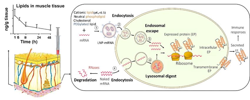
address those formidable obstacles [5] (Figure 1). 1).
UptakeofofmRNA-loaded
Figure1.1.Uptake
Figure mRNA-loadedlipid lipidnanoparticles
nanoparticles(LNPs)
(LNPs)following
followingintramuscular
intramuscularinjection
injectionversus
versusnaked
nakedmRNA
mRNAinto into
the host cells and the subsequent intracellular trafficking. (1) mRNA is incorporated into LNP, which is assembled
the host cells and the subsequent intracellular trafficking. (1) mRNA is incorporated into LNP, which is assembled from from
cationic,
cationic,neutral,
neutral,and
and PEGylated
PEGylated lipids as well
lipids as well as
ascholesterol;
cholesterol;(2)
(2)mRNA-loaded
mRNA-loadedLNPs LNPsareare internalised
internalised into
into thethe host
host cell;cell;
and
and (3) within the cell, mRNA-loaded LNPs undergo endosome escape, and release mRNA for protein
(3) within the cell, mRNA-loaded LNPs undergo endosome escape, and release mRNA for protein synthesis. In contrast,synthesis. In con-
trast, (4) naked mRNA can be partially internalised, but the majority is degraded in lysosomes. In addition, the extracel-
(4) naked mRNA can be partially internalised, but the majority is degraded in lysosomes. In addition, the extracellular
lular mRNA is cleaved by the ribonucleases (RNases) while LNPs at the injection site protect of mRNA from RNases.
mRNA is cleaved by the ribonucleases (RNases) while LNPs at the injection site protect of mRNA from RNases. LNPs
LNPs provide extended local tissue exposure and the pharmacokinetic profile depends on various factors such as the
provide
lipids usedextended local size.
and particle tissue exposure and the pharmacokinetic profile depends on various factors such as the lipids used
and particle size.
Nanoparticles are nanoscale (1–1000 nm) vesicles that have been explored as non-
Nanoparticles are nanoscale (1–1000 nm) vesicles that have been explored as non-viral
viral vectors for the intracellular delivery of mRNA while protecting exogenous mRNA
vectors for the intracellular delivery of mRNA while protecting exogenous mRNA from
from RNase destruction [19]. Compared with viral vectors, non-viral vectors have the ad-
RNase destruction [19]. Compared with viral vectors, non-viral vectors have the advantages
vantages of less immunogenicity, easier production, and higher efficiency to carry the
of less immunogenicity, easier production, and higher efficiency to carry the mRNA [20].
mRNA [20]. Versatile nano-platforms have been investigated to deliver mRNA in the
Versatile nano-platforms have been investigated to deliver mRNA in the body, including
body, including LNPs, polymer-based nanoparticles, polymer-lipid hybrid nanoparticles,
LNPs, polymer-based nanoparticles, polymer-lipid hybrid nanoparticles, poly-peptides,
poly-peptides, protein
protein derivatives mRNA derivatives
complexes, mRNA
and goldcomplexes, and [16,19].
nanoparticles gold nanoparticles
Among these[16,19].
vectors,
Among
LNPs are the most commonly used due to the relative ease of large scaled ease
these vectors, LNPs are the most commonly used due to the relative of large
manufacture,
scaled manufacture,
their ability their
to deliver theability to deliver
encapsulated the encapsulated
mRNA to ribosomes mRNA to ribosomes
(via endosome (via en-
escape), and
dosome escape),
flexibility and flexibility
for surface for surface
modification with a modification with acell
ligand for specific ligand for specific
targeting, cell tar-to
for example
geting,
enhanceforthe
example
uptaketointo
enhance the uptake
dendritic into LNPs
cells [5,18]. dendritic cells [5,18].
are versatile drugLNPs are versatile
delivery systems
drug delivery systems that have been investigated to tackle various disease,
that have been investigated to tackle various disease, including infection and cancer including[21].
As LNPs consist of at least one synthetic or natural lipid layer and an aqueous, oil, solid,
or amorphous core [22], hydrophilic molecules, such as mRNA, small interference RNA
(siRNA) and proteins can be entrapped within the LNP aqueous core, whereas lipophilic
compounds can be incorporated into the lipid layers [18]. Thus, LNPs can be co-loadedEncyclopedia 2021, 1 777
with an immunopotentiator, also known as adjuvants, to enhance the immune response
of the mRNA vaccine. The choice of LNPs for mRNA vaccine delivery has shifted from
traditional liposomes, lipoplexes, and cationic nanoemulsions, to more advanced solid
nanoparticles and nanostructured lipid carriers [20]. LNPs were employed not only by
BNT162b2 and mRNA-1273, but also by other mRNA vaccines in the development of
delivery carriers, which are listed in Table 1.
4.2. Formulation Compositions of Covid-19 mRNA Vaccines
The lipids in LNPs to ferry the mRNA are usually made of four typical components
(Figure 1): an ionisable cationic lipid, a PEGylated lipid, a neutral phospholipid, and
cholesterol. Table 2 lists the lipid components employed in BNT162b2 and mRNA-1273,
and each of these different lipids serves distinct functions:
(1) The ionisable cationic lipids show neutral or mild cationic charges at physiological
pH, and become protonated (cationic) at low pH, therefore, an acid dissociation constant,
or pKa around 6.5 is ideal. At acidic environment, these lipids are able to encapsulate the
negatively charged mRNA through electrostatic interactions during LNPs formation; and
in the physiological environment, it becomes neutral, preventing the rapid removal of the
LNPs from the circulation, and reduces toxicity. The ionisable lipid was reported to be
the primary driver of immunogenicity, pharmacokinetics of the LNPs, and tissue tolera-
bility [23]. Increased biodegradability of the ionisable lipids leads to faster elimination of
lipids in muscle, spleen, and liver without necessarily compromising the immune responses
as little correlation between the protein expression and immunogenicity. In addition, more
biodegradable ionisable lipids are removed from the injection site faster, thus less tissue
irritancy [23]. Furthermore, depending on the lipid structure, some positively charged lipid
can serve as a vaccine adjuvant and stimulate the innate immune response [20]. The ionis-
able lipids used in BNT162b2 and mRNA-1273 are (4-hydroxybutyl)azanediyl)bis(hexane-
6,1-diyl)bis(2-hexyldecanoate) (ALC-0315), and SM-102, respectively. The N:P molar ratio,
defined as nitrogen (N) in the ionizable cationic lipid: phosphate (P) of the nucleotide, is
approximately 6 for both vaccines [8,24].
Table 2. Lipid composition of BNT162b2 and mRNA-1273 [3,4,24].
Lipid BNT162b2 mRNA-1273
Ionisable cationic lipid (A) ALC-0315 SM-102
2-[(polyethylene 1,2-dimyristoyl-rac-glycero3-methoxypolyethylene
PEGylated lipid (B)
glycol)-2000]-N,N-ditetradecylacetamide (ALC-0159) glycol-2000
Neutral lipid (C) DSPC DSPC
A:B:C:cholesterol molar ratios 46.3:1.6:9.4:42.7 50:1.5:10:38.5
(2) A PEGylated lipid consists of a hydrophilic polymer PEG conjugated to a lipid.
In LNPs, the PEG chains anchored on the surface sterically stabilise the LNPs from ag-
gregation, and prevent non-specific binding with proteins (opsonins) and removal by the
reticuloendothelial system in the body [5]. However, the PEG layers could also inhibit the
uptake by the targeted cells as well as interaction with the endosomal membrane, therefore,
the PEG density on the LNP must be carefully tailored and are generally lower than 2%
(mol) and the PEG chain length is limited to 2000.
(3) Neutral lipids are responsible for forming the backbone of the lipid layer and
maintain the LNP structure. 1,2-Distearoyl-sn-glycero-3-phosphocholine (DSPC) was used
in both of the currently approved COVID-19 mRNA vaccines. It is a saturated lipid with a
high melting point, therefore, is able to produce a stable LNPs [20].
(4) Cholesterol in the LNP formulation acts as a stabilising agent of the particles as
well as limiting non-specific LNP-protein interactions [20].Encyclopedia 2021, 1 778
4.3. How Does LNPs Assist mRNA Delivery into the Ribosomes Inside Cells
Following intramuscular injection, LNPs provide extended local tissue exposure
at the injection site [18]. A typical pharmacokinetic profile is shown in Figure 1. The
pharmacokinetics of LNP vaccines depend on their particle size, surface charge, and
colloidal stability of LNPs as well as the biodegradability of the ionisable cationic lipid [23].
These factors in turn determine cellular uptake and lymphatic trafficking as well as the
exposure to liver and spleen [18]. LNPs smaller than 150 nm could be drained via afferent
lymphatic vessels while larger LNPs are readily phagocytosed by immune cells and then
trafficked to the lymph nodes. However, complexity associated with identifying the best
formulation parameters as lymphatic drainage and transfection potency are not the only
parameters determining the immune response.
LNPs have two major roles in mRNA delivery; mediating the uptake of mRNA
into the targeted host cell and promoting the release of mRNA from endosome into the
cytoplasm, thus improve the bioavailability of mRNA to ribosomes where mRNA produces
proteins (antigen) (Figure 1). In addition, LNPs encapsulate the mRNA within the core,
thus protecting the mRNA from being degraded by the extracellular RNases [10].
LNPs facilitate the cellular uptake of the mRNA via various endocytosis routes,
for example, clathrin- and caveolae-mediated endocytosis [5,22]. The efficiency of the
internalization largely depends on the selection of lipids, degree of PEGylation, particle
size, and surface charge of the nanoparticles. After endocytosis, LNPs are first entrapped
inside the endosomes. The ionisable cationic lipids play a crucial role in promoting the
endosomal escape of mRNA. Within the acidic environment of the endosome (pH < ~6.5)
the ionisable lipids become protonated. Those positively charged lipids form ion pairs
with the negatively charged phospholipids in the endosome membrane, disrupt the bilayer
structure and result in endosomal escape [18]. In addition, inclusion of cholesterol in the
LNP structure may contribute to the fusion of the LNPs with the endosome, and promotes
the release of mRNA into the cytoplasm [20]. However, the current LNPs generally have
a low degree of endosome escape (Encyclopedia 2021, 1 779
Table 3. Stability and storage requirement of BNT162b2 and mRNA-1273 vaccines [3,4].
Un-Opened Vials Opened Vials
Vaccines
Abient
Frozen Fridge
Temperature
6 monthsEncyclopedia 2021, 1 780
6. Brenner, S.; Jacob, F.; Meselson, M. An unstable intermediate carrying information from genes to ribosomes for protein synthesis.
Nature 1961, 190, 576–581. [CrossRef] [PubMed]
7. Pardi, N.; Hogan, M.J.; Porter, F.W.; Weissman, D. mRNA vaccines-a new era in vaccinology. Nat. Rev. Drug Discov. 2018, 17,
261–279. [CrossRef]
8. Verbeke, R.; Lentacker, I.; De Smedt, S.C.; Dewitte, H. The dawn of mRNA vaccines: The COVID-19 case. J. Control. Release 2021,
333, 511–520. [CrossRef]
9. Cai, Y.; Zhang, J.; Xiao, T.; Peng, H.; Sterling, S..; Walsh, R., Jr.; Rawson, S.; Rits-Volloch, S.; Chen, B. Distinct conformational states
of SARS-CoV-2 spike protein. Science 2020, 369, 1586–1592. [CrossRef]
10. Zeng, C.; Zhang, C.; Walker, P.G.; Dong, Y. Formulation and delivery technologies for mRNA vaccines. In Current Topics in
Microbiology and Immunology; Springer: Berlin/Heidelberg, Germany, 2020; pp. 1–40.
11. Wadhwa, A.; Aljabbari, A.; Lokras, A.; Foged, C.; Thakur, A. Opportunities and challenges in the delivery of mRNA-based
vaccines. Pharmaceutics 2020, 12, 102. [CrossRef]
12. Blakney, A.K.; Ip, S.; Geall, A.J. An update on self-amplifying mRNA vaccine development. Vaccines 2021, 9, 97. [CrossRef]
13. Kutzler, M.A.; Weiner, D.B. DNA vaccines: Ready for prime time? Nat. Rev. Genet. 2008, 9, 776–788. [CrossRef]
14. Polack, F.; Thomas, S.; Kitchin, N.; Absalon, J.; Gurtman, A.; Lockhart, S. Safety and efficacy of the BNT162b2 mRNA COVID-19
vaccine. N. Engl. J. Med. 2020, 383, 2603–2615. [CrossRef]
15. Baden, L.R.; El Sahly, H.M.; Essink, B.; Kotloff, K.; Frey, S.; Novak, R.; Diemert, D.; Spector, S.A.; Rouphael, N.; Creech, C.B.; et al.
Efficacy and safety of the mRNA-1273 SARS-CoV-2 vaccine. N. Engl. J. Med. 2021, 384, 403–416. [CrossRef] [PubMed]
16. Banerji, A.; Wickner, P.G.; Saff, R.; Stone, C.A.; Robinson, L.B.; Long, A.A.; Wolfson, A.R.; Williams, P.; Khan, D.A.; Phillips, E.;
et al. mRNA vaccines to prevent COVID-19 disease and reported allergic reactions: Current evidence and suggested approach. J.
Allergy Clin. Immunol. Pract. 2021, 9, 1423–1437. [CrossRef]
17. Greenhawt, M.; Abrams, E.M.; Oppenheimer, J.; Leek, T.K.V.; Mack, D.P.; Singer, A.G.; Shaker, M. The COVID-19 pandemic in
2021: Avoiding overdiagnosis of anaphylaxis risk while safely vaccinating the world. J. Allergy Clin. Immunol. Pract. 2021, 9, 1438.
[CrossRef] [PubMed]
18. Reichumuth, A.M.; Oberli, M.; Jaklenec, A.; Langer, R.B.D. mRNA vaccine delivery using lipid nanoparticles. Ther. Deliv. 2016, 7,
319–334. [CrossRef] [PubMed]
19. Lorenz, C.; Fotin-Mleczek, M.; Roth, G.; Becker, C.; Dam, T.C.; Verdurmen, W.P.R.; Brock, R.; Probst, J.; Schlake, T. Protein
expression from exogenous mRNA: Uptake by receptor-mediated endocytosis and trafficking via the lysosomal pathway. RNA
Biol. 2011, 8. [CrossRef]
20. Aldosari, B.N.; Alfagih, I.M.; Almurshedi, A.S.; Hinrichs, J. Lipid nanoparticles as delivery systems for RNA-based vaccines.
Pharmaceutics 2021, 13, 206. [CrossRef]
21. Mitchell, M.J.; Billingsley, M.M.; Haley, R.M.; Wechsler, M.E.; Peppas, N.A.; Langer, R. Engineering precision nanoparticles for
drug delivery. Nat. Rev. Drug Discov. 2021, 20, 101–124. [CrossRef]
22. Li, B.; Zhang, X.; Dong, Y. Nanoscale platforms for messenger RNA delivery. Wiley Interdiscip. Rev. Nanomed. Nanobiotechnol.
2018, 11, 1530. [CrossRef]
23. Hassett, K.J.; Benenato, K.E.; Jacquinet, E.; Lee, A.; Woods, A.; Yuzhakov, O.; Himansu, S.; Deterling, J.; Geilich, B.M.; Ketova, T.;
et al. Optimization of lipid nanoparticles for intramuscular administration of mRNA vaccines. Mol. Ther. Nucleic Acids 2019, 15,
1–11. [CrossRef]
24. Schoenmaker, L.; Witzigmann, D.; Kulkarni, J.A.; Verbeke, R.; Kersten, G.; Jiskoot, W.; Crommelin, D.J.A. mRNA-lipid nanoparticle
COVID-19 vaccines: Structure and stability. Int. J. Pharm. 2021, 601, 120586. [CrossRef] [PubMed]
25. Corbett, K.S.; Edwards, D.K.; Leist, S.R.; Abiona, O.M.; Boyoglu-Barnum, S.; Gillespie, R.A.; Himansu, S.; Schäfer, A.; Ziwawo,
C.T.; DiPiazza, A.T.; et al. SARS-CoV-2 mRNA vaccine design enabled by prototype pathogen preparedness. Nature 2020, 586,
567–571. [CrossRef] [PubMed]
26. Crommelin, D.J.A.; Anchordoquy, T.J.; Volkin, D.B.; Jiskoot, W.; Mastrobattista, E. Addressing the cold reality of mRNA vaccine
stability. J. Pharm. Sci. 2021, 110, 997–1001. [CrossRef] [PubMed]You can also read