Open Fetal Microneurosurgery for Intrauterine Spina Bifida Repair
←
→
Page content transcription
If your browser does not render page correctly, please read the page content below
Research Article

                                                    Fetal Diagn Ther 2021;48:163–173                                       Received: April 16, 2020
                                                                                                                           Accepted: November 23, 2020
                                                    DOI: 10.1159/000513311                                                 Published online: February 12, 2021

Open Fetal Microneurosurgery for
Intrauterine Spina Bifida Repair
Rogelio Cruz-Martínez a, b, c Felipe Chavelas-Ochoa d
Miguel Martínez-Rodríguez a, c Karla Aguilar-Vidales e Alma Gámez-Varela a
Jonahtan Luna-García a Hugo López-Briones a Joel Chávez-Vega e
Ángel Augusto Pérez-Calatayud f Manuel Alejandro Díaz-Carrillo f
Edgar Ahumada-Angulo a Andrea Castelo-Vargas a Eréndira Chávez-González a
Israel Juárez-Martínez a Rosa Villalobos-Gómez a Carlos Rebolledo-Fernández g
aPrenatal Diagnosis and Fetal Surgery Center, Fetal Medicine Mexico and the Fetal Medicine Foundation of Mexico,

Queretaro, Mexico; bInstituto de Ciencias de la Salud (ICSa) , Universidad Autónoma del Estado de Hidalgo (UAEH),
Hidalgo, Mexico; cDepartment of Fetal Surgery, Hospital de Especialidades del Niño y la Mujer Dr. Felipe Núñez
Lara, Queretaro, Mexico; dDepartment of Pediatric Neurosurgery, Hospital de Especialidades del Niño y la Mujer
Dr. Felipe Núñez Lara, Queretaro, Mexico; eDepartment of Anesthesiology, Hospital de Especialidades del Niño y la
Mujer Dr. Felipe Núñez Lara, Queretaro, Mexico; fDepartment of Maternal Intensive Care, Hospital de Especialidades
del Niño y la Mujer Dr. Felipe Núñez Lara, Queretaro, Mexico; gDepartment of Maternal Fetal Medicine, Hospital de
Especialidades del Niño y la Mujer Dr. Felipe Núñez Lara, Queretaro, Mexico

Keywords                                                                  amniotic fluid and uterine volume during the whole surgery.
Fetal surgery · Microneurosurgery · Minihysterotomy ·                     Perinatal outcomes of cases operated with the classic open
Myelomeningocele · Spina bifida                                           fetal surgery technique and open microneurosurgery were
                                                                          compared. Results: Intrauterine SB repair with a complete
                                                                          3-layer correction was successfully performed in 60 cases ei-
Abstract                                                                  ther by classic open fetal surgery (n = 13) or open microneu-
Objectives: The aim of the study was to describe the feasibil-            rosurgery (n = 47). No significant differences were observed
ity of open fetal microneurosurgery for intrauterine spina bi-            in gestational age (GA) at fetal intervention (25.4 vs. 25.1
fida (SB) repair and to compare perinatal outcomes with cas-              weeks, p = 0.38) or surgical times (107 vs. 120 min, p = 0.15)
es managed using the classic open fetal surgery technique.                between both groups. The group with open microneurosur-
Methods: In this study, we selected a cohort of consecutive               gery showed a significantly lower rate of oligohydramnios (0
fetuses with isolated open SB referred to our fetal surgery               vs. 15.4%, p = 0.01), preterm rupture of the membranes (19.0
center in Queretaro, Mexico, during a 3.5-year period (2016–              vs. 53.8%, p = 0.01), higher GA at birth (35.1 vs. 32.7 weeks,
2020). SB repair was performed by either classic open sur-                p = 0.03), lower rate of preterm delivery
hysterotomy site at delivery. Conclusion: Intrauterine spina          15-20 mm, reduced fetal manipulation, and maintenance
repair by open fetal microneurosurgery is feasible and was            of normal amniotic fluid volume during the whole inter-
associated with better perinatal outcomes than classic open           vention. We also compared the perinatal outcomes of
fetal surgery.                           © 2021 S. Karger AG, Basel   such cases with those previously operated with the classic
                                                                      open fetal surgery technique in our center.

                                                                         Methods
   Introduction
                                                                          Subjects
    Spina bifida (SB) is a neural tube malformation that                  Between December 2016 and May 2020, a prospective cohort
represents the most common severely disabling congeni-                of singleton fetuses with confirmed lumbosacral open SB were se-
tal defect. It complicates about 1 in 2,000 live births and           lected for fetal surgery at Hospital de Especialidades del Niño y la
                                                                      Mujer, and Hospital San José, Queretaro, Mexico (a national refer-
is associated with major disabilities such as motor dys-              ral centers for fetal surgery). Detailed fetal morphological ultra-
function or paralysis, mental retardation, hydrocephalus              sound assessment, advanced neurosonography, maternal psycho-
requiring ventriculoperitoneal shunting, bowel and blad-              logical evaluation, and fetal karyotype by amniocentesis were per-
der dysfunction, and long-term neurological handicaps                 formed in all cases.
[1]. Since fetal brain damage is progressive during gesta-                All ultrasound examinations were performed with Voluson E8
                                                                      Expert BT 12.0 or Voluson E10 BT18.0 US equipment with a 6- to
tion due to the chronic exposure of neural tissue to the              2-MHz linear curved-array transducer by 1 of 2 experienced ex-
amniotic fluid, fetal surgery for prenatal myelomeningo-              aminers (R.C.M. or M.M.R.). The inclusion criteria for fetal sur-
cele (MMC) repair was then introduced in an attempt to                gery were singleton pregnancies at
Color version available online
a b
c d
Fig. 1. 15-mm hysterotomy diameter (a) and placement of the self-retaining retractor allowing visualization of
the neural defect and positioning of the enteral feeding tube for continuous infusion of warmed lactated Ringer´s
solution (b). Two lateral sutures (c) were placed in the fetal back (arrow) in order to be fixed against the hyster-
otomy and thus, to expose the portion of the neural tube defect required by the neurosurgeon to perform the
fetal myeloplasty with a complete 3-layer correction (d).
MOMs trial (i.e., complete 3-layer correction through a 6- to 8-cm SB defect located against the uterine wall and in the contralateral
hysterotomy with leakage of amniotic fluid which was replaced side of the placenta. Under ultrasound guidance, two 1-0 absorb-
before uterine closure) while in the last cases, a novel technique able monofilament stitches were placed to plicate the membranes
called open fetal microneurosurgery was performed. to the uterine wall and then a 15- to 20-mm vertical and fundal
In brief, under maternal regional and general anesthesia with midline hysterotomy was performed between the sutures with a
sevoflurane, a maternal Pfannenstiel laparotomy through a low monopolar cautery pencil (Fig. 1a). The amniotic membranes
transverse abdominal incision was performed to allow uterine ex- were opened and plicated to the uterine wall by continuous locking
teriorization from the abdominal cavity. The fetal position was polyglactin 1-0 stitches (Ethicon Inc.). A self-retaining retractor
evaluated by ultrasound and, if necessary, the fetus was moved by (Weitlaner), which includes 3 × 4 blunt outward-curving teeth,
a gentle external manipulation to reach the optimal position, that was used to expose and spread the surgical site and, an 8F enteral
is, a longitudinal position, with cephalic presentation and with the feeding tube was introduced to the amniotic cavity to perform a
Fetal Microneurosurgery for Spina Bifida Fetal Diagn Ther 2021;48:163–173 165
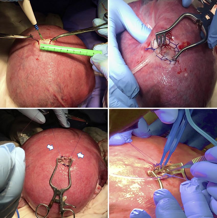
Repair DOI: 10.1159/000513311Color version available online
a b
Fig. 2. Final result after multilayer fetal myeloplasty immediately before (a) and after (b) uterine closure.
continuous infusion of warmed (37°C) lactated Ringer’s solution during the first 48 h after surgery. Prophylactic tocolysis with in-
with antibiotic (2 g cefotaxime per L) in order to maintain the in- domethacin, nifedipine or atosiban was administered immediately
trauterine volume and achieve a normal amniotic fluid index dur- after wound closure and up to 72 h after the procedure. All patients
ing the entire fetal surgery (Fig. 1b). To minimize fetal manipula- were discharged 1 week after fetal surgery and were advised to stay
tion and amniotic fluid loss through the tiny uterine incision, the in the city until delivery. The potential existence of uterine con-
fetal back was fixed against the hysterotomy with 2 monofilament tractions was registered every 24 h by ambulatory cardiotocogra-
4-0 stitches (Fig. 1c). Due to the small hysterotomy diameter, ei- phy. Whenever preterm delivery or PPROM was anticipated, a
ther an HD-3D exoscope system or surgical loupes (4.5×) were course of corticosteroids (betamethasone, 12 mg 2 doses 24 h
used by the pediatric neurosurgeon (FCO) to improve illumina- apart) was administrated. Ultrasound examination to evaluate the
tion, magnification, and visualization of the neural placode, which amount of amniotic fluid, absence of chorioamniotic separation,
was released and positioned within the spina canal and then a fetal estimated weight, and cervical length was performed every
3-layer watertight closure of the neural defect was performed using week until delivery. If a short cervical length was observed during
a monofilament 4-0 suture with a 17-mm needle and biopolar cau- fetal follow-up, vaginal progesterone (200 mg daily) was adminis-
tery (Fig. 1d). In all cases, the size of the neural defect was larger tered until delivery. Fetal neurosonography to identify the regres-
than the hysterotomy incision, and therefore, exposure of the neu- sion of hindbrain herniation and to monitor the lateral ventricle
ral defect was mainly allowed by a gentle mobilization of the uter- diameters was also performed every week. Reversal of Chiari mal-
ine retractor while the fetal back was held against the hysterotomy. formation was defined by fetal ultrasound when the obliterated
In case this maneuver was insufficient, a gentle manipulation of cisterna magna became visible and measurable and the cerebellum
the fetus by the fetal surgery specialist was performed to properly fastigium regressed to a normal position and morphology, that is,
expose the portion of the neural tube defect required by the neu- above the foramen magnum.
rosurgeon. In cases with myeloschisis, a 2.5 × 2.5-cm biological
patch (Lyoplant Onlay) was used, which is an absorbable, liquid- Neonatal Management and Infant Follow-Up
tight, bilayer collagen implant produced from a lyophilized bovine According to our clinical protocol, cesarean delivery was
pericardium designed for dura substitution that can be cut accord- planned at above 38 weeks. All deliveries and neonatal manage-
ing to the shape of the neural defect. Fetal monitoring was con- ment were attended by staff obstetricians, maternal fetal medicine
tinuously performed by a fetal sonographer during the entire sur- specialists, and neonatologists either at our center or at the referral
gical procedure by using fetal Doppler ultrasound. In case of uter- center. Neurological follow-up was performed in our neurodevel-
ine contraction during fetal intervention, maternal nitroglycerin opmental unit which includes neonatal neurohabilitation by a
was used for uterine relaxation [12]. Once fetal myeloplasty was neonatal physical rehabilitator (A.C.B.) and clinical evaluations by
completed, uterine closure was performed by 2 layers (Fig. 2), the a pediatric neurosurgeon, a pediatric orthopedist, and a pediatric
uterus was interiorized into the abdominal cavity, and then the urologist. Endoscopic third ventriculostomy was considered as
abdominal wall was closed in 3 layers. primary treatment in all infants with hydrocephalus showing any
in the following signs of intracranial hypertension: progressive in-
Maternal and Fetal Follow-Up crease in the head circumference accompanied with a bulging fon-
After fetal surgery, the pregnant woman was hospitalized in the tanelle (i.e., above the level of the bone as assessed when the baby
intensive care unit for 24 h. An elastomeric pump was used for is not crying and in an upright position), clinical signs of Chiari
continuous infusion of pain drugs through the epidural catheter malformation (stridor, swallowing difficulties, apnea, and brady-
166 Fetal Diagn Ther 2021;48:163–173 Cruz-Martínez et al.
DOI: 10.1159/000513311Table 1. Maternal and fetal clinical characteristics of the selected pregnancies for intrauterine SB repair by classic
open fetal surgery and open fetal microneurosurgery
Classic open fetal Open fetal p value
surgery, microneurosurgery,
n = 13 n = 47
Maternal age, years 31.4 (7.7) 29.5 (6.1) 0.36
Primiparity, n (%) 7 (53.8) 16 (34.0) 0.19
Previous cesarean section, n (%) 2 (15.4) 20 (42.6) 0.07
BMI, kg/m2 25.2 (4.2) 24.9 (3.9) 0.81
BMI > 35, n (%) 0 (0) 1 (2.1) 0.60
Anterior placenta, n (%) 10 (76.9) 17 (36.2)Table 2. Maternal and fetal studied variables from fetal surgery to delivery
Classic open Open fetal p value
fetal surgery, microneurosurgery,
n = 13 n = 42
GA at fetal surgery, wk 25.1 (1.0) 25.3 (0.82) 0.38
Surgery time, min 120.6 (25.4) 107.3 (30.3) 0.15
Maternal hospital days 6.2 (1.7) 5.6 (1.2) 0.32
Postsurgical maternal pulmonary edema, n (%) 0 (0) 1 (2.4) 0.57
Maternal bleeding requiring blood transfusion, n (%) 1 (7.7) 0 (0) 0.07
Chorioamnionitis, n (%) 1 (7.7) 2 (4.8) 0.68
Placental abruption, n (%) 1 (7.7) 0 (0) 0.07
Oligohydramnios, n (%) 2 (15.4) 0 (0) 0.01
Chorioamniotic separation, n (%) 4 (30.8) 9 (21.4) 0.49
PPROM, n (%) 7 (53.8) 8 (19.0) 0.01
PPROM GA, wk 30.0 (4.2) 31.2 (2.9) 0.41
Intrauterine fetal demise, n (%) 1 (7.7) 0 (0) 0.07
GA at birth, wk 32.7 (4.1) 35.4 (3.0) 0.03
Interval between fetal surgery and delivery, weeks 7.5 (3.8) 9.7 (3.0) 0.04
Cesarean delivery, n (%) 13 (100) 39 (92.9) 0.32
Intact hysterotomy site, n (%) 13 (100) 42 (100) 1.00
Preterm deliveryTable 3. Infant characteristic during follow-up among survivors
Classic open Open fetal p value
fetal surgery, microneurosurgery,
n = 10 n = 40
Infant age at last evaluation, months 32.5 (5.1) 12.2 (9.1) 1 year, n (%) 10 (100) 18 (45.0)terotomy, decreased fetal manipulation, and mainte- microneurosurgery technique nor in Botelho’s study with
nance of a normal amniotic fluid and uterine volume dur- minihysterotomy, the classic open fetal surgery and all
ing the whole fetal intervention and was associated with fetoscopic techniques have showed oligohydramnios
better perinatal outcomes, lower rate of preterm delivery, rates of 23 and 14%, respectively [7, 14–17, 20].
PPROM, and perinatal death compared to the classic Although we expect that minimizing the uterine inci-
open fetal surgery technique. sion may also improve reproductive outcomes by reduc-
Classic open fetal surgery was initially performed ing the risk of uterine rupture and the rate of cesarean
through a 6- to 8-cm hysterotomy, allowing the pediatric section, other obstetrical parameters such as parity, bish-
neurosurgeon a good visualization for a multilayer cor- op score, labor stage, fetal estimated weight, and fetal po-
rection of the neural defect [3]. However, such technique sition must be also considered for an algorithm for a clin-
has showed potential risks of maternal morbidity and ical decision regarding vaginal birth in this population.
complications such as PPROM, preterm delivery, placen- Notably, a few patients treated with our technique showed
tal abruption, chorioamniotic separation, chorioamnion- an unplanned but uneventfully vaginal delivery. This ob-
itis, perinatal death, maternal pulmonary edema, uterine servation may represent the basis for further studies eval-
bleeding requiring maternal blood transfusion, and uter- uating the possibility of a planned vaginal delivery in se-
ine dehiscence at delivery [4, 5]. The concept of decreas- lected cases with open microsurgery.
ing the hysterotomy length for MMC repair in an attempt Our study supports the current evidence demonstrat-
to decrease maternal morbidity and perinatal complica- ing that fetal surgery for SB repair decreases the risk of
tions was first reported by Botelho et al. [10]. In such hydrocephalus and the need of ventriculoperitoneal
study, the outcomes of 39 consecutive fetuses managed shunting, with a low risk of perinatal death [3, 11, 20, 21].
with open fetal surgery through a 2.5–3.5 cm hysterotomy We recognize that our study has also some limitations.
were evaluated showing a 50% decreased risk of PPROM First of all, although similar or higher than that in previ-
and preterm delivery, with no maternal, fetal or neonatal ous published studies, our included sample size is limit-
complications. In agreement with such study, the PPROM ed, and further studies are required for an external vali-
rate, preterm delivery rate, and short-term neonatal out- dation of our results. The lack of long-term neurological
comes in our microneurosurgery technique compare fa- outcomes is also a limitation, and therefore, a long-term
vorably against those in our control group and with the study is required to confirm the potential benefits and
results reported in previous published studies (Table 5). neurological outcomes of our minimally invasive neuro-
In a small cohort of MMC cases, the team at Texas surgical intervention. Of note and contrary to the MOMs
Children’s Hospital recently reported a novel fetoscopic trial, cases with diagnosis or referral between 26 and 28
technique using a two-port approach with an exteriorized weeks of gestation were also selected for fetal interven-
uterus showing good perinatal outcomes with lower rate tion. Although, this is not the first published study in-
of preterm delivery and PPROM than previous published cluding cases above 26 weeks of gestation [10, 11, 15, 16,
techniques [14–17]. In terms of uterine invasion, since 22], a previous study by Peralta et al. [11] showed that
our technique requires a hysterotomy of only 15-20 mm, early GA at surgery was associated with higher rates of
it could be argued that our technique may be as minimal- hindbrain herniation reversal and lower frequency of
ly invasive as the fetoscopic approach which requires 2 or ventriculoperitoneal shunting, and therefore, it would be
3 ports of 5 mm, that is, 2 or 3 uterine incisions of at least expected that fetal interventions above 26 weeks may be
5 mm each. However, our technique had required lower associated with poorer neurological outcomes, but fur-
surgical time than all fetoscopic techniques (Table 5) [8, ther comparison studies between early and late surgical
9, 14–16]. Another potential strength of our technique procedures are required to justify the upper limit of GA
over the fetoscopic approach is that it does not require the at surgery. In addition, it could be argued that a potential
use of carbon dioxide insufflation into the amniotic cav- risk of trauma to the placode may exist during fetal ma-
ity, and thus avoidance its potential theoretical risks [18, nipulation used to expose the neural tube defect through
19]. In addition, our results also suggest that decreasing a tiny uterine incision. However, the fact that amniotic
the hysterotomy diameter, minimizing fetal manipula- membranes were plicated against myometrium makes
tion, and maintaining normal amniotic fluid volume dur- such risk very unlikely. Finally, as a potential limitation
ing fetal surgery may be associated with a lower incidence for the widespread use of this novel approach for intra-
of oligohydramnios during fetal follow-up. While no cas- uterine SB repair, it must be acknowledged that such
es of fetal oligohydramnios were observed neither in our minimally invasion procedure is very challenging be-
170 Fetal Diagn Ther 2021;48:163–173 Cruz-Martínez et al.
DOI: 10.1159/000513311Repair
Fetal Microneurosurgery for Spina Bifida
Table 5. Open fetal microneurosurgery technique compared with other published techniques for intrauterine SB repair
Cruz-Martinez Botelho Adzick Sepulveda Winder Moron Belfort Pedreira Kohl [15]
et al. 2021 et al. [10] et al. [3] et al. [6] et al. [5] et al. [20] et al. [17] et al. [16] n = 51
(current series) n = 39 n = 78 n = 57 n = 40 n = 237 n = 22 n = 45
n = 60*
Approach Open Open mini- Open Open Open Open Fetoscopic- Fetoscopic- Fetoscopic-
microneurosurgery hysterotomy hysterotomy hysterotomy hysterotomy hysterotomy laparotomy percutaneous percutaneous
GA at surgery 22.9–27.6 20.7–26.9 19–25.9 21.0–26.0 19–25.9 24.0–27.0 22.9–25.9 24–28.9 21–29.1
PPROM, n (%) 8 (19.0) 9 (23.1) 36 (46.2) 14 (24.6) 14 (35) 63 (26.6) 5 (22.7) 36 (80.0) 43 (84.3)
Choriamniotic separation, n (%) 0 (0) 1 (2.6) 20 (25.6) 4 (7.0) 12 (30) 49 (20.7) 2 (9.1) – 3 (5.9)
Placental abruption, n (%) 0 (0) 1 (2.6) 5 (6.4) 0 (0) 4 (10) 2 (0.8) 2 (9.1) – 0 (0)
DOI: 10.1159/000513311
Oligohydramnios, n (%) 0 (0) 0 (0) 16 (20.5) – 55 (23.2) 3 (13.6) – 7 (13.7)
Pulmonary edema, n (%) 1 (2.4) 0 (0) 5 (6.4) 2 (3.5) 0 (0) 6 (2.5) 2 (9.1) 0 (0) 1 (1.9)
Chorioamnionitis, n (%) 1 (2.4) 0 (0) 2 (2.6) 1 (1.8) 2 (5) 7 (3.0) 0 (0) 0 (0) 3 (5.9)
Fetal Diagn Ther 2021;48:163–173
Perinatal death, n (%) 2 (4.8) 0 (0) 2 (2.6) 5 (8.6) 0 (0) 5 (2.1) 0 (0) 2 (4.4) 3 (5.9)
Total surgery time, min 107 (64–200) 206 (90–274) 105±21.8 – 139.1±24.1 119.7±7.6 250 (107–434) 193 (83–450) 223 (140–315)
Surgery to delivery interval, weeks 9.7 (1.3–12.9) 10.9 (5.7–17.9) – 9.0 10.8±1.46 7.44±2.4 14.4 (10.3–15.1) 6.1 (1–13) 9.6 (1–14)
GA at birth, weeks 35.4 (26.0–39.6) 35.3 (27.9–39.1) 34.1±3.1 34.0 (24–38.0) 35.5±2.3 33.6±2.4 38.1 (26–40) 32.8±2.5 33.0 (24.6–38.1)
Term deliveries (≥37 wks), n (%) 20 (47.6) 9 (23.1) 16 (20.5) – 14 (35) 31 (13.1) 14 (63.6) – 4 (7.8)
Early preterm deliveries (cause it reduces visualization of the surgical field and Statement of Ethics
thus requires advanced training of the neurosurgeon.
Our research complies with the guidelines for human studies
Nevertheless, using different surgical tools such as the and was conducted ethically in accordance with the World Medi-
self-retaining retractor allows for spreading the surgical cal Association Declaration of Helsinki. All subjects gave their
site to about 5 mm and increases maneuvering through written informed consent, and the study protocol was approved by
the smaller uterine access diameter. In addition, using the by the Hospital Ethics Committee (Hospital de Especialidades
the exoscopic system or surgical loupes enhanced pedi- del Niño y la Mujer Dr. Felipe Núñez Lara, Querétaro, México IRB
077/26042017).
atric neurosurgeon enhanced surgical field understand-
ing and improved hand-eye coordination for a proper
prenatal surgical correction of SB [23], but we learned
Conflict of Interest Statement
that using surgical loupes, which are cheaper than the
exoscopic system, was enough to improve magnification The authors have no conflicts of interests to declare.
and visualization of the surgical field on this high-preci-
sion neurosurgical procedure. In conclusion, our initial
experience suggests that open fetal microneurosurgery Funding Sources
for neural tube defect repair is feasible and is associated
with better perinatal outcomes and similar short-term No funding was obtained for the completion of this study.
neurological outcomes than the classic open fetal surgery
technique.
Author Contributions
R.C.M., F.C.O., M.M.R., K.A.V., A.G.V., J.L.G., H.L.B., J.C.V.,
Acknowledgements and E.A.A. participated in all the fetal interventions. R.C.M.,
F.C.O., M.M.R., and K.A.V. made substantial contributions to de-
Rogelio Cruz-Martinez was supported by the National Council sign the novel fetal surgery technique. R.C.M., F.C.O., M.M.R.,
of Science and technology (Conacyt) and wishes to thank Prof. A.C.V., E.C.G., and I.J.M. did the acquisition of data. R.C.M.,
Waldo Sepulveda, Felipe Otayza, and Juan Carlos Devoto (FE- F.C.O., M.M.R., and E.C.G. did the analysis and interpretation of
TALMED‒ Fetal Surgery group, Santiago, Chile) for supporting data. R.C.M. and R.V.G. drafted the manuscript. All authors listed
the beginning of the national Mexican program of open fetal sur- revised the article for important intellectual content and gave final
gery for intrauterine SB repair. approval of the final version for publication.
References
1 Mitchell LE, Adzick NS, Melchionne J, diagnosis and therapy. Fetal Diagn Ther. 10 Botelho RD, Imada V, Rodrigues da Costa KJ,
Pasquariello PS, Sutton LN, Whitehead AS. 2019;46(3):153–8. Watanabe LC, Rossi Júnior R, De Salles AAF,
Spina bifida. Lancet. 2004 Nov 20–26; 6 Sepulveda W, Corral E, Alcalde JL, Otayza F, et al. Fetal myelomeningocele repair through
364(9448):1885–95. Müller JM, Ravera F, et al. Prenatal repair of a mini-hysterotomy. Fetal Diagn Ther. 2017;
2 Bruner JP, Tulipan N, Paschall RL, Boehm spina bifida: a 2-center experience with open 42(1):28–34.
FH, Walsh WF, Silva SR, et al. Fetal surgery intrauterine neurosurgery in Chile. Fetal Di- 11 Peralta CFA, Botelho RD, Romano ER, Ima-
for myelomeningocele and the incidence of agn Ther. 2020;47(12):873–81. da V, Lamis F, Junior RR, et al. Fetal open
shunt-dependent hydrocephalus. JAMA. 7 Sacco A, Ushakov F, Thompson D, Peebles D, spinal dysraphism repair through a mini-
1999 Nov 17;282(19):1819–25. Pandya P, De Coppi P, et al. Fetal surgery for hysterotomy: influence of gestational age at
3 Adzick NS, Thom EA, Spong CY, Brock JW open spina bifida. Obstet Gynaecol. 2019 Oct; surgery on the perinatal outcomes and post-
3rd, Burrows PK, Johnson MP, et al. A ran- 21(4):271–82. natal shunt rates. Prenat Diagn. 2020; 40(6):
domized trial of prenatal versus postnatal re- 8 Araujo Junior E, Eggink AJ, van den Dobbel- 689–97.
pair of myelomeningocele. N Engl J Med. steen J, Martins WP, Oepkes D. Procedure- 12 Devoto JC, Alcalde JL, Otayza F, Sepulveda
2011 Mar 17;364(11):993–1004. related complications of open vs endoscopic W. Anesthesia for myelomeningocele surgery
4 Sacco A, Van der Veeken L, Bagshaw E, Fer- fetal surgery for treatment of spina bifida in in fetus. Childs Nerv Syst. 2017 Jul; 33(7):
guson C, Van Mieghem T, David AL, et al. an era of intrauterine myelomeningocele re- 1169–75.
Maternal complications following open and pair: systematic review and meta-analysis. Ul- 13 Elbabaa SK, Gildehaus AM, Pierson MJ, Al-
fetoscopic fetal surgery: a systematic review trasound Obstet Gynecol. 2016 Aug; 48(2): bers JA, Vlastos EJ. First 60 fetal in-utero my-
and meta-analysis. Prenat Diagn. 2019 Mar; 151–60. elomeningocele repairs at Saint Louis Fetal
39(4):251–68. 9 Joyeux L, Engels AC, Russo FM, Jimenez J, Care Institute in the post-MOMS trial era: hy-
5 Winder FM, Vonzun L, Meuli M, Moehrlen Van Mieghem T, De Coppi P, et al. Fetoscop- drocephalus treatment outcomes (endoscop-
U, Mazzone L, Krähenmann F, et al. Maternal ic versus open repair for spina bifida aperta: a ic third ventriculostomy versus ventriculo-
complications following open fetal myelome- systematic review of outcomes. Fetal Diagn peritoneal shunt). Childs Nerv Syst. 2017 Jul;
ningocele repair at the zurich center for fetal Ther. 2016;39(3):161–71. 33(7):1157–68.
172 Fetal Diagn Ther 2021;48:163–173 Cruz-Martínez et al.
DOI: 10.1159/00051331114 Degenhardt J, Schürg R, Winarno A, Oehmke 17 Belfort MA, Whitehead WE, Shamshirsaz 21 Houtrow AJ, Thom EA, Fletcher JM, Burrows
F, Khaleeva A, Kawecki A, et al. Percutaneous AA, Bateni ZH, Olutoye OO, Olutoye OA, et PK, Adzick NS, Thomas NH, et al. Prenatal
minimal-access fetoscopic surgery for spina al. Fetoscopic open neural tube defect repair: repair of myelomeningocele and school-age
bifida aperta. Part II: maternal management development and refinement of a two-port, functional outcomes. Pediatrics. 2020 Feb;
and outcome. Ultrasound Obstet Gynecol. carbon dioxide insufflation technique. Obstet 145(2):e20191544.
2014 Nov;44(5):525–31. Gynecol. 2017 Apr;129(4):734–43. 22 Corral E, Sepulveda W, Ravera F, Muller JM,
15 Kohl T. Percutaneous minimally invasive fe- 18 Skinner S, Crossley K, Amberg B, Kashyap A, Tapia M, Reascos M, et al. Use of plastic
toscopic surgery for spina bifida aperta. Part Hooper S, Deprest JA, et al. The effects of par- wound retractor at hysterotomy site in prena-
I: surgical technique and perioperative out- tial amniotic carbon dioxide insufflation in an tal repair of myelomeningocele: a new tech-
come. Ultrasound Obstet Gynecol. 2014 Nov; ovine model. Prenat Diagn. 2018 Dec;38(13): nique. J Matern Fetal Neonatal Med. 2020;
44(5):515–24. 994–1003. 33(17):3010–15.
16 Pedreira DA, Zanon N, Nishikuni K, Moreira 19 Skinner S, DeKoninck P, Crossley K, Amberg 23 Oertel JM, Burkhardt BW. Vitom-3D for exo-
de Sá RA, Acacio GL, Chmait RH, et al. Endo- B, Deprest J, Hooper S, et al. Partial amniotic scopic neurosurgery: initial experience in cra-
scopic surgery for the antenatal treatment of carbon dioxide insufflation for fetal surgery. nial and spinal procedures. World Neurosurg.
myelomeningocele: the CECAM trial. Am J Prenat Diagn. 2018 Dec;38(13):983–93. 2017 Sep;105:153–62.
Obstet Gynecol. 2016 Jan;214(1):111–e11. 20 Moron AF, Barbosa MM, Milani H, Sarmento
SG, Santana E, Suriano IC, et al. Perinatal out-
comes after open fetal surgery for myelome-
ningocele repair: a retrospective cohort study.
BJOG. 2018 Sep;125(10):1280–6.
Fetal Microneurosurgery for Spina Bifida Fetal Diagn Ther 2021;48:163–173 173
Repair DOI: 10.1159/000513311You can also read