PH- and concentration-dependent supramolecular self-assembly of a naturally occurring octapeptide - Beilstein Journal of Organic Chemistry
←
→
Page content transcription
If your browser does not render page correctly, please read the page content below
pH- and concentration-dependent supramolecular self-
assembly of a naturally occurring octapeptide
Goutam Ghosh* and Gustavo Fernández*

  Full Research Paper                                                                                                               Open Access

  Address:                                                             Beilstein J. Org. Chem. 2020, 16, 2017–2025.
  Organisch-Chemisches Institut, Westfälische Wilhelms-Universität     doi:10.3762/bjoc.16.168
  Münster, Correnstraße 40, 48149 Münster, Germany
                                                                       Received: 17 May 2020
  Email:                                                               Accepted: 31 July 2020
  Goutam Ghosh* - gghosh.chem@gmail.com; Gustavo Fernández* -          Published: 17 August 2020
  fernandg@uni-muenster.de
                                                                       This article is part of the thematic issue "Molecular recognition".
  * Corresponding author
                                                                       Associate Editor: N. Sewald
  Keywords:
  aqueous self-assembly; pH-responsive systems; secondary structure;   © 2020 Ghosh and Fernández; licensee Beilstein-Institut.
  self-assembled nanostructures; solid-phase peptide synthesis         License and terms: see end of document.

Abstract
Peptide-based biopolymers represent highly promising biocompatible materials with multiple applications, such as tailored drug
delivery, tissue engineering and regeneration, and as stimuli-responsive materials. Herein, we report the pH- and concentration-de-
pendent self-assembly and conformational transformation of the newly synthesized octapeptide PEP-1. At pH 7.4, PEP-1 forms
β-sheet-rich secondary structures into fractal-like morphologies, as verified by circular dichroism (CD), Fourier-transform infrared
(FTIR) spectroscopy, thioflavin T (ThT) fluorescence spectroscopy assay, and atomic force microscopy (AFM). Upon changing the
pH value (using pH 5.5 and 13.0), PEP-1 forms different types of secondary structures and resulting morphologies due to electro-
static repulsion between charged amino acids. PEP-1 can also form helical or random-coil secondary structures at a relatively low
concentration. The obtained pH-sensitive self-assembly behavior of the target octapeptide is expected to contribute to the develop-
ment of novel drug nanocarrier assemblies.

Introduction
The self-assembly of small molecules is a ubiquitous phenome-            from nonnatural amino acid sequences are also well character-
non in nature [1] and also has key implications for the develop-         ized systems due to their propensity to form secondary struc-
ment of a wide range of functional materials [2-8]. Peptides are         tures, which lead to nanostructured materials [16,17]. Noncova-
arguably the most eminent candidates among all types of bio-             lent interactions, such as hydrogen bonding, van der Waals
logical self-assembling building blocks because of a number of           interactions, hydrophobic interactions, electrostatic interactions,
key properties. This includes a high biocompatibility [9-14], the        and π–π interactions [18-22] are common driving forces in
ease of materials preparation, and the versatility in tuning their       peptide self-assembly. These noncovalent interactions can also
secondary structures (e.g., β-sheet and α-helix) by designing the        be designed to be responsive to various external stimuli, such as
amino acid sequence [15]. Peptidomimetic foldamers designed              heat, pH, light, enzymes, metal ions, and chemical triggers [23-

                                                                                                                                             2017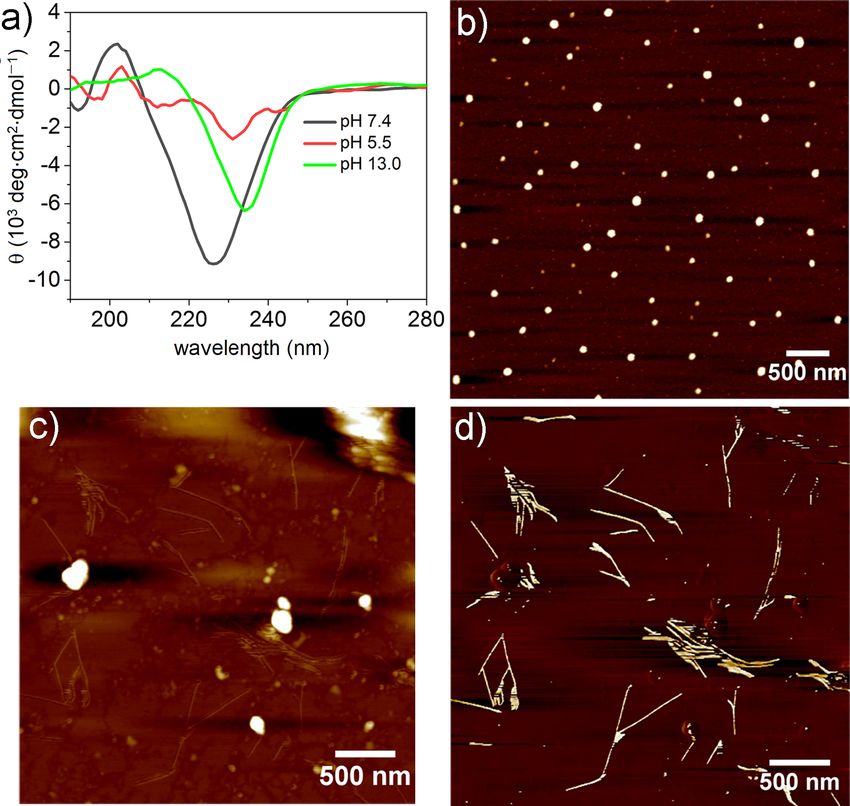
Beilstein J. Org. Chem. 2020, 16, 2017–2025.

39]. In this regard, a particularly relevant property of peptide           considered as suitable carriers for chemotherapeutics [44-46].
assemblies is their ability to undergo significant changes in their        Furthermore, peptides also play an important role as active
morphologies and secondary structures in response to pH stimu-             moieties for many diseases, including cancer [47], peptic ulcer
li. For instance, hydrogen bonding interactions are strongly               [48], asthma [49], cardiovascular diseases [50], and hyperten-
influenced by the pH value, leading to a collapse of the supra-            sion [51]. A particularly important aspect of peptides that
molecular aggregates when an acid or base is added [4]. The                contain both hydrophobic and hydrophilic amino acid residues
pH-responsiveness of self-assembled peptides has been exten-               is their amphiphilicity, which plays a crucial role in the self-
sively exploited for potential applications, with particular focus         assembly process [52]. Keeping these factors in mind, we syn-
on drug delivery systems (DDS) [40], as injectable gels for                thesized an octapeptide, PEP-1, which contains two valine (Val)
tissue engineering [41,42], and biosensing applications [43].              units and one leucine (Leu) unit as the hydrophobic residues
pH-responsive DDS can deliver the drug to a specific tissue or             and one glutamic acid (Glu) residue, two arginine (Arg) units,
organ and protect the payload during the passage through physi-            and one cysteine (Cys) substituent as polar moieties
ological barriers. Most importantly, pH-sensitive DDS are                  (Scheme 1). This choice of amino acids renders PEP-1 amphi-

 Scheme 1: Detailed synthetic scheme for PEP-1. (i) 20% piperidine in DMF, (ii) HBTU, (iii) NMM, (iv) Ac2O/Py/DMF 1:2:3 and (v) TFA/phenol/water/
 TIPS 88:5:5:2.

                                                                                                                                               2018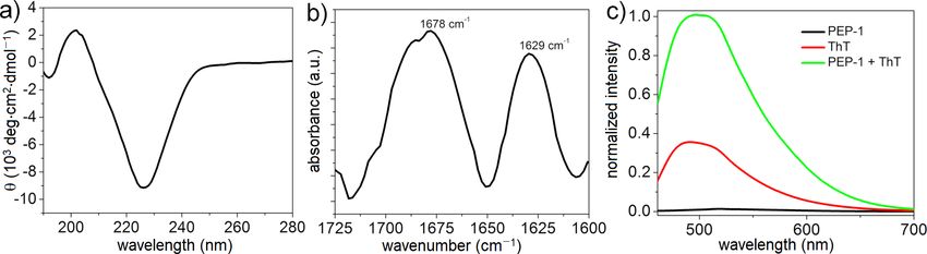
Beilstein J. Org. Chem. 2020, 16, 2017–2025.

philic in nature, which is expected to be beneficial for aqueous      this was stirred for 2 hours following the same procedure as
self-assembly. The polar Glu and Arg motifs are also sensitive        before.
towards basic and acidic pH values, respectively. PEP-1 is actu-
ally a naturally occurring β-strand peptide fragment (residues        At the final stage, we cleaved the peptide from the resin by
16−23 in Figure S1, Supporting Information File 1) of galectin-       using a proper cleavage cocktail: TFA/phenol/water/TIPS
1, a β-sheet lectin protein that is available in bovine spleen [53]   88:5:5:2. DTT was included, as this peptide contains cysteine.
(for a detailed crystal structure see the Protein Data Bank; PDB      After adding the cleavage cocktail to the dried resin and stir-
ID 1SLT). Given the amphiphilic and pH-responsive nature of           ring for 2 hours, the solution was drained off and the resin was
PEP-1, we investigated both the pH- and concentration-depend-         washed with the cocktail and concentrated to dryness in a
ent formation of nanostructures as well as the secondary struc-       round-bottom flask. The peptide was washed several times with
tures.                                                                cold ether, subsequently dissolved in distilled water or glacial
                                                                      acetic acid, and then lyophilized. The lyophilized peptide was
                                                                      dissolved in water/acetonitrile 1:1, v/v, in the presence of 0.1%
Results and Discussion                                                TFA and purified by RP-HPLC using eluent A (10% aceto-
Solid-phase peptide synthesis and                                     nitrile, 90% water containing 0.1% TFA) and eluent B (80%
purification                                                          acetonitrile, 20% water containing 0.1% TFA) in a linear aceto-
The target octapeptide was synthesized in the solid phase             nitrile→water gradient (11% B→50% B in 40 min at 25 °C ) on
following four steps, including: i) deprotection of the Fmoc          a SymmetryPrepTM C18 preparative column (7 µm,
protecting group, ii) coupling of an amino acid, iii) cleavage of     7.8 × 300 mm) at a flow rate of 2 mL/min. Peaks were detected
the peptide from the solid support, and iv) purification of the       at 214 nm. The desired peak was collected, and the purity was
peptide by reversed-phase HPLC. Fmoc-protected Rink amide             checked in an analytical Symmetry C18 column (5 µm,
resin (0.45 g, 0.5 mmol) was swelled in 10 mL of DMF for two          4.6 × 250 mm). The single peak demonstrated the purity of the
hours. After that, the swelled resin was loaded into the special      peptide (Figure S2, Supporting Information File 1). The iden-
apparatus [23], and the solvent was drained off. The swelled          tity of the peptide was confirmed by MALDI–TOF mass spec-
resin was washed with DMF (10 mL × 4). Subsequently, 10 mL            trometry (Figure S3, Supporting Information File 1). The yield
of 20% piperidine in DMF were added to the preswollen resin,          of the purified PEP-1 was 42%.
and the resulting mixture was stirred for 30 minutes under a
nitrogen gas atmosphere. After washing the resin with DMF,            Self-assembly and secondary-structure
the deprotection procedure was repeated, and the resin was thor-      formation
oughly washed with DMF. Subsequently, a Kaiser test [23] was          CD, FTIR spectroscopy, and ThT fluorescence spectroscopy
performed to monitor the deprotection step. A few resin beads         assay were used to investigate the formation of secondary struc-
were placed in a small vial and washed with ethanol, and then,        tures from PEP-1 during the self-assembly. The CD spectrum of
two drops of each of the three solutions were added and heated        PEP-1 at pH 7.4 (PBS buffer, c = 5.0 × 10⁻⁴ M) showed an
to 100 °C for 4–6 min. The color change of the initially color-       intense negative band at around 226 nm that indicated the char-
less beads to blue/purple revealed that the test was positive, in-    acteristic signature of a β-sheet-rich structure (Figure 1a) [54-
dicating that the deprotection was complete and that the system       56].
was ready for the coupling procedure.
                                                                      To support the results obtained from CD spectroscopy, FTIR
The solution of the Fmoc-protected amino acid (2 mmol,                spectroscopy was performed in D2O (pH 7.4). The appearance
4 equiv), HBTU (2 mmol, 4 equiv), and NMM (8 mmol,                    of two intense peaks at 1629 and 1678 cm−1 in the amide I
16 equiv) in 10 mL DMF was added to the resin, and the result-        region (Figure 1b) suggested an intermolecular antiparallel
ing mixture was allowed to stir for two hours under a nitrogen        β-sheet arrangement [57-60]. The band at 1678 cm−1 was the
gas environment. After the completion of the reaction, the            characteristic feature of an antiparallel conformation of the
solution was drained off, and the resin was washed as                 sheet structure or the β-turn structure [61]. To further confirm
mentioned previously (with DMF; 4–6 times for 30 seconds).            the β-sheet formation, we performed a ThT fluorescence spec-
The completion of the coupling procedure was confirmed by a           troscopy assay. ThT is a widely used fluorescent dye that is
Kaiser test. Several cycles of deprotection, coupling,                amyloid-specific and can bind specifically to multistranded
and washing procedures were repeated until the desired                β-sheets [62,63]. PEP-1 is nonemissive due to the absence of
peptide was obtained. The N-terminus of the peptide was acety-        chromophores in its molecular structure, whereas ThT shows a
lated by adding 10 mL of a mixture of acetic anhydride/pyri-          low fluorescence in PBS (pH 7.4) upon excitation at 440 nm.
dine/DMF 1:2:3 to the peptidyl resin at room temperature, and         Interestingly, the fluorescence intensity of ThT significantly in-

                                                                                                                                     2019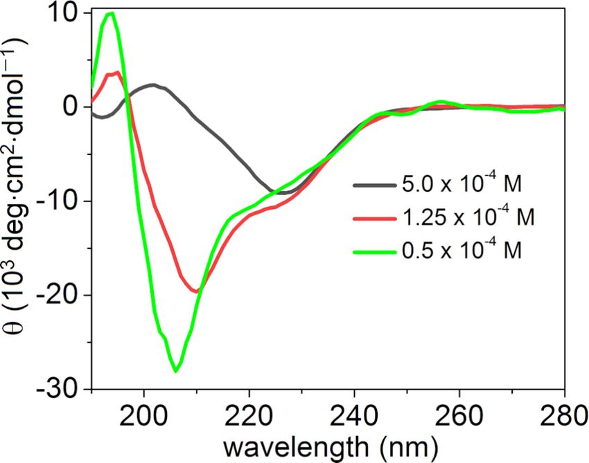
Beilstein J. Org. Chem. 2020, 16, 2017–2025.

 Figure 1: a) CD spectrum; b) FTIR spectrum of the amide I region; and c) ThT fluorescence assay of PEP-1 in PBS at pH 7.4 (c = 5.0 × 10⁻⁴ M).

creases upon mixing with PEP-1 (Figure 1c), confirming the                  evident from the zoomed height and phase images (Figure 2b
formation of a β-sheet structure and supporting the results ob-             and Figure 2d). These results imply that PEP-1 first self-assem-
tained from CD and FTIR spectroscopy.                                       bles into nanobelts, which further assemble into larger struc-
                                                                            tures in a hierarchical process [64].
Microscopic studies by AFM revealed the formation of fractal-
like structures (Figure 2a and Figure 2c) of several micrometers            This behavior may be the result of strong electrostatic attrac-
in length along with discrete short and rigid nanobelts, as                 tion forces between the peptide molecules at a neutral pH value.

 Figure 2: AFM height (a and b) and corresponding phase images (c and d) of PEP-1 at pH 7.4 on mica (c = 5.0 × 10⁻⁴ M).

                                                                                                                                                 2020
Beilstein J. Org. Chem. 2020, 16, 2017–2025.
Even though the pH-responsive behavior of peptides is a well- mation File 1). In contrast, the CD spectrum at highly basic
known phenomenon, fractal-like-structure formation from conditions, such as at pH 13.0, showed a negative CD signal
β-sheets has been rarely observed previously [65]. that was red-shifted (234 nm) and less intense to that observed
at pH 7.4 (226 nm), suggesting the formation of a more twisted
Effect of pH on the self-assembly and distorted β-sheet arrangement [16,66-68]. As the angle be-
The presence of pH-responsive amino acids, such as two Arg tween the two peptides increases in twisted β-sheets, the
(containing free amine groups) and two Glu residues (contain- H-bonding distances increase, weakening the intermolecular
ing free acids) prompted us to investigate the effect of the pH forces. At basic pH values, the carboxyl group of Glu is present
value on the self-assembly and secondary structures. For this, as a negatively charged carboxylate species, thereby inducing
we performed CD and AFM (Figure 3 and Figure S4, Support- weak electrostatic repulsions between Glu− residues, which may
ing Information File 1) both in acidic (pH 5.5 and 2.2) and basic be responsible for the lack of well-defined assemblies. This
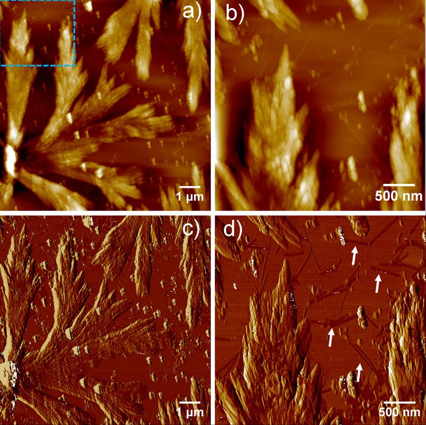
(pH 13.0 and 10.3) media. The pH-dependent CD spectra of phenomenon is also supported by AFM imaging (Figure 3c and
PEP-1 revealed that the β-sheet secondary structure completely Figure 3d), where PEP-1 forms short fibrillar nanostructures
dissociates upon altering the pH from 7.4 to 5.5 (Figure 3a, red with lengths of several hundred nanometers at pH 13.0. Interest-
spectrum), which can be explained by strong electrostatic repul- ingly, at a lower basic pH value, such as 10.3, PEP-1 formed an
sions involving Arg+ moieties. AFM studies at pH 5.5 showed almost similar β-sheet conformation and fractal-like morpholo-
the formation of nanoparticle assemblies, which is in agree- gy as at pH 7.4. This is evidenced by the negative band at
ment with the loss of the secondary structure (Figure 3b). Simi- 227 nm in the CD spectrum, and further supported by AFM
larly, at pH 2.2, PEP-1 also exhibited a structureless CD spec- (Figure S4, Supporting Information File 1). At neutral (pH 7.4)
trum and irregular nanostructures (Figure S4, Supporting Infor- and lower basic (pH 10.3) conditions, the self-assembly
Figure 3: a) pH-dependent CD spectra of PEP-1. b) AFM height image at pH 5.5; c) at pH 13.0; and d) corresponding phase image of c) on mica
(c = 5.0 × 10⁻⁴ M).
2021Beilstein J. Org. Chem. 2020, 16, 2017–2025.
occurred due to the favorable strong electrostatic attraction be- the formation of very stable conformations under the investigat-
tween Glu− and Arg+ residues [69,70]. However, moving to ed conditions. Although the mechanism of transformation is not
more acidic or more basic conditions led to less ordered nano- clear to us at this stage, we hypothesize that multiple intermo-
scale morphologies due to the potential participation of the elec- lecular interactions may play an important role in the transfor-
trostatic repulsions between some of the amino acid residues, as mation. At high concentrations, peptide molecules can come
mentioned above. The pH-responsive self-assembling behavior into closer contact and experience strong intermolecular attrac-
of peptides has a great importance in drug delivery, and since tive forces to facilitate the β-sheet formation, whereas a low
PEP-1 contains rich cationic residues, such as Arg, this system concentration may preferentially favor coiled or helical confor-
can be an interesting potential candidate for DDS and as an mations by salt bridges between positively charged (Arg+) and
antibacterial agent [71]. Biocompatibility, drug delivery, and negatively charged (Glu−) amino acids [73,74]. Mechanistic
antibacterial studies are underway in our laboratory. insights into the observed conformational transformation via
molecular dynamics simulations are underway in our laborato-
Concentration-dependent secondary ry.
structure formation
Apart from the pH-dependent self-assembly, we also investigat- Conclusion
ed the role of the concentration on the self-assembly and sec- In summary, we synthesized a naturally occurring amphiphilic
ondary structure. As shown previously, the CD spectra at peptide fragment, PEP-1, from a β-sheet lectin protein, galectin-
neutral pH disclosed the formation of β-sheet structures at a 1. PEP-1 self-assembles to produce β-sheet-rich structures at
concentration of 5 × 10⁻⁴ M in PBS. Intriguingly, lowering the physiological pH 7.4, as confirmed by CD, FTIR spectroscopy,
concentration to 1.25 × 10⁻⁴ M led to a helical secondary struc- and ThT assay. Microscopy studies revealed the hierarchical
ture (Figure 4, red spectrum), which transformed into a random formation of fractal-like structures from nanobelts. The target
coil structure (Figure 4, green spectrum) upon further decreas- peptide PEP-1 appeared to be highly sensitive towards pH
ing the concentration to 0.5 × 10⁻⁴ M. To the best of our know- changes due to the presence of charged amino acids. Fractal
ledge, this kind of conformational transformation, dependent on networks and the secondary structure can be dissociated under
the concentration of the peptide solution, has rarely been re- acidic conditions (pH 5.5) due to strong electrostatic repulsions.
ported [72]. Under basic conditions (pH 13.0), the electrostatic repulsions
are weakened compared to acidic conditions, but they still have
an effect on the secondary structure and the resulting nanoscale
morphology, leading to twisted β-sheet structures and short
nanofibers. Apart from the pH sensitivity, PEP-1 also showed a
concentration dependency of the secondary structure. At high
concentration, it formed β-sheet-rich structures, which, upon
dilution, transformed into helical structures and further to
random coils. Such pH-responsiveness and concentration-de-
pendent conformational changes may provide access to new
potential peptide candidates for biomedical applications, which
are currently underway in our laboratory.
Experimental
FTIR spectroscopy
FTIR spectra were collected on a PerkinElmer Spectrum 100
FTIR spectrometer. A solution of the peptide (c = 5.0 × 10⁻⁴ M)
Figure 4: CD spectrum of PEP-1 at different concentrations at pH 7.4.
in D 2 O (pH 7.4) was placed in a CaF 2 cell window with a
0.2 mm spacer. The spectra were recorded against the corre-
We also investigated the time-dependent CD for both the sponding solvent background. The scans were between 1800
β-sheet (Figure S5, Supporting Information File 1) and the and 1500 cm −1 , with 200 accumulations at a resolution of
helical conformation (Figure S6, Supporting Information File 1) 0.4 cm −1 .
as well as a ThT assay for the β-sheet (Figure S7, Supporting
Information File 1) to better understand a possible conforma- CD spectroscopy
tional transition over time. However, no changes in the particu- CD experiments were performed on a JASCO J-1500 spec-
lar conformation were found even after 24 h, which suggested tropolarimeter equipped with a Peltier module as a temperature
2022Beilstein J. Org. Chem. 2020, 16, 2017–2025.
controller. The experiments were carried out in buffer solutions Funding
at different pH values. The samples for CD were prepared by G. G. and G. F. thank the European Commission for funding
dissolving the solid peptide in an appropriate buffer solution (ERC-StG-2016 SUPRACOP-715923).
and measured in the far-UV region at 25 °C in the wavelength
range of 280–190 nm. Cuvettes with a path length of 1.0 cm ORCID® iDs
were used. A scan speed of 50 nm/min and a response time of Goutam Ghosh - https://orcid.org/0000-0002-3823-5265
2.0 s were selected. The spectra were averaged over three scans Gustavo Fernández - https://orcid.org/0000-0001-6155-8671
to minimize signal noise.
References
ThT fluorescence spectroscopy assay 1. Whitesides, G. M.; Mathias, J. P.; Seto, C. T. Science 1991, 254,
For the ThT assay, we used a protocol similar to the one previ- 1312–1319. doi:10.1126/science.1962191
2. Dasgupta, A.; Das, D. Langmuir 2019, 35, 10704–10724.
ously described [23]. A diluted solution of ThT at a concentra-
doi:10.1021/acs.langmuir.9b01837
tion of 1.0 × 10⁻³ M was prepared in PBS buffer (pH 7.4) from
3. Chena, J.; Zoua, X. Bioact. Mater. 2019, 4, 120–131.
a concentrated stock solution (5.0 × 10⁻³ M) and then filtered 4. Du, X.; Zhou, J.; Shi, J.; Xu, B. Chem. Rev. 2015, 115, 13165–13307.
with a syringe filter. An aliquot of 50 μL of the diluted ThT doi:10.1021/acs.chemrev.5b00299
solution was added to the peptide solution at 5.0 × 10⁻³ M to 5. Hanabusa, K.; Suzuki, M. Polym. J. 2014, 46, 776–782.
reach a final volume of 2000 μL. After that, the mixture was doi:10.1038/pj.2014.64
6. Dasgupta, A.; Mondal, J. H.; Das, D. RSC Adv. 2013, 3, 9117–9149.
vortexed and kept in the dark for about two hours for binding.
doi:10.1039/c3ra40234g
After two hours, the fluorescence spectra were registered using 7. Jonker, A. M.; Löwik, D. W. P. M.; van Hest, J. C. M. Chem. Mater.
an excitation wavelength of 440 nm on a JASCO Spectrofluo- 2012, 24, 759–773. doi:10.1021/cm202640w
rometer FP-8500 within a wavelength range between 450 and 8. Li, Y.; Rodrigues, J.; Tomás, H. Chem. Soc. Rev. 2012, 41,
700 nm and 5 nm slits. 2193–2221. doi:10.1039/c1cs15203c
9. Mart, R. J.; Osborne, R. D.; Stevens, M. M.; Ulijn, R. V. Soft Matter
2006, 2, 822–835. doi:10.1039/b607706d
AFM studies 10. Tomasini, C.; Castellucci, N. Chem. Soc. Rev. 2013, 42, 156–172.
For microscopy studies, peptide solutions at a concentration of doi:10.1039/c2cs35284b
5 × 10⁻⁴ M were prepared at the desired pH values. AFM sam- 11. Restu, W. K.; Nishida, Y.; Yamamoto, S.; Ishii, J.; Maruyama, T.
ples were prepared by drop-casting 10 μL of the respective sam- Langmuir 2018, 34, 8065–8074. doi:10.1021/acs.langmuir.8b00362
ple onto a mica surface, followed by spin-coating at 1000 rpm, 12. Shigemitsu, H.; Hamachi, I. Acc. Chem. Res. 2017, 50, 740–750.
doi:10.1021/acs.accounts.7b00070
and then, the thin film was dried at room temperature for 24 h.
13. Singh, N.; Kumar, M.; Miravet, J. F.; Ulijn, R. V.; Escuder, B.
After that, images were captured on a Multimode ® 8 SPM Chem. – Eur. J. 2017, 23, 981–993. doi:10.1002/chem.201602624
System (AXS Bruker). Silicon cantilevers with a nominal 14. Habibi, N.; Kamaly, N.; Memic, A.; Shafiee, H. Nano Today 2016, 11,
spring constant of 9 Nm−1 and with a resonant frequency of 41–60. doi:10.1016/j.nantod.2016.02.004
≈150 kHz and a typical tip radius of 7 nm (OMCL-AC200TS, 15. Matson, J. B.; Zha, R. H.; Stupp, S. I.
Curr. Opin. Solid State Mater. Sci. 2011, 15, 225–235.
Olympus) were employed.
doi:10.1016/j.cossms.2011.08.001
16. Martinek, T. A.; Fülöp, F. Chem. Soc. Rev. 2012, 41, 687–702.
Buffer solution preparation doi:10.1039/c1cs15097a
PBS and acetate buffer were used for adjusting pH 7.4 and 17. Trabocchi, A.; Guarna, A. Peptidomimetic Foldamers. Peptidomimetics
5.5, respectively, by following standard protocols. pH 13.0 in Organic and Medicinal Chemistry: The Art of Transforming Peptides
was prepared by adding NaOH into water. 0.15 M sodium in Drugs; John Wiley & Sons, Ltd.: Chichester, UK, 2014; pp 219–229.
doi:10.1002/9781118683033.ch10
chloride were used to avoid the influence of the ionic
18. Aravinda, S.; Shamala, N.; Das, C.; Sriranjini, A.; Karle, I. L.;
strength, and the pH values were precisely measured using a pH Balaram, P. J. Am. Chem. Soc. 2003, 125, 5308–5315.
meter. doi:10.1021/ja0341283
19. Paramonov, S. E.; Jun, H.-W.; Hartgerink, J. D. J. Am. Chem. Soc.
2006, 128, 7291–7298. doi:10.1021/ja060573x
Supporting Information 20. Stendahl, J. C.; Rao, M. S.; Guler, M. O.; Stupp, S. I.
Adv. Funct. Mater. 2006, 16, 499–508. doi:10.1002/adfm.200500161
21. Fichman, G.; Gazit, E. Acta Biomater. 2014, 10, 1671–1682.
Supporting Information File 1
doi:10.1016/j.actbio.2013.08.013
Materials and methods as well as additional figures.
22. Pappas, C. G.; Shafi, R.; Sasselli, I. R.; Siccardi, H.; Wang, T.;
[https://www.beilstein-journals.org/bjoc/content/ Narang, V.; Abzalimov, R.; Wijerathne, N.; Ulijn, R. V.
supplementary/1860-5397-16-168-S1.pdf] Nat. Nanotechnol. 2016, 11, 960–967. doi:10.1038/nnano.2016.169
2023Beilstein J. Org. Chem. 2020, 16, 2017–2025.
23. Ghosh, G.; Barman, R.; Sarkar, J.; Ghosh, S. J. Phys. Chem. B 2019, 48. Ivanikov, I. O.; Brekhova, M. E.; Samonina, G. E.; Myasoedov, N. F.;
123, 5909–5915. doi:10.1021/acs.jpcb.9b02999 Ashmarin, I. P. Bull. Exp. Biol. Med. 2002, 134, 73–74.
24. Appel, R.; Tacke, S.; Klingauf, J.; Besenius, P. Org. Biomol. Chem. doi:10.1023/a:1020621124776
2015, 13, 1030–1039. doi:10.1039/c4ob02185a 49. Hauff, K.; Zamzow, C.; Law, W. J.; De Melo, J.; Kennedy, K.; Los, M.
25. von Gröning, M.; de Feijter, I.; Stuart, M. C. A.; Voets, I. K.; Arch. Immunol. Ther. Exp. 2005, 53, 308–320.
Besenius, P. J. Mater. Chem. B 2013, 1, 2008–2012. 50. Grieco, P.; Gomez-Monterrey, I. Arch. Biochem. Biophys. 2019, 662,
doi:10.1039/c3tb00051f 15–32. doi:10.1016/j.abb.2018.11.021
26. Zou, R.; Wang, Q.; Wu, J.; Wu, J.; Schmuck, C.; Tian, H. 51. Koyama, M.; Hattori, S.; Amano, Y.; Watanabe, M.; Nakamura, K.
Chem. Soc. Rev. 2015, 44, 5200–5219. doi:10.1039/c5cs00234f PLoS One 2014, 9, e105802. doi:10.1371/journal.pone.0105802
27. Sarkar, A.; Kölsch, J. C.; Berač, C. M.; Venugopal, A.; Sasmal, R.; 52. Dehsorkhi, A.; Castelletto, V.; Hamley, I. W. J. Pept. Sci. 2014, 20,
Otter, R.; Besenius, P.; George, S. J. ChemistryOpen 2020, 9, 453–467. doi:10.1002/psc.2633
346–350. doi:10.1002/open.202000017 53. Liao, D. I.; Kapadia, G.; Ahmed, H.; Vasta, G. R.; Herzberg, O.
28. Wan, Y.; Liu, L.; Yuan, S.; Sun, J.; Li, Z. Langmuir 2017, 33, Proc. Natl. Acad. Sci. U. S. A. 1994, 91, 1428–1432.
3234–3240. doi:10.1021/acs.langmuir.6b03986 doi:10.1073/pnas.91.4.1428
29. Falcone, N.; Kraatz, H.-B. Chem. – Eur. J. 2018, 24, 14316–14328. 54. Marchesan, S.; Waddington, L.; Easton, C. D.; Winkler, D. A.;
doi:10.1002/chem.201801247 Goodall, L.; Forsythe, J.; Hartley, P. G. Nanoscale 2012, 4,
30. Kuang, Y.; Gao, Y.; Shi, J.; Li, J.; Xu, B. Chem. Commun. 2014, 50, 6752–6760. doi:10.1039/c2nr32006a
2772–2774. doi:10.1039/c3cc48832b 55. Marchesan, S.; Easton, C. D.; Styan, K. E.; Waddington, L. J.;
31. Minakuchi, N.; Hoe, K.; Yamaki, D.; Ten-no, S.; Nakashima, K.; Kushkaki, F.; Goodall, L.; McLean, K. M.; Forsythe, J. S.; Hartley, P. G.
Goto, M.; Mizuhata, M.; Maruyama, T. Langmuir 2012, 28, 9259–9266. Nanoscale 2014, 6, 5172–5180. doi:10.1039/c3nr06752a
doi:10.1021/la301442f 56. Castelletto, V.; Hamley, I. W.; Harris, P. J. F.; Olsson, U.; Spencer, N.
32. Restu, W. K.; Nishida, Y.; Kataoka, T.; Morimoto, M.; Ishida, K.; J. Phys. Chem. B 2009, 113, 9978–9987. doi:10.1021/jp902860a
Mizuhata, M.; Maruyama, T. Colloid Polym. Sci. 2017, 295, 57. Haris, P. I.; Chapman, D. Biopolymers 1995, 37, 251–263.
1109–1116. doi:10.1007/s00396-017-4093-x doi:10.1002/bip.360370404
33. Draper, E. R.; Adams, D. J. Chem. Commun. 2016, 52, 8196–8206. 58. Jackson, M.; Mantsch, H. H. Crit. Rev. Biochem. Mol. Biol. 1995, 30,
doi:10.1039/c6cc03485c 95–120. doi:10.3109/10409239509085140
34. Zhang, Y.; Gu, H.; Yang, Z.; Xu, B. J. Am. Chem. Soc. 2003, 125, 59. Schneider, J. P.; Pochan, D. J.; Ozbas, B.; Rajagopal, K.; Pakstis, L.;
13680–13681. doi:10.1021/ja036817k Kretsinger, J. J. Am. Chem. Soc. 2002, 124, 15030–15037.
35. Koda, D.; Maruyama, T.; Minakuchi, N.; Nakashima, K.; Goto, M. doi:10.1021/ja027993g
Chem. Commun. 2010, 46, 979–981. doi:10.1039/b920359a 60. Yan, H.; Saiani, A.; Gough, J. E.; Miller, A. F. Biomacromolecules
36. Tanaka, A.; Fukuoka, Y.; Morimoto, Y.; Honjo, T.; Koda, D.; Goto, M.; 2006, 7, 2776–2782. doi:10.1021/bm0605560
Maruyama, T. J. Am. Chem. Soc. 2015, 137, 770–775. 61. Surewicz, W. K.; Mantsch, H. H.; Chapman, D. Biochemistry 1993, 32,
doi:10.1021/ja510156v 389–394. doi:10.1021/bi00053a001
37. Nishida, Y.; Tanaka, A.; Yamamoto, S.; Tominaga, Y.; Kunikata, N.; 62. Groenning, M. J. Chem. Biol. 2010, 3, 1–18.
Mizuhata, M.; Maruyama, T. Angew. Chem., Int. Ed. 2017, 56, doi:10.1007/s12154-009-0027-5
9410–9414. doi:10.1002/anie.201704731 63. Khurana, R.; Coleman, C.; Ionescu-Zanetti, C.; Carter, S. A.;
38. Jones, C. D.; Steed, J. W. Chem. Soc. Rev. 2016, 45, 6546–6596. Krishna, V.; Grover, R. K.; Roy, R.; Singh, S. J. Struct. Biol. 2005, 151,
doi:10.1039/c6cs00435k 229–238. doi:10.1016/j.jsb.2005.06.006
39. Yang, X.; Zhang, G.; Zhang, D. J. Mater. Chem. 2012, 22, 38–50. 64. Wang, W.; Chau, Y. Soft Matter 2009, 5, 4893–4898.
doi:10.1039/c1jm13205a doi:10.1039/b919782f
40. Narayanaswamy, R.; Torchilin, V. P. Molecules 2019, 24, 603. 65. Sneer, R.; Weygand, M. J.; Kjaer, K.; Tirrell, D. A.; Rapaport, H.
doi:10.3390/molecules24030603 ChemPhysChem 2004, 5, 747–750. doi:10.1002/cphc.200301046
41. Saunders, L.; Ma, P. X. Macromol. Biosci. 2019, 19, 1800313. 66. Pashuck, E. T.; Cui, H.; Stupp, S. I. J. Am. Chem. Soc. 2010, 132,
doi:10.1002/mabi.201800313 6041–6046. doi:10.1021/ja908560n
42. Tang, J. D.; Mura, C.; Lampe, K. J. J. Am. Chem. Soc. 2019, 141, 67. Manning, M. C.; Illangasekare, M.; Woody, R. W. Biophys. Chem.
4886–4899. doi:10.1021/jacs.8b13363 1988, 31, 77–86. doi:10.1016/0301-4622(88)80011-5
43. Wong, S.; Shim, M. S.; Kwon, Y. J. J. Mater. Chem. B 2014, 2, 68. Clarke, D. E.; Parmenter, C. D. J.; Scherman, O. A.
595–615. doi:10.1039/c3tb21344g Angew. Chem., Int. Ed. 2018, 57, 7709–7713.
44. Raza, F.; Zhu, Y.; Chen, L.; You, X.; Zhang, J.; Khan, A.; Khan, M. W.; doi:10.1002/anie.201801001
Hasnat, M.; Zafar, H.; Wu, J.; Ge, L. Biomater. Sci. 2019, 7, 69. Zhao, Y.; Yokoi, H.; Tanaka, M.; Kinoshita, T.; Tan, T.
2023–2036. doi:10.1039/c9bm00139e Biomacromolecules 2008, 9, 1511–1518. doi:10.1021/bm701143g
45. Mei, L.; Xu, K.; Zhai, Z.; He, S.; Zhu, T.; Zhong, W. Org. Biomol. Chem. 70. Zhou, X.-R.; Ge, R.; Luo, S.-Z. J. Pept. Sci. 2013, 19, 737–744.
2019, 17, 3853–3860. doi:10.1039/c9ob00046a doi:10.1002/psc.2569
46. Hainline, K. M.; Gu, F.; Handley, J. F.; Tian, Y. F.; Wu, Y.; de Wet, L.; 71. Veiga, A. S.; Sinthuvanich, C.; Gaspar, D.; Franquelim, H. G.;
Vander Griend, D. J.; Collier, J. H. Macromol. Biosci. 2019, 19, Castanho, M. A. R. B.; Schneider, J. P. Biomaterials 2012, 33,
1800249. doi:10.1002/mabi.201800249 8907–8916. doi:10.1016/j.biomaterials.2012.08.046
47. Kurrikoff, K.; Aphkhazava, D.; Langel, Ü. Curr. Opin. Pharmacol. 2019, 72. Ruggeri, F. S.; Byrne, C.; Khemtemourian, L.; Ducouret, G.; Dietler, G.;
47, 27–32. doi:10.1016/j.coph.2019.01.008 Jacquot, Y. J. Pept. Sci. 2015, 21, 95–104. doi:10.1002/psc.2730
73. Meuzelaar, H.; Vreede, J.; Woutersen, S. Biophys. J. 2016, 110,
2328–2341. doi:10.1016/j.bpj.2016.04.015
2024Beilstein J. Org. Chem. 2020, 16, 2017–2025.
74. Walker, K. D.; Causgrove, T. P. J. Mol. Model. 2009, 15, 1213–1219.
doi:10.1007/s00894-009-0482-5
License and Terms
This is an Open Access article under the terms of the
Creative Commons Attribution License
(http://creativecommons.org/licenses/by/4.0). Please note
that the reuse, redistribution and reproduction in particular
requires that the authors and source are credited.
The license is subject to the Beilstein Journal of Organic
Chemistry terms and conditions:
(https://www.beilstein-journals.org/bjoc)
The definitive version of this article is the electronic one
which can be found at:
doi:10.3762/bjoc.16.168
2025You can also read