ViralRecall-A Flexible Command Line Tool for the Detection of Giant Virus Signatures in 'Omic Data - MDPI
←
→
Page content transcription
If your browser does not render page correctly, please read the page content below

Article

ViralRecall—A Flexible Command‐Line Tool for the Detection
of Giant Virus Signatures in ‘Omic Data
Frank O. Aylward * and Mohammad Moniruzzaman

                                         Department of Biological Sciences, Virginia Tech, Blacksburg, VA 24061, USA; monir@vt.edu
                                         * Correspondence: faylward@vt.edu

                                         Abstract: Giant viruses are widespread in the biosphere and play important roles in biogeochemical
                                         cycling and host genome evolution. Also known as nucleo‐cytoplasmic large DNA viruses
                                         (NCLDVs), these eukaryotic viruses harbor the largest and most complex viral genomes known.
                                         Studies have shown that NCLDVs are frequently abundant in metagenomic datasets, and that
                                         sequences derived from these viruses can also be found endogenized in diverse eukaryotic
                                         genomes. The accurate detection of sequences derived from NCLDVs is therefore of great
                                         importance, but this task is challenging owing to both the high level of sequence divergence between
                                         NCLDV families and the extraordinarily high diversity of genes encoded in their genomes,
                                         including some encoding for metabolic or translation‐related functions that are typically found only
                                         in cellular lineages. Here, we present ViralRecall, a bioinformatic tool for the identification of
                                         NCLDV signatures in ‘omic data. This tool leverages a library of giant virus orthologous groups
                                         (GVOGs) to identify sequences that bear signatures of NCLDVs. We demonstrate that this tool can
                                         effectively identify NCLDV sequences with high sensitivity and specificity. Moreover, we show that
                                         it can be useful both for removing contaminating sequences in metagenome‐assembled viral
Citation: Aylward, F.O.;                 genomes as well as the identification of eukaryotic genomic loci that derived from NCLDV.
Moniruzzaman, M. ViralRecall—A           ViralRecall is written in Python 3.5 and is freely available on GitHub:
Flexible Command‐Line Tool for the       https://github.com/faylward/viralrecall.
Detection of Giant Virus Signatures
in ‘Omic Data. Viruses 2021, 13, 150.    Keywords: giant viruses; nucleo‐cytoplasmic large DNA viruses; metagenomics; endogenous viral
https://doi.org/10.3390/v13020150
                                         elements; viral diversity

Received: 14 December 2020
Accepted: 18 January 2021
Published: 20 January 2021
                                         1. Introduction
Publisher’s Note: MDPI stays                  Nucleo‐cytoplasmic large DNA viruses (NCLDVs) are a group of dsDNA viruses of
neutral with regard to jurisdictional    the phylum Nucleocytoviricota [1]. This group includes the largest viruses known, both in
claims in published maps and             terms of physical dimensions and genome length [2]. Some members within the NCLDV
institutional affiliations.              group include those that infect metazoans and have been studied extensively, such as the
                                         Poxviridae, Iridoviridae, and Asfarviridae, while other families of NCLDVs, such as the
                                         Phycodnaviridae, Mimiviridae, Marseilleviridae, and Pithoviridae, have been discovered
                                         relatively recently and are known to infect protist lineages [3–6]. NCLDV genomes encode
Copyright: © 2021 by the authors.
                                         notably diverse functions that include many genes which are otherwise found only in
Licensee MDPI, Basel, Switzerland.
                                         cellular lineages, such as those involved in sphingolipid biosynthesis [7], amino acid
This article is an open access article
                                         metabolism [8], fermentation [9], glycolysis and the tricarboxylic acid (TCA) cycle [10],
distributed under the terms and
                                         structuring of the eukaryotic cytoskeleton [11], and translation [12–14].
conditions of the Creative Commons
Attribution (CC BY) license
                                              Numerous studies have begun to reveal that NCLDVs play key roles in the ecological
(http://creativecommons.org/licenses
                                         and evolutionary dynamics of eukaryotes. Several cultivation‐independent analyses have
/by/4.0/).                               shown that NCLDVs are broadly distributed in the environment, and that they are
                                         particularly diverse and abundant in aquatic systems [10,15–17]. Moreover, analysis of
                                         eukaryotic genomes has revealed that endogenous NCLDVs are common in some
                                         lineages, and thereby play a significant role in host genome evolution [18–22]. There is,

Viruses 2021, 13, 150. https://doi.org/10.3390/v13020150                                                            www.mdpi.com/journal/viruses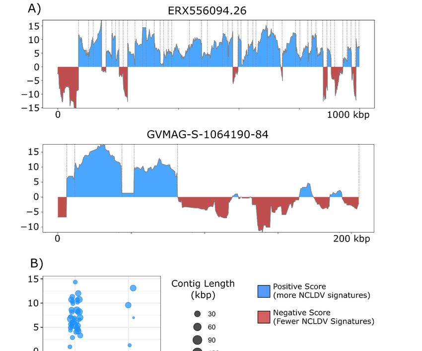
Viruses 2021, 13, 150                                                                                      2 of 14

                        therefore, a need for bioinformatic approaches that facilitate these emerging frontiers of
                        research in NCLDVs. Specifically, tools that can robustly identify signatures of
                        endogenous NCLDVs in eukaryotic genomes will be useful for examining the role of
                        NCLDVs in shaping eukaryotic genome evolution. Furthermore, multiple different
                        approaches for binning NCLDV metagenome‐assembled genomes (MAGs) from diverse
                        environments have been developed [10,23,24], and approaches for quality‐checking these
                        results and removing possible contamination are needed given the unique features of
                        NCLDV genomes.
                             The detection of NCLDV signatures in ‘omic data has proven challenging for a
                        number of reasons. Firstly, NCLDVs are a diverse group of viruses that comprise several
                        divergent lineages; the average amino acid identity between NCLDVs from different
                        families can be as low as ~20%, and in some cases it can be difficult to identify any
                        signatures of homology between disparate NCLDV genomes [10]. Tools such as MG‐
                        Digger [25], Giant Virus Finder [26], and FastViromeExplorer [27] can identify NCLDV
                        sequences in metagenomic data using nucleotide‐level homology searches, and these tools
                        are useful for identifying NCLDVs that are closely related to reference genomes. Given
                        the high degree of genomic diversity encompassed by NCLDV families, tools that
                        leverage NCLDV‐specific protein families are also needed to detect NCLDV sequences
                        that are more divergent compared to those in reference databases. Secondly, NCLDV
                        genomes often encode numerous genes of unknown function, or hypothetical genes for
                        which it is difficult to infer evolutionary provenance based on functional annotations.
                        Lastly, NCLDV genomes are chimeric in that they harbor genes with best matches to
                        homologs present in bacteria, archaea, eukaryotes, and other viruses [28], and homology‐
                        based methods used to classify sequences from these genomes may therefore yield
                        inconclusive results. Indeed, the presence of numerous genes in NCLDV genomes that
                        have previously been identified only in cellular lineages complicates efforts to correctly
                        classify NCLDV sequences.
                             In a previous study, we developed a workflow to identify viral regions in genomic
                        data [18]. This workflow relied on searches against protein families present in the Viral
                        Orthologous Groups database ([29]) to identify genomic loci that derive from viruses. The
                        VOG database contains orthologous groups from numerous viruses; therefore, this tool
                        could not discriminate between different viral groups, and several additional analyses
                        were needed to trace the specific viral provenance of the sequences identified. In this
                        study, we constructed a database of 28,696 giant virus orthologous groups (GVOGs) that
                        represent protein families commonly found in NCLDVs. We also present ViralRecall v.
                        2.0, which leverages these NCLDV‐specific protein families together with several
                        additional features to permit the specific identification and annotation of NCDLV
                        sequences in ‘omic data. We present several benchmarking criteria which establish that
                        ViralRecall v. 2.0 has high specificity and sensitivity and can be used for several
                        applications, including the removal of non‐NCLDV contamination from metagenome‐
                        derived viral genomes and the identification of endogenous NCLDVs in eukaryotic
                        genomes.

                        2. Materials and Methods
                        2.1. NCDLV Genomes Used for Database Construction
                             We compiled a database of 2908 NCLDV genomes, including metagenome‐
                        assembled genomes (MAGs) from two studies [10,23], and reference NCLDV genomes
                        available in NCBI RefSeq as of June 1, 2020 [30]. We dereplicated these genomes by
                        performing pairwise k‐mer comparisons in MASH v. 2.0 (“mash dist” command with
                        parameter ‐k 21, ‐s 300), and combining all genomes with a MASH distance of less than
                        0.05, which corresponded to ≥95% average nucleotide identity (ANI) [31]. NCLDV
                        genomes were combined into clusters using single‐linkage clustering, and the genome
                        with the highest N50 contig size was used as the representative for that cluster. In this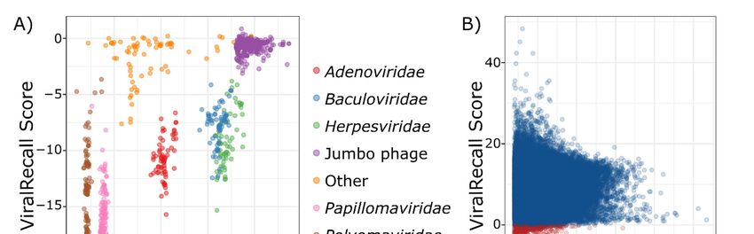
Viruses 2021, 13, 150                                                                                           3 of 14

                        way we generated a nonredundant set of 2436 genomes that we subsequently used for
                        downstream analyses (Supplementary Dataset S1).

                        2.2. Giant Virus Orthologous Groups (GVOGs)
                              To construct orthologous groups (OGs) we used a small subset of high‐quality
                        NCLDV genomes. We did this to eliminate possible contamination present in fragmented
                        NCLDV MAGs and to reduce the computational load necessitated by large‐scale OG
                        construction. Genomes were only included if: 1) we found all four of the single‐copy
                        marker genes A32‐atpase (A32), B‐family DNA Polymerase (PolB), viral late transcription
                        factor 3 (VLTF3), and superfamily II helicase (SFII); 2) we found no duplicate marker
                        genes with < 90% amino acid similarity, which is indicative of the possible binning of
                        multiple NCLDV genomes together; 3) the genome was composed of less than 30 contigs
                        in total; and 4) the genome was not classified as “low quality” by the Schulz et al. study
                        [23]. A total of 888 genomes met all of these criteria, and we calculated OGs from this set.
                        We first predicted proteins using Prodigal v. 2.6.3 [32] (default parameters) and then
                        generated OGs using Proteinortho v. 6.06 with the parameters “‐e=1e‐5 ‐‐identity=20 ‐
                        p=blastp+ ‐‐selfblast ‐‐cov=50 ‐sim=0.95” [33]. Proteins belonging to OGs with >1 member
                        were aligned using Clustal Omega (default parameters) and trimmed using Trimal
                        (parameter ‐gt 0.05), and Hidden Markov Models (HMMs) were subsequently generated
                        using the hmmbuild command in HMMER3 [34–36]. Using this approach, we generated
                        28,992 OGs, which we refer to as giant virus orthologous groups (GVOGs).
                              Annotations were assigned to GVOGs by comparing all proteins comprising each
                        OG to the EggNOG 5.0 and Pfam. v. 32 databases using the hmmsearch command in
                        HMMER3 (e‐value cutoff of 1e‐3 used for EggNOG, ‐‐cut_nc option used for Pfam) [37,38].
                        For each GVOG, an annotation was given only if >50% of the proteins in that GVOG had
                        the same best match in the databases.

                        2.3. Calculation of ViralRecall Scores
                             The workflow implemented in ViralRecall is depicted in Figure 1. ViralRecall
                        calculates scores for input sequences (heretofore referred to as contigs for simplicity)
                        based on the number of encoded proteins that have matches in both the GVOG and Pfam
                        v. 32 databases. Matches are determined using the hmmsearch command in HMMER3,
                        and the e‐value threshold is adjustable in ViralRecall. The scores for contigs are calculated
                        based on the HMMER3 scores for each protein, and they are provided either as a mean
                        for entire contigs, or as a rolling average that can be specified by the user.

                        Figure 1. Diagram of the ViralRecall workflow. Abbreviations: GVOGs, giant virus orthologous
                        groups.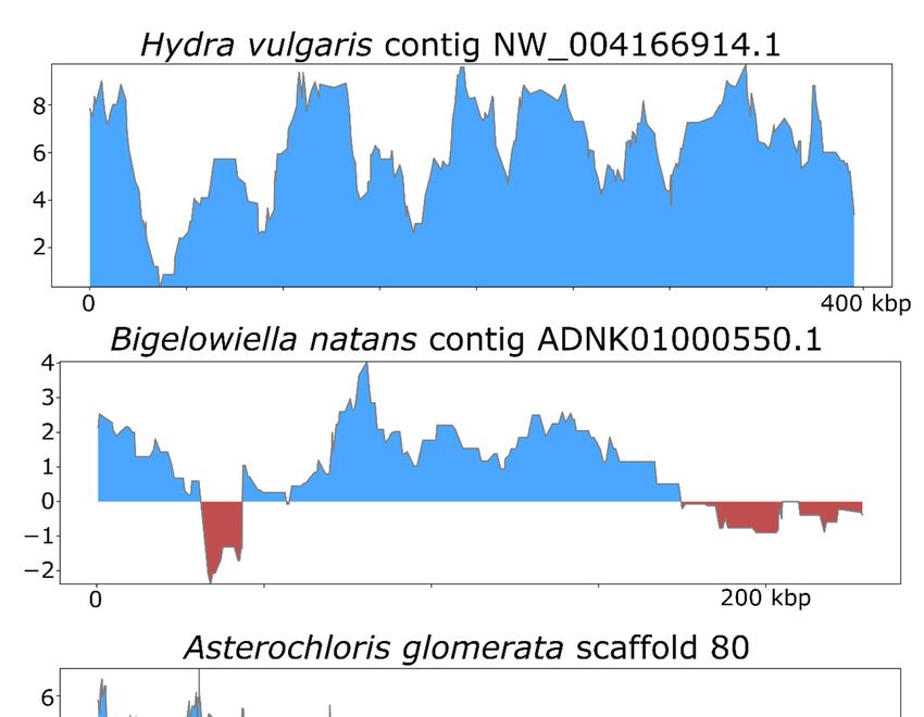
Viruses 2021, 13, 150                                                                                        4 of 14

                             We found that some GVOGs are commonly found in Caudovirales as well as NCLDVs,
                        and we therefore normalized the HMMER score of these GVOGs to avoid false‐positive
                        detection. We did this for Caudovirales because: 1) through manual inspection of some
                        NCLDV MAGs we had detected cases of likely Caudovirales contamination; and 2) jumbo
                        bacteriophages belonging to the Caudovirales had the highest ViralRecall scores among
                        non‐NCLDV dsDNA viruses tested, suggesting that this group of viruses is the most likely
                        source of false positive detections.
                             To identify GVOGs present in Caudovirales, we surveyed a set of 3012 Caudovirales
                        genomes available in NCBI as of July 6, 2020; we predicted proteins from these genomes
                        using Prodigal (default parameters) and identified GVOGs using hmmsearch with an e‐
                        value of 1e‐5. For each GVOG, we calculated scores using the following equation:
                                                  Sgvog = B (PNCLDV + PCaudo / PNCLDV)                           (1)
                        where Sgvog is the final GVOG score, B is the score calculated by HMMER3, PNCLDV is the
                        proportion of NCLDV genomes with hits to that HMM, and PCaudo is the proportion of
                        Caudovirales genomes with hits to that model. In this way, GVOGs that are common in
                        Caudovirales do not lead to high ViralRecall scores when they are detected.
                            Similarly, we normalize matches to Pfam domains present in either >= 1% of the 888
                        high‐quality NCLDV genomes or the 3012 reference Caudovirales genomes using the
                        equation:
                                                 Spfam = B (1−PNCLDV) + PCaudo / PNCLDV                          (2)
                        where Spfam is the final Pfam score, B is the HMMER3 score, PNCLDV is the proportion of
                        NCLDV genomes with hits to that in HMM, and PCaudo is the proportion of Caudovirales
                        genomes with hits to that model. In this way, Pfam domains that are common in NCLDV
                        do not lead to low ViralRecall scores when they are detected, and domains present in both
                        Caudovirales and NCLDV are given a weight proportional to their relative occurrence in
                        these viral groups. A ViralRecall score is then calculated for each ORF predicted by
                        Prodigal using the equation:
                                                         Sfinal = √Sgvog − √Spfam                                (3)
                             Scaling the scores by their square root is necessary to account for differences in
                        overall HMM scores that are obtained for the GVOG and Pfam databases; because the
                        Pfam database is made of domain‐level HMMs, the scores obtained against this database
                        are typically smaller, and outlier GVOG scores can therefore substantially skew the
                        overall score without this normalization.
                             Given this scoring procedure, the presence of NCLDV‐specific GVOGs will lead to
                        higher scores, while the presence of Pfam domains that are typically not found in
                        NCLDVs will lead to lower scores. Final scores for ViralRecall are provided either as a
                        mean for each input contig or replicon, or as a rolling average across a window size that
                        users can adjust (default = 15 ORFs). For simplicity, we use a value of 0 as the cutoff when
                        discriminating between NCLDV and non‐NCLDV sequences, but in principle other
                        values could be used and may be appropriate depending on the situation and the balance
                        between sensitivity and specificity that is desired.

                        2.4. Benchmarking
                             For benchmarking ViralRecall on non‐NCLDV viral sequences, we used a database
                        of 879 non‐NCLDV dsDNA genomes downloaded from NCBI. These genomes were
                        selected because they are reference dsDNA viruses with genomes listed on the ICTV Virus
                        Metadata Resource ([39]) and their genomes were available on NCBI RefSeq. The GVOG
                        and Pfam normalization scores had been generated by using reference Caudovirales
                        genomes in NCBI; therefore, we did not use genomes from this group that were present
                        in the Virus Metadata Resource. Instead, we used a set of 336 jumbo bacteriophages
                        (Caudovirales) that have been reported [40]. Additionally, we did not include any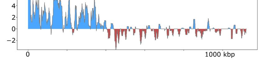
Viruses 2021, 13, 150                                                                                          5 of 14

                        Lavidaviridae (virophage) in this set, because these viruses parasitize giant viruses, and, in
                        some cases, may exchange genes with them [41]. To generate pseudocontigs for
                        benchmarking, we used the gt‐shredder command in genometools ([42]).
                             To benchmark ViralRecall on NCLDV sequences, we used a set of 1548 genomes in
                        the NCLDV database described above. This included all genomes except those used in the
                        construction of GVOGs, because those would not provide an unbiased assessment of the
                        sensitivity of ViralRecall in detecting NCLDV sequences. For benchmarking purposes,
                        ViralRecall was run with default parameters, with the only exception that the ‐c flag was
                        used to generate mean contig‐level scores.
                             For illustrative purposes we selected the following five NCLDV genomes from
                        diverse families and provide the results generated by ViralRecall (shown in Figure 3):
                        Acanthamoeba castellanii Medusavirus [43], Emiliania huxleyi virus 86 [7], Pithovirus
                        sibericum [44], M. separata entomopoxvirus [45], and Hyperionvirus [46]. We also
                        selected the genomes of four non‐NCLDV dsDNA viruses for this purpose; we chose the
                        jumbo bacteriophages with the highest mean score, lowest mean score, and longest length
                        of those tested (FFC_PHAGE_43_1208, M01_PHAGE_56_67, and LP_PHAGE_CIR‐CU‐
                        CL_32_18, respectively), as well as the human herpesvirus 3 strain Dumas [47]. Lastly, we
                        also show the profiles for Yaravirus [48], a virus of A. castelanni with unclear evolutionary
                        provenance, and the Sputnik virophage [41]. In all cases, the viral genomes shown here
                        were not used in the construction of the GVOG database or for score normalization, thus
                        they provide an unbiased assessment of ViralRecall results. For manual inspection of
                        proteins encoded in contigs derived from suspected contamination, we performed
                        homology searches against RefSeq v. 93 using BLASTP+ [49].
                             To illustrate how ViralRecall can be used to identify NCLDV signatures in eukaryotic
                        genomes, we analyzed the Hydra vulgaris, Bigelowiella natans and Asterochloris glomerata
                        genomes. Previous studies have already established NCLDV signatures in these genomes
                        [19,50,51], and our results therefore provide independent verification.

                        3. Results and Discussion
                              We analyzed 879 non‐NCLDV genomes to assess the specificity of ViralRecall in
                        detecting NCLDV sequences. Of these genomes, seven had mean scores >0, and could be
                        considered false positives because they had net positive signatures of NCLDVs (Figure
                        2a). This provides an estimated specificity of 99.2% for ViralRecall when analyzing whole‐
                        genome data. Of the viruses with net positive scores, all had values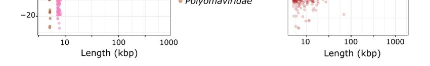
Viruses 2021, 13, 150                                                                                                   6 of 14

      Figure 2. (A) ViralRecall scores and lengths for 879 non‐nucleocytoplasmic large DNA viruses (NCLDV) dsDNA viruses.
      (B) ViralRecall scores and lengths for 38,886 giant virus contigs from 1548 reference and metagenome‐assembled giant
      virus genomes. Contigs with scores 10 kbp in length. We also
                                note that ViralRecall recovered an overall positive for Yaravirus, a eukaryotic virus that
                                may be a divergent member of the NCLDV family [48]; ViralRecall identified 14 GVOG
                                hits out of 70 predicted proteins, providing further evidence that even divergent NCLDVs
                                or related groups can be detected with this tool. Lastly, we recovered a consistently high
                                score for the sputnik virophage (family Lavidaviridae); this is not unexpected given that
                                this virus parasitizes Mimivirus and has likely exchanged genes with NCLDV [41].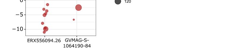
Viruses 2021, 13, 150                                                                                                     7 of 14

      Figure 3. ViralRecall plots of diverse dsDNA viruses. NCLDV genomes are shown on the left, while the right panels show
      other dsDNA viruses, or highly divergent NCLDV in the case of Yaravirus. The jumbo bacteriophages
      LP_PHAGE_COMPLETE_CIR‐CU‐CL_32_18, FFC_PHAGE_43_1208, and M01_PHAGE_56_67 were chosen because they
      have the longest length, highest score, and lowest score, respectively, among the 336 jumbo phages tested.

                                     Overall, these results demonstrate that ViralRecall has high specificity and sensitivity
                                at recovering NCLDV sequences, but this tool should still be used alongside other
                                independent methods for sequence classification. This is because sequences derived from
                                NCLDV, especially if they are fragmented into short contigs, may not contain enough
Viruses 2021, 13, 150                                                                                       8 of 14

                        predicted proteins with matches in the GVOG database to produce a positive overall
                        score. Similarly, other viral groups such as large Caudovirales (jumbo phage) can have a
                        misleadingly high number of GVOG matches. For these reasons, it would still be advisable
                        to use alternative approaches when determining the provenance of sequences in ‘omic
                        datasets, such as phylogenetic analysis of marker genes and homology searches against
                        reference databases. To assist with manual inspection of results, ViralRecall also searches
                        against 10 custom HMMs for NCLDV marker genes and reports the presence of hits to
                        these protein families in the output files. These genes include the five markers commonly
                        used in phylogenetic reconstruction (PolB, A32, VLTF3, SFII, and MCP) as well as five
                        others that are known to be present in many NCLDV genomes (RNA polymerase subunits
                        (RNAPL and RNAPS), the mRNA capping enzyme (mRNAc), ribonucleotide reductase
                        (RNR), and D5 primase/helicase (D5)) [10]. Inspection of the occurrence of these markers
                        can be useful for determining if a sequence derives from NCLDV; for example, of the
                        seven non‐NCLDVs that had scores >0, none encoded proteins with matches to MCP, A32,
                        VLTF3, mRNAc, PolB and D5 markers, although divergent hits with low scores were
                        observed for SFII, and RNR homologs. None of these seven non‐NCLDVs had hits to the
                        RNA polymerase subunits RNAPL or RNAPS, but identification of these markers without
                        subsequent phylogenetic analysis cannot be considered evidence of NCLDV provenance
                        given the universal presence of these protein families in cellular life as well as some
                        Caudovirales [52].
                             The overall incidence of potential contamination in the 1548 NCLDV genomes was
                        low (657 out of 38,896 contigs with scores
Viruses 2021, 13, 150 9 of 14
Figure 4. (A) ViralRecall plots for the giant virus MAGs ERX556094.26 and GVMAG‐S‐1064190.84 demonstrating that both
contain non‐NCLDV contamination. For ERX556096‐26, nine contaminant contigs were detected, while two were found
in GVMAG‐S‐1064190.84. (B) Dot plot of the mean ViralRecall scores for all contigs in ERX556094.26 and GVMAG‐S‐
1064190.84. Contigs with ViralRecall scores < 0 are colored red, and dot sizes are proportional to contig size.
To assess the efficacy of ViralRecall in detecting signatures of NCLDV in eukaryotic
genomes, we analyzed the genomes of Hydra vulgaris, Bigelowiella natans, and Asterochloris
glomerata (Figure 5), which have previously been shown to harbor signatures of
endogenous NCLDVs [19,50,51]. In H. vulgaris, we identified one NCLDV region that
encompassed the entire 396 kbp contig NW_004166914.1 (Figure 5), consistent with
previous findings. This region also encoded proteins with matches to the D5, MCP,Viruses 2021, 13, 150 10 of 14
RNAPL, RNAPS, and VLTF3 markers. For B. natans, we found 35 putative viral regions;
the longest was a 230 kbp region in contig ADNK01000550.1 that also encoded proteins
with matches to the MCP and A32 markers (Figure 5). At the beginning of this contig, we
also found a 22.6 kbp region with NCLDV signatures. Lastly, for A. glomerata, we found
27 putative viral regions; the largest was a 224 kbp region in scaffold 80 (full length 1.2 of
megabases) that encoded homologs to the MCP, A32, SFII, PolB, and D5 marker genes.
These results highlight the variable architecture of endogenous NCLDVs; although some
encompass entire contigs in draft genome assemblies, some span only smaller regions of
chromosomes.
Figure 5. ViralRecall plots for endogenized viral regions identified in Hydra vulgaris, Bigelowiella
natans, and Asterochloris glomerata.
Overall, our results provide evidence that ViralRecall can effectively identify
signatures of NCLDV in a variety of ‘omic datasets, but some limitations remain. For
example, non‐NCLDV sequences belonging to virophages and sometimes Caudovirales
can have misleadingly high ViralRecall scores, and additional analyses will often be
necessary to robustly classify these sequences. Other approaches, such as those that
leverage ribosomal binding site motifs, have successfully been used for the identification
of NCLDV sequences [23], and these methods could be used in addition to ViralRecall toViruses 2021, 13, 150 11 of 14
confirm results. Lastly, ViralRecall relies on large‐scale HMM searches against the Pfam
and GVOG databases, which are time‐consuming and may not be feasible for large
datasets. For example, the application of ViralRecall to assembled metagenomic datasets
may not be advisable; rather, the binning of putative NCLDV MAGs and subsequent
analysis of those bins only is likely to be more efficient. Nonetheless, the benchmarking
results we present are promising, and we anticipate that this tool will be useful for
exploring the signatures of NCLDV in diverse ‘omic datasets and thereby facilitate studies
that expand our knowledge of the ecological and evolutionary roles of these viruses in the
biosphere.
Supplementary Materials: The following are available online at www.mdpi.com/1999‐
4915/13/2/150/s1, Figure S1: Contig‐ lenth, Dataset S1: Tables of genomes used in this study and raw
results of ViralRecall.
Author Contributions: Conceptualization, F.O.A.; software, F.O.A.; validation, F.O.A. and M.M.;
formal analysis, F.O.A. and M.M.; writing: F.O.A. and M.M.
Funding: This research was funded by a Simons Foundation Early Career Award in Marine
Microbial Ecology and Evolution and an NSF IIBR award 1918271 to FOA.
Institutional Review Board Statement: Not Applicable.
Informed Consent Statement: Not Applicable.
Data Availability Statement: All codes are available at https://github.com/faylward/viralrecall.
Acknowledgments: We thank members of the Aylward Lab for their helpful feedback. This work
was performed using compute nodes available at the Virginia Tech Advanced Research and
Computing Center.
Conflicts of Interest: The funders had no role in the design of the study; in the collection, analyses,
or interpretation of data; in the writing of the manuscript, or in the decision to publish the results.
References
1. Koonin, E.V.; Dolja, V.V.; Krupovic, M.; Varsani, A.; Wolf, Y.I.; Yutin, N.; Zerbini, F.M.; Kuhn, J.H. Global Organization and
Proposed Megataxonomy of the Virus World. Microbiol. Mol. Biol. Rev. 2020, 84, doi:10.1128/MMBR.00061‐19.
2. Brandes, N.; Linial, M. Giant Viruses—Big Surprises. Viruses 2019, 11, 404.
3. Raoult, D.; Forterre, P. Redefining viruses: lessons from Mimivirus. Nat. Rev. Microbiol. 2008, 6, 315–319.
4. Sun, T.‐W.; Yang, C.‐L.; Kao, T.‐T.; Wang, T.‐H.; Lai, M.‐W.; Ku, C. Host Range and Coding Potential of Eukaryotic Giant
Viruses. Viruses 2020, 12, doi:10.3390/v12111337.
5. Abergel, C.; Legendre, M.; Claverie, J.‐M. The rapidly expanding universe of giant viruses: Mimivirus, Pandoravirus, Pithovirus
and Mollivirus. FEMS Microbiol. Rev. 2015, 39, 779–796.
6. Aherfi, S.; Colson, P.; La Scola, B.; Raoult, D. Giant Viruses of Amoebas: An Update. Front. Microbiol. 2016, 7, 349.
7. Wilson, W.H.; Schroeder, D.C.; Allen, M.J.; Holden, M.T.G.; Parkhill, J.; Barrell, B.G.; Churcher, C.; Hamlin, N.; Mungall, K.;
Norbertczak, H.; et al. Complete genome sequence and lytic phase transcription profile of a Coccolithovirus. Science 2005, 309,
1090–1092.
8. Moreau, H.; Piganeau, G.; Desdevises, Y.; Cooke, R.; Derelle, E.; Grimsley, N. Marine prasinovirus genomes show low
evolutionary divergence and acquisition of protein metabolism genes by horizontal gene transfer. J. Virol. 2010, 84, 12555–12563.
9. Schvarcz, C.R.; Steward, G.F. A giant virus infecting green algae encodes key fermentation genes. Virology 2018, 518, 423–433.
10. Moniruzzaman, M.; Martinez‐Gutierrez, C.A.; Weinheimer, A.R.; Aylward, F.O. Dynamic genome evolution and complex
virocell metabolism of globally‐distributed giant viruses. Nat. Commun. 2020, 11, 1710.
11. Cunha, V.D.; Da Cunha, V.; Gaia, M.; Ogata, H.; Jaillon, O.; Delmont, T.O.; Forterre, P. Giant viruses encode novel types of
actins possibly related to the origin of eukaryotic actin: the viractins.
12. Abrahão, J.; Silva, L.; Silva, L.S.; Khalil, J.Y.B.; Rodrigues, R.; Arantes, T.; Assis, F.; Boratto, P.; Andrade, M.; Kroon, E.G.; et al.Viruses 2021, 13, 150 12 of 14
Tailed giant Tupanvirus possesses the most complete translational apparatus of the known virosphere. Nat. Commun. 2018, 9,
749.
13. Raoult, D.; Audic, S.; Robert, C.; Abergel, C.; Renesto, P.; Ogata, H.; La Scola, B.; Suzan, M.; Claverie, J.‐M. The 1.2‐megabase
genome sequence of Mimivirus. Science 2004, 306, 1344–1350.
14. Schulz, F.; Yutin, N.; Ivanova, N.N.; Ortega, D.R.; Lee, T.K.; Vierheilig, J.; Daims, H.; Horn, M.; Wagner, M.; Jensen, G.J.; et al.
Giant viruses with an expanded complement of translation system components. Science 2017, 356, 82–85.
15. Hingamp, P.; Grimsley, N.; Acinas, S.G.; Clerissi, C.; Subirana, L.; Poulain, J.; Ferrera, I.; Sarmento, H.; Villar, E.; Lima‐Mendez,
G.; et al. Exploring nucleo‐cytoplasmic large DNA viruses in Tara Oceans microbial metagenomes. ISME J. 2013, 7, 1678–1695.
16. Mihara, T.; Koyano, H.; Hingamp, P.; Grimsley, N.; Goto, S.; Ogata, H. Taxon Richness of “Megaviridae” Exceeds those of
Bacteria and Archaea in the Ocean. Microbes Environ. 2018, 33, 162–171.
17. Endo, H.; Blanc‐Mathieu, R.; Li, Y.; Salazar, G.; Henry, N.; Labadie, K.; de Vargas, C.; Sullivan, M.B.; Bowler, C.; Wincker, P.; et
al. Biogeography of marine giant viruses reveals their interplay with eukaryotes and ecological functions. Nat Ecol Evol 2020, 4,
1639–1649.
18. Moniruzzaman, M.; Weinheimer, A.R.; Martinez‐Gutierrez, C.A.; Aylward, F.O. Widespread endogenization of giant viruses
shapes genomes of green algae. Nature 2020, 588, 141–145.
19. Filée, J. Multiple occurrences of giant virus core genes acquired by eukaryotic genomes: the visible part of the iceberg? Virology
2014, 466‐467, 53–59.
20. Gallot‐Lavallée, L.; Blanc, G. A Glimpse of Nucleo‐Cytoplasmic Large DNA Virus Biodiversity through the Eukaryotic
Genomics Window. Viruses 2017, 9, doi:10.3390/v9010017.
21. Lang, D.; Ullrich, K.K.; Murat, F.; Fuchs, J.; Jenkins, J.; Haas, F.B.; Piednoel, M.; Gundlach, H.; Van Bel, M.; Meyberg, R.; et al.
The Physcomitrella patens chromosome‐scale assembly reveals moss genome structure and evolution. Plant J. 2018, 93, 515–
533.
22. Maumus, F.; Epert, A.; Nogué, F.; Blanc, G. Plant genomes enclose footprints of past infections by giant virus relatives. Nat.
Commun. 2014, 5, 4268.
23. Schulz, F.; Roux, S.; Paez‐Espino, D.; Jungbluth, S.; Walsh, D.A.; Denef, V.J.; McMahon, K.D.; Konstantinidis, K.T.; Eloe‐Fadrosh,
E.A.; Kyrpides, N.C.; et al. Giant virus diversity and host interactions through global metagenomics. Nature 2020, 578, 432–436.
24. Bäckström, D.; Yutin, N.; Jørgensen, S.L.; Dharamshi, J.; Homa, F.; Zaremba‐Niedwiedzka, K.; Spang, A.; Wolf, Y.I.; Koonin,
E.V.; Ettema, T.J.G. Virus Genomes from Deep Sea Sediments Expand the Ocean Megavirome and Support Independent Origins
of Viral Gigantism. MBio 2019, 10, doi:10.1128/mBio.02497‐18.
25. Verneau, J.; Levasseur, A.; Raoult, D.; La Scola, B.; Colson, P. MG‐Digger: An Automated Pipeline to Search for Giant Virus‐
Related Sequences in Metagenomes. Front. Microbiol. 2016, 7, 428.
26. Kerepesi, C.; Grolmusz, V. The “Giant Virus Finder” discovers an abundance of giant viruses in the Antarctic dry valleys. Arch.
Virol. 2017, 162, 1671–1676.
27. Tithi, S.S.; Aylward, F.O.; Jensen, R.V.; Zhang, L. FastViromeExplorer: a pipeline for virus and phage identification and
abundance profiling in metagenomics data. PeerJ 2018, 6, e4227.
28. Boyer, M.; Yutin, N.; Pagnier, I.; Barrassi, L.; Fournous, G.; Espinosa, L.; Robert, C.; Azza, S.; Sun, S.; Rossmann, M.G.; et al.
Giant Marseillevirus highlights the role of amoebae as a melting pot in emergence of chimeric microorganisms. Proc. Natl. Acad.
Sci. U. S. A. 2009, 106, 21848–21853.
29. VOGDB Virus Orthologous Groups. Available Online: https://vogdb.csb.univie.ac.at/ (accessed on 11 January 2021).
30. O’Leary, N.A.; Wright, M.W.; Brister, J.R.; Ciufo, S.; Haddad, D.; McVeigh, R.; Rajput, B.; Robbertse, B.; Smith‐White, B.; Ako‐
Adjei, D.; et al. Reference sequence (RefSeq) database at NCBI: current status, taxonomic expansion, and functional annotation.
Nucleic Acids Res. 2016, 44, D733–45.Viruses 2021, 13, 150 13 of 14
31. Ondov, B.D.; Treangen, T.J.; Melsted, P.; Mallonee, A.B.; Bergman, N.H.; Koren, S.; Phillippy, A.M. Mash: fast genome and
metagenome distance estimation using MinHash. Genome Biol. 2016, 17, 132.
32. Hyatt, D.; Chen, G.‐L.; Locascio, P.F.; Land, M.L.; Larimer, F.W.; Hauser, L.J. Prodigal: prokaryotic gene recognition and
translation initiation site identification. BMC Bioinformatics 2010, 11, 119.
33. Lechner, M.; Findeiss, S.; Steiner, L.; Marz, M.; Stadler, P.F.; Prohaska, S.J. Proteinortho: detection of (co‐)orthologs in large‐
scale analysis. BMC Bioinformatics 2011, 12, 124.
34. Eddy, S.R. Accelerated Profile HMM Searches. PLoS Comput. Biol. 2011, 7, e1002195.
35. Sievers, F.; Wilm, A.; Dineen, D.; Gibson, T.J.; Karplus, K.; Li, W.; Lopez, R.; McWilliam, H.; Remmert, M.; Söding, J.; et al. Fast,
scalable generation of high‐quality protein multiple sequence alignments using Clustal Omega. Mol. Syst. Biol. 2011, 7, 539.
36. Capella‐Gutiérrez, S.; Silla‐Martínez, J.M.; Gabaldón, T. trimAl: a tool for automated alignment trimming in large‐scale
phylogenetic analyses. Bioinformatics 2009, 25, 1972–1973.
37. Huerta‐Cepas, J.; Szklarczyk, D.; Heller, D.; Hernández‐Plaza, A.; Forslund, S.K.; Cook, H.; Mende, D.R.; Letunic, I.; Rattei, T.;
Jensen, L.J.; et al. eggNOG 5.0: a hierarchical, functionally and phylogenetically annotated orthology resource based on 5090
organisms and 2502 viruses. Nucleic Acids Res. 2019, 47, D309–D314.
38. El‐Gebali, S.; Mistry, J.; Bateman, A.; Eddy, S.R.; Luciani, A.; Potter, S.C.; Qureshi, M.; Richardson, L.J.; Salazar, G.A.; Smart, A.;
et al. The Pfam protein families database in 2019. Nucleic Acids Res. 2019, 47, D427–D432.
39. International Committee on Taxonomy of Viruses ICTV. Available online: https://talk.ictvonline.org (accessed on 1
November 2020).
40. Al‐Shayeb, B.; Sachdeva, R.; Chen, L.‐X.; Ward, F.; Munk, P.; Devoto, A.; Castelle, C.J.; Olm, M.R.; Bouma‐Gregson, K.; Amano,
Y.; et al. Clades of huge phages from across Earth’s ecosystems. Nature 2020, 578, 425–431.
41. La Scola, B.; Desnues, C.; Pagnier, I.; Robert, C.; Barrassi, L.; Fournous, G.; Merchat, M.; Suzan‐Monti, M.; Forterre, P.; Koonin,
E.; et al. The virophage as a unique parasite of the giant mimivirus. Nature 2008, 455, 100–104.
42. GenomeTools. Available Online: http://genometools.org/ (accessed on 1 December 2020).
43. Yoshikawa, G.; Blanc‐Mathieu, R.; Song, C.; Kayama, Y.; Mochizuki, T.; Murata, K.; Ogata, H.; Takemura, M. Medusavirus, a
Novel Large DNA Virus Discovered from Hot Spring Water. J. Virol. 2019, 93, doi:10.1128/JVI.02130‐18.
44. Legendre, M.; Bartoli, J.; Shmakova, L.; Jeudy, S.; Labadie, K.; Adrait, A.; Lescot, M.; Poirot, O.; Bertaux, L.; Bruley, C.; et al.
Thirty‐thousand‐year‐old distant relative of giant icosahedral DNA viruses with a pandoravirus morphology. Proc. Natl. Acad.
Sci. U. S. A. 2014, 111, 4274–4279.
45. Thézé, J.; Takatsuka, J.; Li, Z.; Gallais, J.; Doucet, D.; Arif, B.; Nakai, M.; Herniou, E.A. New insights into the evolution of
Entomopoxvirinae from the complete genome sequences of four entomopoxviruses infecting Adoxophyes honmai,
Choristoneura biennis, Choristoneura rosaceana, and Mythimna separata. J. Virol. 2013, 87, 7992–8003.
46. Schulz, F.; Alteio, L.; Goudeau, D.; Ryan, E.M.; Yu, F.B.; Malmstrom, R.R.; Blanchard, J.; Woyke, T. Hidden diversity of soil
giant viruses. Nat. Commun. 2018, 9, 4881.
47. Davison, A.J.; Scott, J.E. The Complete DNA Sequence of Varicella‐Zoster Virus. Journal of General Virology 1986, 67, 1759–1816.
48. Boratto, P.V.M.; Oliveira, G.P.; Machado, T.B.; Andrade, A.C.S.P.; Baudoin, J.‐P.; Klose, T.; Schulz, F.; Azza, S.; Decloquement,
P.; Chabrière, E.; et al. Yaravirus: A novel 80‐nm virus infecting. Proc. Natl. Acad. Sci. U. S. A. 2020, 117, 16579–16586.
49. Camacho, C.; Coulouris, G.; Avagyan, V.; Ma, N.; Papadopoulos, J.; Bealer, K.; Madden, T.L. BLAST+: architecture and
applications. BMC Bioinformatics 2009, 10, 421.
50. Blanc, G.; Gallot‐Lavallée, L.; Maumus, F. Provirophages in the Bigelowiella genome bear testimony to past encounters with
giant viruses. Proc. Natl. Acad. Sci. U. S. A. 2015, 112, E5318–26.Viruses 2021, 13, 150 14 of 14
51. Armaleo, D.; Müller, O.; Lutzoni, F.; Andrésson, Ó.S.; Blanc, G.; Bode, H.B.; Collart, F.R.; Dal Grande, F.; Dietrich, F.; Grigoriev,
I.V.; et al. The lichen symbiosis re‐viewed through the genomes of Cladonia grayi and its algal partner Asterochloris glomerata.
BMC Genomics 2019, 20, 605.
52. Weinheimer, A.R.; Aylward, F.O. A distinct lineage of Caudovirales that encodes a deeply branching multi‐subunit RNA
polymerase. Nat. Commun. 2020, 11, 4506.You can also read