Deletions, Inversions, Duplications: Engineering of Structural Variants using CRISPR/Cas in Mice
←
→
Page content transcription
If your browser does not render page correctly, please read the page content below
Resource

Deletions, Inversions, Duplications: Engineering of
Structural Variants using CRISPR/Cas in Mice
Graphical Abstract                                                 Authors
                                                                   Katerina Kraft, Sinje Geuer, ...,
                                                                   Darı́o G. Lupiáñez, Guillaume Andrey

                                                                   Correspondence
                                                                   lupianez@molgen.mpg.de (D.G.L.),
                                                                   andrey@molgen.mpg.de (G.A.)

                                                                   In Brief
                                                                   Kraft et al. now present a 10-week-long
                                                                   protocol to engineer deletions,
                                                                   inversions, and duplications over a
                                                                   megabase in mice. The authors generate
                                                                   ESCs carrying SVs and show that mice
                                                                   with deletion of Laf4 recapitulate a human
                                                                   malformation.

Highlights
d   CRISVar allows the efficient generation of structural
    variations in ESCs

d   ESCs carry duplications, inversions, deletions, or different
    combinations thereof

d   ESC clones with specific SVs generate germline transmitting
    chimeras

            Kraft et al., 2015, Cell Reports 10, 833–839
            February 10, 2015 ª2015 The Authors
            http://dx.doi.org/10.1016/j.celrep.2015.01.016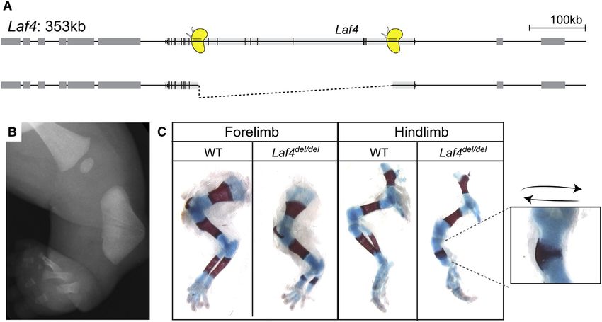
Cell Reports

Resource

Deletions, Inversions, Duplications: Engineering
of Structural Variants using CRISPR/Cas in Mice
Katerina Kraft,1,5 Sinje Geuer,1,2,5 Anja J. Will,1,2 Wing Lee Chan,2 Christina Paliou,1 Marina Borschiwer,1
Izabela Harabula,1 Lars Wittler,1 Martin Franke,1,2 Daniel M. Ibrahim,1,3 Bjørt K. Kragesteen,1,2,3 Malte Spielmann,1,2,3,4
Stefan Mundlos,1,2,3,4 Darı́o G. Lupiáñez,1,6,* and Guillaume Andrey1,6,*
1Max   Planck Institute for Molecular Genetics, 14195 Berlin, Germany
2Institutefor Medical and Human Genetics, Charité Universitätsmedizin Berlin, 13353 Berlin, Germany
3Berlin-Brandenburg Center for Regenerative Therapies (BCRT), Charité Universitätsmedizin Berlin, 13353 Berlin, Germany
4Berlin-Brandenburg School for Regenerative Therapies (BSRT), Charité Universitätsmedizin Berlin, 13353 Berlin, Germany
5Co-first author
6Co-senior author

*Correspondence: lupianez@molgen.mpg.de (D.G.L.), andrey@molgen.mpg.de (G.A.)
 http://dx.doi.org/10.1016/j.celrep.2015.01.016
This is an open access article under the CC BY-NC-ND license (http://creativecommons.org/licenses/by-nc-nd/3.0/).

SUMMARY                                                              sary for proper enhancer-promoter interactions. Such rear-
                                                                     rangements can result in loss of WT interactions and/or ectopic
Structural variations (SVs) contribute to the variability            enhancer-promoter interactions, thereby resulting in gene mis-
of our genome and are often associated with dis-                     expression (Montavon et al., 2012; Spielmann et al., 2012;
ease. Their study in model systems was hampered                      Spielmann and Mundlos, 2013). To discriminate between these
until now by labor-intensive genetic targeting proce-                multiple effects and to study their complex molecular pathology
dures and multiple mouse crossing steps. Here we                     in vivo modeling of SVs is required.
                                                                        SVs can be induced by radiation via the induction of double-
present the use of CRISPR/Cas for the fast (10 weeks)
                                                                     strand breaks, but this is a random process that cannot be tar-
and efficient generation of SVs in mice. We specif-
                                                                     geted to specific genomic regions. So far, allelic series involving
ically produced deletions, inversions, and also dupli-               the remodeling of large DNA segments have been obtained from
cations at six different genomic loci ranging from 1.1               the cis and trans recombination of targeted loxP sites in mouse
kb to 1.6 Mb with efficiencies up to 42%. After PCR-                 chromosomes (Hérault et al., 1998; Ruf et al., 2011; Spitz
based selection, clones were successfully used to                    et al., 2005). However, these approaches are time consuming
create mice via aggregation. To test the practicability              and laborious, involving the targeting of loxP sequences and
of the method, we reproduced a human 500 kb dis-                     subsequent mice crossings with Cre driver animals, a procedure
ease-associated deletion and were able to recapitu-                  taking at least 12 months. More recently, Zinc Finger nucleases
late the human phenotype in mice. Furthermore, we                    (ZNF) or transcription activator-like effector nucleases (TALENs)
evaluated the regulatory potential of a large genomic                have been shown to induce targeted SVs of several hundred ki-
                                                                     lobases in mammalian cells. In vivo inversions and deletions up
interval by deleting a 1.5 Mb fragment. The method
                                                                     to 1 Mb could be obtained in zebrafish (Bauer et al., 2013; Gupta
presented permits rapid in vivo modeling of genomic
                                                                     et al., 2013; Lee et al., 2012; Xiao et al., 2013). However, to our
rearrangements.                                                      knowledge, these methods were not applied to generate large
                                                                     structural variants in mice. This might be due to the process of
INTRODUCTION                                                         generating specific ZNF nucleases or TALEN that is rather slow
                                                                     and requires expert knowledge.
Genomic structural variations (SVs) are large-scale structural          The recent development of the CRISPR/Cas technology has
differences in the genomic DNA ranging in size from a few            led to a wider use of genome editing, opening new possibilities
kilobases to entire chromosomes. SVs may be unbalanced               to engineer SVs in various model systems (Hsu et al., 2014;
as in deletions, duplications, and insertions or balanced as in      Peng et al., 2014; Wang et al., 2013). Indeed, two synthetic guide
inversions and translocations or a combination thereof. SVs          RNAs (sgRNAs), targeted at two different positions of a chromo-
contribute to a large extent to the variability of our genome        some, are able to induce inversions and deletions through non-
and are often associated with disease (Stankiewicz and Lupski,       homologous end joining (NHEJ). For instance, Xiao et al. (2013)
2010). When occurring within the coding sequence of genes,           were able to induce 1.5 kb deletions in zebrafish embryos by in-
they can affect the protein sequence and thereby protein             jecting sgRNAs and Cas9 directly into zygotes. Similarly, larger
function or stability. When encompassing one or several cod-         structural rearrangements involving deletions, inversions, and
ing units, deletions or duplications lead to changes in gene         translocations were obtained in HEK293 or in murine eurythro-
dosage. Furthermore, it was shown that SVs could interfere           leukemia (MEL) cell lines (Canver et al., 2014; Choi and Meyer-
with gene regulation by disrupting genomic architecture neces-       son, 2014).

                                                                  Cell Reports 10, 833–839, February 10, 2015 ª2015 The Authors 833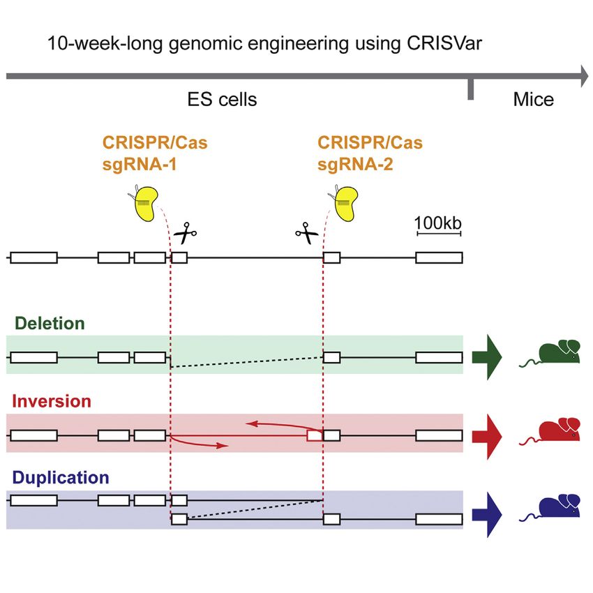
Figure 1. Two sgRNAs Can Induce Structural Variants of the Intermediate Genomic Region
(A) Overview and timing of CRISVar to obtain SVs in mouse.
(B) A 232 kb region at the Pitx1 locus targeted for deletion, inversion, and duplication. Cas9 proteins and sgRNAs are depicted in yellow.
(C) PCR amplifications of breakpoints from the different genetic configurations (DEL, deletion; DUP, duplication; INV A, inversion breakpoint A; INV B, inversion
breakpoint B). BRP (breakpoints) A and B represent the WT product at the CRISPR target sites.
(D) Genetic variability at the rearranged and WT breakpoints. SgRNAs are marked in green or red. Sequences of three clones are shown for each type of junction.
See also Figure S1 and Table S1.

   In this study, we applied the CRISPR/Cas technology in mouse                   RESULTS
embryonic stem cells (ESCs) and developed a 10-week protocol
that we named CRISVar (CRISPR/Cas-induced structural vari-                        CRISPR/Cas for the Induction of SVs in Mouse ESCs
ants) to efficiently produce deletions, inversions, and duplica-                  To test the ability of CRISPR/Cas to generate structural variants
tions in mice. We were able to rearrange targeted genomic                         in mice, we first targeted the pleiotropic and developmentally
intervals ranging from 1 kb to 1.6 Mb using the CRISPR/Cas sys-                   associated locus Pitx1. We designed two sgRNAs in the gene
tem in ESCs (Figure 1A). Moreover, we show that ESCs harboring                    desert adjacent and telomeric to Pitx1 promoter. The two-tar-
these mutations can be used to create chimeric animals. Finally,                  geted sites are separated by 232 kb of intermediate gene desert.
we show at the Laf4 and Epha4 loci the ability of CRISVar                         After transfection of ESCs with the CRISPR constructs, we
to model human pathogenic SVs and to evaluate the regulatory                      screened 288 clones for inversions, deletions, and duplications
influence of a large gene desert, respectively.                                   using a PCR-based approach (Figures 1B and S1). We detected

834 Cell Reports 10, 833–839, February 10, 2015 ª2015 The Authors
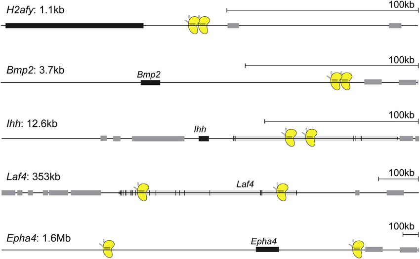
Table 1. Synthesis of Targeted SVs Efficiencies
Name of the Locus                         H2afy             Bmp2           Ihh             Pitx1              Laf4                 Epha4
CRISPR vector                             pX330             pX330          pX459           pX459              pX459                pX330
Size of the rearranged region             1,189 bp          3.7 kb         12.6 kb         32 kb              353kb                1.672 Mb
Deleted                                   11 (3.8%)         12 (6.3%)      121 (42%)       9 (3.1%)           38 (13.2%)           4 (2.1%)
Inverted (two breakpoints mapped)         2 (0.7%)          3 (1.6%)       7 (2.4%)        3 (1%)             12 (4.2%)            3 (1.6%)
Inverted (one breakpoint mapped)          0 (0%)            0 (0%)         9 (3.1%)        8 (2.8%)           20 (6.9%)            1 (0.5%)
Duplicated                                0 (0%)            0 (0%)         0 (0%)          2 (0.7%)           81 (28.1%)           0 (0%)
Deleted/inverted (two breakpoints         1 (0.3%)          1 (0.5%)       3 (1.0%)        0 (0%)             11 (3.8%)            0 (0%)
mapped)
Deleted/inverted (one breakpoint          0 (0%)            1 (0.5%)       2 (0.7%)        1 (0.3%)           9 (3.1%)             0 (0%)
mapped)
Deleted/duplicated                        1 (0.3%)          0 (0%)         1 (0.3%)        4 (1.4%)           28 (10%)             0 (0%)
Inverted/duplicated (two breakpoints      0 (0%)            0 (0%)         0 (0%)          0 (0%)             6 (2.1%)             0 (0%)
mapped)
Inverted/duplicated (one breakpoint       0 (0%)            0 (0%)         0 (0%)          2 (0.7%)           17 (5.9%)            0 (0%)
mapped)
Number of screened clones                 288               192            288             288                288                  192
Successfully aggregated clones            one deletion      -              -               one inversion      two deletions        one inversion
                                          -                 -              -               one deletion       one duplication      one deletion
Numbers and percentages are indicated for each type of rearrangements at every locus. Deletions and inversions were found in all cases. Duplications
where identified in four of six loci. The number of successfully aggregated clones for each allele is indicated in the bottom track.

and sequenced junctions from all three types of structural vari-           with rearrangements of 1.1 kb, 3.7 kb, 12.6 kb, 353 kb, and 1.6
ants (Figures 1C and 1D). Deletions and inversions were found              Mb, respectively (Table 1). Interestingly, the frequency at which
most frequently (5%) followed by duplications (3%). Unexpect-              SVs occurred was different from one locus to another without
edly, not all inversion breakpoints could be mapped (Table 1).             evident relation to the genomic distance separating the two
   SVs did not always segregate with a WT allele but were often            CRISPR sites. We observed deletions and inversions at all sites,
found accompanied by other SVs on the homologous chromo-                   although with a variation in their frequency of around 10-fold be-
some. For instance, one of the clones harbored a deletion and              tween experiments. Duplications were observed at three of
an inversion, indicating that NHEJ occurred simultaneously in              these five rearranged loci.
the two homologous chromosomes. The absence of the WT                         Next, we wanted to test whether the here-produced clones
variant in this clone was confirmed by the loss of PCR product             could be used to create viable animals. We aggregated ESCs
at both sgRNA binding sites (Figure 1C). Another interesting phe-          from eight selected clones to produce animals carrying dele-
nomenon was observed in some of the duplicated alleles. Dupli-             tions, duplication, and inversions at the above-mentioned loci.
cations originate from translocations between homologous                   We were able to produce highly chimeric animals for all rear-
chromosomes; thereby one of the chromosomes loses a copy                   rangement types and loci (Table 1). In our hands, all aggregated
of the rearranged DNA while the other gains it. In our experi-             ESC clones performed to produce chimeric animals.
ments, duplications segregated with a deletion in only half of
the cases. In the others, we found duplications segregating                A 353 kb Intragenic Deletion of Laf4 Recapitulates a
with an inverted or a WT homologous region (Figure 1C). A similar          Human Malformation Syndrome
observation was described using ZNF nucleases in mammalian                 In order to confirm the effectiveness of this genetic tool to
cells (Lee et al., 2012).                                                  generate mouse models, we aimed at reproducing a human dis-
   Sequencing of the junction products revealed diversity of the           ease-associated SV of unknown pathogenicity (Steichen-Gers-
genomic position where the NHEJ had occurred as well as loss               dorf et al., 2008). The SV had occurred de novo, but its functional
of DNA segments (Figure 1D). Double-strand breaks induced                  relevance remained unclear. The patient suffered from multiple
by the Cas9 endonuclease at their target sites were shown to               malformations, including shortening of the femur, aplasia of the
result in local indels, thereby explaining the allele-to-allele vari-      fibula, a triangular tibia, and three toes (Figure 3B). Because of
ability (Canver et al., 2014). Moreover, sequence changes were             the tibia malformation, the diagnosis of a Nievergelt-like syn-
also observed in alleles genotyped as WT, suggesting defective             drome was made. To evaluate the function of LAF4, we first pro-
repair (Figure 1D).                                                        duced a conventional knockout by targeted recombination. We
   After successfully implementing CRISVar at the Pitx1 locus,             could not observe a phenotype in homozygous mice, showing
we targeted other regions of the genome to rule out the possibil-          that inactivation of Laf4 cannot reproduce the human phenotype
ity that the observed effects were locus specific. We aimed at re-         in the mouse (data not shown). We therefore re-evaluated the
gions near the genes H2afy, Bmp2, Ihh, Laf4 and Epha4 (Figure 2)           published data and found that the deletion was smaller than

                                                                        Cell Reports 10, 833–839, February 10, 2015 ª2015 The Authors 835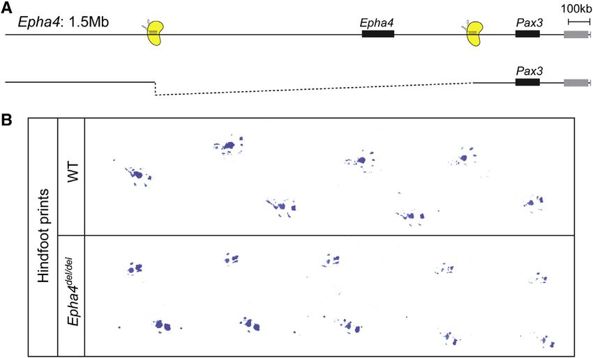
Figure 2. Rearrangements at Other Loci
Summary of the other alleles in which SVs were
induced. The alleles are ordered from the smallest
generated SV (top: H2afy) to the largest (bottom:
Epha4). Sizes of the rearranged regions are indi-
cated next to the locus name.
transcription unit (Figure 4A). Heterozy-
gous clones were used to produce
mice with a 1.5 Mb deletion, which
were subsequently bred to homozygosi-
ty. We observed a neurological pheno-
type in these animals, consisting of a
hopping gait, as previously described
for Epha4 loss of function (Figure 4B;
Movie S1) (Dottori et al., 1998). Other ab-
normalities were not observed in these
mice. In particular, we did not detect
any of the Pax3-associated phenotypes
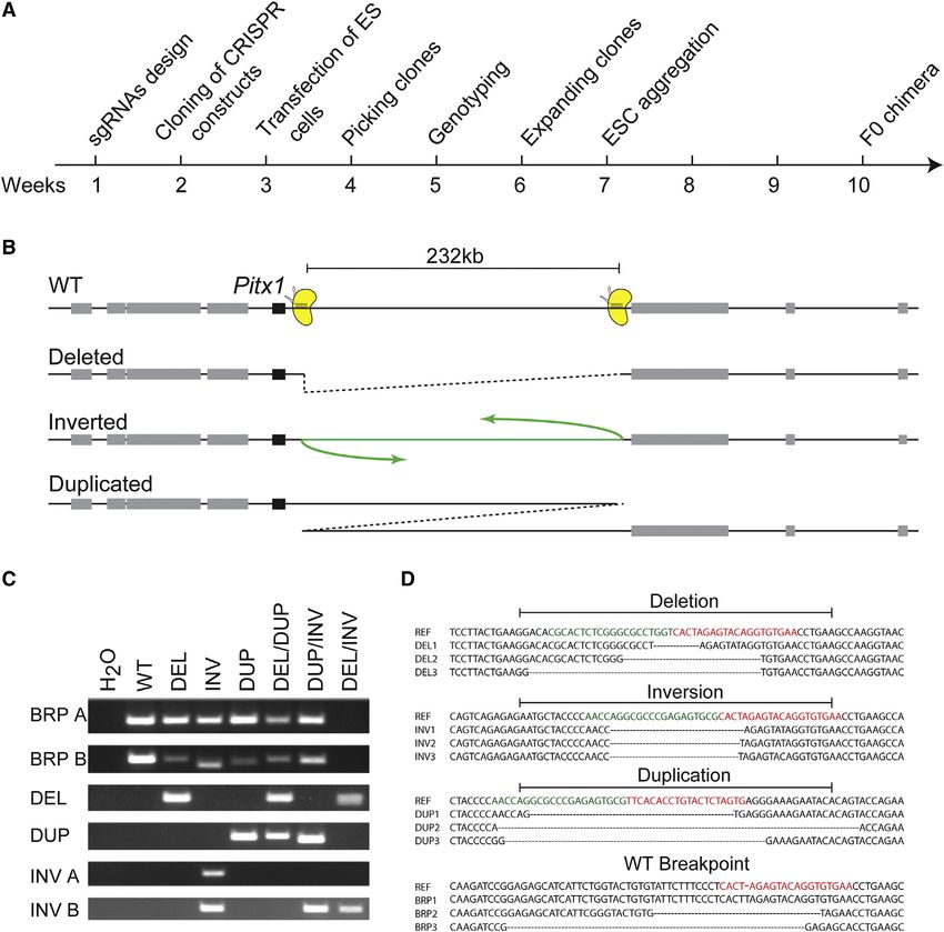
originally thought, encompassing only nine exons of the LAF4 such as pigmentation defects and abnormalities of the spine
gene and not the entire genomic region. The breakpoints of or the brain. Furthermore, in situ hybridization of Pax3 in deletion
this 500 kb deletion were mapped in introns 3 and 12, thus elim- embryos revealed a normal pattern of expression (data not
inating exons 4 to 12 of LAF4 without introducing a frameshift. shown). Our finding that the 1.5 Mb deletion results in a full
The predicted product is a truncated 850 amino acids protein recapitulation of the Epha4 knockout without additional abnor-
lacking a domain predicted to be involved in transcription activa- malities indicates that the region does not contain elements
tion (Ma and Staudt, 1996). To specifically model this SV, we de- essential for Pax3 regulation.
signed sgRNA at the homologous regions in the mouse genome
corresponding to the human breakpoints (Figures 2 and 3A). DISCUSSION
The screening of ESCs revealed clones with heterozygous
and homozygous deletions of the 353 kb homologous region. CRISPR/Cas is a rapid and efficient method to specifically edit
In chimeric animals generated from heterozygous as well as ho- genomes (Cho et al., 2013; Wang et al., 2013). Here we show
mozygous clones, we observed a short zeugopod in the upper that by targeting two sgRNAs at two distal genomic sites we
limb (Figure 3C). The lower-limb abnormalities recapitulated can induce the rearrangement of the intermediate DNA fragment
part of the human phenotype showing a small, triangular ossifi- up to 1.6 Mb in ESCs. We show that duplications, deletions, and
cation center of the tibia and severe hypoplasia of the fibula (Fig- inversions can be obtained in different clones, but they may also
ure 3C). In contrast to the patient, the mice had polydactyly of the occur in various combinations together.
feet with incomplete penetrance. Thus, the specific intragenic We observed extensive variability in the targeting efficiency
deletion in Laf4 generated by CRISPR/Cas recapitulated the from one locus to another. Although the size of the re-arranged
patient’s phenotype, demonstrating the pathogenicity of this DNA region was previously shown to play an important role in
SV. It is likely that the truncated protein exerts a dominant-nega- targeting efficiency, we were not able to confirm this relationship
tive effect, thereby leading to the observed abnormalities of bone (Canver et al., 2014). One of the possibilities is that the chromatin
formation. ‘‘openness’’ at the CRISPR target site affects the cutting effi-
ciency of Cas9 and thereby increases the probability of NHEJ
A 1.5 Mb Deletion of a Gene Desert Encompassing between the two distal breakpoints. In fact, it has been shown
Epha4 Results in Hindlimb Hopping Gait that Cas9 binds better at open chromatin sites, where it has
Large gene deserts are thought to harbor regulatory elements higher off-target effects (Kuscu et al., 2014). In this view, the
and often surround developmentally active genes. We aimed epigenetic status of the targeted region might influence the over-
at using the here-described method to challenge the integrity all efficiency of the rearrangement.
of such a locus. At the Epha4/Pax3 extended locus, two gene Furthermore, we frequently found sequence variations at the
deserts centromeric and telomeric to Epha4 might regulate SV breakpoints. The randomness of NHEJ after a double-strand
either one or both genes. Epha4 has been shown to control break is likely to be accountable for this diversity. This might also
neuronal guidance in hindlimbs. Pax3 is a transcription factor explain why several inverted clones could be mapped at a single
with important function in the migration of muscle progenitors breakpoint only: the other one being too extensively modified for
in the limbs and neural crest migration. Mice with mutations in a proper PCR reaction to occur. This variability in the NHEJ pro-
Pax3 show pigmentation defects, early lethality due to heart de- cess also pinpoints to the limitations of PCR-oriented allele
fects, spina bifida, and exencephaly. We induced a deletion ex- detection; indeed, several false-negative clones with deletions,
tending 1 Mb centromeric and 350 kb telomeric to the Epha4 duplications, or inversions may have been missed because of
836 Cell Reports 10, 833–839, February 10, 2015 ª2015 The AuthorsFigure 3. A 350 kb Deletion at the Laf4 Locus Recapitulates Nievergelt Syndrome
(A) Schematic of the extended Laf4 genomic region. CRISPR/Cas binding sites are depicted in yellow. The induced deletion (350 kb) is shown below.
(B) Radiograph of patient lower leg showing triangular tibia, missing fibula, and oligodactyly.
(C) Skeletal preparations of WT and mutant embryos at E16.5. Cartilage is shown in blue and bone in red. Note the short ulna and radius with small ossification
centers in forelimb and triangular tibia (magnification shown on right) and severely hypoplastic fibula in hindlimb.
See also Table S2.
extensive rearrangements at their breakpoints, inhibiting proper EXPERIMENTAL PROCEDURES
PCR reactions to occur. Because of this uncertainty, these
CRISPR sgRNAs Selection and Cloning
clones were not processed further and discarded. Given the
SgRNAs were designed flanking the regions to rearrange. We used the http://
high efficiency of CRISVar, this was not considered a problem. crispr.mit.edu/ platform to obtain candidate sgRNA sequences with little off-
We also observed that WT alleles might harbor indels induced target specificity. Complementary strands were annealed, phosphorylated,
by CRISPR/Cas. Thus, the WT allele should also be sequenced and cloned into the BbsI site of pX459 or pX330 CRISPR/Cas vector (Addgene;
at the target site. see Table 1).
The limitations of CRISVar can be efficiently bypassed by
a proper screening of ESC clones and subsequent mouse ES Cell Culture and Transfection
crossing. Such extensive screening might be a limiting step if G4 ESCs (300,000) (George et al., 2007) were seeded on CD1 feeders and
transfected with 8 mg of each CRISPR construct using FuGENE technology
the method would be applied directly to mouse zygotes. How-
(Promega). When the construct originated from the pX330 vector, cells were
ever, it is unclear how efficient microinjections of sgRNAs and cotransfected with a puromycine-resistant plasmid. PX459, in contrast,
Cas9 directly into embryos would be. Further testing is thus already contains a puromycine-resistance cassette. After 24 hr, cells were split
required to compare the efficiency of both approaches. The ad- and transferred onto DR4 puro-resistant feeders and selected with puromy-
vantages of producing SVs in ESCs are on the one hand the pos- cine for 2 days. Clones where then grown for 5 to 6 more days, picked, and
sibility of retargeting to introduce additional mutations and on the transferred into 96-well plates on CD-1 feeders. After 2 days of culture, plates
were split in triplicates, two for freezing and one for growth and DNA
other hand the direct use of these cells in culture to investigate
harvesting.
the effects of SVs.
Positive clones were thawed and grown on CD-1 feeders until they reach an
Thus, with CRISVar, large and small SVs of various types can average of four million cells. Three vials were frozen, and DNA was harvested
be produced with high efficiency and in a short time period to from the rest of the cells to confirm genotyping.
study the effect of genomic rearrangements and their pathoge-
netic effect. As shown at the Laf4 locus, human SVs may exert PCR-Based Genotyping
their effects in a specific manner, necessitating an exact repro- Primers were designed on both sides of sgRNAs targets, at a distance of 100–
duction in the mouse genome to evaluate their pathogenicity. 300 bp from the cutting site. Each allele has thus a set of four primers: T1(fwd)/
T2(rev) amplifying one targeted site and T3(fwd)/T4(rev) amplifying the other
CRISPR/Cas-mediated genome editing can be used to effi-
(Figure S1; Table S1). Deletions were mapped using T1 and T4 primers while
ciently create such rearrangements. Finally, as shown in the
inversions were detected with T2/T4 for the proximal breakpoints and T1/T3
Epha4 example, the method can be used to challenge the integ- for the distal one. Finally, duplications were detected with T2/T3 primers. Pos-
rity of genomic regulatory units, as they have recently been itive PCR bands were Sanger sequenced using one of the amplification
proposed (Marinic et al., 2013). primers.
Cell Reports 10, 833–839, February 10, 2015 ª2015 The Authors 837Figure 4. CRISPR/Cas-Induced 1.5 Mb
Deletion at the Epha4 Locus
(A) Schematic of the extended Epha4/Pax3
genomic region. CRISPR/Cas binding sites are
depicted in yellow. The induced deletion (1.5 Mb)
is shown below.
(B) The deletion of a 1.5 Mb region containing the
Epha4 gene and surrounding gene desert results
in a phenocopy of Epha4 knockout mouse but no
other abnormalities. Footprints of hindlimbs show
hopping gait in deletion mutant.
See also Movie S1.
Screening for Laf4 Homozygote Deletion Clones Received: November 17, 2014
Quantification of copy-number variation of the deleted interval in comparison Revised: January 5, 2015
to other genomic positions was performed with a set of qPCR primers (see Accepted: January 7, 2015
Table S2). Published: February 5, 2015
Mouse Aggregation REFERENCES
A frozen ESC vial was seeded on CD-1 feeders, and cells were grown for
2 days. Mice were generated by diploid or tetraploid aggregation (Artus and Artus, J., and Hadjantonakis, A.K. (2011). Generation of chimeras by aggrega-
Hadjantonakis, 2011). All animal procedures were in accordance with institu- tion of embryonic stem cells with diploid or tetraploid mouse embryos.
tional, state, and government regulations (LAGeSo). Methods Mol. Biol. 693, 37–56.
Bauer, D.E., Kamran, S.C., Lessard, S., Xu, J., Fujiwara, Y., Lin, C., Shao, Z.,
Leg Footprints Canver, M.C., Smith, E.C., Pinello, L., et al. (2013). An erythroid enhancer of
Adult mice were encouraged to run over a white paper sheet after their hindfeet BCL11A subject to genetic variation determines fetal hemoglobin level. Sci-
were soaked in blue ink. ence 342, 253–257.
Canver, M.C., Bauer, D.E., Dass, A., Yien, Y.Y., Chung, J., Masuda, T., Maeda,
Skeletal Preparation
T., Paw, B.H., and Orkin, S.H. (2014). Characterization of genomic deletion ef-
E16.5 animals were processed and stained as described previously (Mundlos,
ficiency mediated by clustered regularly interspaced palindromic repeats
2000).
(CRISPR)/Cas9 nuclease system in mammalian cells. J. Biol. Chem. 289,
21312–21324.
SUPPLEMENTAL INFORMATION
Cho, S.W., Kim, S., Kim, J.M., and Kim, J.S. (2013). Targeted genome engi-
neering in human cells with the Cas9 RNA-guided endonuclease. Nat. Bio-
Supplemental Information includes Supplemental Experimental Procedures,
technol. 31, 230–232.
one figure, two tables, and one movie and can be found with this article online
at http://dx.doi.org/10.1016/j.celrep.2015.01.016. Choi, P.S., and Meyerson, M. (2014). Targeted genomic rearrangements using
CRISPR/Cas technology. Nat. Commun. 5, 3728.
AUTHOR CONTRIBUTIONS Dottori, M., Hartley, L., Galea, M., Paxinos, G., Polizzotto, M., Kilpatrick, T.,
Bartlett, P.F., Murphy, M., Köntgen, F., and Boyd, A.W. (1998). EphA4 (Sek1)
K.K. designed, produced, and analyzed the Epha4 1.5 Mb deletion. S.G. de- receptor tyrosine kinase is required for the development of the corticospinal
signed, produced, and analyzed the Laf4 truncation. W.L.C. performed the tract. Proc. Natl. Acad. Sci. USA 95, 13248–13253.
Laf4 knockout. K.K., A.J.W., S.G., M.F., D.M.I., M.S., S.M., D.G.L., and G.A. George, S.H., Gertsenstein, M., Vintersten, K., Korets-Smith, E., Murphy, J.,
designed the research. L.W. performed aggregations. K.K., A.J.W., S.G., Stevens, M.E., Haigh, J.J., and Nagy, A. (2007). Developmental and adult phe-
C.P., M.B., I.H., M.F., D.M.I., B.K.K., D.G.L., and G.A. performed the other ex- notyping directly from mutant embryonic stem cells. Proc. Natl. Acad. Sci.
periments. K.K., S.M., D.G.L., and G.A. wrote the manuscript. USA 104, 4455–4460.
Gupta, A., Hall, V.L., Kok, F.O., Shin, M., McNulty, J.C., Lawson, N.D., and
ACKNOWLEDGMENTS
Wolfe, S.A. (2013). Targeted chromosomal deletions and inversions in zebra-
fish. Genome Res. 23, 1008–1017.
We thank Phillip Grote and Tracie Pennimpede for discussions, advice, and
sharing reagents. We thank Asita Stiege for technical assistance. G.A. is sup- Hérault, Y., Rassoulzadegan, M., Cuzin, F., and Duboule, D. (1998). Engineer-
ported by an Early Postdoc.Mobility grant from the Swiss National Science ing chromosomes in mice through targeted meiotic recombination (TAMERE).
Foundation. D.G.L. is supported by the Fundación Alfonso Martı́n Escudero. Nat. Genet. 20, 381–384.
This research was supported by a grant from the Deutsche Forschungsge- Hsu, P.D., Lander, E.S., and Zhang, F. (2014). Development and applications
meinschaft to S.M. of CRISPR-Cas9 for genome engineering. Cell 157, 1262–1278.
838 Cell Reports 10, 833–839, February 10, 2015 ª2015 The AuthorsKuscu, C., Arslan, S., Singh, R., Thorpe, J., and Adli, M. (2014). Genome-wide Spielmann, M., and Mundlos, S. (2013). Structural variations, the regulatory
analysis reveals characteristics of off-target sites bound by the Cas9 endonu- landscape of the genome and their alteration in human disease. BioEssays
clease. Nat. Biotechnol. 32, 677–683. 35, 533–543.
Lee, H.J., Kweon, J., Kim, E., Kim, S., and Kim, J.S. (2012). Targeted chromo- Spielmann, M., Brancati, F., Krawitz, P.M., Robinson, P.N., Ibrahim, D.M.,
somal duplications and inversions in the human genome using zinc finger nu- Franke, M., Hecht, J., Lohan, S., Dathe, K., Nardone, A.M., et al. (2012). Home-
cleases. Genome Res. 22, 539–548. otic arm-to-leg transformation associated with genomic rearrangements at the
PITX1 locus. Am. J. Hum. Genet. 91, 629–635.
Ma, C., and Staudt, L.M. (1996). LAF-4 encodes a lymphoid nuclear protein
with transactivation potential that is homologous to AF-4, the gene fused to Spitz, F., Herkenne, C., Morris, M.A., and Duboule, D. (2005). Inversion-
MLL in t(4;11) leukemias. Blood 87, 734–745. induced disruption of the Hoxd cluster leads to the partition of regulatory land-
scapes. Nat. Genet. 37, 889–893.
, M., Aktas, T., Ruf, S., and Spitz, F. (2013). An integrated holo-
Marinic
enhancer unit defines tissue and gene specificity of the Fgf8 regulatory land- Stankiewicz, P., and Lupski, J.R. (2010). Structural variation in the human
scape. Dev. Cell 24, 530–542. genome and its role in disease. Annu. Rev. Med. 61, 437–455.
Steichen-Gersdorf, E., Gassner, I., Superti-Furga, A., Ullmann, R., Stricker, S.,
Montavon, T., Thevenet, L., and Duboule, D. (2012). Impact of copy number
Klopocki, E., and Mundlos, S. (2008). Triangular tibia with fibular aplasia asso-
variations (CNVs) on long-range gene regulation at the HoxD locus. Proc.
ciated with a microdeletion on 2q11.2 encompassing LAF4. Clin. Genet. 74,
Natl. Acad. Sci. USA 109, 20204–20211.
560–565.
Mundlos, S. (2000). Skeletal morphogenesis. Methods Mol. Biol. 136, 61–70.
Wang, H., Yang, H., Shivalila, C.S., Dawlaty, M.M., Cheng, A.W., Zhang, F.,
Peng, Y., Clark, K.J., Campbell, J.M., Panetta, M.R., Guo, Y., and Ekker, S.C. and Jaenisch, R. (2013). One-step generation of mice carrying mutations in
(2014). Making designer mutants in model organisms. Development 141, multiple genes by CRISPR/Cas-mediated genome engineering. Cell 153,
4042–4054. 910–918.
Ruf, S., Symmons, O., Uslu, V.V., Dolle, D., Hot, C., Ettwiller, L., and Spitz, F. Xiao, A., Wang, Z., Hu, Y., Wu, Y., Luo, Z., Yang, Z., Zu, Y., Li, W., Huang, P.,
(2011). Large-scale analysis of the regulatory architecture of the mouse Tong, X., et al. (2013). Chromosomal deletions and inversions mediated by
genome with a transposon-associated sensor. Nat. Genet. 43, 379–386. TALENs and CRISPR/Cas in zebrafish. Nucleic Acids Res. 41, e141.
Cell Reports 10, 833–839, February 10, 2015 ª2015 The Authors 839You can also read