Lactobacillus spp. reduces ethanol induced liver oxidative stress and inflammation in a mouse model of alcoholic steatohepatitis
←
→
Page content transcription
If your browser does not render page correctly, please read the page content below
EXPERIMENTAL AND THERAPEUTIC MEDICINE 21: 188, 2021

   Lactobacillus spp. reduces ethanol‑induced liver oxidative stress
   and inflammation in a mouse model of alcoholic steatohepatitis
                               PEI‑SHAN HSIEH, CHING‑WEI CHEN, YI‑WEI KUO and HSIEH‑HSUN HO

                                                    Glac Biotech Co., Ltd., Tainan 74442, Taiwan, R.O.C.

                                                  Received February 14, 2020; Accepted August 19, 2020

                                                                      DOI: 10.3892/etm.2021.9619

Abstract. Alcoholic steatohepatitis (ASH) is a complex multi‑                               in the liver. Collectively, the present results indicated that
factorial disease that can lead to liver fibrosis and cirrhosis if not                      Lactobacillus strains TSP05, TSF331 and TSR332 reduced
treated promptly. Alcohol‑induced oxidative stress and inflam‑                              oxidative stress and inflammatory responses, thus preventing
mation are the main factors that cause steatohepatitis and liver                            ASH development and liver injury.
injury; however, probiotic bacteria in the gastrointestinal tract
have been revealed to regulate immune responses and reduce                                  Introduction
oxidative stress, suggesting that functional probiotics could
help to prevent ASH and liver injury. Despite numerous reports                              Chronic alcohol abuse frequently leads to alcoholic steato‑
on the interactions between ASH and probiotics, the mecha‑                                  hepatitis (ASH) in the liver, with subsequent liver fibrosis and
nisms underlying probiotic‑mediated liver protection remain                                 cirrhosis (1). Alcoholic cirrhosis causes ~1 million mortalities
unknown. Therefore, the aim of the present study was to screen                              per year globally, accounting for 1/2 of all cirrhosis‑related
probiotics with high antioxidant capacity and investigate the                               mortality, while alcohol‑related hepatocellular carcinoma is
ability of different probiotic combinations to reduce alcoholic                             responsible for a further 80,000 deaths (1). The World Health
liver disease (ALD) in a mouse model. It was identified that                                Organization reported that individuals who consume excess
Lactobacillus plantarum (TSP05), Lactobacillus fermentum                                    alcohol are at a high risk of alcoholic liver disease (ALD) and
(TSF331) and Lactobacillus reuteri (TSR332) neutralized                                     that ~6% of all global mortalities can be attributed to alcohol
free radicals and displayed high antioxidant activity in vitro.                             consumption (2‑4); thus, the issue of alcohol abuse requires
In addition, these three functional probiotic strains protected                             immediate attention.
mice from alcohol‑induced liver injury in vivo. Mice treated                                    The liver is the main organ involved in alcohol metabolism,
with the probiotics demonstrated significantly lower alanine                                in which two major enzymes, alcohol dehydrogenase and acet‑
aminotransferase, aspartate aminotransferase and triglyceride                               aldehyde dehydrogenase, participate in the oxidative pathway
levels, which were associated with the downregulation of the                                that converts ethanol to acetaldehyde and causes tissue injury
proinflammatory cytokines TNF‑ α and IL‑6. Furthermore,                                     due to the production of reactive oxygen species (ROS) (5,6).
probiotic treatment upregulated glutathione and glutathione                                 Chronic alcohol abuse has been reported to result in ROS
peroxidase activity, which are bioindicators of oxidative stress                            overproduction and to interfere with lipid metabolism in the
                                                                                            liver, leading to ROS‑mediated liver injury (7). In addition,
                                                                                            the production of large quantities of ROS by ethanol metabo‑
                                                                                            lism may damage the intestinal barrier, leading to barrier
Correspondence to: Dr Hsieh‑Hsun Ho, Glac Biotech Co., Ltd.,                                leakage and allowing endotoxins and metabolites secreted by
Fourth floor, 17 Guoji Road, Xinshi, Tainan 74442, Taiwan, R.O.C.                           gram‑negative bacteria to reach the liver portal circulation via
E‑mail: sam.ho@bioflag.com.tw                                                               the gut‑liver axis, which promotes inflammation and ultimately
                                                                                            results in ALD or ASH (6‑9).
Abbreviations: ALD, alcoholic liver disease; ALT, alanine                                       Certain probiotics have been reported to exert benefi‑
aminotransferase; ASH, alcoholic steatohepatitis; AST, aspartate                            cial effects against ALD and ASH (7,10); for example, the
aminotransferase; CFU, colony‑forming unit; DPPH, Diphenyl                                  Lactobacillus (L.) rhamnosus GG (LGG) strain is often used
picryl hydrazyl; FRAP, ferric reducing antioxidant power; GSH,
                                                                                            for liver protection (11). Forsyth et al (11) revealed that LGG
glutathione; GPx, glutathione peroxidase; H&E, hematoxylin and
                                                                                            supplementation may ameliorate intestinal and liver oxidative
eosin; LAB, lactic acid bacteria; LGG, Lactobacillus rhamnosus
GG; MRS, de Man, Rogosa, and Sharpe; ROS, reactive oxygen                                   stress, improve ethanol‑induced gut leakiness, and reduce
species; TG, triglyceride; GRAS, generally recognized as safe                               inflammatory responses, while Wang et al (12) reported
                                                                                            that L. rhamnosus B10 may decrease the relative liver index
Ke y w o rd s: A L D, a n t i ox i d a n t , a n t i ‑ i n f l a m m a t o r y,             by enhancing superoxide dismutase activity and reducing
L a c t o b a c i l l u s p l a n t a r u m, L a c t o b a c i l l u s f e r m e n t u m,   ROS‑mediated liver injury. Thus, these findings suggest that
Lactobacillus reuteri                                                                       L. strains could enhance antioxidant activity to improve
                                                                                            ALD and hepatitis; however, the mechanism underlying
                                                                                            probiotic‑mediated liver protection is yet to be elucidated.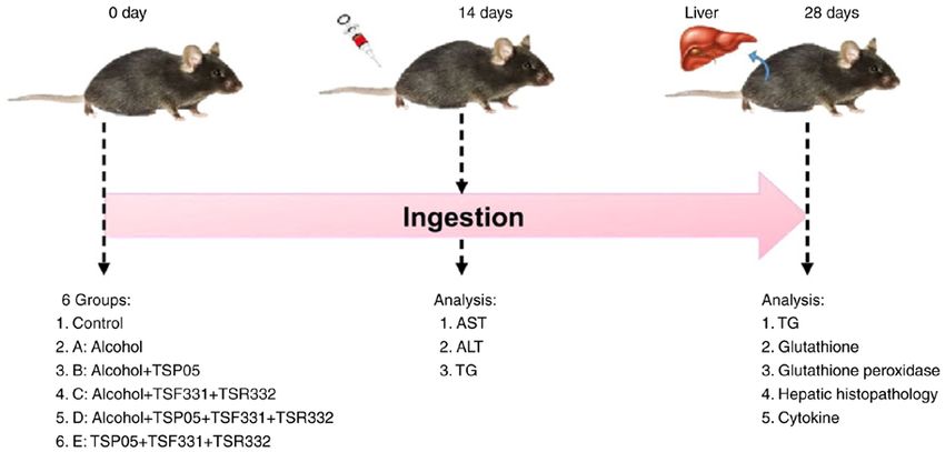
2              HSIEH et al: LACTIC ACID BACTERIA ALLEVIATES ALCOHOLIC STEATOHEPATITIS IN A MOUSE MODEL

    The present study hypothesized that probiotics with             Different FeSO 4·7H 2 O concentrations were prepared to
antioxidant capacity could reduce alcohol‑induced oxidative         construct a standard curve, with vitamin C (2 µg/ml) as a
stress and ultimately liver damage. Therefore, the current          positive control. The samples were analyzed according to the
study screened probiotics with high antioxidant capacity and        standard curve and results are expressed as the reducing power
investigated the ability of different probiotic combinations to     (µg/ml Fe2+).
reduce ALD in a mouse model.
                                                                    Gastric acid and bile tolerance. Gastric acid and bile toler‑
Materials and methods                                               ance was assessed according to the method described by
                                                                    Hyronimus et al (18) and Hassanzadazar et al (19). Activated
Isolation, identification and culture of bacterial strains. A       probiotics were added to eight tubes, one of which was used to
total of 12 lactic acid bacteria (LAB) strains were isolated        count the number of CFUs (1x10⁹ CFU/ml) at 0 h, while the
from the intestines of healthy individuals by Glac Biotech          others were centrifuged at 1,770 x g, 4˚C for 10 min. Each pellet
Co., Ltd., including three L. rhamnosus strains (gl‑105, gl‑165     was mixed well with 5 ml MRS broth (pH 3.5) and incubated
and gl‑201), two L. paracasei strains (gl‑106 and gl‑156), two      at 37˚C for 1‑3 h, with CFUs counted (plate count) in one tube
Enterococcus faecium strains (gl‑212 and gl‑213), L. casei          per hour. The final four tubes were centrifuged at 1,770 x g
subsp. casei (gl‑217), L. gasseri (gl‑102), L. plantarum (TSP05),   4˚C for 10 min before 0.3% bovine bile (Sigma‑Aldrich; Merck
L. fermentum (TSF331) and L. reuteri (TSR332). TSP05 was            KGaA) in MRS broth was added and the tubes were incubated
isolated from pickled vegetables, while TSF331 and TSR332           at 37˚C for 4‑7 h. CFUs were then enumerated in one tube per
were isolated from the intestines of healthy individuals. These     hour for 4‑7 h.
samples included adult stool and pickled vegetables for isola‑
tion, which were mixed in 1% PBS (Sigma‑Aldrich; Merck              Intestinal adhesion assay. The human intestinal epithelial
KGaA). The sample mixtures were plated onto MRS medium              colorectal adenocarcinoma cell line Caco‑2 was obtained from
plate (Difco; BD Biosciences). After 48 h incubation, indi‑         the Bioresource Collection and Research Center BCRC and
vidual colonies were found growing on the plate. The colonies       cultured according to BCRC guidelines. A sterilized cover slip
and bacterial strains were identified using their entire 16S        was placed in the well of a 6‑well dish and incubated at 37˚C,
rDNA sequences as described previously (13‑15) and using            5% CO2 for 3 days with Caco‑2 cells in minimum essential
the National Center for Biotechnology Information nucleotide        medium supplemented with 10% FBS (Merck KGaA) and
Basic Local Alignment Search Tool (NCBI; https://www.ncbi.          1% penicillin‑streptomycin (GE Bio‑Science). The medium
nlm.nih.gov/). Strains TSP05, TSF331 and TSR332 were each           was removed (cell density almost 1.2x10⁶ cells) and the wells
successfully activated three times via incubation in de Man,        were washed twice with PBS before 1.5 ml bacterial suspen‑
Rogosa and Sharpe (MRS) broth (Difco; BD Biosciences)               sion (1x10⁹ CFU/ml) and 1.5 ml MRS medium were added to
at 37˚C for 24 h and then centrifuged at 10,000 x g at 4˚C for      each well and incubated at 37˚C and 5% CO2 for 1‑4 h. The
10 min. The pellets were washed twice with PBS and strains          cells were then washed twice with sterile PBS and fixed with
were mixed in equal proportions (1:1) before being orally           10% methanol at 25˚C for 10 min. The cells were subjected to
administered to mice at a dosage of 8.2x10⁹ colony‑forming          Gram staining at 25˚C for 15 min and enumerated using a light
units (CFU) per kg of body weight.                                  microscope (Nikon Corporation) at 15x100 magnification.

Diphenyl picryl hydrazyl (DPPH) radical‑scavenging                  Experimental animals. Male C57B/6N mice (age, 7 weeks;
activity assay. DPPH radical‑scavenging activity assays             weight, 20‑22 g) were purchased from BioLASCO Taiwan. All
(Sigma‑Aldrich; Merck KGaA) were performed according                mice were housed in plastic cages and fed sterilized food and
to the method by Xing et al (16). Briefly, 150 µl of bacterial      water under 55±5% humidity, controlled temperature (22±2˚C)
cultured solution (1x10⁸ CFU/ml) was mixed with 150 µl              and a 12‑h light/dark cycle, and were acclimated for 1 week prior
DPPH solution (0.2 mM) and incubated at 25˚C in the dark            to experiments. On treatment day 0, the mice were randomly
for 30 min. Distilled water (150 µl) was used as a blank and        allocated to the following six groups (n=7 mice/group): i) Control
2 µg/ml vitamin C was used as a positive control. DPPH              group, fed a regular diet; ii) group A, fed an ethanol‑containing
scavenging was determined by measuring the absorbance               diet (containing 28% ethanol; Dyets, Inc.); iii) group B, fed
(ABS) at 517 nm (BioPhotometer; Eppendorf) and the                  an ethanol‑containing diet + strain TSP05 8.2x10⁹ CFU/kg;
radical‑scavenging capacity was quantified according to             iv) group C, fed an ethanol‑containing diet + strains TSF331 and
the following formula: DPPH radical‑scavenging capacity             TSR332 8.2x10⁹ CFU/kg; v) group D, fed an ethanol‑containing
(%)=ABScontrol‑ABSsample/ABScontrol x100; where ABScontrol and      diet + strains TSP05, TSF331 and TSR332 8.2x10⁹ CFU/kg;
ABSsample represent the ABS of the negative control and sample      and vi) group E, fed a regular diet + strains TSP05, TSF331
at 517 nm, respectively.                                            and TSR332 (Fig. 1). Mice in the control and E groups were
                                                                    fed a Lieber‑DeCarli liquid diet (Dyets, Inc.) without alcohol
Ferric reducing antioxidant power (FRAP) assay. FRAP                ad libitum. To induce alcoholic fatty liver injury, the mice in
assays (Sigma‑Aldrich; Merck KGaA) were conducted                   groups A‑D were fed a Lieber‑DeCarli liquid diet with alcohol,
according to the method of Benzie and Strain (17). Briefly,         as described previously (20). Mice were administered with
750 µl FRAP solution was heated to 37˚C in a water bath and         a single 0.2 ml dose of deionized water (control, group A) or
then 25 µl of bacterial cultured solution and 75 µl ultrapure       0.2 ml deionized water containing the corresponding probiotic(s)
water were added. The mixture was placed in the dark at room        (groups B‑E) daily via oral gavage for 4 weeks. Experiments
temperature for 5 min and the ABS was measured at 593 nm.           were performed in accordance with protocols approved by the
EXPERIMENTAL AND THERAPEUTIC MEDICINE 21: 188, 2021 3
Figure 1. Design of animal experiments. All mice were divide into 6 groups and the experiment was performed for 28 days. Sampling was carried out according
to the experimental design on day 0, 14 and 28. ALT, alanine aminotransferase; AST, aspartate aminotransferase; TG, triglyceride.
Institutional Animal Care and Use Committee of Hung Kuang 10 min, water for 10 min, 0.5% HCl for 5 sec, running water
University (Taichung, Taiwan). for 30 min, 0.5% eosin solution for 2 min, 80% alcohol for
30 sec, 90% alcohol for 30 sec, 100% alcohol for 30 sec,
M ouse seru m biochemical a n alysis. On days 0, xylene:alcohol (1:1) for 30 sec and xylene 30 sec. To histo‑
14 and 28, 0.2 ml of blood samples were collected from the logically evaluate liver injury, the slides were examined and
eye socket of each mouse 2 h after oral gavage with a probiotic images were captured under a bright‑field light microscope
or water. After incubation for 1 h at 25˚C, serum was obtained at 10x20 magnification (TE2000‑S; Nikon Corporation).
and centrifuged at 2,000 x g 4˚C for 10 min. Serum alanine
aminotransferase (ALT), aspartate aminotransferase (AST) Statistical analysis. Data are presented as the mean ± SEM
and triglyceride (TG) levels were determined using an auto‑ obtained from three repeats of per sample. All values from
matic analyzer (Hitachi 7080; Hitachi, Ltd.) at the National different treatment groups were compared using SPSS 17.0
Laboratory Animal Center (Taipei, Taiwan). (SPSS, Inc.), analysing with one‑way ANOVAs followed by
post‑hoc Tukey's tests. P4 HSIEH et al: LACTIC ACID BACTERIA ALLEVIATES ALCOHOLIC STEATOHEPATITIS IN A MOUSE MODEL
Figure 3. Biochemical indicators of liver function. Effects of probiotics
Figure 2. Screening of probiotic antioxidant activities. (A) DPPH assay. on serum (A) ALT and (B) AST concentrations in alcohol‑induced liver
(B) FRAP assay. Vit C (2 µg/ml) was used as a positive control. Data are injury model mice on days 14 and 28. A, alcohol; B, alcohol + TSP05; C,
presented as the mean ± SEM (n=3). DPPH, diphenyl picryl hydrazyl; FRAP, alcohol + TSF331 + TSR332; D, alcohol + TSP05 + TSF331 + TSR332;
ferric reducing antioxidant power; Vit C, vitamin C. E, TSP05 + TSF331 + TSR332. #PEXPERIMENTAL AND THERAPEUTIC MEDICINE 21: 188, 2021 5 Figure 4. Effects of probiotics on serum and hepatic TG content. Effects of probiotics on TG levels in the (A) serum and (B) liver of alcohol‑fed mice. A, alcohol; B, alcohol + TSP05; C, alcohol + TSF331 + TSR332; D, alcohol + TSP05 + TSF331 + TSR332; E, TSP05 + TSF331 + TSR332. # P
6                 HSIEH et al: LACTIC ACID BACTERIA ALLEVIATES ALCOHOLIC STEATOHEPATITIS IN A MOUSE MODEL

                                                                                     Alcohol is metabolized into acetaldehyde, resulting in
                                                                                 the production of free radicals and carcinogens, thereby
                                                                                 stimulating inflammatory responses to release liver injury
                                                                                 indicators (ALT and AST) into the serum (21‑24). Oxidative
                                                                                 damage has been reported to induce lipid peroxidation, thus
                                                                                 disrupting lipid metabolism and affecting liver cells (7,21‑23).
                                                                                 Since enzymatic and non‑enzymatic antioxidant reactions
                                                                                 are necessary to protect against oxidative damage, GPx and
                                                                                 GSH are commonly used as indicators to evaluate the effect of
                                                                                 oxidative stress (23‑25). GSH is the most abundant antioxidant
                                                                                 in liver cells and prevents damage caused by heavy ethanol
                                                                                 consumption (26), while GPx reduces lipid hydroperoxides to
                                                                                 their corresponding alcohols and free hydrogen peroxide to
                                                                                 water (27). Therefore, high GSH levels and GPx activity can
                                                                                 neutralize ROS and prevent oxidative stress (7,23). The present
                                                                                 study identified that feeding mice with probiotics (individually
                                                                                 and in combination) for 4 weeks significantly increased GSH
                                                                                 levels, thus recovering GSH concentrations to a level equivalent
                                                                                 to that of the control group. Moreover, the mice administered
                                                                                 probiotic combinations demonstrated significantly higher GPx
                                                                                 activities, with group E displaying ~3‑fold higher GPx activity
                                                                                 compared with the control group. Thus, these probiotic combi‑
Figure 6. Effects of probiotics on cytokine levels in the liver of alcohol‑fed   nations could stimulate the host antioxidant system and reduce
mice on day 28. (A) TNF‑α, (B) IL‑6 and (C) IL‑10. A, alcohol; B, alcohol        the damage caused by alcohol‑induced oxidation.
+ TSP05; C, alcohol + TSF331 + TSR332; D, alcohol + TSP05 + TSF331 +
TSR332; E, TSP05 + TSF331 + TSR332. ##P
EXPERIMENTAL AND THERAPEUTIC MEDICINE 21: 188, 2021 7
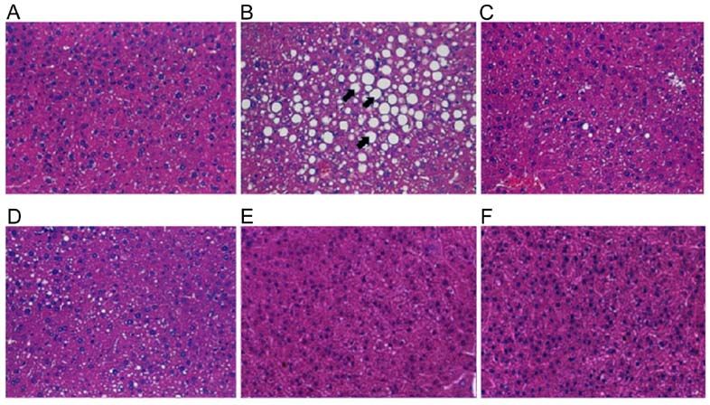
Figure 7. Effects of alcohol and probiotics on hepatic histopathology in alcohol‑fed mice. Hematoxylin and eosin staining in (A) control group; (B) Group
A, alcohol; (C) Group B, alcohol + TSP05; (D) Group C, alcohol + TSF331 + TSR332; (E) Group D, alcohol + TSP05 + TSF331 + TSR332; and
(F) Group E, TSP05 + TSF331 + TSR332. Magnification, x400. Arrows indicates fat inclusions in the liver. TSP05, Lactobacillus plantarum; TSF331,
Lactobacillus fermentum; TSR332, Lactobacillus reuteri.
reasonable to assume that NF‑κB expression may be downregu‑
lated as a result of probiotic supplementation. The antioxidant
response is regulated by a complex network in which Nrf2 is a
key transcription factor, and previous studies have shown that
Nrf2 is activated in the mouse liver by a high‑fat diet (32,33).
In the current study, strong GPx induction was observed in
all probiotic‑treated groups and was attributed to antioxidant
activity; therefore, future studies should investigate whether
GSH and GPx are induced by Nrf2.
Previous studies have reported that probiotics, such as
LGG, could effectively improve ALD (9,28). For instance,
Forsyth et al (11) found that supplementing rats with LGG
(2.5x10⁷ CFU/ml) for 10 weeks improved ALD. Similarly,
Bull‑Otterson et al (34) increased the LGG dose to
1x10⁹ CFU/ml over 2 weeks to examine the effects on liver
improvement in mice, identifying that LGG altered the intes‑
tinal microbiome and reduced alcohol‑induced ALT levels
by 27%. The present results suggested that alcohol‑induced
ALT levels were significantly lower in the probiotic‑treated
groups (B, C and D; 1.6x10⁸ CFU/ml) compared with group A.
Previously, Kim et al (35) reported that L. fermentum LA12
could improve liver function and hepatic steatosis in rats by
restoring the gut barrier, thus preventing endotoxin leakage
into the blood. In addition, LA12 (1x10⁹ CFU/ml; 4 weeks)
reduced ALT and AST levels in the serum by 24 and 18%,
respectively (35). Similarly, Chiu et al (36) demonstrated that
symbiotic (mixture of probiotics and prebiotics) supplementa‑
tion (2x10⁹ CFU/ml) for 4 weeks could attenuate serum ALT
and AST levels by 29 and 13%, respectively. In the present
study, the probiotic‑treated mice in groups B, C and D displayed
Figure 8. Effects of probiotics on the liver and body weight of alcohol‑induced lower ALT (by 28, 37 and 48%, respectively) and AST (by
liver injury mouse models on day 28. Changes in (A) body weight and 14, 20 and 26%, respectively) levels on day 28 compared with
(B) liver/body weight ratio. A, alcohol; B, alcohol + TSP05; C, alcohol + those of group A. Unlike ALT, which is primarily present in
TSF331 + TSR332; D, alcohol + TSP05 + TSF331 + TSR332; E, TSP05 +
TSF331 + TSR332. ###P8 HSIEH et al: LACTIC ACID BACTERIA ALLEVIATES ALCOHOLIC STEATOHEPATITIS IN A MOUSE MODEL
Chronic alcohol consumption increases TG levels in the Ethics approval and consent to participate
serum and liver, eventually leading to fatty liver disease (38,39).
In the current study, the serum TG content did not differ The study protocols were approved by the Institutional Animal
between group A and the control group on day 28, as the stan‑ Care and Use Committee of Hung Kuang University (ethical
dard error in group A was large; however, TG levels were 58% approval no. HK‑10605).
lower in group C compared with in group A. Although serum
TG levels were not decreased to control levels by the probi‑ Patient consent for publication
otic combinations, there were significantly downregulated
alcohol‑induced TG levels in the liver by 34, 36 and 33% in Not applicable.
groups B, C and D, respectively. Moreover, histopathological
analysis of the liver tissue revealed that treatment with probi‑ Competing interests
otics and their combinations (groups B, C and D) significantly
prevented alcohol‑induced fat accumulation in the liver. The authors declare that they have no competing interests.
Collectively, the present results indicated that the LAB strains
TSP05, TSF331 and TSR332 and their combinations could be References
used as supplements to ameliorate ASH and ALD due to their
antioxidant and anti‑inflammatory activities. Furthermore, these 1. Rehm J and Shield KD: Global alcohol‑attributable deaths
probiotics were demonstrated to protect against ethanol‑induced from cancer, liver cirrhosis, and injury in 2010. Alcohol Res 35:
174‑183, 2013.
oxidation, reduce gut barrier damage and suppress the inflamma‑ 2. World Health Organization: Global status report on alcohol
tory response. Although the potential of these probiotics warrants and health 2014. http://www.who.int/substance_abuse/
confirmation in a human clinical study, antioxidants have been publications/global_alcohol_report/msb_gsr_2014_1.pdf.
3. World Health Organization: Global status report on alcohol and
reported to effectively regulate gut permeability and enhance health 2018. https://apps.who.int/iris/bitstream/handle/10665/27
antioxidant activity in patients with ALD (40). The probiotic 4603/9789241565639‑eng.pdf.
strains used in the current study are GRAS and have the potential 4. Bajaj JS: Alcohol, liver disease and the gut microbiota. Nat Rev
Gastroenterol Hepatol 16: 235‑246, 2019.
to be used as therapeutic agents in patients with ALD; however, 5. Lieber CS: Role of oxidative stress and antioxidant therapy in
this study is limited by the fact that fecal microbiota were not alcoholic and nonalcoholic liver diseases. Adv Pharmacol 38:
examined due to inaccurate technology for quantifying and iden‑ 601‑628, 1996.
6. Li F, Duan K, Wang C, McClain C and Feng W: Probiotics and
tifying different microbial species. alcoholic liver disease: Treatment and potential mechanisms.
In conclusion, the present study proposed a novel combina‑ Gastroent Res Pract 2016: 5491465, 2016.
7. Gu Z, Liu Y, Hu S, You Y, Li W and Wang Y: Probiotics for
tion of probiotic L. strains TSP05, TSF331 and TSR332 for the alleviating alcoholic liver injury. Gastroent Res Pract 2019:
improvement of ALD. The current results demonstrated that 9097276, 2019.
this combination could alleviate alcohol‑induced liver injury 8. Schnabl B and Brenner DA: Interactions between the intestinal
microbiome and liver diseases. Gastroenterology 146: 1513‑1524,
and elevate antioxidant activity to attenuate the alcohol‑induced 2014.
oxidative cascade. In addition, these supplements enhanced 9. Zhou Z and Zhong W: Targeting the gut barrier for the treatment
the anti‑inflammatory response to reduce alcohol‑induced of alcoholic liver disease. Liver Res 1: 197‑207, 2017.
10. Kirpich IA, Solovieva NV, Leikhter SN, Shidakova NA,
inflammatory signals. Therefore, this probiotic combination Lebedeva OV, Sidorov PI, Bazhukova TA, Soloviev AG,
provides a new therapeutic option for treating ALD. Barve SS, McClain CJ and Cave M: Probiotics restore bowel
flora and improve liver enzymes in human alcohol‑induced liver
injury: A pilot study. Alcohol 42: 675‑682, 2008.
Acknowledgements 11. Forsyth CB, Farhadi A, Jakate SM, Tang Y, Shaikh M and
Keshavarzian A: Lactobacillus GG treatment ameliorates
The authors would like to thank the Glac Biotech Co., Ltd. alcohol‑induced intestinal oxidative stress, gut leakiness, and
liver injury in a rat model of alcoholic steatohepatitis. Alcohol 43:
consultant, Dr Hau‑Yang Tsen, who provided valuable 163‑172, 2009.
feedback on the manuscript. 12. Wang Y, Gao J, Zhang J and Hu Y: Lactobacillus rhamnosus
B10 treatment ameliorates ethanol‑induced mouse liver injury by
antioxidant pathways. Food Sci 33: 270‑274, 2012.
Funding 13. Hiraishi A: Direct automated sequencing of 16S rDNA amplified
by polymerase chain reaction from bacterial cultures without
No funding was received. DNA purification. Lett Appl Microbiol 15: 210‑213, 1992.
14. Lane DJ: 16S/23S rRNA sequencing. In: Nucleic acid techniques
in bacterial systematics. Stackebrandt E and Goodfellow M (eds.)
Availability of data and materials John Wiley and Sons, New York, pp115‑175, 1991.
15. Weisburg WG, Barns SM, Pelletier DA and Lane DJ: 16S ribo‑
somal DNA amplification for phylogenetic study. J Bacteriol 173:
The datasets used and/or analysed during the current study are 697‑703, 1991.
available from the corresponding author on reasonable request. 16. Xing J, Wang G, Zhang Q, Liu X, Gu Z, Zhang H, Chen YQ
and Chen W: Determining antioxidant activities of lactobacilli
cell‑free supernatants by cellular antioxidant assay: A compar‑
Authors' contributions ison with traditional methods. PLoS One 10: e0119058, 2015.
17. Benzie IFF and Strain JJ: Ferric reducing/antioxidant power
assay: Direct measure of total antioxidant activity of biological
PSH and HHH designed the study. CWC and YWK performed fluids and modified version for simultaneous measurement of
the experiments and analysed the data. PSH, CWC and HHH total antioxidant power and ascorbic acid concentration. Methods
wrote the original manuscript. PSH, CWC and HHH revised Enzymol 299: 15‑27, 1999.
18. Hyronimus B, Le Marrec C, Hadj Sassi A and Deschamps A:
the manuscript. All authors read and approved the final manu‑ Acid and bile tolerance of spore‑forming lactic acid bacteria. Int
script. J Food Microbiol 61: 193‑197, 2000.EXPERIMENTAL AND THERAPEUTIC MEDICINE 21: 188, 2021 9
19. Hassanzadazar H, Ehsani A, Mardani K and Hesari J: 33. Her ná ndez‑Rodas MC, Va lenzuela R, E chever r ía F,
Investigation of antibacterial, acid and bile tolerance properties Rincón‑Cervera MA, Espinosa A, Illesca P, Muñoz P, Corbari A,
of lactobacilli isolated from Koozeh cheese. Vet Res Forum 3: Romero N, Gonzalez‑Mañan D and Videla LA: Supplementation
181‑185, 2012. with docosahexaenoic acid and extra virgin olive oil prevents
20. Lieber CS and DeCarli LM: Liquid diet technique of ethanol liver steatosis induced by a high‑fat diet in mice through
administration: 1989 update. Alcohol Alcohol 24: 197‑211, 1989. PPAR‑ α and Nrf2 upregulation with concomitant SREBP‑1c
21. Guo R and Ren J: Alcohol and acetaldehyde in public health: and NF‑ κ B downregulation. Mol Nutr Food Res: 61, 2017
From marvel to menace. Int J Environ Res Public Health 7: doi: 10.1002/mnfr.201700479.
1285‑1301, 2010. 34. Bull‑Otterson L, Feng W, Kirpich I, Wang Y, Qin X, Liu Y,
22. Tuma DJ and Casey CA: Dangerous byproducts of alcohol break‑ Gobejishvili L, Joshi‑Barve S, Ayvaz T, Petrosino J, et al:
down‑focus on adducts. Alcohol Res Health 27: 285‑290, 2003. Metagenomic analyses of alcohol induced pathogenic alterations
23. Li S, Tan HY, Wang N, Zhang ZJ, Lao L, Wong CW and Feng Y: in the intestinal microbiome and the effect of Lactobacillus rham‑
The role of oxidative stress and antioxidants in liver diseases. Int nosus GG treatment. PLoS One 8: e53028, 2013.
J Mol Sci 16: 26087‑26124, 2015. 35. Kim BK, Lee IO, Tan PL, Eor JY, Hwang JK and Kim SH:
24. Mallikarjuna K, Shanmugam KR, Nishanth K, Wu MC, Protective effect of Lactobacillus fermentum LA12 in an
Hou CW, Kuo CH and Reddy KS: Alcohol‑induced deterioration alcohol‑induced rat model of alcoholic steatohepatitis. Korean
in primary antioxidant and glutathione family enzymes reversed J Food Sci Anim Resour 37: 931‑939, 2017.
by exercise training in the liver of old rats. Alcohol 44: 523‑529, 36. Chiu WC, Huang YL, Chen YL, Peng HC, Liao WH, Chuang HL,
2010. Chen JR and Yang SC: Synbiotics reduce ethanol‑induced
25. Dey A and Lakshmanan J: The role of antioxidants and other hepatic steatosis and inflammation by improving intestinal
agents in alleviating hyperglycemia mediated oxidative stress permeability and microbiota in rats. Food Funct 6: 1692‑1700,
and injury in liver. Food Funct 4: 1148‑1184, 2013. 2015.
26. Pompella A, Visvikis A, Paolicchi A, De Tata V and Casini AF: 37. Wang F, Zhang YJ, Zhou Y, Li Y, Zhou T, Zheng J, Zhang JJ, Li S,
The changing faces of glutathione, a cellular protagonist. Xu DP and Li HB: Effects of beverages on alcohol metabolism:
Biochem Pharmacol 66: 1499‑1503, 2003. Potential health benefits and harmful impacts. Int J Mol Sci 17:
27. Muthukumar K, Rajakumar S, Sarkar MN and Nachiappan V: 354, 2016.
Glutathione peroxidase3 of Saccharomyces cerevisiae protects 38. Enomoto N, Yamashina S, Kono H, Schemmer P, Rivera CA,
phospholipids during cadmium‑induced oxidative stress. Antonie Enomoto A, Nishiura T, Nishimura T, Brenner DA and
van Leeuwenhoek 99: 761‑771, 2011. Thurman RG: Development of a new, simple rat model of early
28. Nanji AA, Khettry U and Sadrzadeh SMH: Lactobacillus feeding alcohol‑induced liver injury based on sensitization of Kupffer
reduces endotoxemia and severity of experimental alcoholic liver cells. Hepatology 29: 1680‑1689, 1999.
(disease). Exp Biol Med 205: 243‑247, 1994. 39. Klop B, do Rego AT and Cabezas MC: Alcohol and plasma
29. Banerjee A and Dhar P: Amalgamation of polyphenols and triglycerides. Curr Opin Lipidol 24: 321‑326, 2013.
probiotics induce health promotion. Crit Rev Food Sci Nutr 59: 40. Varella Morandi Junqueira‑Franco M, Ernesto Troncon L,
2903‑2926, 2019. Garcia Chiarello P, do Rosário Del Lama Unamuno M,
30. Cor tés‑Ma r t í n A, Sel ma M V, Tomás‑Ba rberá n FA, Afonso Jordao A and Vannucchi H: Intestinal permeability and
González‑Sarrías A and Espín JC: Where to look into the puzzle oxidative stress in patients with alcoholic pellagra. Clin Nutr 25:
of polyphenols and health? The postbiotics and gut microbiota 977‑983, 2006.
associated with human metabotypes. Mol Nutr Food Res 64:
e1900952, 2020. This work is licensed under a Creative Commons
31. Soto‑Alarcon SA, Valenzuela R, Valenzuela A and Videla LA: Attribution-NonCommercial-NoDerivatives 4.0
Liver protective effects of extra virgin olive oil: Interaction International (CC BY-NC-ND 4.0) License.
between its chemical composition and the cell‑signaling path‑
ways involved in protection. Endocr Metab Immune Disord Drug
Targets 18: 75‑84, 2018.
32. Valenzuela R, Illesca P, Echever r ía F, Espinosa A,
Rincón‑Cervera MA, Ortiz M, Hernandez‑Rodas MC,
Valenzuela A and Videla LA: Molecular adaptations under‑
lying the beneficial effects of hydroxytyrosol in the pathogenic
alterations induced by a high‑fat diet in mouse liver: PPAR‑ α
and Nrf2 activation, and NF‑κ B down‑regulation. Food Funct 8:
1526‑1537, 2017.You can also read