TWO CASES: CHIROPRACTIC IMAGING - NCMIC
←
→
Page content transcription
If your browser does not render page correctly, please read the page content below
3/17/2021

                               TWO CASES:
                               CHIROPRACTIC IMAGING

                                          STEVEN J GOULD, D.C., D.A.C.B.R.

1

    SURPRISES IN IMAGING

    • Cases to be presented remind us that we need to “do our own work”.
    • Good history and examination should elucidate much information and allow
      us to arrive at a diagnosis prior to treatment or referral.
    • However, there are situations that imaging will disclose findings that change
      our impression and final diagnosis.
    • Decisions to obtain imaging should be based on our history and exam
      findings. (not just by “guidelines” or third party payor mandates)

2

                                                                                             1
3/17/2021

    CASE 1: 8 YOA FEMALE

    • Lower back pain. 8 -9 months duration.
    • Cannot run, constant pain,
    • Prior blue ribbon gymnast. Now not able to perform
      gymnastics.
    • Receiving monthly OMT –osteopathic manipulation for tx.
    • No imaging.

3

    8 YOA FEMALE
    • Patient’s mother reports that she had been seen at a Large
      hospital in KC. DX with Sjogren's and "hypermobility
      syndrome" No spinal x-rays or imaging. Mother says someone
      did hip and knee plain films.

    • Pt. cannot run due to pain in back. Visible gait change with
      splinting or protecting of the back with trying to run, gets to a
      faster walk and appears as though she has adductor muscle
      hypertonicity pulling knees together. (almost like spastic
      gait). Mother says before pain started this kid was an athletic
      gymnast.

4

                                                                                 2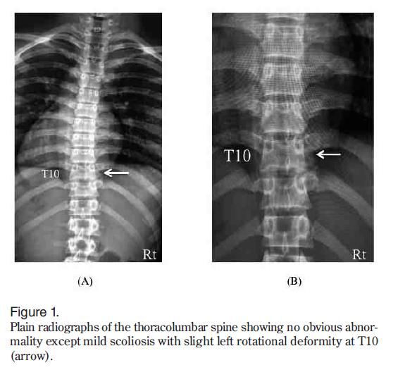
3/17/2021

    8 YOA FEMALE

    • Exam: reduced and painful ROM lumbar spine in all
      movements.
    • + hop test.
    • Crying pain to palpation at L/S junction.
    • Cannot perform fast walking without limping gait.
    • DTR’s +2/4, Myostrength- legs +5/5, No sensory losses.

5

    8 YOA FEMALE
    • Due to patient’s continued pain and range of motion
      deficits and walking difficulties.
    • Lumbar spine radiographs were obtained.
    • Patient’s physical presentation is similar that seen in
      slightly older children with active spondylolysis or
      posterior arch stress reaction.

6

                                                                       3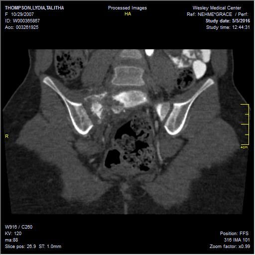
3/17/2021

7

8

           4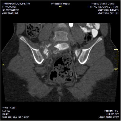
3/17/2021

      8 YOA FEMALE
      • Lumbar spine radiographs revealed increased density of the S1
        segment.
      • Possible radiolucent region at the superior portion of the right
        sacral ala. Bowel gas and content patterns over the region reduce
        visibility.

      • MRI of the lumbar spine was ordered for further evaluation of the
        lower back. Rule out spondylolysis or posterior arch stress
        reaction and for further evaluation of S1.

9

     T2                                    T1

10

                                                                                   5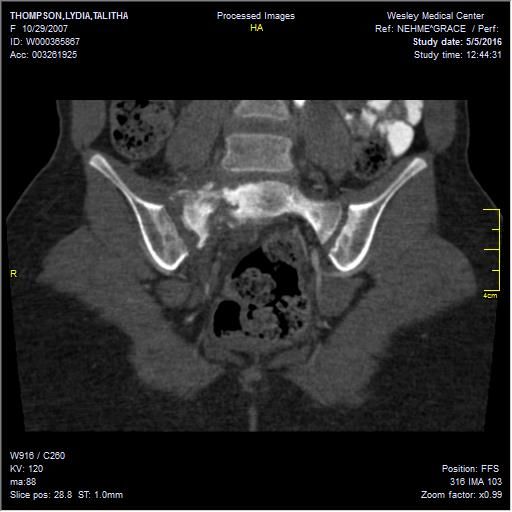
3/17/2021

     IR

11

12

                 6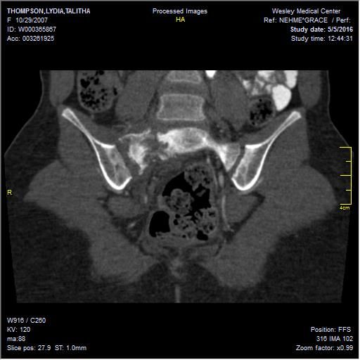
3/17/2021

13

14

            7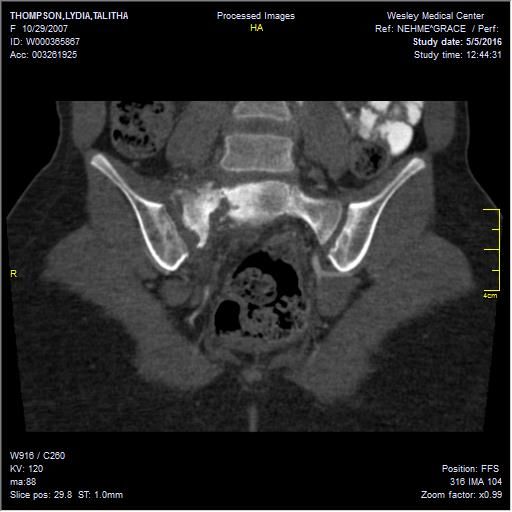
3/17/2021

15

     DIFFERENTIAL DIAGNOSIS
     • Clinical differential diagnosis:
         • Strain of lumbar spine.
         • Active posterior arch stress reaction or spondylolysis.
         • Osseous pathology due to appearance of radiographs – sacrum.

         Imaging Differential Diagnosis:
         Chordoma
         Chondrosarcoma
         Ewing’s Sarcoma
         Osteosarcoma
         Fibrous dysplasia

16

                                                                                 8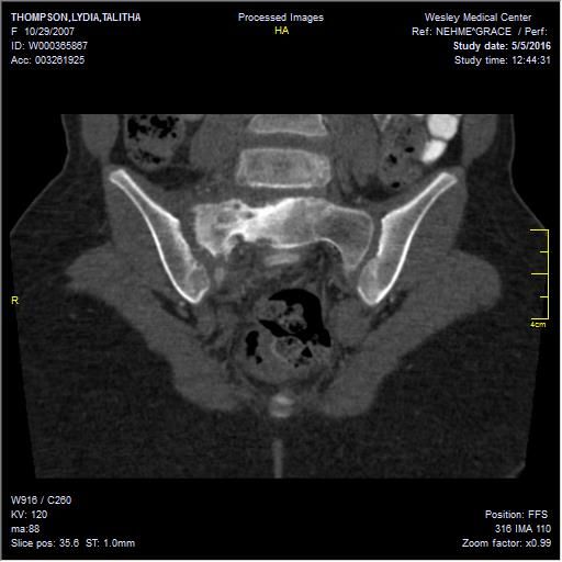
3/17/2021

     DIFFERENTIAL CONSIDERATIONS
     • Spondylolysis – spondylolisthesis in the Adolescent athlete

     • Our young lady is younger than the usual Adolescent athlete that I
       have seen in the office and DX with stress response.

     • In my opinion: Posterior arch stress fractures are an “epidemic” in
       the sports population. The school districts that I cover have this
       injury occur each year. 3 years ago I had 3 football payers at one
       school and another volleyball player at the other school in back
       braces and resting from sport, due to active spondylolysis.

17

                   Case example of stress injury

18

                                                                                    9
3/17/2021

     https://www.ncbi.nlm.nih.gov/pubmed/27040065

19

20

                                                          10
3/17/2021
21
Free article link:
https://www.jstage.jst.go.jp/article/jmi/63/1.2/63_119/_pdf
22
113/17/2021
IMAGING OF SACRAL LESIONS:
• No Good Comes Of A Sacral Mass
• No: Neurogenic tumors, Benign neurofibromas, schwannomas, Malignant peripheral
nerve sheath tumor
• Good: Giant cell tumor
• Comes: Chordoma, Benign notochord cell tumor (BNCT)
• Of: Osteoblastoma
• A: Aneurysmal bone cyst (ABC)
• Sacral: Sarcomas, Ewing’s sarcoma, Osteosarcoma, Chondrosarcoma,
• “Simulators” : Red marrow, Paget disease, Stress fractures
• Mass: Mets, myeloma, lymphoma, Meningocele and Tarlov cysts
https://radiologykey.com/imaging-of-sacral-tumors-and-tumor-simulators-experience-of-the-
mayo-clinic/
23
Age in years Less than 20 20–40 Greater than 40
ABC Giant cell tumor Tarlov cysts
Schwannoma Tumor simulators
Benign Stress fracture
Osteoblastoma
Neurofibroma Paget disease
Red marrow
Ewing’s sarcoma MPNST MPNST, usually3/17/2021
EWIING’S SARCOMA
• Ewing sarcoma in a 20-year-old woman: (a) axial CT, (b) axial T2-w Fat Sat MRI
image, (c) axial T1-w MRI image.
• The lesion appears as a destructive osteolysis with a sclerotic reaction and
invasion of right sacral foramina and sacro-iliac joint
25
ANEURISMAL BONE
CYST
• Aneurismal bone cyst in an 18-year-old boy. (a) Axial CT scan
(soft tissues window) depicts an osteolytic lesion in the right
sacral wing surrounded by a thin sclerotic border. (b) Axial T2-
w MRI: the lesion is multiloculated with fluid–fluid levels, (c)
axial CT scan post-embolization (bone window) shows a
sclerotic ossification within the cyst
26
133/17/2021
SCHWANNOMA
• 33-year-old female with a schwannoma
demonstrate a lytic expansile lesion
centered upon the right S1 neural
foramen. Although the lesion is
detected on the radiograph (thin
arrows), it could easily be confused with
overlying bowel gas.
• The CT shows the lesion to better
advantage and characterizes the benign
nature of the growth pattern with
expansion of the bone and a thin
peripheral rim of sclerosis (thick arrows)
https://radiologykey.com/imaging-of-sacral-tumors-and-tumor-simulators-experience-of-the-mayo-clinic/
27
GIANT CELL TUMOR
• Giant cell tumor of the sacrum in a 29-year-
old female. The lesion demonstrates a
purely lytic pattern of destruction with a
peripheral rim of sclerosis and a large
associated soft tissue mass anteriorly and
posteriorly (arrows). The lesion also crosses
the SI joint (asterisk) and involves the
adjacent ilium.
https://radiologykey.com/imaging-of-sacral-tumors-and-tumor-simulators-experience-of-the-mayo-clinic/
28
143/17/2021
CHORDOMA
61-year-old male show typical imaging features of a chordoma
with a lytic destructive lesion in the lower sacrum that is
centered on the midline, has a large associated exophytic
presacral soft tissue mass.
MR images show a large heterogeneous solid destructive mass
involving the majority of the sacrum and coccyx typical of
chordoma.
The MRI nicely demonstrates both the intraosseous and extra-
osseous extent of the tumor. There are areas of increased
signal within the mass on T1 indicative of intralesional
hemorrhage (asterisk), and the mass shows minimal patchy
enhancement that reflects the myxoid component of the
tumor.
29
• 17-year-old male with an osteoblastoma
show a solid mass with internal
calcifications involving the posterior
elements of S1 and S2 (thin arrows) that is
associated with expansion of the bone
(thick black arrows) as well as surrounding
medullary sclerosis (asterisks)
30
153/17/2021
8 YOA FEMALE
• Dx: biopsy of L/S region was inconclusive
• Bone scan showed uptake in patella.
Biopsy of patella reveals. Langerhans Cell
Histiocytosis.
31
LANGERHANS CELL HISTIOCYTOSIS (EOSINOPHILIC GRANULOMA)
• Langerhans cell histiocytosis (LCH) is a lesion belonging to a group of
disorders now classified by the World Health Organization (WHO) as
histiocytic and dendritic cell disorders (39).
• The incidence in the United States is estimated at 0.05 to 0.5 per
100,000 children per year, with a 2:1 male predominance (20,45,49).
• This disorder represents less than 1% of all biopsy-proven primary bone
lesions
• Histiocyte = tissue macrophage, or a Dendritic cell. Dendritic cells are found in tissue that has
contact with the outside environment such as the over the skin (present as Langerhans cells)
and in the linings of the nose, lungs, stomach and intestines. Immature forms are also found
in the blood.
32
163/17/2021
LANGERHANS CELL HISTIOCYTOSIS;
• Langerhans cell histiocytosis is a disorder in which excess immune
system cells called Langerhans cells build up in the body.
• Langerhans cells, which help regulate the immune system, are
normally found throughout the body, especially in the skin, lymph
nodes, spleen, lungs, liver, and bone marrow.
• In Langerhans cell histiocytosis, excess immature Langerhans cells
usually form tumors called granulomas.
• Many researchers now consider Langerhans cell histiocytosis to be
a form of cancer, but this classification remains controversial.
33
LANGERHANS CELL HISTIOCYTOSIS;
• In approximately 80 percent of affected individuals, one or more granulomas
develop in the bones, causing pain and swelling. The granulomas, which
usually occur in the skull or the long bones of the arms or legs, may cause the
bone to fracture.
• Other signs and symptoms that may occur in Langerhans cell histiocytosis,
depending on which organs and tissues have Langerhans cell deposits, include
swollen lymph nodes, abdominal pain, yellowing of the skin and whites of the
eyes (jaundice), delayed puberty, protruding eyes, dizziness, irritability, and
seizures.
• About 1 in 50 affected individuals experience deterioration of neurological
function (neurodegeneration).
34
173/17/2021
LANGERHANS CELL HISTIOCYTOSIS;
• Langerhans cell histiocytosis is often diagnosed in childhood,
usually between ages 2 and 3, but can appear at any age. Most
individuals with adult-onset Langerhans cell histiocytosis are
current or past smokers; in about two-thirds of adult-onset cases
the disorder affects only the lungs.
• The severity of Langerhans cell histiocytosis, and its signs and
symptoms, vary widely among affected individuals. Certain
presentations or forms of the disorder were formerly considered
to be separate diseases.
35
LANGERHANS CELL HISTIOCYTOSIS (LCH);
• Older names that were sometimes used for forms of
Langerhans cell histiocytosis include;
• Eosinophilic granuloma,
• EG is the most common expression of LCH and is a benign,
solitary lesion of bone.
• It can affect any bone, and is more common in the skull,
mandible, spine, ribs, and long bones.
• In long bones such as the femur, humerus and clavicle, EG
often presents as a modestly destructive lytic lesion with a
characteristic “punched out” appearance. (Herring, 2014)
36
183/17/2021
LANGERHANS CELL HISTIOCYTOSIS (LCH);
• EG – spine:
• Vertebra Plana
• In the spine, immobilization
with a brace has been shown to
be sufficient to allow
remodeling and reconstitution
of vertebral height (Plasschaert,
2002)
37
HAND-SCHÜLLER-CHRISTIAN DISEASE
• Hand-Schüller-Christian disease, a form of LCH
• (multisystem disease without risk organ
involvement)
• This syndrome (15 to 40% of LCH cases) occurs in
children aged 2 to 5 years and in some older children
and adults.
• A disease in which histiocytes start to multiply and
attack the tissues or organs of the patient.
• The most frequent sites of bony involvement are the No Pain, No fever. Beveled appearance “hole within a hole”
flat bones of the skull, ribs, pelvis, and scapula (wing
bone). Case courtesy of Dr Subash Thapa, Radiopaedia.org.
• Chronic otitis media due to involvement of the From the case
mastoid and the temporal bone is common. Diabetes rID: 47957
insipidus affects some patients, mainly children who
have systemic disease.
• Up to 40% of children with it have short stature.
38
193/17/2021
LETTERER-SIWE DISEASE
• Letterer-Siwe disease. (multisystem disease with risk organ involvement)
• This syndrome (10% of LCH cases), a systemic disorder, is the most severe form of Langerhans
cell histiocytosis.
• Typically, a child < 2 years presents with a scaly seborrheic, eczematoid, sometimes purpuric
rash involving the scalp, ear canals, abdomen, and intertriginous areas of the neck and face.
• Denuded skin may facilitate microbial invasion, leading to sepsis.
• Frequently, there is ear drainage, lymphadenopathy, hepatosplenomegaly, and, in severe
cases, hepatic dysfunction with hypoproteinemia and diminished synthesis of clotting factors.
• Anorexia, irritability, failure to thrive, and pulmonary manifestations (eg, cough, tachypnea,
pneumothorax) may also occur.
• Significant anemia and sometimes neutropenia occur; thrombocytopenia is of grave
prognostic significance.
• Parents frequently report precocious eruption of teeth, when in fact the gums are receding to
expose immature dentition.
• Patients may appear abused or neglected.
39
TREATMENT: LANGERHANS CELL HISTIOCYTOSIS
• Supportive care
• Sometimes hormone replacement therapy for hypopituitarism, most commonly diabetes
insipidus
• Chemotherapy for multisystem involvement, single system multifocal involvement, and
involvement in certain sites such as skull-based lesions
• Sometimes surgery, corticosteroid injection, or rarely, radiation therapy (usually for unifocal
bone involvement)
• Because these syndromes are rare and complex, patients are usually referred to institutions
experienced in the treatment of Langerhans cell histiocytosis. The majority of patients should
be treated using protocols developed by the Histiocyte Society
40
203/17/2021
PROGNOSIS: LANGERHANS CELL HISTIOCYTOSIS
• Prognosis is good for patients with Langerhans cell histiocytosis and both of the following:
• Disease restricted to skin, lymph nodes, or bones
• Age > 2 years
• Morbidity and mortality are increased in patients with multisystem involvement, particularly those
with
• Age < 2 years
• Involvement of risk organs (the hematopoietic system, liver, or spleen)
• Involvement of the zygomatic, sphenoid, orbital, ethmoid, or temporal bones denotes a category
of CNS risk lesions that imparts a higher risk of neurodegenerative disease in the skull and front of
the face.
• With treatment, the overall survival rate for patients with multisystem disease without risk organ
involvement is 100%, but event-free survival is about 70%. Disease recurrence may occur. A
chronic remitting and exacerbating course may occur, particularly among adults.
41
CLOSING THOUGHTS ON THIS CASE:
DIFFERENTIAL DIAGNOSTIC CONSIDERATIONS IN CHILDREN AND ADOLESCENTS WITH BACK PAIN.
• Stress reaction/fracture of the pars and posterior elements is a common
condition in adolescents (probably the M.C. pathology)
• Treatment with CMT is not indicated in the “Active” phase when MRI
shows edema.
Chiropractors must keep this diagnosis in the forefront when working with
adolescent patients with back pain.
Children younger than ~12 yoa or pre-adolescent may have a different
etiology of pain, besides stress fracture and this possibility must be kept
in mind.
42
213/17/2021
NEXT CASE
43
CASE 1 NEW MOM
• 26 yoa female
• 6 wks post-partum
• Healthy infant
• Back pain began a couple of weeks prior to presentation.
• Pain with bending and lifting. Increased pain to stand from sitting.
• No falls
• No lower extremity symptoms.
• Prior history + for mini stroke at age 21 from birth control. No
neurologic residual.
44
223/17/2021
• Back pain, significant enough that side posture positioning
for manipulation not tolerated.
• Flexion/distraction motion of Leander table not well
tolerated.
• Passive therapies and stretching used mostly.
• Patient treated for ~ 3 weeks (therapy interrupted, due to
doctor out of town and patient out of town at different
times).
• Not responsive to care.
• MRI ordered.
45
ORDERING/ OBTAINING IMAGING WHEN NEEDED.
• Plain films are commonly the starting point.
• Advanced imaging may be indicated through patient symptoms or clinical
signs.
• MRI
• C.T.
• Ultrasound
• Radionuclide imaging: Bone Scan / PET scan.
• Contrast studies (Barium studies for G.I. / IV contrast – CT or MRI)
• Mammography
• DEXA
46
233/17/2021
47
RADIOGRAPHIC FINDINGS:
• No evidence of fracture or dislocation.
• Vertebral body heights are preserved.
• Lumbar lordosis is reduced.
• Slight right lateral lumbar list.
• SI joints preserved.
• Osteopenia noted. ? Technical in nature or real ?
48
243/17/2021
MRI ORDERED
• Due to continued pain and inability to move/ bend/ lift without
pain.
• MRI of lumbar spine ordered.
49
T1 T2 IR
50
253/17/2021
• T1 MRI
• Compared with
• Lateral Lumbar x-ray
• Endplate depressions
• More notable on MRI
51
SUPERIOR ENDPLATE DEPRESSIONS AT L1, L4, AND L5
52
263/17/2021
SIGNAL T1 & SIGNAL T2 AND IR
53
Review of radiographs and MRI findings
• Plain films:
• Osteoporosis.
• Vertebral body heights preserved.
• MRI:
• Superior endplate depressions at L1, L4, and L5.
• Edema is also present in the sub-endplate zones of the
vertebral bodies
• Findings are consistent with osteoporotic compression
deformities.
54
273/17/2021
FURTHER DISCUSSION WITH PATIENT.
Why does patient’s spine look like a 70 year old –
Osteopenia on X-ray and compression deformities on MRI.
Consider causes of why 26 year old female has osteoporotic
type compressions in her spine.
Pregnancy is a risk factor for osteoporosis.
Medical conditions
Medications
55
COMMON MEDICATION CAUSES OF OSTEOPOROSIS
Steroid therapy (prednisone)
Breast Cancer Drugs (aromatase inhibitors),
Prostate Cancer Drugs (Androgen deprivation therapy)
Heart Burn Medications ( proton pump inhibitors and Aluminum containing antacids)
Depo-Provera (contraceptive)
Excessive Thyroid Hormone replacement (i.e. Synthroid)
Anti-Seizure and Mood altering drugs (Tegretol, Dilantin)
Diuretics (Lasix)
Prostate drugs (Flomax)
Anti-rejection/ immunosupressive therapy (cyclosporine)
Heparin therapy (blood thinners)
Chemotherapy drugs – result in lower testosterone levels
SSRI’s Serotonin reuptake inhibitors (fluoxetine, sertraline, paroxetine, citalopram
Diabetes Med - insulin sensitizers (Thiazolidinediones)
• https://osteoporosis.ca/about-the-disease/what-is-osteoporosis/secondary-osteoporosis/medications-that-can-
cause-bone-loss-falls-andor-fractures/
56
283/17/2021
QUESTION OF MEDICATIONS / OTHER CAUSES OF OSTEOPOROSIS AT YOUNG AGE.
• Patient was treated with heparin and a low molecular weight heparin
alternative (Lovenox) during pregnancy to prevent recurrence of
thrombosis related to pregnancy, due to her prior history.
• Stopped taking heparin prior to visit at chiropractic office, so she didn’t
put it down as a medication on her history form.
57
“SIDE NOTE” FROM EPOCRATES
• Lovenox:
• Black box warnings
• Spinal / epidural hematomas. May occur in anticoagulated pts.
Receiving neuraxial anesthesia or spinal puncture. Hematoma
may result in long term or permanent paralysis; increased
hematoma risk if indwelling epidural catheter use, concomitant
use of drugs affecting hemostasis including NSAIDs, platelet
inhibitors, or other antcoagulants, traumatic or repeated
epidural/spinal puncture hx, spinal deformity, or spinal surgery
hx,
58
293/17/2021
L1
59
HEPARIN OR LMWH INDUCED OSTEOPOROSIS
60
303/17/2021
• 1. Expert Opin Drug Saf. 2005 May;4(3):583-90.
• Minimizing the risk of heparin-induced osteoporosis during pregnancy.
• Hawkins D, Evans J.
• South University, School of Pharmacy, 709 Mall Boulevard, Savannah, GA 31406, USA.
dwhawkins@southuniversity.edu
• Abstract
• Unfractionated heparin (UFH) may lead to symptomatic vertebral fractures in up to 3 out of every 100 people on
long-term therapy. Ten-times that many people will experience a significant reduction in bone density leading to
osteopoenia or osteoporosis. Low molecular weight heparins (LMWH) have been shown to be as effective as UFH
in the prevention and treatment of venous thromboembolism. Several well-established advantages of LMWH over
UFH include increased bioavailability, more predictable dose response, less intensive coagulation monitoring, and
a lower probability of causing immune-mediated thrombocytopenia. There is also some evidence that long-term
LMWH therapy is less likely to cause osteoporotic fractures and significant reductions in bone mass than UFH. Both
UFH and LMWH undergo pharmacokinetic changes during pregnancy, which sometimes necessitates dosage
adjustments. Fondaparinux is a synthetic antithrombotic agent, which specifically binds to antithrombin. It has
been shown to be comparable to, or even more effective than, LMWH in the management of both arterial and
venous thrombosis. Fondaparinux does not appear to have a negative effect on bone metabolism. Therefore,
fondaparinux may be a safe and effective alternative to UFH and LMWH in women who require anticoagulation
during pregnancy.
61
REVIEW OF RADIOGRAPHS
The question I had: Did I miss something on the initial radiographs?
• Go back and review films: No evidence of compression deformities.
• ====
• MRI is more sensitive to disclosing change in anatomy due to sectional
imaging.
• Vertebral compression fractures, endplate depressions, and Schmorl’s nodes
are more conspicuously shown on sectional imaging.
62
313/17/2021
NEXT CASE
63
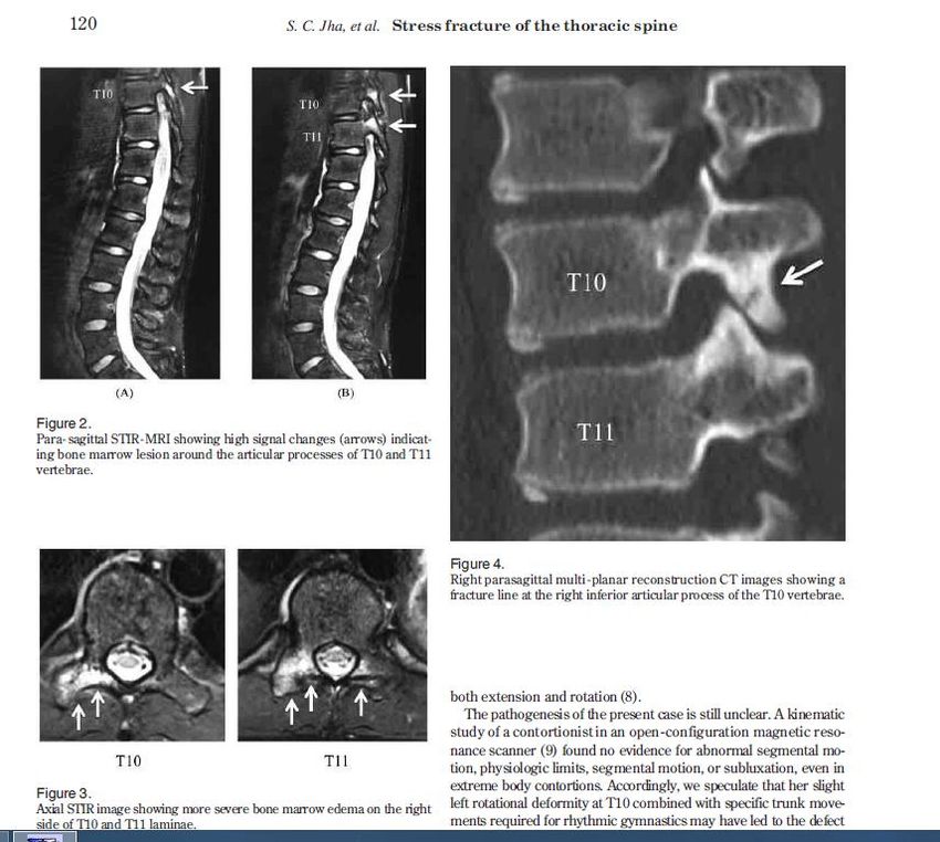
54 YOA FEMALE:
BACK PAIN
T11 subtle compression
Fairly subtle finding:
patient sore at that level.
MRI recommended for
further evaluation.
64
323/17/2021
T2 SAGITTAL MRI
• More conspicuous endplate depression and edema.
• Edema indicates acute or active
compression deformity.
• Fast Spin echo images show high signal
Fluid/water and high signal sub Q fat.
• Hard to differentiate fluid vs fat.
65
• T1 sagittal MRI
• Again, more conspicuous lesion
• on sagittal mri compared to x-ray
66
333/17/2021
• Inversion Recovery (IR) sagittal MRI
• IR images (water weighted images)
differentiate water from fat.
• Fluid / water is bright and
Sub Q fat is low signal
67
NEXT CASE
68
343/17/2021
9 YOA BOY: HEADACHE
HA, loss balance and confusing history of possible fall from tree... 1 month ago or 6
months ago...
Repeat visits to MD and ER. Pediatrian visit for HAs but Ped. didn't want to do CT, due
to too much radiation exposure.
Parents brought him to D.C. and he took plain films of neck and saw shadow anterior to
C1. (acutally rotated C1 due to patient rotation and head tilt, due to patient not being
able to hold his head up well). DC didn’t agree that the appearance on plain film was
due to rotation, so he wanted further imaging. (I didn't have another other history
other than fall from tree 3 weeks ago). So told him to get CT of the neck, to view upper
vert.
When he ordered the neck CT, he decided to get a brain too. Good thing.
69
9 YOA BOY: HEADACHE
DDX so far:
ganglioglioma,
DNET (Dysembryoplastic
neuroepithelial tumour), pilocytic
xanthoastrocytoma,
and less likely
oligodendroglioma.
70
353/17/2021
MED-LEGAL
• Considerations for malpractice
• Missed or delayed diagnosis
71
HOW FINDINGS ARE MISSED
Imaging findings may be missed by:
Perceptual Errors:
• Cause of 70-80 % of missed findings
• Just don’t see the finding, even though it
is there.
Cognitive errors:
• See the finding but ignore it because;
• don’t know what it means
• attribute it to something else
(normal variation or artifact)
72
363/17/2021
“With respect to mistakes or errors in radiographic diagnoses;
- some are cognitive-that is, they result from misinterpretation due to
lack of knowledge or faulty reasoning.
- Perceptual errors, on the other hand, in which radiographic
abnormalities are simply not seen by the radiologist on initial
interpretation, are a far more common cause of radiographic
mistakes, accounting for as many as 80% of them.”
• “The missing of an overt lesion remains as much a mystery and
enigma today as it was 50 years ago.”
73
• “The failure to detect a radiographic abnormality is often attributed to the
subtlety of the radiographic finding. or its poor conspicuity, a term defined by
Potchen and Bisesi, as the ratio between the contrast enhancement of the
lesion or edge relative to the surrounding tissues.”
• “While this definition may adequately explain how a truly subtle lesion can be
missed, it is woefully inadequate to explain how an obvious abnormality can
be missed.”
74
373/17/2021
WHERE’S WALDO
Can’t you see me
75
WHERE’S WALDO
76
383/17/2021
WHERE’S WALDO
77
WHERE’S WALDO
• Findings on imaging may be missed, just as the Waldo may be overlooked
on those cartoon challenges where one tries to find Waldo, in a sea of
similar appearing figures.
• Once Waldo is found, going back to see him again is easy. Just as going
back to an x-ray, knowing where the abnormality is, then it will be easily
seen.
• Radiology interpretation is not practiced retrospectively. We must be able
to discover Waldo on the initial reading.
• Several factors affect our interpretation accuracy: Interruptions – phone,
staff, patients, and any other concentration breakers.
78
393/17/2021
ADDITIONAL FACTORS THAT RESULT IN ERRORS IN
DIAGNOSIS.
• Not getting imaging in the first place.
• Not getting the right images (incomplete series or images of the wrong
body region)
• Technical problems: bad films – technical factors, plain films and digital
processing errors, patient body habitus, overlying artifacts, patient
positioning erros –upright bucky for an ankle study.
79
PERCEPTUAL ERRORS AND NEGLIGENCE
ARTICLE: BERLIN AND HENDRIX. AJR:170, APRIL 1998
• 15 yoa Male. 1 week pain
• PCP saw pt. Ordered x-ray, read as “normal”
• PCP - RX anti-inflammatory,
• 3 weeks pain continued
• Referred to orthopedist
• Dx as Patellar pain syndrome.
• Changed anti-inflammatory med. and Rx exercises
80
403/17/2021
• 5 weeks later (8 weeks Pain)
• Patient fell while riding bike
• Fx distal femur.
• Plaster cast applied, but continued pain and 4 weeks later cast
removed due to swelling
• Tumor found:
• Osteosarcoma.
• Leg amputation.
• Family sued for missed
DX on x-ray.
(radiolucent lesion
In metaphysis)
81
CORRELATION OF HISTORY AND CLINICAL INFORMATION
• Correlation history and clinical information with physical exam for
determination of need for imaging.
• If manipulation is contemplated as a treatment method, then imaging
may be needed.
• Pain severe enough that patient cannot weight bear, then imaging
may be needed.
• Night pain, pain with rest, fever, etc.
• Overt trauma and visible deformity following trauma.
• Repetitive trauma
82
413/17/2021
CORRELATION OF HISTORY AND CLINICAL INFORMATION
• Lower Back Pain – Imaging Guidelines:
• Age > 50 years
• Significant trauma
• Neuromotor deficits
• Unexplained weight loss (10 lb in six months)
• Suspicion of ankylosing spondylitis
• Drug or alcohol abuse // Use of corticosteroids
• History of cancer
• Temperature ≥ 37.8°C (100.0°F)
• Recent visit (within 1 month) for same problem and no improvement
• Patient seeking compensation for back pain
• Deyo RA, Diehl AK. Lumbar spine films in primary care: current use and effects of selective ordering criteria. J Gen
Intern Med 1986;1:20–5. and http://www.aafp.org/afp/1999/1115/p2299.html
83
THE TAKE AWAYS.
• Do your own work – Good history and exam, even if patient has been to
another provider.
• The other provider may have not performed exam or the patient’s condition could have
changed in the interim.
• Order or get imaging when indicated.
• Quality radiographs are required.
• Careful attention to detail in your history, exams, and review of imaging.
• Don’t get hung up on guidelines or “Radiophobia.
84
423/17/2021
•The END
•Thank you !
85
43You can also read