The efficacy of vacuum-ultraviolet light disinfection of some common environmental pathogens
←
→
Page content transcription
If your browser does not render page correctly, please read the page content below
Szeto et al. BMC Infectious Diseases  (2020) 20:127
https://doi.org/10.1186/s12879-020-4847-9

 RESEARCH ARTICLE                                                                                                                              Open Access

The efficacy of vacuum-ultraviolet light
disinfection of some common
environmental pathogens
Wai Szeto1, W. C. Yam2, Haibao Huang3 and Dennis Y. C. Leung1*

  Abstract
  Background: This study is to elucidate the disinfection effect of ozone producing low-pressure Hg vapor lamps against
  human pathogens. Ozone producing low-pressure Hg vapor lamps emit mainly 254 nm ultraviolet light C (UVC) with
  about 10% power of Vacuum-ultraviolet (VUV) light at 185 nm. The combination of UVC and VUV can inactivate airborne
  pathogens by disrupting the genetic materials or generation of reactive oxygen species, respectively. In this study,
  inactivation of common bacteria including Escherichia coli ATCC25922 (E. coli), Extended Spectrum Beta-Lactamase-
  producing E. coli (ESBL), Methicillin-resistant Staphylococcus aureus (MRSA) and Mycobacterium tuberculosis (MTB), and that
  of influenza A viruses H1N1 and H3N2 under the radiation from ozone producing low-pressure Hg vapor lamps was
  examined. Log reduction values at different treatment durations were determined.
  Methods: In vitro tests were carried out. Various bacterium and virus suspensions were added onto nitrocellulose filter
  papers and subjected to the illumination from ozone producing low-pressure Hg vapor lamps. The extents of pathogen
  inactivation at different illumination times were investigated by conducting a series of experiments with increasing
  duration of illumination. log10 reduction in CFU/ml and reduction at log10(TCID50) were respectively measured for
  bacteria and viruses. The disinfection effectiveness of this type of lamps against the pathogens under the environment
  with a moderate barrier to light was therefore evaluated.
  Results: Ozone producing low-pressure Hg vapor lamp successfully inactivated these human pathogens. Nevertheless,
  among these pathogens, disinfection of MTB required more intense treatment. In the best tested situation, 3-log10
  inactivation of pathogens can be achieved with ≤10 min of VUV treatment except MTB which needed about 20 min. This
  demonstrated the high resistance against UV disinfection of MTB.
  Conclusions: Following the criteria that valid germicidal results can be reflected with 3-log10 inactivation for bacteria, 4-
  log10 inactivation for viruses and 5-log10 inactivation for MTB, most of the bacteria required ≤10 min of VUV treatment,
  20 min for the influenza viruses while MTB needed about 30 min VUV treatment. This indicated that VUV light is an
  effective approach against different environmental microorganisms.
  Keywords: Disinfection, Microorganism, Ozone, VUV, IAQ, Influenza, Tuberculosis, ESBL, MRSA

* Correspondence: ycleung@hku.hk
1
 Department of Mechanical Engineering, The University of Hong Kong, Hong
Kong, China
Full list of author information is available at the end of the article

                                      © The Author(s). 2020 Open Access This article is distributed under the terms of the Creative Commons Attribution 4.0
                                      International License (http://creativecommons.org/licenses/by/4.0/), which permits unrestricted use, distribution, and
                                      reproduction in any medium, provided you give appropriate credit to the original author(s) and the source, provide a link to
                                      the Creative Commons license, and indicate if changes were made. The Creative Commons Public Domain Dedication waiver
                                      (http://creativecommons.org/publicdomain/zero/1.0/) applies to the data made available in this article, unless otherwise stated.
Szeto et al. BMC Infectious Diseases (2020) 20:127 Page 2 of 9 Background photocatalysts can utilize and destroy ozone in addition to Indoor air quality (IAQ) has a significant influence on its photocatalytic activity [6]. health, comfort and well-being of building occupants. It has The 254 nm UVC light adopted in conventional infec- been demonstrated that poor IAQ could jeopardize health tion control products can disinfect the illuminated ob- and well-being, which in turn will affect the quality of work jects since the 254 nm radiation can disrupt the genetic and ultimately lower the productivity of workers [1]. materials of airborne pathogens and render them in- One major source of indoor air pollution is the presence viable [7].VUV has an even stronger ionizing power than of micro-organisms, which could cause even more serious UVC light and can generate high concentration reactive problems than some organic and inorganic air contami- species such as ozone and OH radicals [7]. In other nants. This is particularly more phenomenal in cases of words, apart from direct illumination, VUV can inacti- inadequate ventilation, as the condensation in ventilation vate bacterial growth by the radicals generated during system can act as a breeding ground for harmful bacteria VUV irradiation. Therefore, adopting VUV lamps can which are dispensed through the ventilation ducts. Envir- enhance the air disinfection capability of air cleaning onmental airborne bacteria such as Pseudomonas aerugi- systems. A previous study [4] conducted by Huang et al. nosa, Streptomyces albus, Bacillus subtilis and complex demonstrated that 64% toluene removal with VUV ir- populations of micro-organisms within normal flora were radiation alone and the use of photocatalyst enhanced all etiological agents to hypersensitivity pulmonary dis- the toluene removal from 64 to 82%. The experiment eases. Several additional infectious agents such as Legion- adopting UVC lamps and the use of photocatalyst re- ella pneumophila and Mycobacterium tuberculosis (MTB) moved only 14% of toluene. The result demonstrated pose even more grave concerns to the IAQ, as these air- that VUV light could be an effective measure for chem- borne pathogenic bacteria are known to cause severe ill- ical degradation in ventilation systems. When it comes ness in humans. Meanwhile, viruses such as influenza to disinfection, extensive research has been carried out virus were originally thought to be only transmitted from on UVC light and effective destruction of both airborne person to person via aerosols of body fluids. However, in a [8–20] and other human pathogens [21–29] has been recent study conducted by Weistein et al. [2], the produc- shown. Nevertheless, disinfection using VUV light has tion of infectious droplet nuclei of diameter < 5 μm could attracted very little attention. This would be caused by remain suspended and disseminated by air current to in- the relative low prevalence of VUV light sources. Kim fect a susceptible host. A good and reliable disinfection et al. [30] found that the disinfection time required to at- system, therefore, is required to disinfect the airborne mi- tain the same extent of inactivation of aerosolized MS2 croorganisms in order to maintain good IAQ. bacteriophage, using low pressure mercury vapor lamps Adopting vacuum-UV (VUV) lamps, for instance, the with both 254 nm UVC and 185 nm VUV output was ozone producing low-pressure Hg vapor lamps, can be an much shorter than the lamps with 254 nm UVC only. effective mean of disinfecting the airborne microorgan- The disinfection time of ozone only (without UV) process isms. Many existing infection control products use low at ozone concentrations equivalent to the ozone level gen- pressure mercury vapor lamps as light source. This is a erated by the mercury vapor lamps was also significantly source of high energy photons with low cost. Recently, faster than using lamps with 254 nm emission only. Be- pulsed xenon light source technology emitting a broad sides, Huang et al. [4] reported the inactivation of E coli by spectrum (200-300 nm) of UV light is an emerging alter- low pressure mercury vapor lamps. Additionally, some native to low pressure mercury vapor lamps that allows researchers tested the disinfection of water with VUV light much faster surface disinfection because of the high peak and it was reported that the efficiency was quite low com- power [3]. Nevertheless, the pulsed nature of this technol- pared to disinfection with UVC light [31, 32]. The reason ogy would limit its use in continuous air disinfection sys- is due to the low penetration power of VUV light in water tem. Electrical discharge of low pressure mercury vapor [33]. Moreover, the disinfection of human pathogens by mainly emits 254 nm ultraviolet light C (UVC) and 185 VUV light was rarely reported. In our opinion, only Chris- nm VUV light. However, existing products mainly use the tofi et al. [34] reported the disinfection of the microbial lamps with doped quartz envelope that absorbs 185 nm films of 3 types of pathogenic bacteria using ozone pro- photons to prevent the formation of potentially dangerous ducing low-pressure Hg vapor lamps. Therefore, the ef- ozone. Nevertheless, ozone is also a powerful disinfectant fect of VUV light against human pathogens is yet to be and the valuable disinfection opportunity of the 185 nm elucidated. In this study, we evaluated the germicidal VUV light becomes waste heat. effect of VUV light on common bacteria including Escher- Ozone is an issue that bothers on safety if it remains in ichia coli ATCC25922 (E. coli), Extended Spectrum Beta- the output of an air treatment system. However, ozone Lactamase-producing E. coli (ESBL), Methicillin-resistant can be easily destroyed before leaving the air treatment Staphylococcus aureus (MRSA) and Mycobacterium tuber- system if proper catalyst is adopted [4, 5]. Also, some culosis (MTB), and that on influenza viruses H1N1 and
Szeto et al. BMC Infectious Diseases (2020) 20:127 Page 3 of 9
H3N2. Influenza viruses and MTB are inherent airborne Escherichia coli ATCC25922 (E. coli), extended Spectrum
pathogens while E. coli ATCC25922 is always the first Beta-Lactamase-producing E. coli (ESBL) and methicillin-
indicator organism to monitor disinfection efficacy. The resistant Staphylococcus aureus (MRSA)
more drug resistant ESBL and MRSA were chosen as ex- Escherichia coli strain ATCC25922 (E. coli), fully suscep-
amples to monitor disinfection efficacy on human patho- tible to most antibiotics, was purchased from American
gens. Some suspensions of these bacteria and viruses were Type Culture Collection (ATCC). Methicillin-resistant
absorbed into nitrocellulose filter papers during the exper- Staphylococcus aureus strain QC 5618 (MRSA) was pro-
iments and the disinfection under the environment with a vided as a Proficiency Program of Central Public Health
moderate barrier to light was evaluated. Laboratory, Colindale, UK. Extended Spectrum Beta-
Lactamase-producing E. coli strain MM1604 (ESBL) was
provided as a Proficiency Program of Central Public
Methods Health Laboratory Service, Department of Health, Hong
UV irradiation Kong.
To evaluate the biocidal effect of VUV light, bacteria and E. coli and MRSA were inoculated onto Mueller-Hinton
viruses were irradiated with a pair of hot cathode low agar (BD Bioscience, CA, USA) plates and incubated over-
pressure mercury vapor lamps. The lamps were 10 W, U- night at 37 °C to yield single colonies. Overnight cultures
VIX brand, ZW10D15Y, ozone generating. The distance were prepared by inoculating single colonies of each bac-
between the light source and the microorganisms was ap- terial strain into Brain Heart Infusion (BHI) broth (BD
proximately 5 cm and the UV intensities at 254 nm and Bioscience, CA, USA). Bacterial suspension at early expo-
185 nm, respectively measured by a ZDZ-1 UV-C meter nential phase was inoculated into BHI broth at 37 °C for 2
and an ILT1400 radiometer were 21 and 2.3 mW/cm2, re- h. The concentration of the bacterial suspension was then
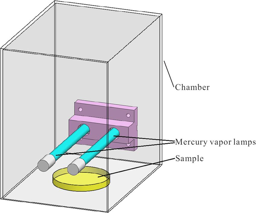
spectively. To reduce the leakage of UV light and lamp- visually adjusted to McFarland standard 0.5. Test suspen-
generated ozone to the surrounding, the lamps and the sion was prepared by diluting the 0.5 McFarland standard
microorganisms under test were contained in a metal inoculum by 10-fold and 100-fold. Actual bacterial
chamber during the experiments as shown in Fig. 1. count was calculated by back titration of the inoculum
suspension. Purity of MRSA was checked by ChromID®
MRSA agar plate (BioMérieux SA, France) and the pu-
Bacterial strains and inoculum preparation rities of E.coli and ESBL-producing E. coli were con-
Following procedures were used to prepare bacterial firmed by MacConkey agar plate (Oxoid™, Thermo
samples for UV irradiation experiments. Scientific, Massachusetts, United States).
Fig. 1 The VUV illumination experimentSzeto et al. BMC Infectious Diseases (2020) 20:127 Page 4 of 9
Mycobacterium tuberculosis (MTB) based on the consideration of the time of disinfection and
MTB H37Rv (ATCC27294) was selected as the model temperature rise of the agar during the course of experi-
organism. Due to the infectivity and the risk of handling ments. As each experiment was carried out inside a Level-2
MTB, the experiments were conducted in the Biosafety Biosafety Cabinet, the 2 mL added suspension was carefully
Level-3 Laboratory of The University of Hong Kong. adjusted so that the filter remained moisted at the end of
MTB was first inoculated onto non-selective Middleb- irradiation as dryness will reduce the viable count recovered
rook 7H11 agar (BD Bioscience, CA, USA) supplemented from the filter.
with 10% Oleic acid-Albumin-Dextrose-Catalase (OADC) The illuminated bacterial suspension and the nitrocel-
and incubated at 37 °C with 5% CO2 until single colonies lulose filter were vigorously washed by 10 mL Phosphate-
were obtained. Mycobacterial colonies were resuspended buffered saline (PBS). The suspension was then serially
into glass-bead Phospate-Buffered Saline with 0.1% Tween diluted with PBS from 100 to 10− 4, and 100 μL of each of
80. Inoculum was vortexed for 30 s to homogenize the the serially diluted bacterial suspensions was spread onto a
bacterial suspension. Bacterial concentration was then ad- Mueller-Hinton agar plate. Meanwhile, bacterial test suspen-
justed to optical density at 600 nm = 0.15–0.17, which is sions without VUV illumination were spread onto Mueller
equivalent to 0.5 McFarland standard. Two test suspen- Hinton agar to obtain the initial colony-forming units (CFU)
sions were prepared, which were 0.5 McFarland standard before the use of VUV light disinfection as control.
inoculum and 10-fold diluted 0.5 McFarland suspensions. All Mueller-Hinton agar plates were incubated over-
Actual MTB count was calculated by back titration of the night at 37 °C. The resultant CFU in each test suspen-
inoculum suspension on Middlebrook 7H11 agar. Purity sion reflected the viable bacterial count after different
of MTB was checked by culturing the inoculums on blood disinfection durations. The disinfection assay was carried
agar to ensure no fungal and bacterial contamination, and out in triplicate for each bacterial strain.
on non-selective Middlebrook 7H11 agar to ensure there
was no contamination by nontuberculous mycobacteria. VUV disinfection experiments of Mycobacterium
tuberculosis
Virus strains and cell lines To investigate the minimum time required by VUV light
H1N1 and H3N2 for optimal MTB disinfection, test sets were used in
Following procedures were used to prepare viral samples which 2 mL concentration-adjusted MTB inoculums,
for UV irradiation experiments. added onto nitrocellulose filter papers, were illuminated
H1N1 was isolated from the first swine flu patient in by VUV for 10, 20, 30 and 45 min.
Hong Kong in 2009 by the Department of Microbiology, The illuminated bacterial suspension and nitrocellu-
The University of Hong Kong. H3N2, a seasonal flu in lose filter were vigorously washed by 10 mL PBS, and
Hong Kong, was generously provided by Prof. H.L. the suspension was serially diluted (100–10− 4). A total of
Chen, Department of Microbiology, The University of 100 μL of each diluted bacterial suspension was spread
Hong Kong. MDCK (Madin-Darby canine kidney) cell onto selective Middlebrook 7H11 agar supplemented
line provided by CDC, USA, was used to cultivate H1N1 with 10% oleic albumin dextrose (OADC), 200,000 unit/
and H3N2 viruses. L Polymyxin B, 50 mg/L Carbenicillin, 10 mg/L Ampho-
Both seasonal influenza A viruses were cultured in MDCK tericin B and 20 mg/L Trimethoprim Lactate. Bacterial
cells in MEM (GiBCO) supplemented with TPCK-trypsin inoculum without VUV illumination was used as MTB
(Sigma-Aldrich, MO, USA). Virus-infected cells were har- growth control and to determine the original viable bac-
vested when almost all MDCK cells exhibited cytopathic terial count. Each test set was conducted in triplicate.
effects. Infected cells and the conditioned media underwent
one freeze-thaw cycle to release viral particles. The suspen- VUV disinfection experiments of influenza viruses H1N1 and
sion was then centrifuged at 3000 rpm for 5 min, and super- H3N2
natant containing viral particles was collected. Tissue To analyze the virucidal effect of VUV light, 2 mL virus
culture infective dose 50 (TCID50) was determined in a 96- samples at ~ 1 × 10⁶ TCID50/mL were added onto nitrocel-
well tissue culture plate using Reed Muench method. Virus lulose filter papers and irradiated by vacuum ultraviolet light
stock was stored at − 80 °C prior usage. (VUV) for 5, 10, 15 and 20 min at an illumination distance
of 5 cm at 25 °C. The illuminated viral suspension and nitro-
UV disinfection experiments cellulose filter were vigorously washed, and the suspension
VUV disinfection experiments of E. coli, ESBL and MRSA was then serially diluted (100–10− 8) by Minimum Essential
To analyze the bactericidal effect of VUV light, 2 mL of Medium (MEM) supplement with TPCK-trypsin. Each di-
bacterial suspension was added onto the nitrocellulose luted sample was used to infect Madin-Darby Canine Kid-
filter and irradiated by VUV for 2, 5, 10 and 15 min at a ney (MDCK) cells in the presence of TPCK-trypsin at 37 °C
distance of 5 cm at 25 °C. This distance was selected for 3 days. The end point of cytopathic effects (CPE) asSzeto et al. BMC Infectious Diseases (2020) 20:127 Page 5 of 9
small, round and degeneration was recorded. Virus sample insufficient bactericidal activity with an average 2.4-log10
without VUV illumination was used to infect MDCK as growth reduction and 99.57% inhibition of bacterial growth
positive control and to determine the original viral load. (Fig. 2a and b). The results suggested that VUV light disin-
Each test was conducted in triplicate. fection is much more effective against lower E. coli bacterial
concentration. At 15 min disinfection, complete inhibition
Data analysis of bacterial growth was also observed in 10-fold diluted 0.5
For bacteria, log10 reduction of viable bacterial count in McFarland standard inoculum, resulting in at least 6-log10
CFU/mL was calculated by comparing control and post growth reduction (Fig. 2a and b).
irradiation filters.
For influenza viruses, reductions at log10 (TCID50) Extended Spectrum Beta-Lactamase-producing E. coli (ESBL)
was calculated similarly. Initial bacterial counts of ESBL for 10-fold diluted and
For each test, outliers were removed by Dixon’s Q test 100-fold diluted 0.5 McFarland standard inoculums across
at 95% significance level. The resultant log10 reduction in triple experimental sets, presented in the Additional file 1
CFU/ml of each bacterial strain and the resultant log10 as Expt. 3 and Expt. 4, were (2.7 ± 0.3) × 107 CFU/mL and
reduction in TCID50 for each viral strain were plotted (3.2 ± 0.7) × 106 CFU/mL, respectively. It was observed
against disinfection durations, and error bars showing the that after 15-min disinfection, both 10-fold diluted and
data of the experiments that deviate from the correspond- 100-fold diluted 0.5 McFarland standard inoculums were
ing mean value were also provided. MS Excel was used in able to achieve complete inhibition of bacterial growth,
all calculations and graph generation. A spreadsheet file resulting in at least 6-log10 growth reduction (Fig. 2a and
containing raw data and intermediate calculations is pro- b). However, at 10-min of disinfection time, although, the
vided in as a supplementary information file. device was able to produce at least 6-log10 reduction of
bacterial growth for the 100-fold diluted inoculum, VUV
Results light was only able to produce a borderline to insufficient
Escherichia coli ATCC25922 (E. coli) bactericidal effect for the 10-fold diluted 0.5 McFarland
Initial inoculum sizes for E. coli in 10-fold diluted and standard inoculum. The test only demonstrated an aver-
100-fold diluted 0.5 McFarland standard inoculums across age of 2.96-log10 reduction with 99.63% growth inhib-
triplicate experiment sets, presented in the Additional file 1 ition. The results have demonstrated that VUV light is
as Expt. 1 and Expt. 2, were (1.9 ± 0.6) × 107 CFU/mL and more effective against a lower concentration of ESBL.
(2.4 ± 0.2) × 106 CFU/mL, respectively. At 10 min VUV
light disinfection, the device was able to produce at least Methicillin-resistant Staphylococcus aureus (MRSA)
6-log10 reduction in viable bacterial count for 100-fold Initial bacterial counts of MRSA for 10-fold diluted and
diluted 0.5 McFarland standard inoculum. However, 10 100-fold diluted 0.5 McFarland standard inoculums across
min VUV light disinfection for 10-fold diluted 0.5 McFar- triple experiment sets, presented in the Additional file 1 as
land standard inoculum can only achieve a borderline to Expt. 5 and Expt. 6, were (3.7 ± 0.9) × 106 CFU/mL and
Fig. 2 VUV light disinfection against E. coli, ESBL and MRSA. Both 10-fold (a) and 100-fold (b) diluted 0.5 McFarland standard inoculums of E. coli
(denoted by E. coli with the dilution ratio behind), ESBL (denoted by ESBL with the dilution ratio behind) and MRSA (denoted by MRSA with the
dilution ratio behind) were subjected to VUV light disinfection. The log10 (CFU/mL reduction) were plotted against the time of disinfection. Data
were plotted as the means of triplicate biological replicates ±errorSzeto et al. BMC Infectious Diseases (2020) 20:127 Page 6 of 9
(3.8 ± 1.7) × 105 CFU/mL, respectively. At 10 min of VUV
light disinfection, the bacteria of the 10-fold diluted and
the 100-fold diluted 0.5 McFarland standard inoculums
were completely inhibited, resulting in at least 5-log10
growth reduction (Fig. 2a and b).
Mycobacterium tuberculosis (MTB)
As defined in previous sections, disinfection time against
bacteria was considered sufficient when a minimum 3-
log10 reduction of viable bacterial count was observed.
For mycobactericidal activity, a 5-log10 reduction in viable
bacterial load is required due to the highly infectious na-
ture of MTB. In other words, a minimum of 5-log10 viable
bacterial load would be required for a valid experimental
set. The average bacterial concentration for McFarland
standard 0.5 MTB inoculum was only (3–5) × 106 CFU/
mL according to our previous experiments (data not
shown). When the bacterial inoculum was diluted by 100-
fold, the bacterial concentration would only be around
104 CFU/mL. The bacterial load could be too low and it Fig. 3 VUV light disinfection against MTB. The experimental sets
were conducted on 0.5 McFarland standard inoculum (denoted by
was incapable of illustrating 5-log10 growth reduction.
MTB 1) and 10-fold diluted 0.5 McFarland standard inoculum
The experiment was therefore conducted with a higher (denoted by MTB 10). The log10 (CFU/mL reduction) were plotted
bacterial concentration and more detailed disinfection against the time of disinfection. Data were plotted as the means of
time as compared to the tests of other bacteria. 0.5 McFar- triplicate biological replicates ±error
land standard and 10-fold diluted 0.5 McFarland stand-
ard inoculums were used and irradiated by VUV for 10,
20, 30 and 45 min. Initial bacterial counts for 0.5 indistinguishable from test agent-induced cytotoxic effects.
McFarland standard and the 10-fold diluted 0.5 McFar- VUV light disinfection time against viruses would be con-
land standard MTB inoculums were (4.4 ± 1.7) × 106 CFU/ sidered sufficient when a minimum of 3-log10 reduction in
mL and (1.2 ± 0.2) × 105 CFU/mL, respectively, presented viral-induced cytotoxicity in titer was achieved. Therefore,
in the Additional file 1 as Expt. 7 and Expt. 8. the infectious viruses recovered from the positive controls
Gradual reduction in bacterial count was observed with must be ≥4-log10 for valid viricidal test results. To deter-
prolonged VUV disinfection time. Complete inhibition of mine the disinfection efficacy of VUV light against seasonal
bacterial growth was observed after 30 min VUV light dis- influenza viruses, two common influenza A viruses, H3N2
infection. At 20 min VUV illumination, VUV light was able and H1N1, causing seasonal epidemics were used. In the
to produce an average of 4-log10 and 3.6-log10 reduction current study, initial viral loads for both H1N1 and H3N2,
in 0.5 McFarland standard and the 10-fold diluted 0.5 presented in the Additional file 1 as Expt. 9 and Expt. 10,
McFarland standard inoculums, respectively (Fig. 3). were 5.4 ± 0.4 log10(TCID50/mL) and 5.1 ± 0.8 log10(T-
In the present study, we have demonstrated that VUV CID50/mL), respectively.
light disinfection can achieve complete inactivation of MTB For samples with log10(TCID50/mL) less than 1.5, the
growth after 30 min disinfection regardless of the bacterial titer was treated as 0.5 for log reduction calculation and
concentration. Meanwhile at 20 min, VUV light disinfection graph plotting purpose.
can only result in a minimum of 3-log10 reduction in bac- At 5 min of illumination, VUV light can inactivate
terial count, which is much longer when compared to the E H1N1 and H3N2 by 2.2- and 3.0-log10 folds viral load
coli, ESBL and MRSA experiments described in previous (TCID50), respectively (Fig. 4). When the VUV illumin-
sections. Previous studies [19, 35, 36] showed that myco- ation time was extended to 20 min, more than 4-log10
bacterial species are generally more resistant to UV disin- reductions in TCID50 of both seasonal influenza A vi-
fection, but are subject to a better disinfection effect under ruses were observed.
VUV light. It seemed that VUV light disinfection was less
effective against MTB at a lower bacterial concentration. Discussion
High-energy vacuum-UV light is efficient in disinfection.
Influenza viruses H1N1 and H3N2 Similar to other UV disinfection mechanisms, direct illumin-
Meanwhile for viral disinfection, test results were consid- ation of VUV could result in the formation of new bonds be-
ered acceptable when the viral-induced cytotoxic effect is tween adjacent nucleotides, causing photochemical damageSzeto et al. BMC Infectious Diseases (2020) 20:127 Page 7 of 9
criterion for mycobacterial disinfection. It was only after
30 min of disinfection that the required 5-log10 reduc-
tion of Mycobacterium tuberculosis viable bacterial load
could be achieved regardless of the bacterial concentra-
tion. This is concordant to previous studies [19, 35, 36]
where mycobacterial species were generally more resist-
ant to UV disinfection. This is probably accounted by
the thicker lipid cell wall in Mycobacterium species.
The tested variations in concentrations of bacteria did
not manifest a trend in the rate of inactivation. For E.
coli and ESBL, higher bacterial concentration resulted in
lower rates of inactivation. Experiments with MTB
showed a different trend. Nevertheless, no obvious trend
was showed in the experiments with MRSA.
From literature, various research teams reported the
UV dosages required attaining 99.9% (3-log) inactivation
of various bacteria or viruses under light from low pres-
sure mercury vapour lamps. For example, the UV dos-
Fig. 4 VUV light disinfection against H1N1 and H3N2 influenza A ages in mJ/cm2 for 3-log inactivation of T7 phage, E
viruses. The log10 (TCID50/mL reduction) was plotted against coli., Staphylococcus aureus, Mycobacterium avium and
disinfection time Mycobacterium phlei are 10 [37], 5 [37], 9 [34], 18 [20]
and 158 [34], respectively. Most of their experiments
on DNA strands and eventually inactivating the replication were conducted with bacteria and viruses virtually un-
of microorganisms. protected. In our experiment, attaining 3-log inactivation
In addition, the high-energy VUV could also lead to the typically required 10 min. Considering that our equip-
formation of both OH radicals and O3, which diffuse into ment provided 21 and 2.3 mW/cm2 light power at 254
anywhere that is shielded from direct UV irradiation and nm and 185 nm, and the total UV power is ~ 23 mW/
inhibit the growth of microorganism. This explained the cm2. The UV dosage of 10 min illumination is ~ 14,000
excellent bactericidal efficiency of VUV light disinfection mJ/cm2, far higher than the usual values. This could be
even in the presence of the opaque nitrocellulose filter. the consequence of our testing condition created by
Our result has further revealed the potential of VUV light loading the suspended bacteria or viruses onto nitrocellu-
to provide a thorough disinfection, even for dust particles lose filter paper. Some bacteria were actually protected
and large aerosols contaminated with pathogens where from direct UV light by the shading effect of filter paper
direct UV illumination cannot penetrate. which is different from the testing setup in the literature.
In this study, we demonstrated that VUV light disinfec- In order to provide sufficient disinfection against all
tion is effective against Escherichia coli, Extended Spectrum the microorganisms we included in this study, we sug-
Beta-Lactamase-producing E. coli and Methicillin-resistant gested the use of Mycobacterium reduction as a bench-
Staphylococcus aureus. For the best tested situation, with mark test for future disinfection instrument designs that
the criterion of 3-log10 inactivation of bacteria, valid germi- incorporates the VUV light system.
cidal result can be achieved with ≤10 min of VUV treat- Although, the disinfection under the environment with
ment. Additionally, more than 5-log10 reduction in viable a moderate barrier to light was successful, there are limi-
plate count can be attained below 15 min of disinfection. tations in the present study. The current pilot study on
In the disinfection tests against seasonal influenza vi- the disinfection efficacy of VUV light disinfection was
ruses H1N1 and H3N2, we also demonstrated that viral conducted in laboratory-controlled conditions. For ex-
load could be effectively reduced by 4-log10 folds after ample, due to safety consideration, device type testing
20 min VUV illumination and this also satisfied the cri- on aerosolized bacteria and viruses is not possible. All
terion of valid germicidal result. Additionally, more than bacterial and viral inoculums were prepared in liquid
3-log10 reduction in viral load can be attained with < 10 suspension and illuminated by VUV on a Petri dish,
min of treatment. which differed from actual environmental settings.
Mycobacterium tuberculosis, on the other hand, re-
quired a more intense disinfection. Conclusion
At 20 min disinfection, VUV light disinfection could Airborne pathogens are important indoor air quality
only result in a 3-log10 reduction in viable plate count. concerns. A good and reliable disinfection system is a
This is insufficient according to our 5-log10 reduction must to maintain good indoor air quality. Vacuum-UVSzeto et al. BMC Infectious Diseases (2020) 20:127 Page 8 of 9
lamps with ozone production were found to be effective Competing interests
for inactivating various human pathogens. With the best The authors declare that they have no competing interests.
tested situation, 3-log10 inactivation of Escherichia coli, Author details
Extended Spectrum Beta-Lactamase-producing E. coli, 1
Department of Mechanical Engineering, The University of Hong Kong, Hong
Methicillin-resistant Staphylococcus aureus and seasonal Kong, China. 2Department of Microbiology, The University of Hong Kong,
Hong Kong, China. 3School of Environmental Science and Engineering, Sun
influenza viruses can be achieved with ≤10 min of VUV Yat-sen University, Guangzhou, China.
treatment except Mycobacterium tuberculosis which
needed about 20 min. This demonstrated the high resist- Received: 29 July 2019 Accepted: 3 February 2020
ance against UV disinfection of MTB. Valid germicidal
results, reflected with 3-log10 inactivation for bacteria,
4-log10 inactivation for viruses and 5-log10 inactivation References
1. Kosonen R, Tan F. The effect of perceived indoor air quality on productivity
for MTB, can be obtained with all tested pathogens. The
loss. Energy Buildings. 2004;36(10):981–6.
duration of VUV treatment required for valid germicidal 2. Weinstein RA, Bridges CB, Kuehnert MJ, Hall CB. Transmission of influenza:
result of most of the bacteria was ≤10 min while MTB implications for control in health care settings. Clin Infect Dis. 2003;37(8):
needed about 30 min. 20 min was adequate for the influ- 1094–101.
3. Wang T, MacGregor S, Anderson J, Woolsey G. Pulsed ultra-violet
enza viruses. This indicated that VUV light is an effective inactivation spectrum of Escherichia coli. Water Res. 2005;39(13):2921–5.
approach against different environmental and pathogenic 4. Huang H, Leung DYC, Li G, Leung MK, Fu X. Photocatalytic destruction of air
microorganisms, and can potentially be used for air- pollutants with vacuum ultraviolet (VUV) irradiation. Catal Today. 2011;
175(1):310–5.
purifying units in future ventilation systems. 5. Batakliev T, Georgiev V, Anachkov M, Rakovsky S. Ozone decomposition.
Interdiscip Toxicol. 2014;7(2):47–59.
6. Wu M, Leung DYC, Zhang Y, Huang H, Xie R, Szeto W, Li F. Toluene
Supplementary information degradation over Mn-TiO2/CeO2 composite catalyst under vacuum
Supplementary information accompanies this paper at https://doi.org/10.
ultraviolet (VUV) irradiation. Chem Eng Sci. 2019;195:985–94.
1186/s12879-020-4847-9.
7. Schalk S, Adam V, Arnold E, Brieden K, Voronov A, Witzke H-D. UV-lamps for
disinfection and advanced oxidation-lamp types, technologies and
Additional file 1. This file contains all data supporting the findings in applications. IUVA News. 2005;8(1):32–7.
this study which is a spreadsheet file containing raw data and 8. Brickner PW, Vincent RL, First M, Nardell E, Murray M, Kaufman W. The
intermediate calculations. application of ultraviolet germicidal irradiation to control transmission of
airborne disease: bioterrorism countermeasure. Public Health Rep. 2003;
118(2):99.
Abbreviations
9. Escombe AR, Moore DA, Gilman RH, Navincopa M, Ticona E, Mitchell B,
ATCC: American type culture collection; BHI: Brain heart infusion;
Noakes C, Martínez C, Sheen P, Ramirez R. Upper-room ultraviolet light and
CFU: Colony-forming units; CPE: Cytopathic effect; E. coli: Escherichia coli;
negative air ionization to prevent tuberculosis transmission. PLoS Med.
ESBL: Extended spectrum beta-lactamase; IAQ: Indoor air quality;
2009;6(3):e1000043.
MDCK: Madin-Darby canine kidney; MEM: Minimum essential medium;
10. Xu P, Peccia J, Fabian P, Martyny JW, Fennelly KP, Hernandez M, Miller SL.
MRSA: Methicillin-resistant Staphylococcus aureus; MTB: Mycobacterium
Efficacy of ultraviolet germicidal irradiation of upper-room air in inactivating
tuberculosis; O3: Ozone; OADC: Oleic acid-albumin-dextrose-catalase;
airborne bacterial spores and mycobacteria in full-scale studies. Atmos
OH: Hydroxyl radical; PBS: Phosphate-buffered saline; TCID50: Tissue culture
Environ. 2003;37(3):405–19.
infective dose 50; TPCK: 6-(1-tosylamido-2-phenyl) ethyl chloromethyl ketone;
11. Goldstein MA, Tauraso NM. Effect of formalin, β-propiolactone, merthiolate,
UV: Ultraviolet; UVC: Ultraviolet C; VUV: Vacuum ultraviolet
and ultraviolet light upon influenza virus infectivity, chicken cell
agglutination, hemagglutination, and antigenicity. Appl Environ Microbiol.
Acknowledgements 1970;19(2):290–4.
Not applicable. 12. Walker CM, Ko G. Effect of ultraviolet germicidal irradiation on viral aerosols.
Environ Sci Technol. 2007;41(15):5460–5.
Authors’ contributions 13. Green CF, Scarpino PV. The use of ultraviolet germicidal irradiation (UVGI) in
WS was a major contributor in writing and editing the manuscript. WCY was disinfection of airborne bacteria. Environ Eng Policy. 2001;3(1):101–7.
responsible for the experimental investigation and analysis. HH provided 14. Peccia J, Werth HM, Miller S, Hernandez M. Effects of relative humidity on
comments on the experiment setup and manuscript. DYCL coordinated the the ultraviolet induced inactivation of airborne bacteria. Aerosol Sci Technol.
test and approved the final manuscript. All authors read and approved the 2001;35(3):728–40.
final manuscript. 15. Chumpolbanchorn K, Suemanotham N, Siripara N, Puyati B, Chaichoune K.
The effect of temperature and UV light on infectivity of avian influenza virus
Funding (H5N1, Thai field strain) in chicken fecal manure; 2006.
Funding was provided by the National Natural Science Foundation of China 16. Hollaender A, Oliphant JW. The inactivating effect of monochromatic
(NSFC) and The Research Grants Council (RGC) of Hong Kong. The funding ultraviolet radiation on influenza virus. J Bacteriol. 1944;48(4):447.
bodies were not involved in the preparation of the manuscript. 17. Lai K-M. Using selective media to assess aerosolization damage and
ultraviolet germicidal irradiation susceptibility of Serratia marcescens.
Availability of data and materials Aerobiologia. 2005;21(3–4):173–9.
All data supporting the findings in this study are contained within the 18. Nardell EA, Bucher SJ, Brickner PW, Wang C, Vincent RL, Becan-McBride K,
supplementary information files. James MA, Michael M, Wright JD. Safety of upper-room ultraviolet
germicidal air disinfection for room occupants: results from the tuberculosis
Ethics approval and consent to participate ultraviolet shelter study. Public Health Rep. 2008;123(1):52–60.
Not applicable. 19. Collins FM. Relative susceptibility of acid-fast and non-acid-fast bacteria to
ultraviolet light. Appl Microbiol. 1971;21(3):411–3.
Consent for publication 20. Shin G-A, Lee J-K, Freeman R, Cangelosi GA. Inactivation of Mycobacterium
Not applicable. avium complex by UV irradiation. Appl Environ Microbiol. 2008;74(22):7067–9.Szeto et al. BMC Infectious Diseases (2020) 20:127 Page 9 of 9
21. Rutala WA, Gergen MF, Weber DJ. Room decontamination with UV
radiation. Infect Control Hosp Epidemiol. 2010;31(10):1025–9.
22. Truman RW, Gillis TP. The effect of ultraviolet light radiation on
Mycobacterium leprae. Int J Lepr Other Mycobact Dis. 2000;68(1):11–7.
23. Mofidi AA, Baribeau H, Rochelle PA, De Leon R, Coffey BM, Green JF.
Disinfection of Cryptosporidium parvum with polychromatic UV light. J-Am
Water Works Assoc. 2001;93(6):95–109.
24. Yaun BR, Sumner SS, Eifert JD, Marcy JE. Response of Salmonella and
Escherichia coli O157: H7 to UV energy. J Food Prot. 2003;66(6):1071–3.
25. Zimmer J, Slawson R, Huck P. Inactivation and potential repair of
Cryptosporidium parvum following low-and medium-pressure ultraviolet
irradiation. Water Res. 2003;37(14):3517–23.
26. Jinadatha C, Villamaria FC, Restrepo MI, Ganachari-Mallappa N, Liao I-C,
Stock EM, Copeland LA, Zeber JE. Is the pulsed xenon ultraviolet light no-
touch disinfection system effective on methicillin-resistant Staphylococcus
aureus in the absence of manual cleaning? Am J Infect Control. 2015;43(8):
878–81.
27. Hassen A, Mahrouk M, Ouzari H, Cherif M, Boudabous A, Damelincourt JJ.
UV disinfection of treated wastewater in a large-scale pilot plant and
inactivation of selected bacteria in a laboratory UV device. Bioresour
Technol. 2000;74(2):141–50.
28. Sommer R, Lhotsky M, Haider T, Cabaj A. UV inactivation, liquid-holding
recovery, and photoreactivation of Escherichia coli O157 and other
pathogenic Escherichia coli strains in water. J Food Prot. 2000;63(8):1015–20.
29. Jinadatha C, Quezada R, Huber TW, Williams JB, Zeber JE, Copeland LA.
Evaluation of a pulsed-xenon ultraviolet room disinfection device for impact
on contamination levels of methicillin-resistant Staphylococcus aureus. BMC
Infect Dis. 2014;14(1):187.
30. Kim J, Jang J. Inactivation of airborne viruses using vacuum ultraviolet
photocatalysis for a flow-through indoor air purifier with short irradiation
time. Aerosol Sci Technol. 2018;52(5):557–66.
31. Ramsay IA, Niedziela J-C, Ogden ID. The synergistic effect of excimer and
low-pressure mercury lamps on the disinfection of flowing water. J Food
Prot. 2000;63(11):1529–33.
32. Wang D, Oppenländer T, El-Din MG, Bolton JR. Comparison of the
disinfection effects of vacuum-UV (VUV) and UV light on Bacillus subtilis
spores in aqueous suspensions at 172, 222 and 254 nm. Photochem
Photobiol. 2010;86(1):176–81.
33. Zoschke K, Börnick H, Worch E. Vacuum-UV radiation at 185 nm in water
treatment–a review. Water Res. 2014;52:131–45.
34. Christofi N, Misakyan M, Matafonova G, Barkhudarov E, Batoev V, Kossyi I,
Sharp J. UV treatment of microorganisms on artificially-contaminated
surfaces using excimer and microwave UV lamps. Chemosphere. 2008;73(5):
717–22.
35. Peccia J, Hernandez M. UV-induced inactivation rates for airborne
Mycobacterium bovis BCG. J Occup Environ Hyg. 2004;1(7):430–5.
36. Bohrerova Z, Linden KG. Assessment of DNA damage and repair in
Mycobacterium terrae after exposure to UV irradiation. J Appl Microbiol.
2006;101(5):995–1001.
37. Bohrerova Z, Shemer H, Lantis R, Impellitteri CA, Linden KG. Comparative
disinfection efficiency of pulsed and continuous-wave UV irradiation
technologies. Water Res. 2008;42(12):2975–82.
Publisher’s Note
Springer Nature remains neutral with regard to jurisdictional claims in
published maps and institutional affiliations.BioMed Central publishes under the Creative Commons Attribution License (CCAL). Under the CCAL, authors retain copyright to the article but users are allowed to download, reprint, distribute and /or copy articles in BioMed Central journals, as long as the original work is properly cited.
You can also read