Microbial Growth Inhibition by Alternating Electric Fields䌤
←
→
Page content transcription
If your browser does not render page correctly, please read the page content below
ANTIMICROBIAL AGENTS AND CHEMOTHERAPY, Oct. 2008, p. 3517–3522                                                                      Vol. 52, No. 10
0066-4804/08/$08.00⫹0 doi:10.1128/AAC.00673-08
Copyright © 2008, American Society for Microbiology. All Rights Reserved.

            Microbial Growth Inhibition by Alternating Electric Fields䌤
           Moshe Giladi,1 Yaara Porat,1 Alexandra Blatt,1 Yoram Wasserman,2 Eilon D. Kirson,2
                                    Erez Dekel,3 and Yoram Palti2,4*
      NovoBiotics Limited1 and NovoCure Limited,2 Matam Advanced Technology Centre, Haifa 31905, Weizmann Institute of
                        Science, P.O. Box 26, Rehovot 76100,3 and B. Rappaport Faculty of Medicine,
                              Technion—Israel Institute of Technology, Technion City, Haifa 32000,4 Israel
                             Received 22 May 2008/Returned for modification 7 July 2008/Accepted 20 July 2008

             Weak electric currents generated using conductive electrodes have been shown to increase the efficacy of
          antibiotics against bacterial biofilms, a phenomenon termed “the bioelectric effect.” The purposes of the

                                                                                                                                                      Downloaded from http://aac.asm.org/ on April 22, 2021 by guest
          present study were (i) to find out whether insulated electrodes that generate electric fields without “ohmic”
          electric currents, and thus are not associated with the formation of metal ions and free radicals, can inhibit
          the growth of planktonic bacteria and (ii) to define the parameters that are most effective against bacterial
          growth. The results obtained indicate that electric fields generated using insulated electrodes can inhibit the
          growth of planktonic Staphylococcus aureus and Pseudomonas aeruginosa and that the effect is amplitude and
          frequency dependent, with a maximum at 10 MHz. The combined effect of the electric field and chloramphen-
          icol was found to be additive. Several possible mechanisms underlying the observed effect, as well as its
          potential clinical uses, are discussed.

   The use of physical means as an aid for modern medicine in                   currents, mostly direct current (DC) (1, 5, 20, 31, 32), as well
the continuous battle against pathogenic microorganisms holds                   as alternating electric fields of as much as 10 MHz (4, 18), can
new prospects that only recently have begun to be widely rec-                   enhance the efficacy of antibacterial agents against bacterial
ognized. Light sources of various types are being used for                      biofilms. In all of these studies, the electric currents were
photodynamic therapy in dentistry and dermatology (10, 13,                      generated using conductive electrodes, allowing for the forma-
33). Ultrasound waves are used for human dental plaque re-                      tion of metal ions and free radicals at the electrode surface.
moval (16) and, in combination with antibiotics, for the erad-                  Like cisplatin, these products are toxic to human cells, and
ication of bacterial biofilms in vitro and in vivo (3, 7, 19, 21). In           therefore the use of such electric currents was limited.
addition, thermotherapy, originally developed as a tool against                    Recently it was demonstrated that low-intensity alternat-
cancerous tumors, has been found to be effective against cu-                    ing electric fields at frequencies of 100 to 200 kHz can
taneous leishmaniasis (22).                                                     inhibit the growth of proliferating cancerous cell lines, both
   The major drawback of the methods mentioned above is                         in vitro and in vivo, without affecting normal quiescent cells (11,
their limited selectivity; thus, ultrasonic waves and thermother-               12). These fields, termed “tumor-treating fields” (TTFields),
apy nonspecifically produce heat that may cause severe collat-                  were generated by means of electrically insulated ceramic
eral damage. Similarly, the illumination of the photosensitizers
                                                                                electrodes, thus ensuring that during the application of the
in photodynamic therapy can harm tissues in the vicinity of the
                                                                                field there is no electrolysis and that no biocides or ions are
target area. Other downsides of photodynamic therapy include
                                                                                produced at the electrode surface and released into the
the need to deliver the photosensitizers to the treated area and
                                                                                medium. Clinical investigations, supported by in vitro stud-
the low tissue penetration of the radiation, limiting the appli-
                                                                                ies, have demonstrated the safety of the use of TTFields.
cation of this treatment to topical infections (13, 14).
   The use of an additional physical means, weak electric cur-                  Evidence was presented indicating that the mechanism at
rents, to inhibit bacterial growth was suggested by Rosenberg                   the basis of this inhibitory effect was related to the unidi-
et al. (24), who observed that electrolysis resulted in the arrest              rectional dielectrophoresis forces produced by the nonho-
of Escherichia coli cell division. Further investigation of this                mogeneous electric fields generated in the vicinity of the
phenomenon revealed that transition platinum complexes pro-                     cleavage plain that gradually develops and separates the
duced at the platinum electrodes during electrolysis were re-                   newly formed daughter cells from each other. Since a similar
sponsible for the bacterial growth inhibition. These derivatives                process occurs in rapidly replicating prokaryotic organisms,
were found not to be specific to bacteria; they were also toxic                 it is reasonable to assume that they can be targeted by
to human cells. In fact, this work eventually led to the discovery              appropriately tuned electric fields. The field parameters re-
of the known chemotherapeutic agent cisplatin (25). In the                      quired for affecting bacteria are expected to be significantly
years to follow, it was demonstrated that low-intensity electric                different from those affecting mammalian cells due to the
                                                                                significant differences in size (see Kirson et al. [11]).
                                                                                   In view of these considerations, the objectives of the
 * Corresponding author. Mailing address: B. Rappaport Faculty of               present study were to test the feasibility of using alternating
Medicine, Technion—Israel Institute of Technology, Technion City,
                                                                                electric fields generated by insulated electrodes for the in-
Haifa 32000, Israel. Phone: 972-4-8500232. Fax: 972-4-8501207. E-mail:
Yoram@Novo-cure.com.                                                            hibition of planktonic bacteria and to define the effective
 䌤
   Published ahead of print on 28 July 2008.                                    field parameters for the inhibition process.

                                                                         3517
3518 GILADI ET AL. ANTIMICROB. AGENTS CHEMOTHER.
two exposed tips positioned 1 cm apart. The probe was connected, through a
coaxial cable, to a floating input oscilloscope (190B; Fluke, The Netherlands).
Field intensities were measured at the end of each treatment by dipping the
probe in the culture medium, such that the two measuring points were in parallel
with the lines of the electric field. Field intensities are expressed as peak-to-peak
volts per centimeter of distance.
Application of AMFields to bacteria. Overnight bacterial cultures were diluted
in fresh LB broth to an optical density (OD) corresponding to bacterial counts
of 1 ⫻ 107 CFU/ml. Petri plates containing the AMFields chamber (Fig. 1B)
were filled with 7 ml of the diluted cultures and placed inside an incubator set to
maintain the proper culture temperature of 37°C once the fields were applied
(FOC 225I; Velp Scientifica). Fields were applied for 2 h for S. aureus and 2.5 h
for P. aeruginosa. Preliminary experiments indicated that these treatment dura-
tions were sufficient to allow for the growth of the control culture group (not
exposed to AMFields) by approximately 1 order of magnitude. Control bacterial
groups, placed in inactive AMFields chambers, were positioned in a prewarmed
incubator at 37°C. The AMFields chamber temperature reached 37.0 ⫾ 0.2°C
Downloaded from http://aac.asm.org/ on April 22, 2021 by guest
within 5 min for both the treated and control groups. At the end of treatment,
cultures were suspended by pipetting. Four 250-l aliquots were dispensed into
a 96-microwell plate (Nunclon⌬; Nunc, Denmark), and the OD at 750 nm
(OD750) was determined spectrophotometrically with a microplate reader (Infi-
nite 200; Tecan, Austria). The percentage of growth for each well was calculated
as (OD750 of the treated well/OD750 of the control well) ⫻ 100.
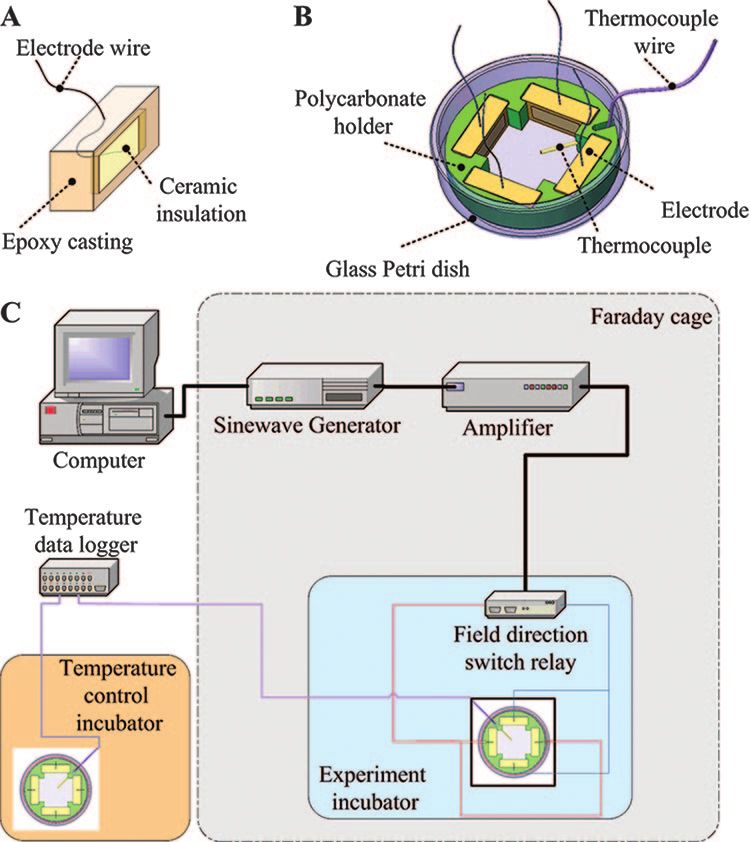
FIG. 1. Schematic design of an insulated AMFields electrode (A), Combined effect of AMFields and antibiotics. Chloramphenicol was obtained
an AMFields bacterium-treating chamber (B), and the AMFields gen- as a powder (Sigma, Israel) and dissolved in 95% ethanol (Frutarom, Israel). All
eration system (C). the stock solutions were filter sterilized and held at ⫺20°C until use. Serial
twofold dilutions of each antibiotic agent were prepared by following the CLSI
guidelines.
The MIC of an antibiotic was defined as the lowest concentration that com-
MATERIALS AND METHODS pletely inhibited the growth of the organism. MICs were determined using
Reagents. Unless stated otherwise, all reagents were purchased from Sigma microdilution susceptibility tests. Briefly, a 100-l bacterial suspension (approx-
(Israel). Dehydrated culture media were purchased from Difco Laboratories imately 5 ⫻ 105 CFU/ml) in LB medium was added to 100 l of culture medium
(Detroit, MI). containing either no antibiotic or an antibiotic at one of several concentrations
Test strains and growth conditions. Pseudomonas aeruginosa strain PAO1 was (serial twofold dilutions) in 96-well plates. Inhibition of proliferation was deter-
a generous gift from Shiri Navon-Venezia (Division of Infectious Diseases, mined by measurement of the OD750 after overnight incubation at 37°C. The
Sourasky Medical Center, Tel Aviv, Israel). Staphylococcus aureus strain SH1000 MIC of AMFields was defined as the lowest intensity that inhibited growth by
was a generous gift from Yair Aharonowitz (Department of Molecular Micro- 80% or more relative to the growth of the control, as determined using the
biology and Biotechnology, Tel Aviv University). All strains were grown in LB microplate reader.
medium (1.0% Bacto tryptone, 0.5% yeast extract, 1.0% NaCl) (Frutarom). Drug interactions with AMFields were assessed according to the checkerboard
Broth cultures of freshly plated bacterial strains were grown in 3 ml of liquid method, with the following modifications. S. aureus inocula were diluted in LB
medium at 37°C for 16 h in an orbital shaker (220 rpm; New Brunswick Scientific, medium containing the antibiotic to a final concentration of 0.5 ⫻ 105 to 1.0 ⫻
NJ) and diluted in fresh LB broth to a predetermined absorbance at 595 nm 105 CFU/ml. The final concentrations of chloramphenicol ranged from 0.25 to 4
(Biowave cell density meter; WPA, United Kingdom), which yielded the desired g/ml. AMFields-treated plates and control plates were handled as described for
CFU per ml. the AMFields application experiments, except that the treatment time was 6 h.
AMFields generation system. AMFields (antimicrobial fields) were generated At the end of the treatment, cultures were sampled and tested for bacterial
inside a 50-mm-diameter glass petri dish by pairs of parallel 15-mm-long, 5-mm- growth inhibition as described above. The percentage of growth for each well was
high electrodes placed 23 mm apart (Fig. 1A). The metal electrodes were com- calculated as described above. Samples were also subjected to serial 10-fold
pletely insulated from the medium by a ceramic material (lead magnesium dilutions from which 20-l aliquots were plated onto LB agar plates (1.5% agar,
niobate–lead titanate [PMN-PT]) with a very high dielectric constant (ε, ⬎5,000) 1.0% Bacto tryptone, 1% NaCl, 0.5% yeast extract). CFU counts were performed
such that the capacitance of the electrodes was approximately 10 nF each. The after overnight incubation at 37°C.
back of the electrodes was insulated by a 5-mm-thick layer of 353ND medical- To evaluate the effect of the combination treatment, the fractional inhibitory
grade epoxy (Epoxy Technology, Billerica, MA). Each chamber contained two concentration (FIC) (6) was calculated for the AMFields and for each antibiotic.
electrode pairs positioned perpendicularly to each other so as to generate se- The following formulas were used to calculate the FIC index: FIC of AMFields ⫽
quentially, in the medium between them, electric fields at 90° to each other. The (MIC of AMFields in combination)/(MIC of AMFields alone); FIC of chloram-
four electrodes were held in position by a polycarbonate structure, as shown in phenicol ⫽ (MIC of chloramphenicol in combination)/(MIC of chloramphenicol
Fig. 1B. The electrodes were connected to a radio frequency amplifier (75A250; alone); FIC index ⫽ (FIC of AMFields) ⫹ (FIC of chloramphenicol). Synergy
AR Worldwide, Souderton, PA) activated by a sine wave generator (model 662; was defined as a FIC index of ⱕ0.5. Indifference was defined as a FIC index of
OR-X, Israel). The entire AMFields-generating system was placed inside a ⬎0.5 but ⱕ4. Antagonism was defined as a FIC index of ⬎4.
Faraday cage in order to meet the guidelines of the International Non-Ionizing Finite-element simulations of electric-field distribution in bacteria. Numerical
Radiation Committee (INIRC) for limiting exposure to time-varying electric, calculations, based on a finite-element mesh, were used to reconstruct the elec-
magnetic, and electromagnetic fields. The temperature at the center of the tric-field distribution inside dividing P. aeruginosa and S. aureus cells. The fol-
chamber was monitored continuously using an insulated T-type thermocouple lowing geometries and parameters were used for the calculations. P. aeruginosa
(Omega, Stamford, CT). (A diagram of the system is given in Fig. 1C.) At the was considered an ellipse, with a large radius of 2.0 m and a small radius of 0.6
highest frequencies used (30 to 50 MHz), the fields interfered with the temper- m, having two membranes (external and internal) 8 nm thick. The two mem-
ature measurements. Therefore, under these conditions, temperatures were branes were assumed to be separated by a periplasmic space of 50 nm. The
measured periodically with the field turned off briefly. dividing bacterium furrow diameter was taken as 0.2 m, and the applied exter-
Because AMFields are associated with heat production, the chamber temper- nal field was 20 V/cm. Since no data regarding the electric properties of P.
ature was kept constant at the desired level by computer feedback control of the aeruginosa have been published, we used the following data for the electric
waveform amplitude. The field generation was switched between the two per- properties of E. coli in the calculations: inner membrane conductivity, 1 S/m;
pendicular directions every 300 ms by activating two pairs of perpendicular outer membrane conductivity, 3 mS/m; medium conductivity, 0.5 S/m; cytoplasm
electrodes. These cycle times were found to minimize the creation of thermal conductivity, 0.5 S/m; conductivity of the periplasmic space, 50 mS/m (9, 26). S.
gradients within the treated area that could affect the bacterial growth rate. The aureus was considered a sphere with a radius of 0.6 m and a membrane
intensity of the electric field was measured using a shielded coaxial probe with thickness of 8 nm. The bacterial cell wall thickness was 20 nm. The dividing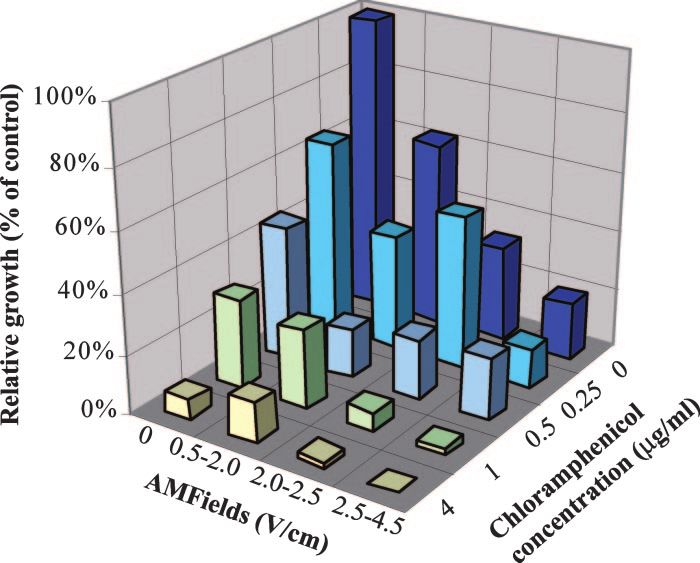
VOL. 52, 2008 MICROBIAL GROWTH INHIBITION BY ELECTRIC FIELDS 3519
FIG. 3. Relative growth of S. aureus (strain SH1000) after a 6-h
exposure to 10-MHz AMFields of various intensities. The relative
growth, based on OD measurements, is expressed as a percentage of
the growth of the heat control (mean ⫾ standard error for at least
three independent experiments). The initial S. aureus concentration in
these experiments was 0.5 ⫻ 105 to 1 ⫻ 105 CFU/ml.
Downloaded from http://aac.asm.org/ on April 22, 2021 by guest
value similar to those reported by the CLSI. The separate and
combined effects of AMFields and chloramphenicol on the
growth of S. aureus, as determined by OD measurements, are
given in Fig. 4. Similar results were obtained using CFU counts
FIG. 2. Average relative growth of microorganisms exposed to (data not shown). As seen, in the presence of 4 V/cm, 10-MHz
AMFields at various frequencies. (A) S. aureus (strain SH1000) after a
2-h treatment; (B) P. aeruginosa (strain PAO1) after 2.5 h of treat- AMFields, much lower concentrations of chloramphenicol (1
ment. The effect, based on OD measurements, is expressed as a per- g/ml) are sufficient to produce ⬎95% inhibition of the growth
centage of the growth of the heat control. Averages for at least 16 of S. aureus. The FIC index was found to be 0.6, indicating that
independent experiments ⫾ SD are presented. The solid lines indicate there is an additive effect for the combined exposure to AM-
the corresponding average field intensities ⫾ SD.
Fields and chloramphenicol.
Finite-element simulations of the electric-field distribution.
The electric-field distribution in and around P. aeruginosa and
bacterium furrow diameter was taken as 0.2 m, and the applied external field S. aureus, calculated using the finite-element mesh method, is
was 20 V/cm. In the simulation, the membrane conductivity was 1 S/m and the
cell wall conductivity was 10 mS/m. The conductivity of the medium was 0.5 S/m,
shown in Fig. 5. In the simulation (Fig. 5A), it is seen that
and the conductivity of the cytoplasm was 0.8 S/m (26). inside the dividing bacterium, close to the furrow, the electric
field is strongest and is nonuniform. The nonuniformity gen-
erates dielectrophoresis forces that approach a maximal value
RESULTS
at AMFields frequencies of ⬃2.0 MHz for P. aeruginosa (Fig.
Inhibition of bacterial growth as a function of AMFields 5B) and ⬃7.0 MHz for S. aureus (Fig. 5C).
frequency. The frequency dependence of the effect of the AM-
Fields was tested between 100 kHz and 50 MHz. The results in DISCUSSION
Fig. 2 show that AMFields at 2 to 4 V/cm inhibit the growth of
the two bacterial species tested. The effect is frequency depen- The use of electric current as a bacterial growth inhibitor
dent, with maximum growth inhibition at 10 MHz for both S. was reported more than 40 years ago (24), with many subse-
aureus and P. aeruginosa. Note that the AMFields-generating quent reports over the years (27, 30). Most of the literature on
system was designed to maintain a constant temperature in the the subject deals with enhancement of the efficacy of antibiot-
chamber by adjusting the field intensity. Therefore, the inten-
sity of the fields varied between different frequencies within a
range of ⫾15%. The results presented are means ⫾ standard
deviations (SD) for at least 16 pooled samples. Analysis of
variance using XLSTAT (version 2008.5.01; Addinsoft) dem-
onstrated the high significance of the frequency dependency (P ⬍
0.0001) for both species. Higher field frequencies were not
tested due to equipment limitations.
Inhibition of S. aureus growth as a function of AMFields
intensity. Once we had identified 10 MHz as the most effective
frequency against S. aureus, we tested the relationship between
the intensity of AMFields and bacterial growth inhibition at 10
MHz. As seen in Fig. 3, the growth inhibition is field intensity
dependent, with larger inhibitory effects at higher intensities.
The results presented are means ⫾ standard errors for at least
FIG. 4. Separate and combined effects of 10-MHz AMFields of
three independent experiments (P ⬍ 0.001). various intensities and chloramphenicol at different concentrations on
Combined effect of AMFields and antibiotics. The MIC of the growth of S. aureus (strain SH1000) after 6 h of exposure. The
chloramphenicol against S. aureus was found to be 4 g/ml, a calculations are based on OD measurements.3520 GILADI ET AL. ANTIMICROB. AGENTS CHEMOTHER.
can lead to electrolysis, the production of toxic derivatives and
free radicals, modification of the pH (28), and alterations in
bacterial biofilm structure (29). High-intensity pulsed electric
currents (generating electric fields of ⬎1,000 V/cm) cause elec-
troporation (17, 34).
High-frequency alternating currents (10 MHz) were re-
ported by Caubet et al. (4) to enhance the susceptibility of
bacterial E. coli biofilms to antibiotics and to decrease the
number of bacteria in biofilms by ⬎60%, even in the absence
of an antibiotic. These effects were attributed by the authors to
changes induced in the biofilm’s exopolysaccharide matrix due
to the interaction of the electromagnetic field with charged
particles present in that matrix. However, since these experi-
ments were carried out using conductive electrodes, the effects
Downloaded from http://aac.asm.org/ on April 22, 2021 by guest
may be due in part to the production of toxic elements at the
electrode surface.
Unlike all the reports discussed above, in which electric
currents were applied using conductive electrodes, in the
present study we investigated bacterial growth inhibition by
high-frequency, low-intensity electric fields generated by com-
pletely insulated electrodes. Thus, the electric fields were not
associated with electrolysis or the production of free radicals,
toxic metal ions, etc., at the electrode surface. Furthermore,
the low intensity of the fields applied here (0.5 to 4 V/cm) rules
out the possibility of electroporation, which occurs at field
intensities in the range of 1,000 V/cm. The continuous control
of the medium temperature eliminates the possibility of ther-
mal effects. In addition, the reported enhancement of the
transfer of ions and antibiotics through biofilms by electric
currents is not relevant to the present study, because the bac-
teria treated here were planktonic and the inhibitory effect was
observed even in the absence of antibiotics.
Since the commonly accepted mechanisms mentioned above
are unlikely to be responsible for the bacterial growth inhibi-
tion reported in the present study, we must look for alternative
mechanisms to explain the reported effects. In analogy to the
mechanism suggested by Kirson et al. (11, 12) for mammalian
cells, it seems logical that high-frequency alternating electric
fields affect dividing bacteria during cytokinesis due to the
nonhomogeneous electric fields generated near the bridge sep-
arating the daughter cells. These nonhomogeneous fields exert
unidirectional dielectrophoresis forces on charged and polar
particles and molecules and thus may result in their movement
toward the furrow (12). In order to evaluate whether AM-
Fields can induce such effects in dividing bacteria, the field
distribution and the associated electric forces were modeled
FIG. 5. (A) Electric field inside and around a dividing rod-like
using finite-element mesh simulations. The results of these
bacterium. The simulation is based on 10-MHz electric fields with an
intensity of 20 V/cm. The dividing bacterium has a furrow diameter of simulations (Fig. 5) indicate that the gradient of the electric
0.2 m, located at 0 m on the x axis. (B and C) Magnitude of the force field inside a dividing bacterium exerts forces of 10⫺2 to 10⫺3
acting on a dipole of 3,000 debye units inside a dividing P. aeruginosa pN directed toward the furrow on small dipoles. This force in
(B) or S. aureus (C) cell when the field intensity is 20 V/cm as a a bacterial cell, which is of the same order of magnitude as the
function of the AMFields frequency.
calculated force in a mammalian cell (11, 12), is sufficient to
induce particle and macromolecule distortion and movement
at frequencies shown to be effective against bacterial growth.
ics against microbial biofilms by the application of weak DC Considering the bacterial cytoplasm viscosity to be 1 centi-
currents, a phenomenon termed “the bioelectric effect” by poise, the terminal velocity of a small dipole is expected to be
Costerton et al. (5). Several mechanisms were suggested for ⬃4 m/s for P. aeruginosa, and ⬃0.6 m/s for S. aureus, per
this inhibition, depending on the nature of the current: rela- V/cm of external electric field. Since the electric force is di-
tively low intensity direct current and low-frequency alternat- rected toward the furrow, it will lead to the accumulation of
ing electric fields, when applied using conductive electrodes, molecules around the furrow within a few seconds. Such forcesVOL. 52, 2008 MICROBIAL GROWTH INHIBITION BY ELECTRIC FIELDS 3521
may affect the structural integrity of the cells or interfere with efficacy in combination with AMFields. One of the major draw-
processes in which electrostatic forces play a role. In eukary- backs of treating pathogens with antibacterial agents is the
otic cells, the cellular structures that are expected to be mostly ability of bacteria to develop resistance to antibiotics. So far,
affected are the highly polar spindle microtubules. Kirson et al. there is no evidence that the bacterial strains used in this study
(12) demonstrated that an alternating electric field interfered acquired resistance to the inhibitory effect of the fields (data
with the orientation of the spindle microtubules as well as with not shown). Assuming that the effect is a result of the nonho-
the polymerization-depolymerization processes involved in the mogeneous fields created at the bridge separating the daughter
chromosome separation process. While the common paradigm cells of the dividing bacterium, in order for a bacterium to
is that bacteria lack the highly organized microtubules present escape inhibition by AMFields, its physical properties would
in eukaryotic cells, a tubulin homologue, FtsZ, has been found have to change radically—an unlikely outcome.
to be present in all bacteria (2). The structural similarity be- There are three main limitations to the use of DC currents,
tween FtsZ and tubulin regarding their large dipole moment, including weak currents, for the treatment of infection. The
as calculated by the protein dipole moment server (8), as well first is that such currents may stimulate nerves and muscles,
as the analogy between the repeated nature of the Z-ring and causing pain and muscular contractions in the patient. The
Downloaded from http://aac.asm.org/ on April 22, 2021 by guest
the spindle apparatus, raises the possibility that FtsZ is a target second relates to the spread of the currents in the body, which
for the action of AMFields. Of course, other polar and struc- can be regarded as a volume conductor. Thus, unless the lesion
turally oriented molecules may be influenced by the AMFields, is superficial or unless there is a conductor leading from the
thus disrupting various cellular processes within the cell. surface to a deeply situated lesion, a current density of suffi-
An additional explanation for the frequency-dependent in- cient intensity at the target can be obtained only when the
hibition of bacterial growth relates to the suggested effect of density near the electrodes is of a damaging and stimulating
alternating electric fields on the enzyme-substrate reaction magnitude. The third limitation is that DC currents cannot be
equilibrium (23). According to the suggested model, the elec- generated by insulated electrodes and are therefore always
tric charge distribution on many enzymes varies with the con- associated with electrolysis, metal ions, free radicals, etc. A
formational changes associated with enzyme-substrate interac- recent report demonstrated the applicability of weak DC cur-
tion. Because the AMFields may affect molecular charge
rents in reducing the level of pin tract infections associated
distributions, they could interfere with some enzymatic reac-
with external fixators in a goat model (31). The authors were
tions. The effect is expected to be larger for membrane en-
able to deliver the DC current to the infected implants because
zymes, because the membrane prevents the enzyme from ro-
the pins protruded externally, thus allowing for a direct con-
tating and thus escaping the effect of the field, and because the
nection with the current source. The currents used were far too
electric field is magnified in the membrane. Assuming that one
low to induce nerve stimulation; however, the risks of electrol-
or more pivotal enzymes are indeed influenced by the electric
ysis and the formation of toxic derivatives still remain, requir-
fields, exposure of these enzymes to the properly tuned fre-
ing long-term, thorough follow-up to ensure the safety of the
quency can inhibit bacterial growth by depleting the cell of the
use of such currents.
enzymes’ products. Two exemplary proteins that are expected
The relatively high frequencies at which the AMFields effect
to be influenced by external electric fields and that are present
in P. aeruginosa and S. aureus are glycerol-3-phosphate dehy- was observed allow for the application of high intensities re-
drogenase (36) and FtsK (2, 15). Glycerol-3-phosphate dehy- quired for deep treatment without nerve or muscle stimulation.
drogenase is involved in respiration, glycolysis, and phospho- Thus, the door is open for future applications of AMFields as
lipid biosynthesis, and FtsK is an essential cell division protein. a treatment for resistant infections and difficult-to-treat
Both are membrane proteins with large dipole moments (1,793 chronic conditions such as infected diabetic ulcers and infec-
and 1,579 debye units, respectively, as calculated by the protein tions associated with implants. Infected ulcers could be treated
dipole moment server [8]). These values are very similar to the by placing the infected body part in a container filled with a
dipole moment of tubulin, the structure and function of which high-electric-impedance solution. Insulated electrodes, con-
during mitosis have been shown to be disrupted by alternating nected to the AMFields generator, would generate the neces-
electric fields of a few hundred kHz (11, 12). sary fields. Alternatively, the AMFields could be delivered to
The combined effect of AMFields and chloramphenicol infected orthopedic implants by means of properly positioned
against S. aureus was investigated in detail and was found to be surface electrodes. Similar methodology is currently being used
additive. The combination of chloramphenicol and electric in treating recurrent glioblastoma patients with TTFields (11).
currents had not been tested before; however, the majority of Note also that because the bacterial division cycle is about 20
studies reported that the combined effect of other antibiotics min long, effective treatment is achieved within a few hours (in
and electric current was synergistic (4, 35). The synergism contrast to cancer cell treatment). Thus, AMFields can be used
reported could be the result of the strong effect of the biocides to accelerate the treatment of common infections, such as
generated during the application of direct electric current us- tonsillitis, pharyngitis, and otitis, in parallel with antibiotics.
ing conductive electrodes. The production of biocides consti- In summary, AMFields constitute a promising new antimi-
tutes a major obstacle to the clinical use of electric-current crobial modality in the continuous battle against microbial
treatment due to the nonspecific nature of these toxic deriva- pathogens. Unlike the electric currents whose use has been
tives. This stands in contrast to the specific and safe use of reported previously, AMFields are not expected to have any
AMFields applied by insulated electrodes with frequencies toxicity for human cells. The efficacy of AMFields against bac-
that have no known effect on human cells (11, 12). The results terial pathogens will most likely be enhanced in vivo by the
of these preliminary experiments indicate enhanced antibiotic activity of the immune system as well as by cotreatment with3522 GILADI ET AL. ANTIMICROB. AGENTS CHEMOTHER.
various antibiotics, making AMFields a potential new antiin- 16. Mourad, P. D., F. A. Roberts, and C. McInnes. 2007. Synergistic use of
ultrasound and sonic motion for removal of dental plaque bacteria. Com-
fection treatment modality. pend. Contin. Educ. Dent. 28:354–358.
17. Oshima, T., and M. Sato. 2004. Bacterial sterilization and intracellular pro-
ACKNOWLEDGMENTS tein release by a pulsed electric field. Adv. Biochem. Eng. Biotechnol. 90:
113–133.
Y. Palti has a minority holding in NovoBiotics, Ltd., and is a member 18. Pareilleux, A., and N. Sicard. 1970. Lethal effects of electric current on
of the company’s board of directors. M. Giladi, A. Blatt, and Y. Porat Escherichia coli. Appl. Microbiol. 19:421–424.
are employed in full by NovoBiotics, Ltd. E. Dekel is a consultant to 19. Pitt, W. G., M. O. McBride, J. K. Lunceford, R. J. Roper, and R. D. Sagers.
NovoBiotics, Ltd. 1994. Ultrasonic enhancement of antibiotic action on gram-negative bacte-
ria. Antimicrob. Agents Chemother. 38:2577–2582.
20. Rabinovitch, C., and P. S. Stewart. 2006. Removal and inactivation of Staph-
REFERENCES ylococcus epidermidis biofilms by electrolysis. Appl. Environ. Microbiol. 72:
1. Blenkinsopp, S. A., A. E. Khoury, and J. W. Costerton. 1992. Electrical 6364–6366.
enhancement of biocide efficacy against Pseudomonas aeruginosa biofilms. 21. Rediske, A. M., N. Rapoport, and W. G. Pitt. 1999. Reducing bacterial
Appl. Environ. Microbiol. 58:3770–3773. resistance to antibiotics with ultrasound. Lett. Appl. Microbiol. 28:81–84.
2. Carballido-López, R., and J. Errington. 2003. A dynamic bacterial cytoskel- 22. Reithinger, R., M. Mohsen, M. Wahid, M. Bismullah, R. J. Quinnell, C. R.
eton. Trends Cell Biol. 13:577–583. Davies, J. Kolaczinski, and J. R. David. 2005. Efficacy of thermotherapy to
3. Carmen, J. C., B. L. Roeder, J. L. Nelson, B. L. Beckstead, C. M. Runyan, treat cutaneous leishmaniasis caused by Leishmania tropica in Kabul, Af-
G. B. Schaalje, R. A. Robison, and W. G. Pitt. 2004. Ultrasonically enhanced ghanistan: a randomized, controlled trial. Clin. Infect. Dis. 40:1148–1155.
Downloaded from http://aac.asm.org/ on April 22, 2021 by guest
vancomycin activity against Staphylococcus epidermidis biofilms in vivo. 23. Robertson, B., and R. D. Astumian. 1990. Michaelis-Menten equation for an
J. Biomater. Appl. 18:237–245. enzyme in an oscillating electric field. Biophys. J. 58:969–974.
4. Caubet, R., F. Pedarros-Caubet, M. Chu, E. Freye, M. de Belem Rodrigues, 24. Rosenberg, B., L. Vancamp, and T. Krigas. 1965. Inhibition of cell division
J. M. Moreau, and W. J. Ellison. 2004. A radio frequency electric current in Escherichia coli by electrolysis products from a platinum electrode. Nature
enhances antibiotic efficacy against bacterial biofilms. Antimicrob. Agents 205:698–699.
Chemother. 48:4662–4664. 25. Rosenberg, B., L. VanCamp, J. E. Trosko, and V. H. Mansour. 1969. Plati-
5. Costerton, J. W., B. Ellis, K. Lam, F. Johnson, and A. E. Khoury. 1994. num compounds: a new class of potent antitumour agents. Nature 222:385–
Mechanism of electrical enhancement of efficacy of antibiotics in killing 386.
biofilm bacteria. Antimicrob. Agents Chemother. 38:2803–2809. 26. Sanchis, A., A. P. Brown, M. Sancho, G. Martinez, J. L. Sebastian, S. Munoz,
6. Eliopoulos, G. M., and R. C. Moellering. 1991. Antimicrobial combinations, and J. M. Miranda. 2007. Dielectric characterization of bacterial cells using
p. 432–492. In V. Lorian (ed.), Antibiotics in laboratory medicine, 3rd ed. dielectrophoresis. Bioelectromagnetics 28:393–401.
The Williams & Wilkins Co, Baltimore, MD. 27. Spadaro, J. A., T. J. Berger, S. D. Barranco, S. E. Chapin, and R. O. Becker.
7. Ensing, G. T., B. L. Roeder, J. L. Nelson, J. R. van Horn, H. C. van der Mei, 1974. Antibacterial effects of silver electrodes with weak direct current.
H. J. Busscher, and W. G. Pitt. 2005. Effect of pulsed ultrasound in combi- Antimicrob. Agents Chemother. 6:637–642.
nation with gentamicin on bacterial viability in biofilms on bone cements in 28. Stewart, P. S., W. Wattanakaroon, L. Goodrum, S. M. Fortun, and B. R.
vivo. J. Appl. Microbiol. 99:443–448. McLeod. 1999. Electrolytic generation of oxygen partially explains electrical
8. Felder, C. E., J. Prilusky, I. Silman, and J. L. Sussman. 2007. A server and enhancement of tobramycin efficacy against Pseudomonas aeruginosa biofilm.
database for dipole moments of proteins. Nucleic Acids Res. 35:W512– Antimicrob. Agents Chemother. 43:292–296.
W521. 29. Stoodley, P., D. deBeer, and H. M. Lappin-Scott. 1997. Influence of electric
9. Hölzel, R. 1999. Non-invasive determination of bacterial single cell proper- fields and pH on biofilm structure as related to the bioelectric effect. Anti-
ties by electrorotation. Biochim. Biophys. Acta 1450:53–60. microb. Agents Chemother. 41:1876–1879.
10. Jori, G., C. Fabris, M. Soncin, S. Ferro, O. Coppellotti, D. Dei, L. Fantetti, 30. Valle, A., E. Zanardini, P. Abbruscato, P. Argenzio, G. Lustrato, G. Ranalli,
G. Chiti, and G. Roncucci. 2006. Photodynamic therapy in the treatment of and C. Sorlini. 2007. Effects of low electric current (LEC) treatment on pure
microbial infections: basic principles and perspective applications. Lasers bacterial cultures. J. Appl. Microbiol. 103:1376–1385.
Surg. Med. 38:468–481. 31. van der Borden, A. J., P. G. Maathuis, E. Engels, G. Rakhorst, H. C. van der
11. Kirson, E. D., V. Dbaly, F. Tovarys, J. Vymazal, J. F. Soustiel, A. Itzhaki, D. Mei, H. J. Busscher, and P. K. Sharma. 2007. Prevention of pin tract
Mordechovich, S. Steinberg-Shapira, Z. Gurvich, R. Schneiderman, Y. Was- infection in external stainless steel fixator frames using electric current in a
serman, M. Salzberg, B. Ryffel, D. Goldsher, E. Dekel, and Y. Palti. 2007. goat model. Biomaterials 28:2122–2126.
Alternating electric fields arrest cell proliferation in animal tumor models 32. van der Borden, A. J., H. van der Werf, H. C. van der Mei, and H. J.
and human brain tumors. Proc. Natl. Acad. Sci. USA 104:10152–10157. Busscher. 2004. Electric current-induced detachment of Staphylococcus epi-
12. Kirson, E. D., Z. Gurvich, R. Schneiderman, E. Dekel, A. Itzhaki, Y. Was- dermidis biofilms from surgical stainless steel. Appl. Environ. Microbiol.
serman, R. Schatzberger, and Y. Palti. 2004. Disruption of cancer cell rep- 70:6871–6874.
lication by alternating electric fields. Cancer Res. 64:3288–3295. 33. Wainwright, M. 1998. Photodynamic antimicrobial chemotherapy (PACT).
13. Maisch, T. 2007. Anti-microbial photodynamic therapy: useful in the future? J. Antimicrob. Chemother. 42:13–28.
Lasers Med. Sci. 22:83–91. 34. Weaver, J. C. 1993. Electroporation: a general phenomenon for manipulat-
14. Maisch, T., C. Bosl, R. M. Szeimies, N. Lehn, and C. Abels. 2005. Photody- ing cells and tissues. J. Cell. Biochem. 51:426–435.
namic effects of novel XF porphyrin derivatives on prokaryotic and eukary- 35. Wellman, N., S. M. Fortun, and B. R. McLeod. 1996. Bacterial biofilms and
otic cells. Antimicrob. Agents Chemother. 49:1542–1552. the bioelectric effect. Antimicrob. Agents Chemother. 40:2012–2014.
15. Massey, T. H., C. P. Mercogliano, J. Yates, D. J. Sherratt, and J. Lowe. 2006. 36. Yeh, J. I., U. Chinte, and S. Du. 2008. Structure of glycerol-3-phosphate
Double-stranded DNA translocation: structure and mechanism of hexameric dehydrogenase, an essential monotopic membrane enzyme involved in res-
FtsK. Mol. Cell 23:457–469. piration and metabolism. Proc. Natl. Acad. Sci. USA 105:3280–3285.You can also read