Assessing effectiveness of Komagataeibacter strains for producing surface microstructured cellulose via guided assembly based biolithography - Nature
←
→
Page content transcription
If your browser does not render page correctly, please read the page content below
www.nature.com/scientificreports
OPEN Assessing effectiveness
of Komagataeibacter
strains for producing
surface‑microstructured cellulose
via guided assembly‑based
biolithography
Marcello Brugnoli1, Francesco Robotti2, Salvatore La China1, Kavitha Anguluri1,
Hossein Haghighi1, Simone Bottan2, Aldo Ferrari2* & Maria Gullo1*
In this study, a medical device made of surface microstructured bacterial cellulose was produced using
cellulose-producing acetic acid bacteria wild-type strains in combination with guided assembly-based
biolithography. The medical device aims at interfering with the cell’s focal adhesion establishment
and maturation around implantable devices placed in soft tissues by the symmetrical array on its
surface. A total of 25 Komagataeibacter strains was evaluated over a three-step selection. In the
first step, the ability of strains to produce a suitable bacterial cellulose layer with high production
yield was examined, then nine strains, with a uniform and smooth layer of bacterial cellulose, were
cultured in a custom-made silicone bioreactor and finally the characteristics of the symmetrical array
of topographic features on the surface were analysed. Selected strains showed high inter and intra
species variability in bacterial cellulose production. The devices obtained by K2G30, K1G4, DSM
46590 (Komagataeibacter xylinus), K2A8 (Komagataeibacter sp.) and DSM 15973T (Komagataeibacter
sucrofermentas) strains were pouched-formed with hexagonal surface pattern required for reducing
the formation of fibrotic tissue around devices, once they are implanted in soft tissues. Our findings
revealed the effectiveness of the selected Komagataeibacter wild-type strains in producing surface
microstructured bacterial cellulose pouches for making biomedical devices.
The search for biopolymers with innovative attributes is a challenge for the biotechnological industry. In this
frame, bacterial cellulose (BC) in the native and functionalized form has received extensive attention due to fea-
tures, such as high-water holding capacity, high light transparency, non-toxicity, purity and b iocompatibility1,2.
Based on the structural and safety characteristics of BC and its derivates, this biopolymer has been proposed
in several fields such as food, textile, pharmaceutical, biomedical, cosmetic, environmental and engineering
applications3–9. According to the United States Food and Drug Administration (FDA), BC is is “generally recog-
nized as safe” (GRAS)10,11. Recently, the European Food Safety Authority (EFSA) Scientific Panel on Biological
Hazards (BIOHAZ) included the species Komagataeibacter sucrofermentans in the list of QPS-recommended
biological agents, intentionally added to f ood12. Previous studies highlighted the absence of BC cytotoxicity on
mouse fibroblast cells and its suitability as the carrier of active medical and cosmetic formulations13,14.
For instance, BC could be used as a vehicle for antibiotics or medicines, allowing their transfer to the wound.
At the same time, it acts as a physical barrier against external infections3,15. Moreover, BC is adaptable to the
wound surface and provides an exudate absorption thus it is possible to use it as a matrix for the epithelialization
of burns even of third-degree15,16. BC grafts might potentially reduce the rejection rates of transplanted corneas
and improve the treatment of eye diseases (e.g., age-related macular degeneration) mainly due to the augmenting
local neovascularization, diminishing side effects, and surgical recovery i ntervals17. However, it is widely known
that the human body recognizes the foreign material immediately after implantation, and it could trigger an
1
Department of Life Sciences, University of Modena and Reggio Emilia, 42123 Reggio Emilia, Italy. 2Hylomorph
AG, Zurich, Switzerland. *email: af@hylomorph-medical.com; maria.gullo@unimore.it
Scientific Reports | (2021) 11:19311 | https://doi.org/10.1038/s41598-021-98705-2 1
Vol.:(0123456789)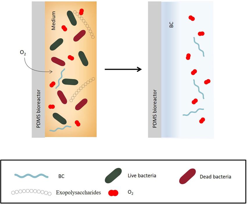
www.nature.com/scientificreports/
Strain Dried BC (g) Native BC in liquid cultivation Isolation source Species
K1A1826 0.0168n ± 0.0001 Uniform and smooth Liquid kombucha tea fraction Komagataeibacter sp.
K1G4 = UMCC 294731 0.2096a ± 0.0001 Uniform and smooth Liquid kombucha tea fraction K. xylinus
K1G23 26
0.0629b ± 0.0001 Uniform and smooth Liquid kombucha tea fraction Komagataeibacter sp.
K2A826 0.0401e ± 0.0001 Uniform and smooth Liquid kombucha tea fraction Komagataeibacter sp.
K2A10 = UMCC 296526 0.0162n ± 0.0001 Uniform Liquid kombucha tea fraction Komagataeibacter sp.
K2A8 26
0.0293g ± 0.0002 Uniform and smooth Liquid kombucha tea fraction Komagataeibacter sp.
K2G826 0.0296g ± 0.0001 Uniform and smooth Liquid kombucha tea fraction Komagataeibacter sp.
K2G1026 0.0230l ± 0.0001 Uniform Liquid kombucha tea fraction Komagataeibacter sp.
K2G1426 0.0317f ± 0.0001 Uniform Liquid kombucha tea fraction Komagataeibacter sp.
K2G1526 0.0264i ± 0.0001 Uniform Liquid kombucha tea fraction Komagataeibacter sp.
K2G30 = UMCC 275633 0.0519d ± 0.0002 Uniform and smooth Pellicle kombucha tea fraction K. xylinus
K2G39 = UMCC 297026 0.0540c ± 0.0002 Uniform and smooth Liquid kombucha tea fraction Komagataeibacter sp.
K2G41 = UMCC 297126 0.0247k ± 0.0001 Uniform and smooth Liquid kombucha tea fraction Komagataeibacter sp.
K2G44 = UMCC 297226 0.0132p ± 0.0001 Uniform Pellicle kombucha tea fraction Komagataeibacter sp.
DSM 15973 T 34
0.0164n ± 0.0001 Uniform and smooth Black cherry K. sucrofermentas
DSM 200435 0.0294g ± 0.0001 Uniform Unknown source K. xylinus
DSM 2325 35
0.0276h ± 0.0006 Uniform Unknown source K. xylinus
DSM 46590 0.0255j ± 0.0001 Uniform and smooth Unknown source K. xylinus
DSM 46591 0.0194m ± 0.0002 Uniform and smooth Unknown source K. xylinus
DSM 4660236 0.0064q ± 0.0001 Fragmented Vinegar K. xylinus
DSM 4660336 0.0127p ± 0.0001 Fragmented Unknown source K. xylinus
DSM 4660437 0.0155° ± 0.0005 Uniform and smooth Unknown source K. xylinus
DSM 4660536 0.0041r ± 0.0004 Fragmented Vinegar brew K. xylinus
DSM 6513T 37 0.0047r ± 0.0002 Fragmented Mountain ash berries K. xylinus
DSM 5602T 38 ND Fragmented Vinegar K. hansenii
Table 1. Weight of dried BC and shape of native BC produced by AAB strains used in this study and their
isolation source. Values are given as mean ± standard deviation (n = 3). Different lowercase letters in the same
column indicate significant differences (p < 0.05). Bold fonts refer to strains that were chosen for surface-
microstructured BC production. ND not detectable.
inflammatory response followed by a sequence of events that lead to the deposition of fibrotic t issue18,19. Such
an event is correlated with several health risks for patients, both during control interventions and after, when
the functionality of the implanted device is r equired20.
The intrinsic characteristics of BC, such as the non-toxic chemical composition, the purity, the high porosity,
the bulk mechanical properties, and the matrix-like morphology, make it advantageous in biomedical applica-
tions to prevent the fibrotic adhesion, as documented by several s tudies15,21–23. Indeed, it was previously reported
that the adverse conditions for adhesion, between cells and BC, are established by an isotropic distribution of
topographical features, which physically interfere with the establishment and maturation of focal a dhesion21,24.
The micro-structuration of BC’s surface is possible using a Guided Assembly-Based Biolithography (GAB), which
is a powerful replica molding methodology to transfer on-demand functional topographies to the surface of BC
nanofiber textures. BC nanofibers are assembled in a three-dimensional network reproducing the hexagonal
pattern imposed by the mold20,24,25. Thus, the formation of the pattern on the surface of BC inhibits the initial
biological recognition22 avoiding the formation of fibrotic tissue around implantable devices placed in soft tissues.
Among BC producing organisms, acetic acid bacteria (AAB) of the Komagataeibacter genus comprise spe-
cies such as Komagataeibacter xylinus, K. hansenii and K. sucrofermentas with high production yield which are
reported as native or functionalized biomaterial pruducers1.
In the present work, 25 AAB wild-type strains belonging to the Komagataeibacter genus were evaluated for
their suitability to synthesize a medical device made of surface microstructured BC in combination with GAB.
Due to high intraspecific variability within AAB in producing B C26, 9 strains were selected based on BC
production yield and shape. Selected strains were cultured inside the bioreactor and their characteristics of the
symmetrical array on the surface were analysed. Promising outcomes have been obtained by 5 strains, which
produced the required surface-microstructured BC. This study provides new evidence for the use of wild-type
AAB strains as biological machineries for producing biomedical devices.
Results and discussion
Assessment of bacterial cellulose production by Komagataeibacter strains. The amount and
shape of BC produced by AAB strains are presented in Table 1. AAB species considered for this study were K.
xylinus, K. hansenii, and K. sucrofermentans, which are reported as the highest BC producers among the species
of the Komagataeibacter genus27–32. BC was produced by all the strains; however, a great variability was observed
not only on the macroscopic structure of the native BC (Supplementary Fig. S1), but also in terms of weight.
Scientific Reports | (2021) 11:19311 | https://doi.org/10.1038/s41598-021-98705-2 2
Vol:.(1234567890)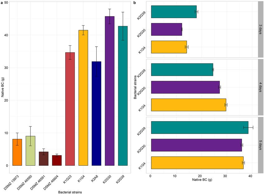
www.nature.com/scientificreports/
Figure 1. Weight of native BC produced by screened strains. BC weight was obtained after incubation at 28 °C
for 7 (a) and 3, 4, 5 (b) days inside bioreactor. Values are given as mean ± standard deviation (n = 3).
Some strains produced a uniform and homogenous BC layer, smooth on the surface, which was easily removed
from the culture broth without damaging its shape. Whereas other strains produced a fragmented BC layer with
a heterogenous and compact macrostructure. Once removed from the culture broth the cellulosic matrix has
lost its original shape.
Considering this study aimed to synthesize a medical device made of surface-microstructured BC, 9 strains
producing uniform and smooth BC layers of different weights (Supplementary Fig. S2) were selected for further
investigation. Among them, 5 strains were isolated from Kombucha tea, one from black cherry and 3 from
unknown isolation sources (Table 1).
The variability in BC production has been previously observed for strains of the genus Komagataeibacter
and within strains of the same species (e.g., K. xylinus). In the Komagataeibacter genus, differences in cellulose
synthase (CS) complex have been correlated to a different ability in BC p roduction39. The main reason for this
difference is due to the number of bcs operons, which generally span from 1 to 3 in members of the Komaga-
taeibacter genus39,40. However in K. xylinus species two strains were described to possess a fourth copy of bcsAB
gene33,41. We previously obtained and analysed K2G30 and K1G4 genomes. The K2G30 genome possesses three
bcs operons and a fourth copy of bcsAB gene, that encodes the catalytic core of CS. Whereas K1G4 analysis
revealed the presence of the two bcs operon types structurally completed and a third copy of bcsAB g ene31,33.
These features can explain the high amount of BC produced by these two strains.
Other factors that contribute to strain variability in BC production are, the isolation source and handling of
culture. Most of the strains of this study originated from food matrices, especially Kombucha tea, which is con-
sidered a selective source for the recovery of BC producing AAB21, while others were originally collected from
sugared and acidic products. The laboratory culturing of strains also affects the stability of phenotypic traits. This
phenomenon is already observed for AAB when they are continuously cultivated and preserved by short-time
preservation methods, which increase the formation of high rate of spontaneous mutants42,43.
Production of surface‑structured bacterial cellulose with guided assembly‑based biolithogra‑
phy. Cultures derived from the nine selected strains were tested in the polydimethylsiloxane (PDMS) biore-
actor, using the same conditions as during the previous tests (Hestrin–Schramm44 (HS) broth; 5% v/v inoculum;
incubation at 28 °C for 7 days). Outcomes confirmed the great variability in native BC weight (Fig. 1a) (Sup-
plementary Table S1) and characteristics. Some strains differed from the others to produce non-optimal BC
pouches. K1G4, K2G30 and K2G39 strains produced the highest amount of BC, but the formed pouch did not
have optimal attributes. Also, the visual analysis of transparency and thickness of the pouches varied among
strains (Data not shown). To assess the suitability of the strains for the purpose of this study, it has been hypoth-
Scientific Reports | (2021) 11:19311 | https://doi.org/10.1038/s41598-021-98705-2 3
Vol.:(0123456789)www.nature.com/scientificreports/
Flask (30 mL) Bioreactor (55 mL)
Strain Yield (g/L) ± st dev (g/L) Yield (g/L) ± st dev (g/L)
K1G4 6.9867a ± 0.0033 3.7020bc ± 0.4153
b
K1G23 2.0967 ± 0.0033 2.8348cd ± 0.2950
c
K2G39 1.8011 ± 0.0069 3.8764b ± 0.4344
d
K2G30 1.7311 ± 0.0051 4.8939a ± 0.2734
e
K2A8 1.3367 ± 0.0033 2.5028d ± 0.5575
T f
DSM 15973 0.5456 ± 0.0019 0.8077e ± 0.0387
DSM 46604 0.5156g ± 0.0168 0.4907e ± 0.0621
DSM 46590 0.8511h ± 0.0019 0.8721e ± 0.2112
DSM 46591 0.6478i ± 0.0051 0.4992e ± 0.0705
Table 2. Dried BC yield (g/L) produced after incubation at 28 °C for 7 days inside flasks and bioreactor.
Values are given as mean ± standard deviation (n = 3). Different lowercase letters in the same column indicate
significant differences (p < 0.05).
esized to reduce the incubation time to 3, 4, and 5 days for the highest producers (K2G30, K2G39, and K1G4)
(Fig. 1b). Results showed that the optimal production required 3 days of cultivation. The yield of BC production
and its characteristics are also influenced by the type of vessel in which the microbial strains grow. Considering
AAB strictly aerobic organisms, in the static cultivation regime, BC is formed at specific sites of the air surface
of liquids45. Therefore, production in terms of yield is dependent on the ratio between the surface exposed to air
and the volume (S/V ratio) of the vessel, in which bacteria grow.
The bioreactor used in this study allows the transfer of oxygen, which is a key factor in the production of BC
by the Komagataeibacter sp. The constant diffusion of oxygen is permitted by the non-polar nature of the P DMS46.
Indeed, the lower the polarity of a material is, the higher is the permeability to the oxygen, as reported for other
materials, such as film composites with whey protein isolate and p ectin47,48. Therefore, a higher BC yield was
reached for most of the culture strains due to the bigger gas–liquid interface (Table 2). Moreover, oxygen inside
the bioreactor was further increased by the high permeability of PDMS49,50.
Considering the cultivation method used in this study, the gas–liquid interface area of the 100 mL flask was
approximately 9.42 cm2, whereas in the case of the bioreactor it was nearly 120 cm2, of which just 9.5 cm2 of the
free interface. Consequently, by approximating the geometric structure of the 100 mL flask to a truncated cone
with diameters of 6.60 cm and 6.00 cm, and by approximating the structure of the bioreactor to a parallelepiped,
the S/ V ratios were 0.60 c m2/cm3 and 1.75 c m2/cm3, respectively.
Even though the higher BC production occurred inside the PDMS bioreactors, the size of the pouches pro-
duced by strain DSM 46604 was smaller than that required, and it collapsed, not standing at the level at which the
bacterial culture had been inoculated. Once the strain was grown inside the PDMS bioreactor, the structure of
the pocket composed by BC, to support its sidewalls, has nothing but the BC itself. The collapse of the structure
could be correlated to the slowness of the BC pace synthesis by the microbial strain, which leads to the absence
of a unique and packed biofilm BC formation inside the bioreactor. Therefore, the DSM 46604 strain did not
produce satisfactory results from a structural point of view.
As already mentioned, through a continuous exchange of oxygen, bacteria can produce BC also within the
walls of the bioreactor (Fig. 2), even though more slowly than the upper part, which is directly in contact with the
air. Therefore, a thick layer of BC was observed for almost all strains at the open liquid–air interface at the end of
incubation time. A sort of closed cap, sealing the top of the surface-microstructured BC pouch was formed. That
cap was easily removed from the rest of the BC and the devices assumed the pouch-shape, without any damage
to the BC. Most of tested strains formed the removable cap with two exceptions. In surface-microstructured BC
produced by DSM 46591 and DSM 46604, the thicker layer was not visible and, consequently, not possible to
remove without damaging the rest of the BC.
Our outcomes highlight that the use of defined AAB strains is a versatile strategy that allows obtaining cos-
tumed devices, modulating the growth conditions. Moreover, for some strains a reduction in cultivation time
was proved, which is a further milestone for using AAB in industry. Based on these results, K2G30 and the K1G4
(3 days of cultivation), and K2A8, DSM 1 5973T, and DSM 46590 (7 days of cultivation) are candidate strains for
surface-microstructured BC production assisted by GAB.
Characterization of surface‑structured bacterial cellulose with guided assembly‑based bio‑
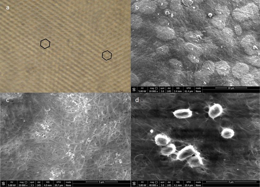
lithography. Since the surface micropattern designed by Hylomorph AG has characteristic dimensions in
the sub-micron range (1–10 µm), brightfield microscopy (BF) and scanning electron microscopy (SEM) were
used to investigate the presence of the surface micropattern on the pouches. All the 5 candidate strains (K2G30,
K1G4, K2A8, DSM 1 5973T and DSM 46590) showed the hexagonal pattern on the surface using BF (Fig. 3a).
In parallel, SEM analysis with high magnification was performed to observe the surface of the matrix and to
confirm the fibrous network of BC (Fig. 3b,c). Moreover, to visualize how bacterial cells are dispersed into the
BC matrix (Fig. 3d), a K2A8 sample, as representative of the pool of strains, was differently treated for SEM
experiment, by turning the BC pouch inside out and reducing the washing steps. Results are consistent with the
literature describing AAB of Komagataeibacter genus as short rods with an average width of 0.65 µm (ranging
Scientific Reports | (2021) 11:19311 | https://doi.org/10.1038/s41598-021-98705-2 4
Vol:.(1234567890)www.nature.com/scientificreports/
Figure 2. Formation of BC layer inside the PDMS bioreactor.
from 0.5 to 0.8 µm) and an average length of 2 µm (ranging from 1.0 to 3.00 μm), occurring singly, in pairs or
in chain51.
Conclusions
In this work, 25 Komagataeibacter strains were tested for producing a BC device suitable for biomedical purposes.
The medical device was manufactured in the form of a pouch, synthesized by AAB at the liquid–air interface,
after a period of incubation inside a PDMS bioreactor. Among studied strains, K2G30, K1G4, K2A8, DSM
15973T and DSM 46590 produced optimal surface-microstructured BC and they were designated as candidate
strains for the purpose of this study. Although a further research step is required to evaluate the biocompatibility,
the durability of the device and the reduction of fibrotic tissue, results of this study open new horizons toward
applying wild-type AAB strains in the biomedical field.
Materials and methods
Materials. HS broth was prepared following the recipe: D-glucose 2% w/v, Yeast extract 1% w/v, Polypeptone
0.5% w/v, Disodium phosphate anhydrous ( NaHPO4) 0.27% w/v, Citric acid 0.115% w/v)44. Whereas the 105
broth was prepared according to DSMZ instructions: D-glucose 10% w/v, calcium carbonate (CaCO3) 2% w/v,
Yeast extract 1% w/v. Sterilization was conducted in an autoclave at 121 °C for 20 min.
Bacterial strains and cultivation conditions. AAB strains used in this study were supplied by UMCC
(Unimore Microbial Culture Collection, Italy) and DSMZ (Deutsche Sammlung von Mikroorganismen und
Zellkulturen GmbH, Braunschweig, Germany) culture collections, respectively (Table 1). All the strains were
handled by UMCC culture collection, according to the standard procedures of the Microbial Resource Research
Infrastructure—Italian Joint Research Unit (MIRRI-IT)52.
DSMZ strains were revitalized according to manufacturer instructions, using 105 broth and UMCC strains on
HS broth. After revitalization, a pre-inoculum was performed on 5 mL HS broth. Then an aliquot of inoculum
(5% v/v) was transferred in a 100 mL Erlenmeyer flask containing 30 mL HS broth. Incubation was performed
at 28 ˚C for 7 days, under static conditions.
Qualitative and quantitative tests of BC. BC production was qualitatively estimated following the
method proposed by Navarro et al., 1 99953. Pellicle was collected from the broth culture after 7 days of incuba-
tion at 28 °C, treated with 4 mL of 5% NaOH and boiled for 2 h. BC production has been confirmed when the
pellet did not dissolve after boiling. K. xylinus K2G30 was used as a positive control.
Scientific Reports | (2021) 11:19311 | https://doi.org/10.1038/s41598-021-98705-2 5
Vol.:(0123456789)www.nature.com/scientificreports/
Figure 3. K2A8 strain as representative of the pool of strains. (a) Micro-pattern characterization. Hexagonal
pattern of the surface-microstructured BC manufactured using biolithography. Hexagonal pattern-formation
was observed by BF microscopy at high magnification ×20 using native BC. (b) SEM image of surface-
microstructured BC. (c) SEM image of microstructured BC fibrous network. (d) SEM image of K2A8 present
among microstructured BC fibrous network.
Estimation of BC yield was carried out following the method proposed by Hwang et al. 199954. Briefly, native
BC from culture broth was collected and washed with distilled water four times with a time-lapse of 15 min and
additional washing with 1 M NaOH. Washed BC films were kept at 90 °C for 30 min inside the solution of NaOH
1 M. Finally, BC was rinsed using distilled water four times and then dried at 25 °C until a constant weight was
reached. The weighting of the dried BC film was performed using an analytical balance (Gibertini E42S, Milan
Italy). The yield of BC was expressed as grams of dried BC per liter (g/L).
Growth of AAB in 3D bioreactors. 3D bioreactors manufactured in PDMS were used as vessels for
assessing the development of surface microstructured BC by Komagataeibacter genus strains. Cultivation in the
bioreactor was carried out with 55 mL of HS broth and 5% v/v inoculum. Bioreactors were covered with sterile
gauze on the top and incubated at 28 °C for 7 days. Then, each sample was washed using at first distilled water,
then a solution of NaOH 1 M and rinsed with distilled water several times. The last washing in distilled water
was conducted in shaking conditions. BC production within the bioreactor was evaluated by the formation of a
homogeneous film with a cap at an open liquid–air interface. The sealing of the pouch was removed aseptically
and avoiding the damaging of the BC pouch-shaped device. The yield of BC produced was controlled as previ-
ously described and expressed in grams of dried BC per liter (g/L).
Microscopy. The surface microstructure of BC pouches was examined using a field emission scanning elec-
tron microscope (NovaNano SEM 450, FEI, USA). Samples were cut (5 × 5 mm2) and mounted on a stainless-
steel stub with double-sided tape. The analysis was performed in a low vacuum mode (80 kPa) with an accelera-
tion voltage of 10 kV55. The surface microstructure pattern of the films was obtained through a BF microscopy
Eclipse Ts2 inverted microscope (Nikon, Tokyo, Japan).
Statistics analysis. The statistical analysis of the data was performed through analysis of variance
(ANOVA) using multcompView package implemented in R v 4.0.456. The experiment was performed in 3 rep-
licates. The differences between means were evaluated by Tukey HSD test (p < 0.05). The data were expressed as
the mean ± standard deviation (SD).
Scientific Reports | (2021) 11:19311 | https://doi.org/10.1038/s41598-021-98705-2 6
Vol:.(1234567890)www.nature.com/scientificreports/
Data availability
All data generated or analysed during this study are included in this published article (and its Supplementary
Information file).
Received: 29 June 2021; Accepted: 7 September 2021
References
1. Gullo, M., La China, S., Falcone, P. M. & Giudici, P. Biotechnological production of cellulose by acetic acid bacteria: Current state
and perspectives. Appl. Microbiol. Biotechnol. 102, 6885–6898 (2018).
2. George, J., Ramana, K. V., Sabapathy, S. N., Jagannath, J. H. & Bawa, A. S. Characterization of chemically treated bacterial (Aceto-
bacter xylinum) biopolymer: Some thermo-mechanical properties. Int. J. Biol. Macromol. 37, 189–194 (2005).
3. Chawla, P. R., Bajaj, I. B., Survase, S. A. & Singhal, R. S. Microbial cellulose: Fermentative production and applications. Food
Technol. Biotechnol. 47, 107–124 (2009).
4. Grande, C. J., Torres, F. G., Gomez, C. M. & Carmen Bañó, M. Nanocomposites of bacterial cellulose/hydroxyapatite for biomedical
applications. Acta Biomater. 5, 1605–1615 (2009).
5. Gallegos, A. M. A., Carrera, S. H., Parra, R., Keshavarz, T. & Iqbal, H. M. N. Bacterial cellulose: A sustainable source to develop
value-added products - A review. BioResources 11, 5641–5655 (2016).
6. Ullah, H., Santos, H. A. & Khan, T. Applications of bacterial cellulose in food, cosmetics and drug delivery. Cellulose 23, 2291–2314
(2016).
7. Gomes, R. J., de Borges, M. F., de Rosa, M. F., Castro-Gómez, R. J. H. & Spinosa, W. A. Acetic acid bacteria in the food industry:
Systematics, characteristics and applications. Food Technol. Biotechnol. 56, 139–151 (2018).
8. Vigentini, I. et al. Set-Up of bacterial cellulose production from the genus Komagataeibacter and its use in a gluten-free bakery
product as a case study. Front. Microbiol. 10, 1–13 (2019).
9. Mubashir, M. et al. Cellulose acetate-based membranes by interfacial engineering and integration of ZIF-62 glass nanoparticles
for CO2 separation. J. Hazard. Mater. 415 (2021).
10. Shi, Z., Zhang, Y., Phillips, G. O. & Yang, G. Utilization of bacterial cellulose in food. Food Hydrocoll. 35, 539–545 (2014).
11. Bourdichon, F. et al. Food fermentations: Microorganisms with technological beneficial use. Int. J. Food Microbiol. 154, 87–97
(2012).
12. Koutsoumanis, K. et al. Update of the list of QPS-recommended biological agents intentionally added to food or feed as notified
to EFSA 9: Suitability of taxonomic units notified to EFSA until september 2018. EFSA J. 17, 1–46 (2019).
13. Volova, T. G., Prudnikova, S. V., Sukovatyi, A. G. & Shishatskaya, E. I. Production and properties of bacterial cellulose by the strain
Komagataeibacter xylinus B-12068. Appl. Microbiol. Biotechnol. 102, 7417–7428 (2018).
14. Almeida, T., Silvestre, A. J. D., Vilela, C. & Freire, C. S. R. Bacterial nanocellulose toward green cosmetics: Recent progresses and
challenges. Int. J. Mol. Sci. 22, 1–25 (2021).
15. Czaja, W., Krystynowicz, A., Bielecki, S. & Brown, R. M. Microbial cellulose—The natural power to heal wounds. Biomaterials 27,
145–151 (2006).
16. Jiji, S., Udhayakumar, S., Rose, C., Muralidharan, C. & Kadirvelu, K. Thymol enriched bacterial cellulose hydrogel as effective
material for third degree burn wound repair. Int. J. Biol. Macromol. 122, 452–460 (2019).
17. Picheth, G. F. et al. Bacterial cellulose in biomedical applications: A review. Int. J. Biol. Macromol. 104, 97–106 (2017).
18. Bongiorni, M. G. et al. Il rischio iatrogeno connesso all’impianto di pacemaker e defibrillatori. G. Ital. Cardiol. 10, 395–406 (2009).
19. Nagmetova, G., Berthold-Pluta, A., Garbowska, M., Kurmanbayev, A. & Stasiak-Rózańska, L. Antibacterial activity of biocellulose
with oregano essential oil against Cronobacter strains. Polymers (Basel). 12, 1–10 (2020).
20. Robotti, F. et al. Microengineered biosynthesized cellulose as anti-fibrotic in vivo protection for cardiac implantable electronic
devices. Biomaterials 229, 119583 (2020).
21. Robotti, F. Surface microstructuring for control of cellular activities and bio-synthesized cellulose biolithography. ETH Zurich
https://doi.org/10.3929/ethz-b-000171210 (2017).
22. Cacicedo, M. L. et al. Progress in bacterial cellulose matrices for biotechnological applications. Bioresour. Technol. 213, 172–180
(2016).
23. Anton-Sales, I. et al. In vivo soft tissue reinforcement with bacterial nanocellulose. Biomater. Sci. 9, 3040–3050 (2021).
24. Bottan, S. et al. Surface-structured bacterial cellulose with guided assembly-based biolithography (GAB). ACS Nano 9, 206–219
(2015).
25. Robotti, F. et al. A micron-scale surface topography design reducing cell adhesion to implanted materials. Sci. Rep. 8, 1–13 (2018).
26. La China, S. et al. Kombucha tea as a reservoir of cellulose producing bacteria: Assessing diversity among Komagataeibacter isolates.
Appl. Sci. 11, 1595 (2021).
27. Tsouko, E. et al. Bacterial cellulose production from industrial waste and by-product streams. Int. J. Mol. Sci. 16, 14832–14849
(2015).
28. Fijałkowski, K., Zywicka, A., Drozd, R., Kordas, M. & Rakoczy, R. Effect of Gluconacetobacter xylinus cultivation conditions on
the selected properties of bacterial cellulose. Polish J. Chem. Technol. 18, 117–123 (2016).
29. Chen, S. Q. et al. Characterisation of bacterial cellulose from diverse Komagataeibacter strains and their application to construct
plant cell wall analogues. Cellulose 24, 1211–1226 (2017).
30. Gullo, M. et al. Increased production of bacterial cellulose as starting point for scaled-up applications. Appl. Microbiol. Biotechnol.
101, 8115–8127 (2017).
31. La China, S. et al. Genome sequencing and phylogenetic analysis of K1G4: A new Komagataeibacter strain producing bacterial
cellulose from different carbon sources. Biotechnol. Lett. 42, 807–818 (2020).
32. La China, S., Zanichelli, G., De Vero, L. & Gullo, M. Oxidative fermentations and exopolysaccharides production by acetic acid
bacteria: A mini review. Biotechnol. Lett. 40, 1289–1302 (2018).
33. Gullo, M., La China, S., Petroni, G., Di Gregorio, S. & Giudici, P. Exploring K2G30 genome: A high bacterial cellulose producing
strain in glucose and mannitol based media. Front. Microbiol. 10, 58 (2019).
34. Toyosaki, H. et al. The characterization of an acetic acid bacterium useful for producing bacterial cellulose in agitation cultures:
The proposal of Acetobacter xylinum subsp. sucrofermentans subsp. nov.. J. Gen. Appl. Microbiol. 41, 307–314 (1995).
35. Mamlouk, D. & Gullo, M. Acetic acid bacteria: Physiology and carbon sources oxidation. Indian J. Microbiol. 53, 377–384 (2013).
36. Gillis, M. & De Ley, J. Intra- and intergeneric similarities of the ribosomal ribonucleic acid cistrons of Acetobacter and Glucono-
bacter. Int. J. Syst. Bacteriol. 30, 7–27 (1980).
37. Semjonovs, P. et al. Cellulose synthesis by Komagataeibacter rhaeticus strain P 1463 isolated from Kombucha. Appl. Microbiol.
Biotechnol. 101, 1003–1012 (2017).
38. Yamada, Y. Systematics of acetic acid bacteria. in Acetic Acid Bacteria: Ecology and Physiology. 1–50. https://doi.org/10.1007/978-
4-431-55933-7_1 (Springer, 2016).
Scientific Reports | (2021) 11:19311 | https://doi.org/10.1038/s41598-021-98705-2 7
Vol.:(0123456789)www.nature.com/scientificreports/
39. Römling, U. & Galperin, M. Y. Bacterial cellulose biosynthesis: Diversity of operons, subunits, products, and functions. Trends
Microbiol. 23, 545–557 (2015).
40. Valera, M. J., Torija, M. J., Mas, A. & Mateo, E. Cellulose production and cellulose synthase gene detection in acetic acid bacteria.
Appl. Microbiol. Biotechnol. 99, 1349–1361 (2015).
41. Liu, M. et al. Complete genome analysis of Gluconacetobacter xylinus CGMCC 2955 for elucidating bacterial cellulose biosynthesis
and metabolic regulation. Sci. Rep. 8, 6266 (2018).
42. Azuma, Y. et al. Whole-genome analyses reveal genetic instability of Acetobacter pasteurianus. Nucleic Acids Res. 17, 5768–5783
(2009).
43. Gullo, M., Mamlouk, D., De Vero, L. & Giudici, P. Acetobacter pasteurianus strain AB0220: Cultivability and phenotypic stability
over 9 years of preservation. Curr. Microbiol. 6, 576–580 (2012).
44. Hestrin, S. & Schramm, M. Synthesis of cellulose by Acetobacter xylinum. II. Preparation of freeze-dried cells capable of polymer-
izing glucose to cellulose. Biochem. J. 58, 345–352 (1954).
45. Steel, R. & Walker, T. K. A comparative study of cellulose-producing cultures and celluloseless mutants of certain Acetobacter spp..
J. Gen. Microb. 17, 445–453 (1957).
46. Hu, L. et al. In-situ grafting to improve polarity of polyacrylonitrile hollow fiber-supported polydimethysiloxane membranes for
CO2 separation. J Colloid Interface Sci. 510, 12–19 (2018).
47. Zhou, Y. et al. Characterization of whey protein isolate and pectin composite film catalyzed by small laccase from Streptomyces
coelicolor. Environ. Technol. Innov. 19, 100999 (2020).
48. Niu, X. et al. Small Laccase from Streptomyces coelicolor catalyzed chitosan-pectin blending film for hazardous gas removal. Environ.
Technol. Innov. 23, 101690 (2021).
49. Shiku, H. et al. Oxygen permeability of surface-modified poly(dimethylsiloxane) characterized by scanning electrochemical micros-
copy. Chem. Lett. 35, 234–235 (2006).
50. Wolf, M. P., Salieb-Beugelaar, G. B. & Hunziker, P. PDMS with designer functionalities—Properties, modifications strategies, and
applications. Prog. Polym. Sci. 83, 97–134 (2018).
51. Yamada, Y. et al. Description of Komagataeibacter gen. nov., with proposals of new combinations (Acetobacteraceae). J. Gen. Appl.
Microbiol. 58, 397–404 (2012).
52. De Vero, L. et al. Preservation, characterization and exploitation of microbial biodiversity: The perspective of the italian network
of culture collections. Microorganisms 7, 685 (2019).
53. Navarro, R. R. & Komagata, K. Differentiation of Gluconacetobacter liquefaciens and Gluconacetobacter xylinus on the basis of
DNA base composition, DNA relatedness, and oxidation products from glucose. J. Gen. Appl. Microbiol. 45, 7–15 (1999).
54. Hwang, J. W., Yang, Y. K., Hwang, J. K., Pyun, Y. R. & Kim, Y. S. Effects of pH and dissolved oxygen on cellulose production by
Acetobacter xylinum BRC5 in agitated culture. J. Biosci. Bioeng. 88, 183–188 (1999).
55. Haghighi, H. et al. Characterization of bio-nanocomposite films based on gelatin/polyvinyl alcohol blend reinforced with bacterial
cellulose nanowhiskers for food packaging applications. Food Hydrocoll. 113, 106454 (2021).
56. Wickham, H. Ggplot2. Wiley Interdiscip. Rev. Comput. Stat. 3, 180–185 (2011).
Acknowledgements
Part of this research was supported by FAR Fondo di Ateneo per la Ricerca UNIMORE 2020. Hylomorph AG
provided a financial contribution. Luciana De Vero is acknowledged for managing bacterial strains within UMCC
culture collection.
Author contributions
M.B.: Methodology; Data curation, Writing original draft, Review & editing; F.R.: Methodology, Review & edit-
ing; S.L.: Methodology, Review; K.A.: Review & editing; H.H.: Methodology, Review; S.B.: Methodology, Review
& editing; A.F.: Review & editing; M.G.: Conceptualization, Funding acquisition, Review & editing, Supervision.
Competing interests
The authors declare no competing interests.
Additional information
Supplementary Information The online version contains supplementary material available at https://doi.org/
10.1038/s41598-021-98705-2.
Correspondence and requests for materials should be addressed to A.F. or M.G.
Reprints and permissions information is available at www.nature.com/reprints.
Publisher’s note Springer Nature remains neutral with regard to jurisdictional claims in published maps and
institutional affiliations.
Open Access This article is licensed under a Creative Commons Attribution 4.0 International
License, which permits use, sharing, adaptation, distribution and reproduction in any medium or
format, as long as you give appropriate credit to the original author(s) and the source, provide a link to the
Creative Commons licence, and indicate if changes were made. The images or other third party material in this
article are included in the article’s Creative Commons licence, unless indicated otherwise in a credit line to the
material. If material is not included in the article’s Creative Commons licence and your intended use is not
permitted by statutory regulation or exceeds the permitted use, you will need to obtain permission directly from
the copyright holder. To view a copy of this licence, visit http://creativecommons.org/licenses/by/4.0/.
© The Author(s) 2021
Scientific Reports | (2021) 11:19311 | https://doi.org/10.1038/s41598-021-98705-2 8
Vol:.(1234567890)You can also read