Protocatechuic acid attenuates cerebral aneurysm formation and progression by inhibiting TNF-alpha/Nrf-2/NF-kB-mediated inflammatory mechanisms in ...
←
→
Page content transcription
If your browser does not render page correctly, please read the page content below
Open Life Sciences 2021; 16: 128–141
Research Article
Gang Xiao*#, Mei Zhang#, Xing Peng, Guangyuan Jiang
Protocatechuic acid attenuates cerebral
aneurysm formation and progression by
inhibiting TNF-alpha/Nrf-2/NF-kB-mediated
inflammatory mechanisms in experimental rats
https://doi.org/10.1515/biol-2021-0012 inflammatory cytokine and chemokine levels in a dose-depen-
received July 15, 2020; accepted September 16, 2020 dent manner. We found that PCA treatment exerts protective
Abstract: Our current research aims to examine whether effects by suppressing the development and progression of
protocatechuic acid (PCA) can be used as a therapeutic CA through the inhibition of inflammatory responses in
agent for the development of cerebral aneurysm (CA) and macrophages via TNF-α/NF-κB/Nrf-2 signaling pathways,
to elucidate the mechanisms behind this. We assessed the thus demonstrating that PCA can act as a treatment for CA.
effects of PCA at 50 and 100 mg/kg on the activation of Keywords: cerebral aneurysm, protocatechuic acid,
signaling pathways for tissue necrosis factor (TNF)-α/ macrophages, inflammatory response mechanisms, chemo-
nuclear factor (NF)-κB/nuclear factor erythroid 2 (Nrf-2) on kines, TNF-α signaling, Nrf-2
progression and development in an elastase-induced CA
model, accompanied by a high-salt diet to induce hyper-
tension. The expression of inflammatory cytokines, chemo-
kines, tumor necrosis factor-α, interleukins (IL)-8, IL-17, 1 Introduction
IL-6, IL-1β, and matrix metalloproteinase (MMP)-2 and
MMP-9 was analyzed by ELISA, western blot, and reverse Cerebral aneurysm (CA) is the most common cerebrovas-
transcriptase quantative polymerase chain reaction. The cular disease with pathological dilations of cerebral arteries
expression levels of antioxidant enzymes and translocation occurring in more than 5% of the population [1]. CA can
of Nrf-2 were also determined. The group treated with PCA appear as unruptured [2] with a symptom of chronic head-
demonstrated a significant (P < 0.05) decrease in the aneur- ache [3] and as ruptured forms, giving rise to hypertension
ysmal size in rats compared to the CA-induced group. causing a condition called subarachnoid hemorrhage (SAH)
We found that PCA treatment suppressed the invasion of [4,5]. This leads to morbidity and mortality worldwide.
macrophage and activation of TNF-α/NF-κB/Nrf-2 signaling The etiology of CA is unclear, but several factors, such
pathways. There was a significant decrease (P < 0.05) in pro- as chronic inflammation, hemodynamic changes with
endothelial dysfunction [6], oxidative stress, and apop-
tosis, may contribute to the development of CA [7,8]. It
is hypothesized that release of inflammatory cytokines
# These authors contributed equally to this work. and free radicals disrupts the endothelial intima of
blood vessel, causing extracellular matrix remodeling
dysfunction [6–9].
* Corresponding author: Gang Xiao, Department of Neurosurgery,
Several studies highlighted the important role of
Chongqing Traditional Chinese Medicine Hospital, No. 6 Panxi 7
Branch Road, Jiangbei District, Chongqing 400021, People’s chronic inflammation in the formation and progression
Republic of China, tel: +86-023-6798-3716, fax: +86-023-6798- of CA [10–14]. In addition, reactive oxygen species (ROS)
3716, e-mail: gangxiao21@outlook.com, cqxiaogang3@163.com can damage vascular endothelium contributing to the
Mei Zhang: Department of Dermatology, Daping Hospital, formation and rupture of CA [15,16]. It is known that
Army Medical University, Chongqing 400042, China
ROS plays a significant role in inflammation and vascular
Xing Peng, Guangyuan Jiang: Department of Neurosurgery,
Chongqing Traditional Chinese Medicine Hospital, No. 6 Panxi 7
smooth muscle dysfunction involving transcriptional factor,
Branch Road, Jiangbei District, Chongqing 400021, nuclear factor erythroid 2 (Nrf-2), because of its endogenous
People’s Republic of China antioxidant effect. Under inflammatory conditions, Nrf-2
Open Access. © 2021 Gang Xiao et al., published by De Gruyter. This work is licensed under the Creative Commons Attribution 4.0
International License.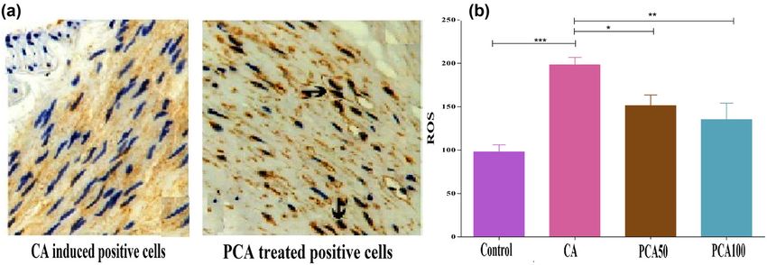
PCA prevents CA by anti-inflammatory pathway 129
translocates from the cytoplasm into the nucleus. This (JNK) pathway causing Nrf-2 to translocate from the cyto-
results in the expression of endogenous anti-inflammatory plasm into the nucleus, which results in the suppression of
and antioxidant substances [17]. However, the pathogenesis ROS generation [36,37]. In addition, PCA has anti-inflam-
of Nrf-2 in the formation and progression of CA is unclear. matory effect in lipopolysaccharide (LPS) induced BV2
Inflammatory responses are also influenced by the microglia, acting on the activation of NF-κB and MAPK
recruitment of nuclear factor (NF)-kB-mediated macrophages. signaling pathways [38].
This induces the expression of inflammation-related genes In patients previously pretreated with PCA, inflam-
such as monocyte chemoattractant protein-1 (MCP-1), inter- mation was noticeably restrained. Moreover, pretreart-
leukin (IL)-1β, inducible nitric oxide synthase (iNOS), and ment had an inhibitory effect on the production of NO
matrix metalloproteinases (MMPs) [18]. NF-kB-mediated sig- and TNF-α secretion in the macrophages, which was
naling through iNOS and IL-1β expression induces apoptosis induced by LPS-induced acute inflammation and oxida-
of vascular smooth muscle causing endothelium damage tive stress by inhibiting iNOS/NO and cyclooxygenase-2
with the disruption of elastic lamina [19]. It also mediates (COX-2) expression/PGE2 expression involving the NF-κB
the expression of MMP-2 and MMP-9 causing degradation of and MAPK signaling pathways [39]. PCA also demon-
collagen leading to vascular wall remodeling [20]. strated a novel anti-cancer activity that involves the
Studies have shown that increased levels of expression downregulation of the RAS/Akt/NF-kB pathway. This
of tissue necrosis factor (TNF)-α are implicated in the pro- works by targeting RhoB activation and reducing MMP-
gression of CA. This can be induced by the injection of mediated cell involvement in cancer cells [39]. Chen et al.
elastase into the coronary arteries and the aorta of animal, demonstrated that oral administration of 50 mg/kg of
which results in the fragmentation of lamina [21–24]. Chronic PCA in mice reached a plasma peak of 73.6 μM [40].
inflammation mediated by TNF-α causes disruption of vessel Another study by Leila Safaeian et al. showed that PCA
walls and deposition of plaques. The secretion of TNF-α by T- at 50, 100, and 200 mg/kg has antioxidant and antihyper-
cells also stimulates other immune cells to secrete TNF-α [25]. tensive effects against dexamethasone-induced hyperten-
These plaques formed at the intersection of cerebral sion [41]. In addition, the study by Bhattacharjee et al. [42]
arteries and their curvatures [26] weaken the endothe- reported that the treatment with PCA at 50 and 100 mg/kg,
lium causing migration of leukocytes [27], which causes p.o. significantly (P < 0.05–0.01) promotes glucose metabo-
further injury to the vascular smooth muscle through the lism in the skeletal muscle, controlling glycemic and lipid
expression of MMPs. This disrupts the tight endothelial levels. This also causes a decrease in the secretion of pro-
junctions between the vascular walls of cerebrum [25]. In inflammatory cytokines and improved myocardial phy-
addition, several other conditions such as hemodynamic siology to near normalcy in type II diabetic rats.
stress and increase in blood pressure exacerbate the With the aforementioned effects, we investigated the
expression of TNF-α leading to the progression of CA. protective effects of PCA in CA-induced rats with stereo-
However, there currently exist no noninvasive thera- taxic administration of elastase in cerebrospinal fluid to
pies, and hence the need to develop new therapies to histologically imitate human CA, with inhibition of TNF-
address the formation and progression of CA. In a routine alpha/Nrf-2/NF-kB-mediated inflammatory mechanisms
drug screen for various diseases, the therapeutic use of for the suppression of CA formation and SAH at doses
phytochemicals is growing in popularity, because they of 50 and 100 mg/kg, respectively.
have either no or low side effects. Protocatechuic acid
(PCA) is a phenolic acid, commonly present in green tea,
is one of the main metabolites of catechins found in humans
after ingestion of green tea infusions. Its antioxidant and 2 Materials and methods
anti-inflammatory effects have been widely recognized
[28–31]. It was proven to show antihypertensive and anti-
oxidant effects against high-salt induced hypertension [32]. 2.1 Rat IA model
PCA has also been shown to be efficient in treating asthma
airway remodeling [33]. Through anti-inflammatory, antiox- Wistar rats of either sex weighing between 150 and 170 g
idant, and antiapoptotic mechanisms, PCA was used in were treated. Elastase (CAS Number 39445-21-1) and PCA
protecting against methotrexate (MTX) induced hepatorenal (CAS Number: 99-50-3) were procured from Sigma Aldrich,
toxicity [34]. It has also proved its effectiveness in reducing China. Rats were initially divided into four groups: vehicle
paw edema, cotton pellet-induced granuloma, and index of control, CA induced, PCA treated (50 mg/kg, orally), and
arthritis in rat models [35]. PCA exhibits an antiapoptotic PCA treated (100 mg/kg, orally), with 16 rats in each group.
effect through activation of the c-Jun N-terminal kinase PCA was administered orally to rats 1 week before the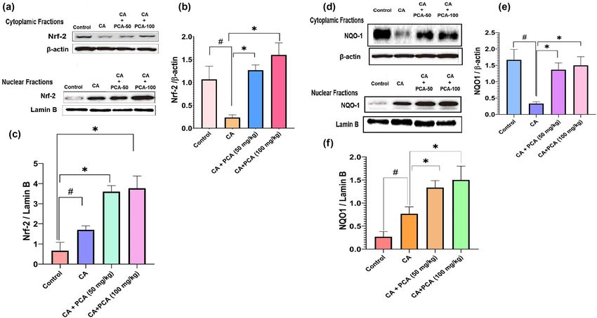
130 Gang Xiao et al.
induction of CA and continued until the end of the experi- PCR (Applied Bio-systems Real-Time PCR, Thermo-Fisher
ment. All test groups were treated with ketamine (80 mg/ Scientific, China) analysis using SYBR® Green (Sigma
kg) for inducing anesthesia. Before subjecting the rats to left Aldrich, China). Thirty to forty cycles were used for per-
internal carotid artery ligation, a single stereotaxic elastase forming qRT-PCR. The first phase involved denaturation
injection was administered into the cerebrospinal fluid of cDNA at 90°C for 30 s preceded by annealing for 30 s at
in the right basal cistern as previously illustrated [43]. For 50°C; finally, the method was extended for 40 s at 70°C. The
16 weeks, body weights and body mass index (BMI) of rats ΔCt method was used to measure the expression of tran-
were recorded in all four groups. scripts normalizing to the internal control of glyceraldehyde
To cause hypertension, a high-salt diet (4% NaCl in 3-phosphate dehydrogenase (GAPDH). The primers (Eurofins
standard animal chow and allowed to drink water) was MWG Operon, Bangalore, Karnataka, India) used for quanti-
implemented continuously for all the groups for 4 weeks fying specific genes in this study are listed in Table 1.
[44]. The tail-cuff procedure was used in rats to assess the
systolic blood pressure both before and during treatment,
and again for the next 4 weeks. After 90 days, a blue dye
containing bromophenol was administered into the lungs, 2.4 Western blotting assay
the Willis circle, and major branches. The vascular wall
thickness was determined, and the degree of aneurysm Protein lysates from the entire COW were obtained, incu-
was calculated. Hematoxylin and eosin (H&E) staining bated with ice-cold lysis buffer containing 1 mm phenyl-
was used to perform degenerative transformation evalua- methanesulfonyl fluoride (PMSF) and centrifuged at 4°C
tions of vessel endothelium. for 15 min. The protein concentration was determined
using Pierce™ BCA Protein Assay kit (CAT No. 23225;
Ethical approval: The research related to animal use has Thermo Fisher Scientific, China). Proteins were then sepa-
been complied with all the relevant national regulations rated using a 10% sodium dodecyl sulfate (SDS) - polyacryl-
and institutional policies for the care and use of animals. amide-based discontinuous gel and western transferred
onto 0.3 μm polyvinylidene fluoride or polyvinylidene
difluoride (PVDF) membranes. After blocking with 4% (w/v)
bovine serum albumin in a mixture of tris-buffered saline
2.2 ELISA measurements
and Tween 20 at room temperature, the membranes were
incubated overnight at a temperature of 4°C with primary
The standard ELISA kits (Catalogue number, CA8563518G3;
antibodies. After washing, membranes were incubated with
Chongqing Mbio Technology Co., Ltd, Chongqing, China)
horseradish peroxidase-conjugated secondary antibodies at
were used for assessing cytokine levels (IL-1β, IL-2, IL-17,
room temperature for 1 h. To visualize proteins, ECL western
IL-8, IL-6, and TNF-α). Tissue samples were obtained from
blotting substrate (BioVision, Inc, Milpitas, CA, USA) was
all groups around the COW tissues to determine the con-
centrations of specific cytokines, including IL-1β, IL-2, IL-17,
IL-8, IL-6, and TNF-α, as per the instructions of the supplier Table 1: Oligonucleotides used in this study
(Wuhan Fine Biotech Co., Ltd, China). Similarly, the values
for the parameters MMP-2 and MMP-9 were also obtained Gene Primer Sequence
using the commercial kits (Chongqing Mbio Technology
TNF-α 5′ forward 3′ CATCCGTTCTCTACCCAGCC
Co., Ltd, Chongqing, China). 3′ reverse 5′ AATTCTGAGCCCGGAGTTGG
NF‑κB 5′ forward 3′ ACGATCTGTTTCCCCTCATC
3′ reverse 5′ TGC TTCTCTCCCCAGGAATA
MMP‑2 5′ forward 3′ CTGATAACCTGGATGCAGTCGT
2.3 RNA isolation and quantitative real-time
3′ reverse 5′ CCAGCCAGTCCGATT TGA
polymerase chain reaction (qRT-PCR) MMP-9 5′ forward 3′ TTCAAGGACGGTCGGTATT
analysis 3′ reverse 5′ CTCGAGCCTAGACCCAACTTA
GAPDH 5′ forward 3′ AAGAAGGTGGTGAAGCAGGC
3′ reverse 5′ TCCACCACCCTGTTGCTGTA
The samples were collected around the COW region from Nrf-2 5′ forward 3′ CACATCCAGTCAGAAACCAGTGG
control and experimental rat groups. Total mRNA was 3′ reverse 5′ GGAATGTCTGCGCCAAAAGCTG
extracted using TRIzol® reagent (Sigma Aldrich, China) INF-γ 5′ forward 3′ GGCAAAAGGACGGTAACACG
according to the manufacturer’s instructions and stored 3′ reverse 5′ TCTGTGGGTTGTTCACCTCG
SM22 5′ forward 3′ ATCCTATGGCATGAGCCGTG
at −80°C. The cDNA was synthesized using ABScript II
3′ reverse 5′ CAGGCTGTTCACCAACTTGC
synthesis kit (Sigma Aldrich, China) to conduct the qRT-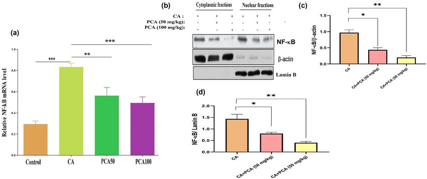
PCA prevents CA by anti-inflammatory pathway 131
used. The following primary antibodies were used to perform To evaluate the production of macrophages in CA
this assay: NF‑κB (#N8523; 1:1,000, Sigma Aldrich, China), walls, the brain tissue in the area typically performed
β-actin (#SAB3500350; 1:5,000, Sigma Aldrich, China), Lamin with CA was extracted and split into 5 μm sections.
B (#SAB5700147; 1:10,000, Sigma Aldrich), MMP-9 (#SAB5700152; These areas were initially hydrated and then stained
1:2,000, Sigma Aldrich, China), and GAPDH (#AB2302; with primary antibody CD68 (CAS No. 3F103; 1:200 dilu-
1:5,000, Sigma Aldrich, China). To examine the expression tion, Santa Cruz Biotech, Shanghai, China), and conse-
of nuclear transcription factor Nrf-2, nuclear and cytoplasmic quently several secondary antibodies (CAS No. sc-2354;
proteins were isolated separately. Gel electrophoresis was goat anti-mouse IgG, peroxidase-linked antibody, 1:5,000
used to differentiate the lysates of cells and then transferred dilution) were counterstained with hematoxylin. To ensure
to the PVDF. The membranes were probed with anti-Nrf- that a consistent number of macrophage cells were produced
2 (#ABE1845; 1:500, Sigma Aldrich, China), anti-NADPH in the CA walls, the number of labeled positive cells was
(#ab109225; 1:2,000, ElabScience, Hubei, China), and anti- counted in a 100 μm square area surrounding the CA walls.
αSMA antibodies (#ab5694; 1:5,000, ElabScience, Hubei,
China). The membranes were visualized with improved
chemiluminescence, followed by X-ray film for imaging.
The results were analyzed using ImageJ. 2.6 Statistical analysis
Mean ± SE was used to statistically measure the effects
2.5 H&E and immunohistochemical staining from multiple observations. Student’s t-test is used to
deduce the significance of two mean values between two
The extracted tissues were sliced into 5 μM sections and different groups. One-way ANOVA method was used for
placed on polylysine-coated slides until treatment with multiple comparisons. GraphPad Prism software was used
H&E. The tissues were then stained with hematoxylin to determine the significance with P < 0.05.
solutions for 7 h at temperatures between 50 and 70°C
and then washed with tap water till the water was color-
less. Next, 10% acetic acid and 85% ethanol were used for
2 and 10 h for differentiation of the tissue, and tissues 3 Results
were then washed with tap water. We saturated the tissue
in the lithium carbonate solution for 10 h and then washed it
with tap water. Eventually, staining was done for 48 h with 3.1 PCA suppresses CA formation and
the eosin solution. The tissue slices were dehydrated twice progression in rats
for 30 min with 95% ethanol and then immersed in xylene at
50–70°C for 1 h, accompanied by paraffin for 10 h. The In this study, we attempted to understand the inhibitory
stained tissues were then imaged using microscopy. effect of PCA against CA progression, after the adminis-
Paraffin-embedded sections in Histoclear solution tration of elastase. The development of aneurysm was
were heated overnight to 50°C and deparaffinized for identified along the Willis circle or its major branches,
5–10 min. The sections were rehydrated using 100% which is consistent with previous studies [25–27]. Most
alcohol twice for 10 min, followed by 95%, 90%, 70% aneurysms were larger in size, with multiple aneurysms
of alcohol for 10 min respectively. Subsequently the sec- around three to four times larger than their parent arteries.
tions were re-hydrated with water for 5 min. The activity Before triggering CA, the experimental group of rats was
of endogenous peroxidase was inactivated by immersion treated with PCA. We found that the control group had an
in 3% H2O2 peroxide for 25 min. Antigen unmasking was aneurysm size of 26.5 ± 0.76 μm, constantly increasing in
accomplished by incorporating slides for 1 h at 95°C in size to 45.5 ± 1.67 μm (P < 0.001) in the group induced by
the target recovery solution. Tissue portions were intracranial aneurysm over a period of 16 weeks, whereas
blocked with 10% normal goat serum in phosphate-buf- the group of rats pretreated with PCA had an aneurysm size
fered saline after rapid cooling to room temperature. significantly decreased to 31.33 ± 1.10 μm (P < 0.05) and
Antibodies for Nrf2 (#C20, Santa Cruz Biotechnology) 32.33 ± 2.06 μm (P < 0.01) at doses of 50 and 100 mg/kg,
were used for immunohistochemistry staining at dilution respectively, of PCA relative to the CA group (Table 2) in
(1:2,000). 3,3-Diaminobenzidine (DAB) was used to develop a dose-dependent manner. While taking into consideration
and display colors. With hematoxylin, the tissue slices were the systolic blood pressure (SBP) levels, the CA group
gently counterstained, dehydrated, and covered. Using a showed significant (P < 0.001) variation in their levels after
light microscope, the slides were imaged. 8 weeks of CA induction. Similarly, the group pretreated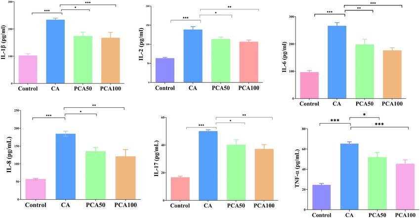
132 Gang Xiao et al.
Table 2: Progression of CA size in control, induced, and PCA-50 and 100 mg/kg administered experimental group of rats monitored for 16
weeks
Control CA PCA-50 PCA-100
0 week 28.33 ± 0.88 28.5 ± 0.76 27.5 ± 0.76 28.5 ± 0.76
4 weeks 26 ± 0.97 28.5 ± 0.76 29.5 ± 0.76 27.5 ± 0.76
8 weeks 26.5 ± 0.76 36.5 ± 0.76*** 33 ± 0.97* 29.5 ± 0.76***
12 weeks 26.5 ± 0.76 38.5 ± 0.76*** 31.33 ± 1.99** 30.67 ± 1.71**
16 weeks 26.5 ± 0.76 45.5 ± 1.67*** 31.33 ± 1.10* 32.33 ± 2.06**
Values are expressed as mean ± SE (n = 16). Statistical significance is expressed as ***P < 0.001 CA compared to vehicle (control) and PCA-
treated groups; *P < 0.05, PCA-50 compared to CA rats; and **P < 0.01 PCA-100 compared to CA rats.
with PCA displayed a significant decrease in SBP levels after that an endothelial cell lining with a thin layer of smooth
8 weeks in a dose-dependent manner when compared to muscle cells as described in Figure 2a and b shows mod-
the CA group (Figure 1), besides the degree of elevated erately thick vascular walls with inflammatory infiltration
aneurysm scores (Table 3). Treatment with PCA suppressed of the cells, thus displaying distortion of the vessel wall
vessel wall thickness in rats (P < 0.05) relative to the CA with evidence of myointimal hyperplasia. The thickness
group (Table 4) in a dose-dependent manner. After CA of the tunica media in the PCA-treated groups was sig-
induction, the body weight and BMI were substantially nificantly decreased compared to the CA group (Figure 2c
decreased after 4 weeks when compared to the control group. and d, Table 4) at 50 and 100 mg/kg in a dose-dependent
However, the groups receiving PCA-50 and 100 mg/kg treat- manner.
ments demonstrated a gain in body weight and BMI to This study was able to analyze the effect of PCA
normal control groups after 4 weeks. Furthermore, they on macrophage infiltration. Recruitment for macrophage
retained the body weight and BMI of rats in groups receiving infiltration was significantly (P < 0.05) elevated 4 weeks
PCA treatments in a dose-dependent manner until 16 weeks after aneurysm induction (Table 7, Figure 2f). Macro-
of the study duration (Tables 5 and 6). phage staining of the normal control group using anti-
The effect of PCA was examined in rats with H&E CD68 antibody in the cerebral artery showed lack of
staining to assess the formation and progression of CA. inflammatory cells and macrophage infiltration (Figure 2e).
A cerebral artery from the CA-induced group revealed Chronic macrophage-related inflammation plays a major
role in CA progression and can be seen in Figure 2f. This is
further demonstrated with staining with anti-CD68 anti-
bodies. The positive cells show that a majority of leuko-
cytes in the intracranial aneurysms were macrophages.
Figure 2f shows numerous leukocytes in intracranial
aneurysms, and the distribution of macrophages was
identical to that of leukocytes. Significantly, a fewer number
of cells were observed at the inflammatory site as compared
to CA after PCA treatments in a dose-dependent manner
Table 3: Effect of Nrf-2 activation on IA formation and progression
with aneurysmal scores in control and PCA treatment groups
CA PCA-50 PCA-100
Aneurysmal score 3 ± 0.39 1.8 ± 0.21* 1.5 ± 0.2**#
Figure 1: Effect of PCA on systolic blood pressure in control, CA-induced, Values are expressed as mean ± SE (n = 16). Statistical significance
and experimental group of 16-week-monitored rats administered PCA-50 is expressed as *P < 0.05, PCA-50 compared to CA rats; **P < 0.01,
and 100 mg/kg. Values are expressed as mean ± SE (n = 16). Statistical PCA-100 compared to CA rats. #P < 0.05, PCA-100 compared with
significance is expressed as **P < 0.01; ***P < 0.01 compared to the PCA-50 treatment. Aneurysmal scores in the CA group were much
control group, #P < 0.05 PCA-50 compared to CA rats, and ΨP < 0.01 higher than that in the PCA group, corresponding to aneurysmal
PCA-100 compared to CA rats. score of 1–5.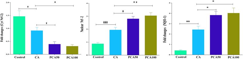
PCA prevents CA by anti-inflammatory pathway 133
Table 4: Wall thickness ratio
Control CA PCA-50 PCA-100
Wall 38.5 ± 1.26 73.33 ± 1.71*** 48.50 ± 4.0* 51.33 ± 2.81**
thickness
Values are expressed as mean ± SE (n = 16). Statistical significance is expressed as ***P < 0.001 CA compared to vehicle (control) and PCA-
treated groups; *P < 0.05, PCA-50 compared to CA rats; and **P < 0.01 PCA-100 compared to CA rats.
(P < 0.05) at 50 and 100 mg/kg, respectively (Table 7, Figure It’s been proved that the NF‑κB signaling was quite
2g and h). crucial in macrophage infiltration, and as a result, the
levels of downstream MMP-2 and MMP-9 in the walls
of aneurysm were measured. It was found that the
levels of MMP-2 and MMP-9 proteins show an increase
in the CA group and were reduced with PCA treatment
3.2 Effect of PCA on inflammatory cytokines, (Figure 4).
MMP-2, and MMP-9 analyzed by ELISA, Growing evidence suggests that in chronic inflam-
qRT-PCR, and western blot analysis mation, there are molecular pathways causing TNF-α
stimulation which further leads to nuclear translocation
The cytokine levels (Figure 3) in the group of rats induced of NF-κB. This is also demonstrated in our studies where
with CA were elevated in comparison to the control we found that NF-κB protein levels shown by western blot
group. The cytokines include IL-1β (P < 0.05), IL-2 (P < analysis (Figure 5) in aneurysmal walls of the CA were
0.05), IL-17 (P < 0.05), IL-8 (P < 0.01), IL-6 (P < 0.01), and decreased in the rats treated with PCA-50 and 100 mg/
TNF-α (P < 0.05). With the decline in the inflammatory kg. The data suggest that the TNF-α stimulated NF-κB
cytokines in groups pretreated with PCA, the CA growth pathway in macrophages was altered by PCA causing sup-
signaling was substantially (P < 0.05) reduced. pression of CA formation. Because of this, potentially,
Table 5: Effect of PCA-50 and 100 mg/kg administered to the experimental group of rats monitored for 16 weeks on body weight
Control CA PCA-50 PCA-100
0 week 162.33 ± 2.45 165.5 ± 1.76 157.5 ± 2.36 158.5 ± 1.86
4 weeks 167.6 ± 1.97 138.5 ± 1.46** 159.5 ± 1.37* 157.5 ± 1.91*
8 weeks 163.5 ± 1.76 136.5 ± 2.46*** 153.43 ± 1.97* 159.5 ± 2.76**
12 weeks 166.5 ± 1.36 135.5 ± 1.86*** 161.33 ± 2.99** 168.67 ± 1.71***
16 weeks 167.5 ± 2.76 137.7 ± 2.67*** 167.3 ± 1.10** 169.33 ± 2.06***
Values are expressed as mean ± SE (n = 16). Statistical significance is expressed as ***P < 0.001 CA compared to vehicle (control) and PCA-
treated groups; *P < 0.05, PCA-50 compared to CA rats; and **P < 0.01 PCA-100 compared to CA rats.
Table 6: Effect of PCA-50 and 100 mg/kg administered experimental group of rats monitored for 16 weeks on BMI
Control CA PCA-50 PCA-100
0 week 0.54 ± 0.005 0.55 ± 0.006 0.54 ± 0.003 0.54 ± 0.005
4 weeks 0.55 ± 0.007 0.50 ± 0.004** 0.54 ± 0.007** 0.54 ± 0.009**
8 weeks 0.54 ± 0.007 0.51 ± 2.46*** 0.53 ± 0.009** 0.54 ± 0.006***
12 weeks 0.54 ± 0.003 0.50 ± 1.86** 0.54 ± 0.008** 0.55 ± 0.001***
16 weeks 0.55 ± 0.006 0.51 ± 0.006*** 0.55 ± 0.002*** 0.55 ± 0.006***
Values are expressed as mean ± SE (n = 16). Statistical significance is expressed as ***P < 0.001 CA compared to vehicle (control) and PCA-
treated groups; *P < 0.05, PCA-50 compared to CA rats; and **P < 0.01 PCA-100 compared to CA rats.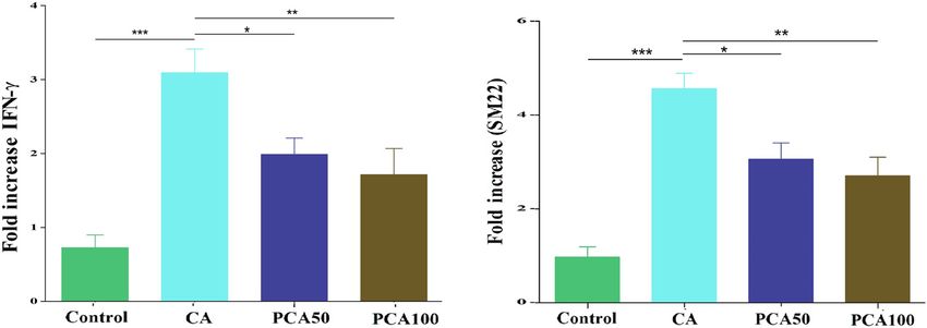
134 Gang Xiao et al.
the NF‑κB pathway is involved in PCA to inhibit the
progression of CA within macrophages (Figure 5).
To provide evidence of the protective role of PCA to
CA, mRNA levels of transgelin (SM22) and inflammatory
cytokines were analyzed with qPRC (Figures 3 and 6).
The group of rats induced by the CA had elevated levels
of cytokines TNF-α, IL-6, IFN-γ, and SM22. In contrast,
a significant decrease in cytokine levels was observed in
the PCA-treated groups (P < 0.01). Here, we validate the
PCA’s protective action by inhibiting cytokines, thus
proving it to be effective in controlling CA and therefore
can be considered as a novel treatment (Figure 6).
Nrf-2 pathway is involved in the mechanisms of
antioxidant defense system. For the investigation of
antioxidant enzymes such as NQO-1, qRT-PCR and wes-
tern blot analysis were used. Figures 7 and 8 indicate
that PCA increased the gene expression of antioxidant
enzymes (Figure 8d–f), triggering Nrf-2 to substantially
downregulate cytokine levels (P < 0.05) in both qRT-
PCR and western blot analysis. Furthermore, our find-
ings revealed that the activation of Nrf-2 with PCA in
cytoplasm and nucleus as shown in Figure 7 shows a
remarkable decrease in cytoplasmic Nrf-2 expression
(Figure 8a and b) and an increase in nuclear Nrf-2
expressions (Figure 8a and c) after treatment with
PCA, respectively (P < 0.05).
3.3 PCA causes downregulation and nuclear
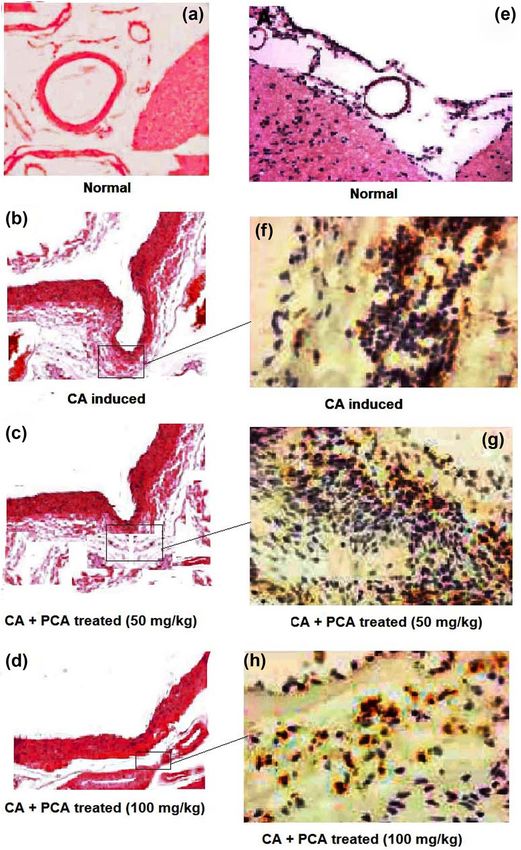
Figure 2: H&E staining of normal cerebral artery, CA induced by translocation of Nrf-2 with decreased
elastase, and CA treated with PCA-50 and 100 mg/kg (a–h). cellular ROS levels
Squares in f–h images are enlarged to show staining with anti-
CD68-positive cells revealing that a majority of leukocytes in intra-
cranial aneurysms were macrophages. 2G shows numerous leuko-
Nrf-2 expression was significantly decreased in the con-
cytes noticed in intracranial aneurysms indicating the distribution trol group stained with DAB when compared to PCA treat-
of macrophages was identical to that of leukocytes. Scale bar for a ment. There was a difference in expression between the
and e: 500 µm. Scale bar for b–d: 100 µm. Scale bar for f–h: 20 µm. cytoplasmic and nucleus cellular compartments in CA
(Figure 9a). In the CA group, the amount of Nrf-2 found
Table 7: Macrophage infiltration (number of cells) in control, induced, and PCA-50 and 100 mg/kg administered experimental group of rats
monitored for 16 weeks
Control CA PCA-50 PCA-100
0 week 2.45 ± 0.12 2.26 ± 0.07 2.24 ± 0.06 2.35 ± 0.11
4 weeks 3.85 ± 0.09 4.09 ± 0.13 4.05 ± 0.13 4.05 ± 0.07
8 weeks 3.39 ± 0.08 7.02 ± 0.25*** 5.57 ± 0.50* 5.37 ± 0.52**
12 weeks 4.78 ± 0.12 8.84 ± 0.19*** 6.29 ± 0.68* 5.69 ± 0.81**
16 weeks 5.85 ± 0.12 8.92 ± 0.2*** 6.93 ± 0.69* 6.41 ± 0.45**
Values are expressed as mean ± SE (n = 16). Statistical significance is expressed as ***P < 0.001 CA compared to vehicle (control) and PCA-
treated groups; *P < 0.05, PCA-50 compared to CA rats; and **P < 0.01 PCA-100 compared to CA rats.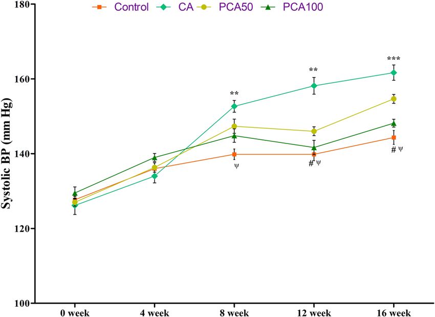
PCA prevents CA by anti-inflammatory pathway 135
Figure 3: Effect of PCA on inflammatory cytokines in control, CA-induced, and experimental group of rats administered PCA-50 and
100 mg/kg. Values are expressed as mean ± SE (n = 16). Statistical significance is expressed as ***P < 0.01 compared to the control group,
*P < 0.05 PCA-50 compared to CA rats, and **P < 0.01 PCA-100 compared to CA rats.
in the nucleus was greater than that of the group treated treatment with PCA. These findings confirm that activa-
with PCA. Figure 9b shows that the CA group showed an tion of Nrf-2 causes suppression of intracellular oxidative
increase in ROS levels visualized by an elevated fluores- stress.
cence intensity (three-fold) when compared to the control
group. This was substantially (P < 0.05) reduced by the
4 Discussion
This is the first study to investigate the effect of PCA on
CA in rats. CA is a chronic inflammatory condition caused
by excessive hemodynamic disturbance in arterial walls.
It may cause serious SAH, a severe type of stroke.
PCA possesses wide pharmacological activities such as
anti-inflammatory, antioxidant, antiapoptotic, and anti-
cancer [28–31]. Our findings demonstrated that PCA
suppresses the formation and progression of CA via
the inhibition of macrophage infiltration in rats when
upon elastase administration. This mechanism has yet
to be elucidated; however, several factors, namely
hemodynamic stress, inflammation, degeneration of
extracellular matrix, ROS generation, etc., contribute
to the formation of CA [45]. In the CA group, elastic
Figure 4: Effect of PCA on relative expression of MMP-2 and MMP-9 tissues with swollen aneurysmal dilation were absent.
in control, CA-induced, and experimental group of rats administered
Furthermore, there was an increase in aneurysm size
PCA-50 and 100 mg/kg. Values are expressed as mean ± SE (n = 16).
Statistical significance is expressed as ***P < 0.001 compared to (Table 2) demonstrating the connection between the
the control group, *P < 0.05 PCA-50 compared to CA rats, and **P < increase in size with the number of weeks after elastase
0.01 PCA-100 compared to CA rats. administration. Our results showed that PCA treatment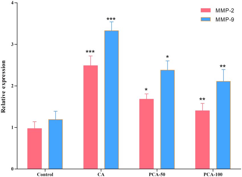
136 Gang Xiao et al. Figure 5: Effect of PCA on relative mRNA expression of NF-κB in control, CA-induced, and experimental group of rats administered PCA-50 and 100 mg/kg. Values are expressed as mean ± SE (n = 16). Statistical significance is expressed as ***P < 0.01 compared to the control group, **P < 0.01 PCA-50 compared to CA rats, and ***P < 0.001 PCA-100 compared to CA rats. Figure 6: Effect of PCA on inflammatory cytokines IFN-γ and SM22 in control, CA-induced, and experimental group of rats administered PCA- 50 and 100 mg/kg. Values are expressed as mean ± SE (n = 16). Statistical significance is expressed as ***P < 0.01 compared to the control group, *P < 0.05 PCA-50 compared to CA rats, and **P < 0.01 PCA-100 compared to CA rats. Figure 7: Effect of PCA on mRNA expressions of cytoplasm and nuclear fractions Nrf-2, antioxidant enzyme NQO-1 in control, CA-induced, and experimental group of rats administered PCA-50 and 100 mg/kg. Values are expressed as mean ± SE (n = 16). Statistical significance is expressed as ***P < 0.01 compared to control group, *P < 0.05 PCA-50 compared to CA rats, and **P < 0.01 PCA-100 compared to CA rats.
PCA prevents CA by anti-inflammatory pathway 137
Figure 8: Effect of PCA on (a) and (d) western blot and (b, c, e, and f) densitometry analysis of cytoplasm and nuclear fractions Nrf-2 and
antioxidant enzyme NQO-1 in control, CA-induced, and experimental group of rats administered PCA-50 and 100 mg/kg. Values are
expressed as mean ± SE (n = 16). Statistical significance is expressed as #P < 0.01 compared to the control group, *P < 0.05 PCA-50
and 100 mg/kg compared to CA rats.
was able to reduce the effects of elastase significantly of 16 weeks. PCA treatment reduced the high blood pres-
(P < 0.05) decreasing the aneurysm size, thereby showing sure restoring readings comparable to control rats. The
the role of inflammatory cytokines, IL-1β, IL-2, IL-17, IL-8, formation of CA was regulated by MMPs, namely MMP-2
IL-6 and TNF-α in the development of lesions in and MMP-9, which caused the deterioration of the endo-
CA-induced rats (Figure 3) over 16 weeks. thelium of cerebral arteries via degradation of elastic lamina
Further validation of aneurysm induction using elas- resulting in inflammation and hypertension. The over-
tase was obtained by measuring systolic blood pressure expression of MMP-2 and MMP-9 in CA-induced rats rein-
in all animal groups. Our findings noted high blood pres- forced the effect of CA induction with elastase treatment
sure because of the induction of aneurysms over a period relative to PCA groups. These overexpressions of MMPs
Figure 9: (a) Translocation of Nrf-2 from positive cells and number of positive cells analyzed (n = 4) showing H&E staining in CA-induced and
PCA (100 mg/kg)-treated groups. (b) Intracellular ROS in the control, CA-induced, and experimental group of rats administered PCA-50 and
100 mg/kg. Values are expressed as mean ± SE (n = 16). Statistical significance is expressed as ***P < 0.01 compared to the control group,
*P < 0.05 PCA-50 compared to CA rats, and **P < 0.01 PCA-100 compared to CA rats.138 Gang Xiao et al. Figure 10: Schematic representation of the study. that are involved in the progression of CA concur with of aneurysm [53–55]. The increase in pro-inflammatory results of other studies [46–48]. We investigated the role cytokines downregulated SM22, which in turn promoted that the pro-inflammatory cytokine, TNF-α, plays in the the destruction and formation of the cytoskeleton and development of intracranial aneurysm. The mRNA expres- accelerated the formation and rupture of an aneurysm sion of pro-inflammatory cytokines, TNF-α, IL-17 IL-6, and [56]. The treatment of PCA at 50 and 100 mg/kg showed IL-8 with activated infiltration of macrophages in response a significant reduction in the levels of pro-inflammatory to IFN- γ, [49] showed a significant increase in CA-induced cytokines in a dose-dependent manner. This demon- rats (Figure 6). The increase in IL-6 levels correlated strates that potentially TNF-α signaling mechanism is with cerebral damage via the rupture of aneurysm and involved in the development and progression of CA. thus the development of CA [50]. Increased IL-1β levels In addition, PCA was shown to inhibit NF-κB expres- in CA-induced rats demonstrated the deterioration of extra- sion, subsequently exerting anti-inflammatory action on cellular matrix, resulting in damage of the vascular endo- aneurysms. Our results show an increase in systolic pres- thelium [51]. The IL-1β levels were suppressed with PCA sure denoting induction of hemodynamic stress in animal treatments and inhibited the development of an aneurysm, models within the intracranial arteries, thereby triggering thus reducing the progression of vascular remodeling inflammation with progression of CA, similar to the patho- [52]. The recruitment of macrophage infiltration was genesis of CA in humans [57]. demonstrated by an increase in IL-17 and IL-8 levels Although several studies reported the role of NF-κB leading to the production of cytokines. These markers as a key transcription factor in the regulation of the expres- of inflammation result in the formation and development sion of MMPs, production and release of pro-inflammatory
PCA prevents CA by anti-inflammatory pathway 139
cytokines, and macrophage infiltration in advancing the NF-κB, and Nrf-2 activation pathways. The underlying
progression of CA [58–60], the effect of PCA on the CA mechanism involves a decreased expression of pro-inflam-
progression has not been reported. The treatment with matory cytokines, upregulation of antioxidant enzyme acti-
PCA at the two different doses shows suppression in vities, and reduced intracellular ROS formation. We propose
aneurysmal wall degeneration, prevention in macrophage that PCA can be an alternative and novel therapeutic for the
infiltration, and inflammatory cytokines. This potentially treatment of CA by inhibiting TNF-α, NF-κB, and Nrf-2 acti-
involves the role of NF-κB and MCP‑1 signaling pathways vation pathways.
and could be a therapeutic target in the treatment of CA.
In addition, this study noticed significant reductions in Funding: The authors state no funding involved.
the expressions of MMP-2 and MMP-9 in PCA-treated
groups. While CA pathogenesis involving MMPs is debated, Conflict of interest: The authors state no conflict of
our findings concur with a previous study, showing an interest.
increased expression of MMP-2 and MMP-9 in human serum
and aneurysmal walls [61]. Data availability statement: The datasets generated during
Inflammation and oxidative stress are possibly involved and/or analyzed during the current study are available from
in causing endothelial dysfunction. The transcription factor, the corresponding author on reasonable request.
Nrf-2, is involved in the modulation of cellular oxidative
processes via antioxidant and detoxifying genes. Under oxi-
dative stress, Nrf-2 translocates from the cytoplasm to the
nucleus. Thus, we hypothesized that the potential role of References
Nrf-2 activation is to inhibit the oxidative stress and offer
protective effects against CA formation and progression. [1] Monique HV, Ale A, Raya B, Gabriël JR. Prevalence of unrup-
Our results showed reduced aneurysmal scores and pro- tured intracranial aneurysms, with emphasis on sex, age,
gression of CA without rupturing after treatment with comorbidity, country, and time period: a systematic review
PCA at 50 and 100 mg/kg, respectively, thus indicating and meta-analysis. Lancet Neurol. 2011;10(7):626–36.
[2] Etminan N, Rinkel G. Unruptured intracranial aneurysms:
the role of Nrf-2 pathway activation in the suppression
development, rupture and preventive management. Nat Rev
of CA progression. This is consistent with another study Neurol. 2016;12:699–713.
that reported elevated Nrf-2 levels in the inhibition of [3] Eric PBD. Headache, cerebral aneurysms, and the use of trip-
acute aortic dissection formation. Our immunohistochem- tans and ergot derivatives. Headache. 2015;55(5):739–47.
ical findings showed the downregulation and Nrf-2 nuclear [4] Schievink WI. Intracranial aneurysms. N Engl J Med.
1997;336(1):28–40.
translocation in aneurysm walls (Figure 9).
[5] Anderson CS, Feigin V, Bennett D, Lin RB, Hankey G,
We show that PCA decreases the inflammatory responses
Jamrozik K. Active and passive smoking and the risk of sub-
in the formation of CA by inhibiting the NF‑κB pathway and arachnoid hemorrhage. Stroke. 2004;35:633–7.
macrophage infiltration. NF‑κB pathway inhibition can be a [6] Francesco S, Sapir S, Loreto G, Sophie C, Félix T, Cyril PM, et al.
potential target for the prevention and treatment of CA. We Hemodynamic stress, inflammation, and intracranial
also confirmed that Nrf-2 signaling inhibits CA formation aneurysm development and rupture: a systematic review.
World Neurosurg. 2018;115:234–44.
and development, via acting on Nrf-2 translocation into
[7] Hashimoto T, Meng H, Young WL. Intracranial aneurysms: links
the nucleus. TNF-α signaling initiates the procedure in the among inflammation, hemodynamics and vascular remo-
CA-induced group of rats by development and maturation deling. Neurol Res. 2006;28(4):372–80.
of the CA. We found that with the treatment of PCA, it [8] Chalouhi N, Hoh BL, Hasan D. Review of cerebral aneurysm
is possible to control aneurysms as well as inhibit the formation, growth and rupture. Stroke. 2013;44(12):
3613–22.
aneurysm rupture in the brain (Figure 10).
[9] Liu P, Song Y, Zhou Y, Liu Y, Qiu T, An Q, et al. Cyclic
mechanical stretch induced smooth muscle cell changes in
cerebral aneurysm progress by reducing collagen type IV and
collagen type VI levels. Cell Physiol Biochem.
5 Conclusion 2018;45(3):1051–60.
[10] Pera J, Korostynski M, Krzyszkowski T, Czopek J, Slowik A,
Dziedzic T, et al. Gene expression profiles in human ruptured
This study suggests that formation and progression of
and unruptured intracranial aneurysms: what is the role of
CA induced by inflammation and oxidative stress can inflammation? Stroke. 2010;41:224–31.
be inhibited with the treatment of PCA in a dose-depen- [11] Hosaka K, Hoh BL. Inflammation and cerebral aneurysms.
dent manner. This involves the regulatory roles of TNF-α, Transl Stroke Res. 2014;5(2):190–8.140 Gang Xiao et al.
[12] Tuttolomondo A, Riccardo DS, Domenico DR, Pedone C, accumulation in diabetic nephropathy. Biomed Pharmacother.
Placa SL, Pinto A, et al. Effects of clinical and laboratory vari- 2018;98:18–22.
ables and of pretreatment with cardiovascular drugs in acute [28] Jang SA, Song HS, Kwon JE, Baek HJ, Koo HJ, Sohn EH, et al.
ischaemic stroke: a retrospective chart review from the GIFA Protocatechuic acid attenuates trabecular bone loss in ovar-
study. Int J Cardiol. 2011;151(3):318–22. iectomized mice. Oxid Med Cell Longev. 2018;2018:7280342.
[13] Raimondo DD, Tuttolomondo A, Buttà C, Miceli S, Licata G, doi: 10.1155/2018/7280342.
Pinto A. Effects of ace-inhibitors and angiotensin receptor [29] Molehin OR, Adeyanju AA, Adefegha SA, Akomolafe SF.
blockers on inflammation. Curr Pharm Des. Protocatechuic acid mitigates adriamycin-induced reproduc-
2012;18(28):4385–413. tive toxicities and hepatocellular damage in rats. Comp Clin
[14] Licata G, Tuttolomondo A, Corrao S, Raimondo DD, Pathol. 2018;27:1681–9.
Fernandez P, Caruso C, et al. Immunoinflammatory activation [30] Jang SE, Choi JR, Han MJ, Kim DH. The preventive and curative
during the acute phase of lacunar and non-lacunar ischemic effect of cyanidin-3β-D-glycoside and its metabolite proto-
stroke: association with time of onset and diabetic state. Int J catechuic acid against TNBS-induced colitis in mice. Nat Prod
Immunopathol Pharmacol. 2006;19(3):639–46. Sci. 2016;22(4):282–6.
[15] Starke RM, Chalouhi N, Ding D, Daniel MSR, Mckisic MS, [31] Safaeiana L, Emamia R, Hajhashemia V, Haghighatian Z.
Gary KO, et al. Vascular smooth muscle cells in cerebral Antihypertensive and antioxidant effects of protocatechuic
aneurysm pathogenesis. Transl Stroke Res. acid in deoxycorticosterone acetate-salt hypertensive rats.
2014;5(3):338–46. Biomed Pharmacother. 2018;100:147–55.
[16] Starke RM, Chalouhi N, Ali MS, Pascal MJ, Stavropoula IT, [32] Lende AB, Kshirsagar AD, Deshpande AD, Muley MM, Patil RR,
Fernando GL, et al. The role of oxidative stress in cerebral Bafna PA, et al. Anti-inflammatory and analgesic activity of
aneurysm formation and rupture. Curr Neurovasc Res. protocatechuic acid in rats and mice. Inflammopharmacology.
2013;10(3):247–55. 2011;19(5):255–63.
[17] Keleku-Lukwete N, Suzuki M, Yamamoto M. An overview of the [33] Liu YD, Sun X, Zhang Y, Wu HJ, Wang H, Yang R, et al.
advantages of KEAP1-NRF2 system activation during inflam- Protocatechuic acid inhibits TGF-β1-induced proliferation and
matory disease treatment. Antioxid Redox Signal. migration of human airway smooth muscle cells. J Pharmacol
2018;29(17):1746–55. Sci. 2019;139(1):9–14. doi: 10.1016/j.jphs.2018.10.011.
[18] Aoki T, Kataoka H, Shimamura M, Nakagami H, Wakayama K, [34] Owumi SE, Ajijola IJ, Agbeti. OM. Hepatoremal protective
Moriwaki T, et al. nF-kappaB is a key mediator of cerebral effects of Protocatechuic acid In rats administered with anti-
aneurysm formation. Circulation. 2007;116(24):2830–40. cancer drug methotrexate. Hum Exp Toxicol.
[19] Moriwaki T, Takagi Y, Sadamasa N, Aoki T, Nozaki K, 2019;38(11):1254–65.
Hashimoto N. Impaired progression of cerebral aneurysms in [35] Varì R, Scazzocchio B, Santangelo C, Filesi C, Galvano F,
interleukin-1β deficient mice. Stroke. 2006;37(3):900–5. D’Archivio M, et al. Protocatechuic acid prevents oxLDL-
[20] Aoki T, Kataoka H, Morimoto M, Nozaki K, Hashimoto N. induced apoptosis by activating JNK/Nrf2 survival signals in
Macrophage-derived matrix metalloproteinase-2 and -9 macrophages. Oxid Med Cell Longev. 2015;2015:351827.
promote the progression of cerebral aneurysms in rats. [36] Varì R, D’Archivio M, Filesi C, Carotenuto S, Scazzocchio B,
Stroke. 2007;38(1):162–9. Santangelo C, et al. Protocatechuic acid induces antioxidant/
[21] Wanderer S, Waltenspuel C, Gruter BE, Remonda L, Fandino J, detoxifying enzyme expression through JNK-mediated Nrf2
Marbacher S, et al. Arterial pouch microsurgical bifurcation activation in murine macrophages. J Nutr Biochem.
aneurysm model in the rabbit. J Vis Exp. 2020;159:32478731. 2011;22(5):409–17.
doi: 10.3791/61157 [37] Wang HY, Wang H, Wang JH, Wang Q, Ma QF, Chen YY.
[22] de Oliveira IA. Main models of experimental saccular aneurysm in Protocatechuic acid inhibits inflammatory responses in LPS-
animals. Neurosurgery. 2012;9(7):10264–8. doi: 10.5772/50310. stimulated BV2 Microglia via NF-kappaB and MAPKs signaling
[23] Rowinska Z, Gorressen S, Merx MW, Koeppel TA, Liehn EA, pathways. Neurochem Res. 2015;40:1655–60.
Zernecke A. Establishment of a new murine elastase-induced [38] Jangho L, Su JH, Hye JL, Min JK, Jin HK, Yun TK, et al. Protective
aneurysm model combined with transplantation. PLoS One. effect of Tremella fuciformis berk extract on LPS-induced acute
2014;9:7. inflammation via inhibition of the NF-κB and MAPK pathways.
[24] Jayaraman T, Paget A, Shin YS, Li X, Mayer J, Chaudhry HW, Food Funct. 2016;7(7):3263–72.
et al. TNF-alpha-mediated inflammation in cerebral aneurysms: [39] Hui-Hsuan L, Jing-Hsien C, Fen-Pi C, Chau-Jong W.
a potential link to growth and rupture. Vasc Health Risk Manag. Protocatechuic acid inhibits cancer cell metastasis involving
2008;4(4):805–17. the down-regulation of Ras/Akt/NF-kB pathway and MMP-2
[25] Liu Z, Ajimu K, Yalikun N, Zheng Y, Xu F. Potential therapeutic production by targeting RhoB activation. Br J Pharmacol.
strategies for intracranial aneurysms targeting aneurysm 2011;162(1):237–54.
pathogenesis. Front Neurosci. 2019;13:1238. doi: 10.3389/ [40] Chen W, Wang D, Wang LS, Bei D, Wang J, See WA, et al.
fnins.2019.01238. Pharmacokinetics of protocatechuic acid in mouse and its
[26] Olmos G, Lladó J. Tumor necrosis factor alpha: a link between quantification in human plasma using LC‐tandem mass spec-
neuroinflammation and excitotoxicity. Mediators Inflamm. trometry. J Chromatogr B. 2012;908:39–44. doi: 10.1016/
2014;2014:861231. doi: 10.1155/2014/861231. j.jchromb.2012.09.032.
[27] Ma Y, Chen F, Yang S, Chen B, Shi J. Protocatechuic acid [41] Safaeian L, Hajhashemi V, Javanmard SH, Naderi HS. The effect
ameliorates high glucose-induced extracellular matrix of protocatechuic acid on blood pressure and oxidative stressPCA prevents CA by anti-inflammatory pathway 141
in glucocorticoid-induced hypertension in rat. Iran J Pharm of cerebral aneurysm: contribution of interleukin-1beta and
Res. 2016;15(Special issue):83–91. nuclear factor-kappaB. Arterioscler Thromb Vasc Biol.
[42] Bhattacharjee N, Dua TK, Khanra R, Joardar S, Nandy A, Saha A, 2009;29(7):1080–6.
et al. Protocatechuic acid, a phenolic from Sansevieria rox- [52] Moriwaki T, Takagi Y, Sadamasa N, Aoki T, Nozaki K,
burghiana leaves, suppresses diabetic cardiomyopathy via Hashimoto N. Impaired progression of cerebral aneurysms in
stimulating glucose metabolism, ameliorating oxidative interleukin-1beta-deficient mice. Stroke. 2006;37(3):900–5.
stress, and inhibiting inflammation. Front Pharmacol. [53] Toth G, Cerejo R. Intracranial aneurysms: review of current
2017;8:251. doi: 10.3389/fphar.2017.00251. science and management. Vasc Med. 2018;23(3):276–88.
[43] Nuki Y, Tsou TL, Kurihara C, Kanematsu M, Kanematsu Y, [54] Cummings TJ, Johnson RR, Diaz FG, Michael DB. The relation-
Hashimoto T. Elastase-induced intracranial aneurysms in ship of blunt head trauma, subarachnoid hemorrhage, and
hypertensive mice. Hypertension. 2009;54(6):1337–44. rupture of pre-existing intracranial saccular aneurysms.
[44] Allen Linda A, Schmidt James R, Thompson Christopher T, Neurol Res. 2000;22(2):165–70.
Carlson Brian E, Beard Daniel A, Lombard Julian H. High salt [55] Chalouhi N, Points L, Pierce GL, Ballas Z, Jabbour P, Hasan D.
diet impairs cerebral blood flow regulation via salt-induced Localized increase of chemokines in the lumen of human
angiotensin-II suppression. Microcirculation. cerebral aneurysms. Stroke. 2013;44(9):2594–7.
2019;26(3):e12518. [56] Shen J, Yang M, Ju D, Jiang H, Zheng JP, Xu Z, et al. Disruption
[45] Weir B. Unruptured aneurysms. J Neurosurg. of SM22 promotes inflammation after artery injury via
2002;97(5):1011–3. nuclear factor kappaB activation. Circ Res.
[46] Seo JH, Guo S, Lok J, Navaratna D, Whalen MJ, Kim KW, et al. 2010;106(8):1351–62.
Neurovascular matrix metalloproteinases and the blood-brain [57] Jou LD, lee DH, Morsi H, Mawad ME. Wall shear stress on
barrier. Curr Pharm Des. 2012;18(25):3645–8. ruptured and unruptured intracranial aneurysms at the
[47] Zhang X, Ares WJ, Taussky P, Ducruet AF, Grandhi R. Role of internal carotid artery. Am J Neuroradiol. 2008;29(9):1761–7.
matric metalloproteinases in the pathogenesis of intracranial [58] Chalouhi N, Ali MS, Jabbour PM, Tjoumakaris SI, Gonzalez IF,
aneurysms. Neurosurg Focus. 2019;47:E4. doi: 10.3171/ Rosenwasser RH, et al. Biology of intracranial aneurysms:
2019.4. role of inflammation. J Cereb Blood Flow Metab.
[48] Pannu H, Kim DH, Guo D, King TM, Van Ginhoven G, Chin T, 2012;32(9):1659–76.
et al. The role of MMP-2 and MMP-9 polymorphisms in [59] Starke RM, Raper DM, Ding D, Chalouhi N, Owens GK, Hasan DM.
sporadic intracranial aneurysms. J Neurosurg. Tumor necrosis factor-α modulates cerebral aneurysm formation
2006;105(3):418–23. and rupture. Transl Stroke Res. 2014;5(2):269–77.
[49] Mantovani A, Sica A, Locati M. Macrophage polarization comes [60] Starke RM, Chalouhi N, Ali MS, Jabbour PM, Tjoumakaris SI,
of age. Immunity. 2005;23(4):344–6. Gonzalez LF, et al. The role of oxidative stress in cerebral
[50] Morgan L, Cooper J, Montgomery H, Kitchen N, Humphries SE. aneurysm formation and rupture. Curr Neurovasc Res.
The interleukin-6 gene -174G [{GT}]C and -572G[{GT}]C pro- 2013;10(3):247–55.
moter polymorphisms are related to cerebral aneurysms. [61] Jin D, Sheng J, Yang X, Gao B. Matrix metalloproteinases and
J Neurol Neurosurg Psychiatry. 2006;77(8):915–7. tissue inhibitors of metalloproteinases expression in human
[51] Aoki T, Kataoka H, Ishibashi R, Nozaki K, Morishita R, cerebral ruptured and unruptured aneurysm. Surg Neurol.
Hashimoto N. Reduced collagen biosynthesis is the hallmark 2007;68(Suppl 2):S11–6.You can also read