U2AF homology motifs: protein recognition in the RRM world
←
→
Page content transcription
If your browser does not render page correctly, please read the page content below
REVIEW
U2AF homology motifs: protein
recognition in the RRM world
Clara L. Kielkopf,1,3 Stephan Lücke,2 and Michael R. Green2,4
1
Department of Biochemistry and Molecular Biology, Johns Hopkins University Bloomberg School of Public Health,
Baltimore, Maryland 21205, USA; 2Howard Hughes Medical Institute, Programs in Gene Function and Expression and
Molecular Medicine, University of Massachusetts Medical School, Worcester, Massachusetts 01605, USA
Recent structures of the heterodimeric splicing factor U2 sistent with this idea, recent structures of the heterodi-
snRNP auxiliary factor (U2AF) have revealed two unex- meric splicing factor U2 snRNP auxiliary factor (U2AF)
pected examples of RNA recognition motif (RRM)-like have revealed two unexpected examples of RRM-like do-
domains with specialized features for protein recogni- mains with specialized features for protein recognition
tion. These unusual RRMs, called U2AF homology mo- (Kielkopf et al. 2001; Selenko et al. 2003). In light of this
tifs (UHMs), represent a novel class of protein recogni- structural information, we call these unusual RRMs
tion motifs. Defining a set of rules to distinguish tradi- U2AF homology motifs (UHMs) to reflect their distinct
tional RRMs from UHMs is key to identifying novel role in protein recognition. Here, the critical sequence
UHM family members. Here we review the critical se- features necessary to mediate protein–UHM interactions
quence features necessary to mediate protein–UHM in- are reviewed and formulated in a manner that has per-
teractions, and perform comprehensive database mitted a comprehensive database search designed to
searches to identify new members of the UHM family. identify members of the UHM family. The resulting im-
The resulting implications for the functional and evolu- plications for the functional and evolutionary relation-
tionary relationships among candidate UHM family ships among candidate members of the UHM family are
members are discussed. discussed. This review represents a first step toward dis-
tinguishing canonical RRMs from UHMs, and thereby
contributes toward a major goal of the postgenomic era
The processes of RNA splicing, transport, capping, edit- (Thornton et al. 2000): to convert genomic sequences
ing, and polyadenylation are heavily dependent on pro- into testable functional hypotheses.
tein factors that recognize the pre-mRNA and assemble
the appropriate pre-mRNA processing complexes. Sur-
prisingly, the many different protein factors that guide Structural features of RNA recognition
pre-mRNA modification pathways are composed of a by canonical RRMs
limited number of conserved, modular RNA-binding do- The RNA-binding function of the canonical RRM do-
mains (Burd and Dreyfuss 1994). Of these, the RNA rec- main has been extensively investigated over the last two
ognition motif (RRM) domain is by far the most abun- decades. The most conserved RRM signature sequence is
dant type of eukaryotic RNA-binding motif. In addition an eight-residue motif called ribonucleoprotein 1 (RNP1;
to associations between protein and RNA, protein–pro- Adam et al. 1986; Sachs et al. 1986), which has the con-
tein interactions are essential to recruit catalytic com- sensus [RK]-G-[FY]-[GA]-[FY]-[ILV]-X-[FY] (where X is
ponents to sites of RNA modification and to coordinate any amino acid). A second six-residue region of homol-
pre-mRNA processing with other cellular pathways. In- ogy, called RNP2, is typically located ∼30 residues N-
terestingly, traditional protein interaction domains, terminal to RNP1 (Lahiri and Thomas 1986; Dreyfuss et
such as SH2, SH3, and WW motifs, are rarely observed in al. 1988), and has the consensus [ILV]-[FY]-[ILV]-X-N-L.
pre-mRNA processing factors (e.g., see Shatkin and Man- Additional conserved amino acids define an ∼80-residue
ley 2000; Zhou et al. 2002), implying that the ability to domain that encompasses the RNA-binding function
interact with other proteins may reside in the sequences (Query et al. 1989; Scherly et al. 1989; Birney et al. 1993).
previously thought to be involved in RNA binding. Con- The three-dimensional structure of the canonical
RRM domain was first determined for the RRM of U1A
(Nagai et al. 1990; Hoffman et al. 1991). The RRM fold is
[Keywords: U2AF; RNA recognition motif; protein–protein interaction;
RNA-binding domain; PUMP; splicing factor]
composed of two ␣-helices packed against four antipar-
Corresponding authors. allel -strands with topology ␣␣, which form an ␣/
3
E-MAIL ckielkop@jhsph.edu; FAX (410) 955-2926. sandwich (Fig. 1). The RNP consensus motifs form two
4
E-MAIL michael.green@umassmed.edu; FAX (508) 856-5473.
Article and publication are at http://www.genesdev.org/cgi/doi/10.1101/ central -strands, with RNP1 in 3 and RNP2 in 1.
gad.1206204. Because of the alternating side-chain conformations of
GENES & DEVELOPMENT 18:1513–1526 © 2004 by Cold Spring Harbor Laboratory Press ISSN 0890-9369/04; www.genesdev.org 1513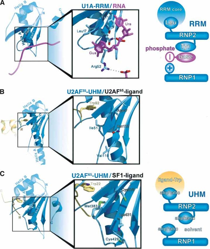
Kielkopf et al.
U2AF was identified as a factor that binds to pre-mRNA
consensus sequences at the 3⬘ splice site (3⬘SS), and is
required for stable association of the U2 snRNP core spli-
ceosome particle with the pre-mRNA branch point se-
quence (BPS) during the first ATP-dependent step of the
splicing process (Complex A; Ruskin et al. 1988; Zamore
and Green 1989). The importance of U2AF in vitro was
soon corroborated by the discovery that both subunits
are essential in Drosophila melanogaster (Kanaar et al.
1993; Rudner et al. 1996, 1998b) and Caenorhabditis el-
egans (Zorio and Blumenthal 1999b). Moreover, U2AF65
is an essential protein in Schizosaccharomyces pombe
(Potashkin et al. 1993), and U2AF35 is necessary for ver-
Figure 1. Representative canonical RRM fold, from the struc- tebrate development (Golling et al. 2002). Because U2AF
ture of the U1A/RNA complex (PDB code 1URN). The N- and commits the pre-mRNA to the first critical ATP-depen-
C-terminal ends of the RRM are indicated. The position and dent step of splicing, its binding is often regulated during
orientation of the RNA are represented with a ribbon diagram. alternative splicing (Smith and Valcarcel 2000). In hu-
mans, the products of five U2AF35-like open reading
frames and the single U2AF65 subunit may form distinct
the pleated -sheet, some of the consensus residues heterodimers with different functional activities (Tupler
maintain the core fold, whereas others are displayed on et al. 2001; Shepard et al. 2002). In addition to U2AF,
the surface for nucleic acid recognition. Structures of other non-snRNP protein factors are required for forma-
single RRMs complexed with RNA have been deter- tion of Complex A, including Splicing Factor 1 (SF1) and
mined for the U1A-RRM bound to a hairpin loop of U1 Splicing Factor 3b (SF3b), a multisubunit component of
snRNA (Oubridge et al. 1994; Price et al. 1998; Deo et al. the U2 snRNP (Kramer and Utans 1991).
1999; Handa et al. 1999; Allain et al. 2000; Wang and To perform its role in RNA splicing, two central ca-
Tanaka Hall 2001), and for a ternary complex of the U2 nonical RRM domains of U2AF65 recognize the poly-
snRNP proteins U2B⬙-RRM/U2A⬘ with a U2 snRNA pyrimidine tract (Py-tract) in the pre-mRNA (Fig. 2).
hairpin loop (Price et al. 1998). In contrast to the isolated Binding of U2AF65 to the Py-tract is strengthened by
RRM of U1A, in most cases multiple RRMs are observed cooperative protein–protein interactions with SF1 at the
within a single polypeptide, with an average of two upstream BPS (Berglund et al. 1998; Rain et al. 1998) and
RRMs per protein (Letunic et al. 2004). The structures of with U2AF35, which contacts the downstream 3⬘SS con-
several proteins composed of two tandem RRMs com-
plexed with single-stranded RNA oligonucleotides have
been determined, including the alternative splicing fac-
tor Sxl (Handa et al. 1999), PAB (Deo et al. 1999), pre-
rRNA packaging protein nucleolin (Allain et al. 2000),
and translation regulatory protein HuD (Wang and
Tanaka Hall 2001). A comparison of these six different
structures has revealed some common themes, as well as
differences, in the mode of canonical RRM/RNA recog-
nition. When all the structures are superimposed, struc-
tural equivalent hydrogen-bonds or stacking interactions
are observed between single-stranded RNA and residues
in the RNP1 and RNP2 motifs. A variety of sequences
and RNA conformations are recognized by a variety of
complementary hydrogen bonds with specific bases and
differing arrangements of single or multiple RRMs.
Figure 2. Diagram of protein–protein interactions mediated by
U2AF, the UHM prototype the U2AF heterodimer during the initial stages of pre-mRNA
During pre-mRNA splicing, U2AF and other essential splicing. The U2AF heterodimer (mediated by the U2AF35-
UHM/U2AF65-ligand interaction) binding to the poly-pyrimi-
factors facilitate sequential association of small nuclear
dine tract (Py-tract) and 3⬘-splice site (3⬘SS) is facilitated by co-
RNP particles (snRNPs), including U1, U2, U4, U5, and
operative interactions between the U2AF65-UHM and SF1 at
U6 snRNPs, with the borders of intervening pre-mRNA the branchpoint sequence (BPS). Subsequently, SF1 interactions
sequences (for review, see Brow 2002). Following assem- with the U2AF65-UHM are replaced by SAP155. The N- and
bly of the functional spliceosome, the intron is excised C-terminal ends of the proteins are indicated; the conserved
as a branched lariat by two catalytic steps, and adjacent ligand Trp residue (W) is also shown (discussed further in the
exons are joined together to form the spliced mRNA. text).
1514 GENES & DEVELOPMENT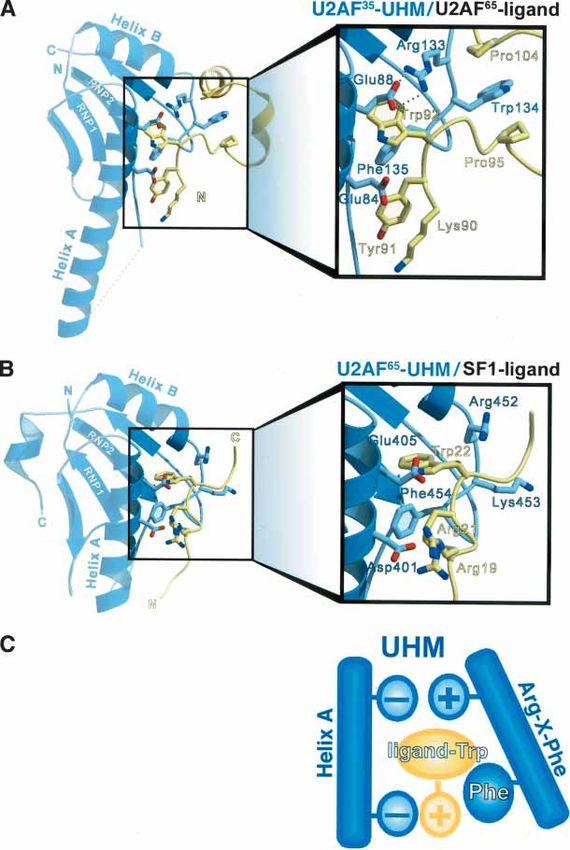
Protein recognition by U2AF homology motifs
sensus (Merendino et al. 1999; Wu et al. 1999; Zorio and et al. 2001; Selenko et al. 2003). In addition to aliphatic
Blumenthal 1999a). The C-terminal UHM domain of residues, a conserved Arg–X–Phe motif (where X is any
U2AF65 interacts with the N-terminal domain of SF1 amino acid; see below) on the loop connecting the last
(U2AF65-UHM/SF1-ligand; Rain et al. 1998). At the op- ␣-helix (Helix B) and -strand of the UHM fold contrib-
posite end of the large subunit, the N-terminal domain of utes to the Trp-binding pocket. The Arg residue in the
U2AF65 provides a ligand that interacts with the central loop (U2AF35-Arg 133 or U2AF65-Arg 452) forms an in-
UHM domain of U2AF35 (U2AF35-UHM/U2AF65-ligand; tramolecular salt bridge with the last Glu residue of He-
Zhang et al. 1992; Rudner et al. 1998b). Subsequently, lix A (U2AF35-Glu 88 or U2AF65-Glu 405) that shields
entry of the U2 snRNP displaces SF1 by interacting with one face of the ligand-Trp, whereas the Phe residue
the BPS via the U2 snRNA (Nelson and Green 1989; Wu (U2AF35-Phe 135 or U2AF65-Phe 454) encloses the oppo-
and Manley 1989; Zhuang and Weiner 1989; Query et al. site Trp face. In addition to the extensive interface with
1994), and with the U2AF65 C-terminal domain via the the ligand-Trp, a series of acidic residues in Helix A of
SF3b subunit, SAP155 (Gozani et al. 1998; Habara et al. the UHM interacts with basic residues at the N terminus
1998). Once the U2 snRNP has contacted the pre- of the protein ligand. Specifically, electrostatic interac-
mRNA, U2AF is dissociated by conformational rear- tions between U2AF35-Glu 84 and U2AF65-Lys 90 as
rangements of the spliceosome components (Bennett et well as U2AF65-Asp 401 and SF1-Arg 21 are observed at
al. 1992; Chiara et al. 1997). In summary, key protein– similar positions for both structures. The essential na-
protein interactions are mediated by the U2AF65-UHM, ture of acidic residues within Helix A, Phe 454, and the
which interacts with SF1 and subsequently SAP155, and Trp-binding pocket was confirmed for the U2AF65-
by the U2AF35-UHM, which interacts with the U2AF65 UHM/SF1-ligand complex by site-directed mutagenesis
N terminus. of the U2AF65-UHM or SF1-ligand followed by pull-
down assays (Selenko et al. 2003). Likewise, the U2AF65-
ligand-Trp 92 was found to contribute two orders of mag-
Structural features of protein–protein interactions
nitude to the affinity of the U2AF35-UHM/U2AF65-li-
by UHMs
gand complex by isothermal titration calorimetry.
Based on primary sequence analysis, both the U2AF65 In the U2AF35-UHM, a distinctive Trp residue
C-terminal domain and the central domain of U2AF35 (U2AF35-Trp 134) is observed at the X position of the
were suspected to contain unusual variations of the Arg–X–Phe motif on the last loop of the UHM domain.
RRM fold (Birney et al. 1993). However, the borders of Bulky aromatic residues such as Trp at this solvent-ex-
the U2AF-UHM domains could not be assigned accu- posed position are especially rare among canonical RRM
rately because of sequence insertions in the first helix of domains (1% of 676 annotated RRM domains in the
the fold (Helix A) and the absence of aromatic amino SWISS-PROT database). The most frequently observed
acids in the RNP-like motifs that are normally critical residues at the corresponding position of canonical
for RNA recognition. The independent determination of RRMs are highly charged, including Glu (16%) and Lys
the X-ray structure of the U2AF35-UHM/U2AF65-ligand (15%), as is also observed for the U2AF65-UHM (Arg–
complex (Kielkopf et al. 2001) and NMR structure of the Lys–Phe). The unusual U2AF35-Trp 134 inserts between
U2AF65-UHM/SF1-ligand complex (Selenko et al. 2003) a series of unique Pro residues at the C terminus of the
confirmed that both the C-terminal U2AF65 and central U2AF65-ligand, which are completely absent from the
U2AF35 protein interaction domains adopt the ␣␣ SF1-ligand of the U2AF65-UHM. The additional Trp/Pro
RRM-fold topology. Within the RRM-like fold, the se- interaction significantly contributes to the high affinity
quence insertions separating the RNP-like motifs in- of the U2AF heterodimer (1.7 nM Kd; Kielkopf et al.
crease the length of Helix A from three turns observed 2001). Because the U2AF65-UHM/SF1-ligand complex
among canonical RRMs to five or eight turns for U2AF65 lacks the corresponding Trp/Pro interaction, the affinity
and U2AF35, respectively; the functional role of these is relatively weak (∼100 nM Kd; Selenko et al. 2003). The
sequence insertions, if any, is unclear. The parallel use of sequence differences in the ligands recognized by the
an RRM-like fold to recognize similar peptide ligands U2AF35-UHM and U2AF65-UHM domains reflect the
implies that the U2AF35-UHM and U2AF65-UHM do- different functional roles of the complexes, which, re-
mains represent a new type of protein–protein interac- spectively, maintain the constitutive U2AF35-UHM/
tion motif, hitherto undetected amid the many canoni- U2AF65 heterodimer (Zhang et al. 1992; Rudner et al.
cal RRMs of pre-mRNA processing factors. 1998b) or form a transient U2AF65/SF1 intermediate dur-
Three-dimensional structural information revealed ing spliceosome assembly (Rutz and Seraphin 1999).
unanticipated sequence features of U2AF35-UHM and The structures of the U2AF35-UHM/U2AF65-ligand
U2AF65-UHM domains that enable interaction with and U2AF65-UHM/SF1-ligand complexes revealed sev-
short protein ligands. Despite low primary sequence eral sequence features that distinguish UHMs from ca-
identity (23%), ligand recognition by the different UHM nonical RRM domains. One striking feature of UHM do-
domains is very similar (Fig. 3). In both the U2AF35- mains is their atypical RNP-like motifs. The first residue
UHM/U2AF65-ligand and U2AF65-UHM/SF1-ligand of the RNP1-like motif and the second residue of the
structures, a critical Trp residue in the ligand sequence RNP2-like motif are unusual in that they are exposed on
inserts into a tight hydrophobic pocket between the the -sheet surface rather than directly involved in RNA
␣-helices and the RNP1- and RNP2-like motifs (Kielkopf binding. Residues in these positions consist of aliphatic
GENES & DEVELOPMENT 1515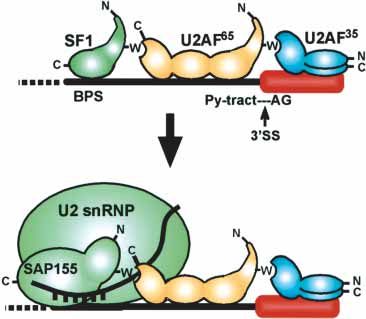
Kielkopf et al.

Figure 3. Structures of U2AF-UHM/ligand
complexes. The UHM is shown in blue, the
protein ligand is shown in yellow. Key inter-
acting side chains are drawn in ball-and-stick
representation. (A) The U2AF35-UHM bound
to the U2AF65-ligand. (B) The U2AF65-UHM
bound to the SF1-ligand. (C) Schematic repre-
sentation of signature UHM protein–protein
interactions shared by the two complexes.

amino acids (U2AF35-Ala 47, Val 110, or U2AF65-Cys           tinguish UHM from canonical RRM domains, because
379, Cys 429) as opposed to the basic and aromatic resi-     they also serve to preserve the RRM fold (Birney et al.
dues used for RNA recognition by canonical RRM do-           1993). One exception is the last aliphatic residue of the
mains. Other prominent distinguishing sequences in-          RNP2 motif (U2AF35-Ile 51 or U2AF65-Met 383), which
clude the Arg–X–Phe motif and acidic residues in Helix       contributes to the Trp-binding pocket and consequently
A (especially U2AF35-Glu 84/Glu 88 and U2AF65-Asp            differs from the conserved RNP2-Leu residue within the
401/Glu 405). As a consequence of the acidic nature of       hydrophobic core of canonical RRM domains. Thus, at
Helix A and lack of a basic RNP1 residue that usually        least three major sequence differences required for
contacts the RNA, the isoelectric points of UHM do-          UHM–protein interactions distinguish UHMs from ca-
mains are remarkably low (pI 4.1 for the U2AF35-UHM          nonical RRM domains: (1) atypical RNP-like motifs, (2)
and pI 4.3 for the U2AF65-UHM) compared with the typi-       an Arg–X–Phe motif in the last loop, and (3) an acidic
cally basic character of canonical RRMs (pI >9) that func-   character of Helix A.
tion to bind anionic RNA ligands. The majority of the
aliphatic residues lining the Trp-binding pocket (includ-
                                                             Identifying novel UHM family members
ing U2AF35-UHM Leu 48, Val 85, Leu 130, and Ile 140
and their U2AF65-UHM counterparts Leu 380, Val 402,          The discovery of two examples of RRM-like domains
Leu 449, and Val 459), however, cannot be used to dis-       with specialized sequence characteristics for protein–

1516       GENES & DEVELOPMENT
Protein recognition by U2AF homology motifs
protein interactions raised the question of whether the ing that these proteins may have independently evolved
U2AF35-UHM and U2AF65-UHM domains represented a UHM-like sequence motifs. Additional proteins from di-
larger family of modular protein interaction domains. verse eukaryotes displayed UHM signature sequences,
Examples of proteins with domains similar to the but were considered homologs of other UHM candidates
U2AF65 or U2AF35-UHM had been previously noted based on high sequence identity (Table 2). Given the dif-
(Kielkopf et al. 2001; Selenko et al. 2003), including the ficulty of distinguishing UHMs from canonical RRM do-
C-terminal homodimerization domain of PUF60, which mains based on primary sequence comparisons alone,
has previously been referred to as the PUF60, U2AF65, additional UHM protein interaction domains may be
MUD2 protein–protein interaction (PUMP) domain hidden within the RRM superfamily.
(Page-McCaw et al. 1999). Several search strategies were In a few cases, UHM candidates that share a similar
used to further extend the UHM family. An initial con- domain organization may represent homologs despite
sensus pattern for the U2AF65, U2AF35, PUF60, and Tat- low sequence identities and/or a lack of consistent func-
SF1 UHM domains, defined automatically using the pro- tional data, including SPF45 and DRT111; HCC1 and
gram PRATT (Brazma et al. 1996), proved too stringent PAD1; TAT-SF1, UAP2, and CUS2; and MUD2 and
as it only matched homologs of these proteins in a Scan- U2AF65. In a well-studied example, MUD2 is the S. cer-
ProSite search of the SWISS-PROT/TrEMBL databases evisiae homolog of U2AF65 based on similar functional
(Gattiker et al. 2002). Therefore, a target pattern ([ILM- interactions with the Py-tract, U2 snRNP, and SF1 (Abo-
VFC]-X-[LIFV]-X-[NSHT]-[ILMVC]-X(6,40)-[VLIT]-X(2)- vich et al. 1994; Rain et al. 1998). Despite low sequence
[ED]-X(4,5)-G-X-[IVA]-X(4)-[VIL]-X(4,25)-[GV]-X-[VIAL]- identity (16%), heterologous complexes between MUD2
[FY]-[VIL]-X-[FYC]-X(6,12)-[AC]-[LVMIC]-X-X-[LMIF]-X- and human SF1, or between S. cerevisiae SF1 and human
[NG]-R-[WYKM]-[FY]-X-G-X(4,8)-[IVL]) was defined U2AF65 have not been observed (Rain et al. 1998) indi-
manually based upon conserved residues that either cating that the ligand specificity of the human U2AF65-
maintain the RRM-like fold (Birney et al. 1993) or medi- UHM has diverged from the MUD2-UHM. These differ-
ate protein–protein interactions in the structures of ences in protein–protein interaction specificity are con-
U2AF35-UHM/U2AF65-ligand and U2AF65-UHM/SF1-li- sistent with functional divergence of MUD2 from other
gand (Kielkopf et al. 2001; Selenko et al. 2003). A search U2AF large subunits. For example, MUD2 is dispensable
of the SWISS-PROT/TrEMBL databases with this target for viability in S. cerevisiae (Abovich et al. 1994),
pattern identified several novel UHM candidates. The whereas the UHM domain of the S. pombe U2AF65 ho-
UHM family was further extended by manually inspect- molog (which shares 31% sequence identity with human
ing RRM family alignments (Prosite PS50102) and the U2AF65) is required in vivo (Banerjee et al. 2004). The
results of iterative PHI-PSI BLAST searches (Altschul et U2AF65-UHM interacts with an N-terminal domain of
al. 1997) for similarities to the signature Arg–X–Phe mo- the SAP155 subunit of the U2 snRNP that is absent from
tif observed in the last loop of the prototype U2AF-UHM the S. cerevisiae homolog of SAP155 (Gozani et al. 1998).
domains. Moreover, S. cerevisiae lacks an ortholog of the U2AF
Sequence comparisons revealed that the principal fea- small subunit, indicating that MUD2 functions in the
tures that distinguish UHM candidates from canonical absence of the heterodimeric partner. These differences
RRMs are conserved among 12 novel UHM candidates between S. cerevisiae and other U2AF homologs,
(Table 1; Fig. 4A), including (1) poor conservation of coupled with the identification of eight human UHM
amino acids in the RNP1- and RNP2-like consensus mo- candidates and 35 more homologs in a variety of higher
tifs that would normally bind RNA (first/third and sec- eukaryotes compared with only three convincing yeast
ond positions, respectively); (2) an Arg–X–Phe motif in UHM candidates, suggests that the UHM diverged from
the last loop of the RRM-like fold; and (3) conserved the canonical RRM late in the evolutionary timeframe to
acidic residues in the predicted Helix A and a low iso- serve the complicated pre-mRNA processing require-
electric point (average pI ∼4.5). Seven additional UHM ments of multicellular organisms.
candidates displayed a subset of the UHM characteris- The 12 candidate UHM domains are found in the con-
tics. To further investigate the evolutionary relationship text of a variety of domain arrangements within their
among members of the UHM and RRM families, a phy- protein sequences; a subset is detailed in Table 3. With
logenetic tree of the candidates was constructed using the exception of the central URP-UHM, the UHM do-
neighbor joining with correction for multiple substitu- mains often occur near the C terminus of the candidate
tions (Fig. 4B; Thompson et al. 1997). A comparison with proteins, providing an exposed position to facilitate mo-
canonical RRMs whose role in RNA recognition has lecular recognition. Many of the UHM candidates also
been established by structure determination (including contain motifs frequently observed in splicing factors,
U1A, SXL, PAB, HuD, and nucleolin) revealed that the such as canonical RRMs, arginine–serine (RS) domains,
12 convincing UHM candidates occupy a phylogenetic zinc fingers, and Gly-rich regions. Additional unex-
branch distinct from canonical RRMs that diverged from pected domains are also observed, including the LAP2-
a common ancestral domain. The dendrogram also con- Emerin-Man1 (LEM) protein–protein interaction domain
firms that several of the putative UHM candidates (i.e., of MAN1 and kinase domain of KIS.
those that displayed only a subset of the UHM charac- The diverse functional domains of the UHM candi-
teristics) are more closely related to canonical RRM do- dates are accompanied by an array of different biological
mains than to U2AF or other UHM candidates, indicat- functions (Table 3). Like U2AF65 and U2AF35, many of
GENES & DEVELOPMENT 1517Kielkopf et al.
Table 1. Signature sequences of UHM candidates compared with representative canonical RRMs
Accession RXF Acidic
UHM candidate number RNP2 RNP1 motif residuesa pI
35
U2AF Q01081 IALLNI VGNVYVKF RWF y/y 4.1
U2AF65 P26368 LCLMNM CGKIFVEF RKF y/y 4.3
UHM candidates closely related to U2AF
URP BAA08533 LLIKSM RGNVYVQY RWY y/y 4.3
KIS Q8TAS1 LRLLNV RGQVFVEY RMF y/y 4.2
PUF60 AAF05605 MVLRNM IVKIFVEF RWF y/y 4.6
SPF45 AAH39322 VLLRNM AVRIFLEF RYF y/y 4.5
DRT111 AAA73382 LLLRNM AVRIFVQF RYF y/y 4.4
TAT-SF1 AAB18823 VIIKNM DGVASVSF RWF y/y 4.6
UAP2 CAB38682 VVLKHI DGVVTVRF RYF y/y 4.5
CUS2 CAA96203 VIFANV KGEATVVF RYF y/y 4.2
MAN1 AAF73293 LKIRNM EGCVYVKC SWF y/y 5.8
HCC1 Q14498 FQLSNM QGNVYVKC RWF y/y 5.7
PAD1 AAD22102 VVLHNM AGDIYLKF RYF y/y 4.8
MUD2 CAA81911 LLLLNC AGNIYIKF TQF y/y 4.2
UHM candidates distantly related to U2AF
PRP24 AAA76605 VFVSNL STVALVEF KYF n/n 5.8
NGR1 CAA85176 ADLLSL RCFGFVRF KWF n/n 9.9
SAP49 AAH04273 IFIGNL KGYAFINF QYL y/n 4.4
MATRIN3 P43243 IHLSNL KSQAFIEM LWF n/n 9.5
SRP38 AAN65380 LFVRNV RGFAYVQF KWI y/y 7.0
PES4 P39684 IFIKNL YLWAFVTY FYF y/n 9.7
TIA1 P31483 VFVGDL KGYGFVSF QWL y/n 9.2
Canonical RRMs with known RNA comples structures
U1A-RRM1 JQ1528 IYINNL RGQAFVIF FPF n/n 10.0
SXL-RRM1 AAO39587 LIVNYL YGYAFVDF ITV y/n 9.7
SXL-RRM2 AAO39587 LYVTNL RGVAFVRY VIP n/n 9.2
PAB-RRM1 P11940 LYVGDL LGYAYVNF DVI n/n 7.0
PAB-RRM2 P11940 IFIKNL KGYGFVHF MLL n/n 8.4
HUD-RRM1 AAA58396 LIVNYL LGYGFVNY LRL y/n 9.3
HUD-RRM2 AAA58396 LYVSGL RGVGFIRF QKP y/n 9.0
Nucleolin-RRM1 A27441 LFIGNL RKFGYVDF LKV y/n 5.2
Nucleolin-RRM2 A27441 LLAKNL KGIAYIEF AIE y/n 4.4
RRM consensusb ⌿⍜⌿XXL ⊕G⍜⍀⍜⌿X⍜ XXX n/n >9
UHM consensusb ⌿⌿⌿XX⌿ XGX⌿⍜⌿X⍜ ⊕ [WX] F y/yProtein recognition by U2AF homology motifs
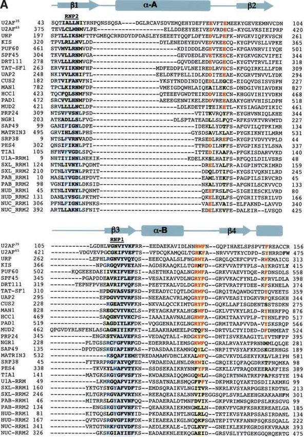
Figure 4. Identification of candidate UHM-containing proteins. (A) Structure-based alignment of candidate UHM sequences, U2AF35-
UHM, U2AF65-UHM, and representative canonical RRMs whose structures in complex with RNA are known. Structures were aligned
using the program TOPP from the CCP4 suite (Collaborative Computational Project, no. 4, 1994). Secondary structure elements are
indicated above the sequences. The positions of key residues that distinguish UHM from RRM domains are highlighted yellow.
Consensus UHM residues including acidic residues and the Arg–X–Phe loop are red. Residues in the RNP-like motifs that match the
canonical RRM consensus are blue. (B) Phylogenetic tree of candidate UHM domains.
UHM domains via a similar arrangement of basic and cally recognizes a single target, or could promiscuously
Trp residues. A search for the ligand consensus pattern interact with the intended ligand of a different UHM in
[RK]-X-[RK]-W, shared by both the SF1 and U2AF65 li- the absence of temporal or spatial regulation. Toward
gands, found >1000 matches within the SWISS-PROT answering this question, the U2AF65-UHM has been
database, indicating that predicting protein ligands of found not to interact with the N terminus of the
candidate UHMs is impractical without further experi- U2AF65-ligand in two-hybrid assays (Tronchere et al.
mental information to identify their functional binding 1997) and pull-down experiments (C. Kielkopf, unpubl.),
partners. Given that all 12 compelling UHM candidates suggesting that individual UHMs may, indeed, specifi-
possess the signature sequences predicted to recognize cally recognize a unique target. By analogy with other
ligands containing the [RK]-X-[RK]-W motif, it remains modular peptide binding domains (Pawson and Nash
an open question whether each UHM domain specifi- 2003), UHM sequences flanking the Trp-binding pocket
GENES & DEVELOPMENT 1519Kielkopf et al.
Table 2. Representative homologs of UHM candidates residues (Kielkopf et al. 2001). Other UHM sequences
vary from similar Arg–Tyr–Phe motifs (SPF45, DRT111,
Sequence
Human Accession identityb
UAP2, CUS2, and PAD1) to divergent Lys (U2AF65) and
protein number Homologa Source organism (%) Met (KIS) residues. Besides recognizing the ligand C ter-
minus via the Arg–X–Phe loop, distinct U2AF65-UHM or
U2AF35 Q01081 Q9D883 M. musculus 100 U2AF35-UHM residues make specific contacts with N-
AAM34646 D. rerio 97 terminal ligand residues. In particular, the bulky
AAB17271 D. melanogaster 82
U2AF65-ligand Tyr 91 stacks against unique U2AF35 aro-
EAA08115 A. gambiae 82
matic residues (Tyr 52 and Phe 81), and forms a specific
AAM97101 A. thaliana 54
CAB55137 C. elegans 66 hydrogen-bond with His 77 of the U2AF35-UHM
(Kielkopf et al. 2001). Similar or identical residues in the
U2AF65 P26368 P26369 M. musculus 99 URP-UHM (Phe 206, Phe 239, and Gln 235) suggest that
AAH65869 D. rerio 85
a bulky, hydrophobic residue preceding the consensus
A48249 D. melanogaster 72
ligand-Trp would be recognized in an analogous manner,
EAA08228 A. gambiae 61
E96634 A. thaliana 42 consistent with an interaction between hURP and
AAC26982 C. elegans 59 U2AF65 in pull-down and yeast two-hybrid assays
(Tronchere et al. 1997). The smaller size of the corre-
KIS Q8TAS1 AAH58732 M. musculus 99
sponding U2AF65-UHM residues (Ile 398 and Val 384)
PUF60 AAF05605 AAH10601 M. musculus 95 would leave the hydrophobic side chain of a ligand-Tyr
CAD61099 D. rerio 76 in an unfavorable, solvent-exposed environment
EAA09944 A. gambiae 53 (Selenko et al. 2003). Considering the variety of cellular
AAF47501 D. melanogaster 49 roles played by UHM candidates and the consequent re-
AAF60676 C. elegans 48
quirement to recognize diverse protein ligands, it will be
SPF45 AAH39322 AAC64085 M. musculus 100 important to determine whether variation in the posi-
AAH45473 D. rerio 78 tions corresponding to U2AF35 Tyr 52, Phe 81, and His
EAA15153 A. gambiae 39 77, and the central position of the Arg–X–Phe loop en-
EAA46008 D. melanogaster 36 ables recognition of distinct ligand sequences by UHM
CAA97799 C. elegans 35
domains.
Tat-SF1 AAB18823 AAH37711 M. musculus 100 In addition to recognizing short peptide ligands, UHM
AAH55565 D. rerio 55 domains can self-associate to form protein homodimers.
AAF51719 D. melanogaster 45 For example, the PUF60-UHM domain interacts with it-
EAA10753 A. gambiae 44 self in two-hybrid assays (Poleev et al. 2000) and forms
AAK29956 C. elegans 37
SDS-resistant homodimers during electrophoresis (Page-
HCC1 Q14498 Q8VH51 M. musculus 99 McCaw et al. 1999). The U2AF35-UHM has been shown
AAH44487 D. rerio 79 to form weak homodimers by gel filtration, analytical
AAF52478 D. melanogaster 61 ultracentrifugation, dynamic light scattering (Kielkopf et
EAA12873 A. gambiae 53 al. 2001), and two-hybrid assays (Wentz-Hunter and Pot-
AAM97977 C. elegans 47
ashkin 1996), whereas homodimers of the U2AF65-UHM
AAM20703 A. thaliana 42
have not been observed (Tronchere et al. 1997). Homo- or
MAN1 AAF73293 Q9WU40 M. musculus 70 heterotypic oligomerizations also have been observed for
a
Candidates identified from Homo sapiens, Mus musculus, classical protein–protein interaction domains, with sev-
Danio rerio, Anopheles gambiae, Drosophila melanogaster, eral different effects on ligand recognition. For example,
Arabidopsis thaliana, and Caenorhabditis elegans, based on the nNOS-PDZ/syntrophin heterodimer prohibits pep-
similar domain structure and >35% sequence identity in match- tide recognition (Hillier et al. 1999), whereas GRIP or
ing regions identified by the PSI-BLAST program (Altschul et al. Shank PDZ homodimers leave the peptide-binding pock-
1997). ets free (Im et al. 2003a,b) and the Eps8-SH3 homodimer
b
Sequence identity between the human candidate and the ho- alters the ligand specificity (Kishan et al. 1997). Al-
molog from the indicated source organism. though a U2AF35-UHM homodimer can be modeled
with the solvent exposed Arg–Trp–Phe loop binding to
the Trp-binding site on a second UHM domain, alterna-
may ensure specific and directional interactions (N-to-C tive interfaces are possible that would allow the oligo-
orientation) with the ligand. mer to simultaneously recognize peptide ligands, as ob-
In support of this analogy, structure-based modeling served for established protein–protein interaction do-
suggests that variation of the central X residue in the mains.
UHM Arg–X–Phe loop may provide one mechanism for
UHM recognition of diverse C-terminal ligand se-
Do UHM domains recognize RNA?
quences. Several of the UHM candidates (hURP, PUF60,
Tat-SF1, and HCC1) share a Trp residue within the Arg– Modeling of the U2AF-UHM/ligand structures with
X–Phe loop that is essential in the U2AF35-UHM for RNA has revealed that peptide binding to the helical
specific recognition of C-terminal U2AF65-ligand-Pro surface of the RRM-like fold is not predicted to physi-
1520 GENES & DEVELOPMENTProtein recognition by U2AF homology motifs
Table 3. Function of human UHM candidates and their potential disease relevance
Domains are annotated according to CDD (Marchler-Bauer et al. 2002). (RS) Arg–Ser rich; (Zn) zinc binding; (S/T kinase) Ser/Thr
phosphokinase; (G-patch) Gly rich; (acidic) Glu/Asp rich; (TM) transmembrane region; (LEM) LAP2-Emerin-Man1 homology.
cally interfere with putative RNA interactions on the canonical RRM-RNA structures, conserved aromatic
opposite -sheet face (Kielkopf et al. 2001; Selenko et al. Phe/Tyr residues at the third RNP1 position or second
2003). Although the U2AF35-UHM/U2AF65-ligand com- RNP2 position stack with RNA bases or sugars, and a
plex binds RNA weakly (Kd >6 µM), accessory protein basic Arg/Lys residue at the first position of the RNP1
factors and adjacent domains in the full-length U2AF35 motif frequently forms a salt bridge with the phosphate
sequence (e.g., flanking zinc fingers and an RS domain) backbone (Fig. 5A; Oubridge et al. 1994; Price et al. 1998;
are required to assist the weak interaction (Rudner et al. Deo et al. 1999; Handa et al. 1999; Allain et al. 2000;
1998a; J. Valcarcel, pers. comm.). Likewise, the U2AF65- Wang and Tanaka Hall 2001). In contrast, the corre-
UHM domain is not required for Py-tract recognition sponding U2AF35-UHM (Fig. 5B) and U2AF65-UHM (Fig.
(Banerjee et al. 2003), and does not appear to interact 5C) residues are replaced with aliphatic substitutions
with RNA (Selenko et al. 2003). These results indicate that are not predicted to interact favorably with RNA.
that UHM domains are not likely to be involved in RNA Moreover, UHMs display unexpectedly low isoelectric
interactions. points for optimal binding of basic peptides.
Instead, the UHM family has evolved sequence char- In addition to poor conservation of RNP-like motifs
acteristics that have no benefit for RNA binding, while and overall negative charge, RNA binding by the
optimizing the interaction with peptide ligands. In most U2AF65-UHM structure is further inhibited by a C-ter-
GENES & DEVELOPMENT 1521Kielkopf et al. Figure 5. RNA recognition by an RRM domain compared with the U2AF-UHM domains. The UHM or RRM domains are shown in blue, protein ligands are yellow, and RNA ligands are purple. (A) The U1A-RRM recognizing an RNA oligonucleotide. The RNP1-Arg recognizes the RNA phosphates, a nucleotide base stacks on RNP2-Tyr, and RNP2-Leu is folded in the hydrophobic core. (B) The U2AF35-UHM/U2AF65-ligand complex (PDB code 1JMT) rotated 180° about the Y-axis with respect to Figure 3 to show the putative RNA interaction surface. (C) Similar view of the U2AF65-UHM/SF1-ligand structure (PDB code 1O0P). The RNP2-Ile packs with the ligand-Trp. The small aliphatic residues in the RNP-like motifs are predicted to be unable to form favorable RNA interactions. Schematic representation of RNP residues that differ between UHMs compared with canonical RRMs is shown to the right. minal ␣-helix that forms a tight hydrophobic interface U2AF65-UHM structure, Phe 433 in the RNP1-like motif with the putative RNA-binding surface of the RRM-like and Tyr 463 within the preceding turn interact with Tyr fold (Selenko et al. 2003). In contrast, the C-terminal 469, Phe 474, and Trp 475 in the C-terminal ␣-helix extensions of canonical RRMs more often strengthen (Selenko et al. 2003). Although counterparts of Phe 474 rather than inhibit RNA binding. For example, the C- and Trp 475 are absent among the UHM candidates, aro- terminal helical extension of the N-terminal U1A-RRM matic residues at positions corresponding to Phe 433, not only contributes to RNA binding (Oubridge et al. Tyr 463, and Tyr 469 are observed for the UHM domains 1994; Zeng and Hall 1997) but also mediates dimer for- of KIS, PUF60, SPF45, and HCC1. This raises the possi- mation for recognition of tandem RNA elements (Klein bility that some of the UHM candidates may have a hy- Gunnewiek et al. 2000; Varani et al. 2000). Based on the drophobic C-terminal extension that may either inter- 1522 GENES & DEVELOPMENT
Protein recognition by U2AF homology motifs
fere with RNA binding as for the U2AF65-UHM, or con- RRMs, for other fold families whose members play di-
tribute to homodimer formation in a manner similar to verse functional roles.
U1A.
Acknowledgments
Conclusions We thank S. Evans for editorial assistance, and J. Bender, M.
Matunis, M. Swenson, and J. Wedekind for careful reading of the
The canonical RRM domain was a relatively late evolu- manuscript. Funding for C.L.K. is provided by the Johns Hop-
tionary addition to the array of RNA-binding folds that kins University Center for AIDS Research grant #P30 AI42855.
emerged in response to the needs of complex pre-mRNA
processing pathways (Anantharaman et al. 2002). As pro-
cesses that were originally based on the RNA world be- References
came progressively more regulated and reliant on protein
interactions, the RRM fold further developed specialized Abovich, N., Liao, X.C., and Rosbash, M. 1994. The yeast
MUD2 protein: An interaction with PRP11 defines a bridge
sequence characteristics for protein recognition to form
between commitment complexes and U2 snRNP addition.
the UHM subfamily. These UHM signature sequences Genes & Dev. 8: 843–854.
included divergent residues in the RNP-like motifs, an Adam, S.A., Nakagawa, T., Swanson, M.S., Woodruff, T.K., and
Arg–X–Phe loop sequence, and key acidic residues that Dreyfuss, G. 1986. mRNA polyadenylate-binding protein:
collectively recognized the Trp residue and positive Gene isolation and sequencing and identification of a ribo-
charge of the protein ligand. Convincing UHM candi- nucleoprotein consensus sequence. Mol. Cell. Biol. 6: 2932–
dates have been discovered in association with a variety 2943.
of fundamental cellular processes, ranging from pre- Allain, F.H., Bouvet, P., Dieckmann, T., and Feigon, J. 2000.
mRNA splicing to transcription, DNA repair, and signal Molecular basis of sequence-specific recognition of pre-ribo-
transduction. The large number of proteins that share somal RNA by nucleolin. EMBO J. 19: 6870–6881.
Altschul, S.F., Madden, T.L., Schaffer, A.A., Zhang, J., Zhang,
the signature protein–protein interaction residues of
Z., Miller, W., and Lipman, D.J. 1997. Gapped BLAST and
UHM domains supports the proposal that the U2AF- PSI-BLAST: A new generation of protein database search pro-
UHMs represent a novel family of modular protein in- grams. Nucleic Acids Res. 25: 3389–3402.
teraction domains. Anantharaman, V., Koonin, E.V., and Aravind, L. 2002. Com-
Because protein interaction domains are attractive parative genomics and evolution of proteins involved in
modules for communication among a network of path- RNA metabolism. Nucleic Acids Res. 30: 1427–1464.
ways, the UHM domain may be an evolutionary exten- Auboeuf, D., Honig, A., Berget, S.M., and O’Malley, B.W. 2002.
sion of RRMs that couples pre-mRNA processing with Coordinate regulation of transcription and splicing by ste-
other nuclear processes. Protein recognition by so-called roid receptor coregulators. Science 298: 416–419.
RNA-binding domains is an emerging theme in molecu- Auboeuf, D., Dowhan, D.H., Kang, Y.K., Larkin, K., Lee, J.W.,
Berget, S.M., and O’Malley, B.W. 2004. Differential recruit-
lar recognition. An early example of RRM–protein inter-
ment of nuclear receptor coactivators may determine alter-
actions was observed in the structure of U2B⬙/U2A⬘, in native RNA splice site choice in target genes. Proc. Natl.
which the ␣-helical surface of the U2B⬙-RRM interacts Acad. Sci. 101: 2270–2274.
with the U2A⬘ leucine-rich repeat motif (Price et al. Banerjee, H., Rahn, A., Davis, W., and Singh, R. 2003. Sex lethal
1998). Several recent structures of the -sheet surfaces of and U2 small nuclear ribonucleoprotein auxiliary factor
heterodimeric RRM domains interacting with ␣-helical (U2AF65) recognize polypyrimidine tracts using multiple
protein ligands have revealed a second mode of RRM– modes of binding. RNA 9: 88–99.
protein recognition distinct from that of UHM/protein Banerjee, H., Rahn, A., Gawande, B., Guth, S., Valcarcel, J., and
complexes (Fribourg et al. 2003; Lau et al. 2003; Shi and Singh, R. 2004. The conserved RNA recognition motif 3 of
Xu 2003; Kadlec et al. 2004). In addition to distinguish- U2 snRNA auxiliary factor (U2AF65) is essential in vivo but
dispensable for activity in vitro. RNA 10: 240–253.
ing protein recognition domains within the RRM family,
Bennett, M., Michaud, S., Kingston, J., and Reed, R. 1992. Pro-
a growing list of fold families such as the Sterile ␣-Motif tein components specifically associated with prespliceo-
(SAM; Kim and Bowie 2003), LEM (Cai et al. 2001; Laguri some and spliceosome complexes. Genes & Dev. 6: 1986–
et al. 2001), Pumilio/HEAT-repeat domains (Wang et al. 2000.
2002), and zinc fingers (Morgan et al. 1997) have been Berglund, J.A., Abovich, N., and Rosbash, M. 1998. A coopera-
found to bind either nucleic acids or protein ligands tive interaction between U2AF65 and mBBP/SF1 facilitates
through slight variations of a common scaffold. Further- branchpoint region recognition. Genes & Dev. 12: 858–867.
more, RS domains have been shown to contact the pre- Bieche, I., Manceau, V., Curmi, P.A., Laurendeau, I., Lachkar, S.,
mRNA during splicing (Valcarcel et al. 1996; Shen et al. Leroy, K., Vidaud, D., Sobel, A., and Maucuer, A. 2003.
2004), and have also been reported to mediate protein– Quantitative RT–PCR reveals a ubiquitous but preferen-
tially neural expression of the KIS gene in rat and human.
protein interactions (Wu and Maniatis 1993). Because a
Brain Res. Mol. Brain Res. 114: 55–64.
major goal of the “postgenomic” era is the ability to Birney, E., Kumar, S., and Krainer, A.R. 1993. Analysis of the
predict protein functions even in the absence of corrobo- RNA-recognition motif and RS and RGG domains: Conser-
rating experimental results (Thornton et al. 2000), it will vation in metazoan pre-mRNA splicing factors. Nucleic Ac-
be essential to compile a lexicon of signature sequences, ids Res. 21: 5803–5816.
such as those that distinguish UHMs from canonical Boehm, M., Yoshimoto, T., Crook, M.F., Nallamshetty, S.,
GENES & DEVELOPMENT 1523Kielkopf et al. True, A., Nabel, G.J., and Nabel, E.G. 2002. A growth factor- Hoffman, D.W., Query, C.C., Golden, B.L., White, S.W., and dependent nuclear kinase phosphorylates p27Kip1 and regu- Keene, J.D. 1991. RNA-binding domain of the A pro- lates cell cycle progression. EMBO J. 21: 3390–3401. tein component of the U1 small nuclear ribonucleoprotein Brazma, A., Jonassen, I., Ukkonen, E., and Vilo, J. 1996. Discov- analyzed by NMR spectroscopy is structurally similar ering patterns and subfamilies in biosequences. Proc. Int. to ribosomal proteins. Proc. Natl. Acad. Sci. 88: 2495– Conf. Intell. Syst. Mol. Biol. 4: 34–43. 2499. Brow, D.A. 2002. Allosteric cascade of spliceosome activation. Im, Y.J., Lee, J.H., Park, S.H., Park, S.J., Rho, S.H., Kang, G.B., Annu. Rev. Genet. 36: 333–360. Kim, E., and Eom, S.H. 2003a. Crystal structure of the Shank Burd, C.G. and Dreyfuss, G. 1994. Conserved structures and PDZ–ligand complex reveals a class I PDZ interaction and a diversity of functions of RNA-binding proteins. Science novel PDZ–PDZ dimerization. J. Biol. Chem. 278: 48099– 265: 615–621. 48104. Cai, M., Huang, Y., Ghirlando, R., Wilson, K.L., Craigie, R., and Im, Y.J., Park, S.H., Rho, S.H., Lee, J.H., Kang, G.B., Sheng, M., Clore, G.M. 2001. Solution structure of the constant region Kim, E., and Eom, S.H. 2003b. Crystal structure of GRIP1 of nuclear envelope protein LAP2 reveals two LEM-domain PDZ6-peptide complex reveals the structural basis for class structures: One binds BAF and the other binds DNA. EMBO II PDZ target recognition and PDZ domain-mediated mul- J. 20: 4399–4407. timerization. J. Biol. Chem. 278: 8501–8507. Chiara, M.D., Palandjian, L., Feld Kramer, R., and Reed, R. 1997. Imai, H., Chan, E.K., Kiyosawa, K., Fu, X.D., and Tan, E.M. Evidence that U5 snRNP recognizes the 3⬘ splice site for 1993. Novel nuclear autoantigen with splicing factor motifs catalytic step II in mammals. EMBO J. 16: 4746–4759. identified with antibody from hepatocellular carcinoma. J. Collaborative Computational Project, number 4. 1994. The Clin. Invest. 92: 2419–2426. CCP4 suite: Programs for protein crystallography. Acta Jung, D.J., Na, S.Y., Na, D.S., and Lee, J.W. 2002. Molecular Crystallogr. D50: 760–763. cloning and characterization of CAPER, a novel coactivator Dean, W., Bowden, L., Aitchison, A., Klose, J., Moore, T., Me- of activating protein-1 and estrogen receptors. J. Biol. Chem. neses, J.J., Reik, W., and Feil, R. 1998. Altered imprinted 277: 1229–1234. gene methylation and expression in completely ES cell-de- Kadlec, J., Izaurralde, E., and Cusack, S. 2004. The structural rived mouse fetuses: Association with aberrant phenotypes. basis for the interaction between nonsense-mediated mRNA Development 125: 2273–2282. decay factors UPF2 and UPF3. Nat. Struct. Mol. Biol. Dendouga, N., Callebaut, I., and Tomavo, S. 2002. A novel DNA 11: 330–337. repair enzyme containing RNA recognition, G-patch and Kanaar, R., Roche, S.E., Beall, E.L., Green, M.R., and Rio, D.C. specific splicing factor 45-like motifs in the protozoan para- 1993. The conserved pre-mRNA splicing factor U2AF from site Toxoplasma gondii. Eur. J. Biochem. 269: 3393–3401. Drosophila: Requirement for viability. Science 262: Deo, R.C., Bonanno, J.B., Sonenberg, N., and Burley, S.K. 1999. 569–573. Recognition of polyadenylate RNA by the poly(A)-binding Kielkopf, C.L., Rodionova, N.A., Green, M.R., and Burley, S.K. protein. Cell 98: 835–845. 2001. A novel peptide recognition mode revealed by the X- Dreyfuss, G., Swanson, M.S., and Pinol-Roma, S. 1988. Heter- ray structure of a core U2AF35/U2AF65 heterodimer. Cell ogeneous nuclear ribonucleoprotein particles and the path- 106: 595–605. way of mRNA formation. Trends Biochem. Sci. 13: 86–91. Kim, C.A. and Bowie, J.U. 2003. SAM domains: Uniform struc- Fong, Y.W. and Zhou, Q. 2001. Stimulatory effect of splicing ture, diversity of function. Trends Biochem. Sci. 28: 625– factors on transcriptional elongation. Nature 414: 929– 628. 933. Kishan, K.V., Scita, G., Wong, W.T., Di Fiore, P.P., and New- Fribourg, S., Gatfield, D., Izaurralde, E., and Conti, E. 2003. A comer, M.E. 1997. The SH3 domain of Eps8 exists as a novel novel mode of RBD–protein recognition in the Y14–Mago intertwined dimer. Nat. Struct. Biol. 4: 739–743. complex. Nat. Struct. Biol. 10: 433–439. Kitagawa, K., Wang, X., Hatada, I., Yamaoka, T., Nojima, H., Gattiker, A., Gasteiger, E., and Bairoch, A. 2002. ScanProsite: A Inazawa, J., Abe, T., Mitsuya, K., Oshimura, M., Murata, A., reference implementation of a PROSITE scanning tool. Appl. et al. 1995. Isolation and mapping of human homologues of Bioinformatics 1: 107–108. an imprinted mouse gene U2AF1-RS1. Genomics 30: 257– Golling, G., Amsterdam, A., Sun, Z., Antonelli, M., Maldonado, 263. E., Chen, W., Burgess, S., Haldi, M., Artzt, K., Farrington, S., Klein Gunnewiek, J.M., Hussein, R.I., van Aarssen, Y., Palacios, et al. 2002. Insertional mutagenesis in zebrafish rapidly iden- D., de Jong, R., van Venrooij, W.J., and Gunderson, S.I. 2000. tifies genes essential for early vertebrate development. Nat. Fourteen residues of the U1 snRNP-specific U1A protein are Genet. 31: 135–140. required for homodimerization, cooperative RNA binding, Gozani, O., Potashkin, J., and Reed, R. 1998. A potential role for and inhibition of polyadenylation. Mol. Cell. Biol. 20: 2209– U2AF–SAP155 interactions in recruiting U2 snRNP to the 2217. branch site. Mol. Cell. Biol. 18: 4752–4760. Kramer, A. and Utans, U. 1991. Three protein factors (SF1, SF3 Habara, Y., Urushiyama, S., Tani, T., and Ohshima, Y. 1998. and U2AF) function in pre-splicing complex formation in The fission yeast prp10+ gene involved in pre-mRNA splic- addition to snRNPs. EMBO J. 10: 1503–1509. ing encodes a homologue of highly conserved splicing factor, Kuldau, G.A., Raju, N.B., and Glass, N.L. 1998. Repeat-induced SAP155. Nucleic Acids Res. 26: 5662–5669. point mutations in PAD1, a putative RNA splicing factor Handa, N., Nureki, O., Kurimoto, K., Kim, I., Sakamoto, H., from Neurospora crassa, confer dominant lethal effects on Shimura, Y., Muto, Y., and Yokoyama, S. 1999. Structural ascus development. Fungal Genet. Biol. 23: 169–180. basis for recognition of the tra mRNA precursor by the Sex- Laguri, C., Gilquin, B., Wolff, N., Romi-Lebrun, R., Courchay, lethal protein. Nature 398: 579–585. K., Callebaut, I., Worman, H.J., and Zinn-Justin, S. 2001. Hillier, B.J., Christopherson, K.S., Prehoda, K.E., Bredt, D.S., and Structural characterization of the LEM motif common to Lim, W.A. 1999. Unexpected modes of PDZ domain scaffold- three human inner nuclear membrane proteins. Structure ing revealed by structure of nNOS–syntrophin complex. Sci- (Camb) 9: 503–511. ence 284: 812–815. Lahiri, D.K. and Thomas, J.O. 1986. A cDNA clone of the 1524 GENES & DEVELOPMENT
Protein recognition by U2AF homology motifs

    hnRNP C proteins and its homology with the single-                       Osada, S., Ohmori, S.Y., and Taira, M. 2003. XMAN1, an inner
    stranded DNA binding protein UP2. Nucleic Acids Res.                        nuclear membrane protein, antagonizes BMP signaling by
    14: 4077–4094.                                                              interacting with Smad1 in Xenopus embryos. Development
Lallena, M.J., Chalmers, K.J., Llamazares, S., Lamond, A.I., and                130: 1783–1794.
    Valcarcel, J. 2002. Splicing regulation at the second catalytic          Oubridge, C., Ito, N., Evans, P.R., Teo, C.H., and Nagai, K. 1994.
    step by Sex-lethal involves 3⬘ splice site recognition by                   Crystal structure at 1.92 Å resolution of the RNA-binding
    SPF45. Cell 109: 285–296.                                                   domain of the U1A spliceosomal protein complexed with an
Lau, C.K., Diem, M.D., Dreyfuss, G., and Van Duyne, G.D.                        RNA hairpin. Nature 372: 432–438.
    2003. Structure of the Y14–Magoh core of the exon junction               Page-McCaw, P.S., Amonlirdviman, K., and Sharp, P.A. 1999.
    complex. Curr. Biol. 13: 933–941.                                           PUF60: A novel U2AF65-related splicing activity. RNA
Letunic, I., Copley, R.R., Schmidt, S., Ciccarelli, F.D., Doerks,               5: 1548–1560.
    T., Schultz, J., Ponting, C.P., and Bork, P. 2004. SMART 4.0:            Pang, Q., Hays, J.B., and Rajagopal, I. 1993. Two cDNAs from
    Towards genomic data integration. Nucleic Acids Res.                        the plant Arabidopsis thaliana that partially restore recom-
    32: D142–D144.                                                              bination proficiency and DNA-damage resistance to E. coli
Li, X.Y. and Green, M.R. 1998. The HIV-1 Tat cellular coacti-                   mutants lacking recombination-intermediate-resolution ac-
    vator Tat-SF1 is a general transcription elongation factor.                 tivities. Nucleic Acids Res. 21: 1647–1653.
    Genes & Dev. 12: 2992–2996.                                              Pawson, T. and Nash, P. 2003. Assembly of cell regulatory sys-
Lin, F., Blake, D.L., Callebaut, I., Skerjanc, I.S., Holmer, L., Mc-            tems through protein interaction domains. Science
    Burney, M.W., Paulin-Levasseur, M., and Worman, H.J.                        300: 445–452.
    2000. MAN1, an inner nuclear membrane protein that                       Poleev, A., Hartmann, A., and Stamm, S. 2000. A trans-acting
    shares the LEM domain with lamina-associated polypeptide                    factor, isolated by the three-hybrid system, that influences
    2 and emerin. J. Biol. Chem. 275: 4840–4847.                                alternative splicing of the amyloid precursor protein mini-
Liu, J., He, L., Collins, I., Ge, H., Libutti, D., Li, J., Egly, J.M., and      gene. Eur. J. Biochem. 267: 4002–4010.
    Levens, D. 2000. The FBP interacting repressor targets TFIIH             Potashkin, J., Naik, K., and Wentz-Hunter, K. 1993. U2AF ho-
    to inhibit activated transcription. Mol. Cell 5: 331–341.                   molog required for splicing in vivo. Science 262: 573–575.
Liu, J., Akoulitchev, S., Weber, A., Ge, H., Chuikov, S., Libutti,           Price, S.R., Evans, P.R., and Nagai, K. 1998. Crystal structure of
    D., Wang, X.W., Conaway, J.W., Harris, C.C., Conaway,                       the spliceosomal U2B⬙–U2A⬘ protein complex bound to a
    R.C., et al. 2001. Defective interplay of activators and repres-            fragment of U2 small nuclear RNA. Nature 394: 645–650.
    sors with TFIH in xeroderma pigmentosum. Cell 104: 353–                  Query, C.C., Bentley, R.C., and Keene, J.D. 1989. A common
    363.                                                                        RNA recognition motif identified within a defined U1 RNA
Marchler-Bauer, A., Panchenko, A.R., Shoemaker, B.A., Thies-                    binding domain of the 70K U1 snRNP protein. Cell 57: 89–
    sen, P.A., Geer, L.Y., and Bryant, S.H. 2002. CDD: A data-                  101.
    base of conserved domain alignments with links to domain                 Query, C.C., Moore, M.J., and Sharp, P.A. 1994. Branch nucleo-
    three-dimensional structure. Nucleic Acids Res. 30: 281–                    phile selection in pre-mRNA splicing: Evidence for the
    283.                                                                        bulged duplex model. Genes & Dev. 8: 587–597.
Maucuer, A., Camonis, J.H., and Sobel, A. 1995. Stathmin in-                 Rain, J.C., Rafi, Z., Rhani, Z., Legrain, P., and Kramer, A. 1998.
    teraction with a putative kinase and coiled-coil-forming pro-               Conservation of functional domains involved in RNA bind-
    tein domains. Proc. Natl. Acad. Sci. 92: 3100–3104.                         ing and protein–protein interactions in human and Saccha-
Maucuer, A., Le Caer, J.P., Manceau, V., and Sobel, A. 2000.                    romyces cerevisiae pre-mRNA splicing factor SF1. RNA
    Specific Ser-Pro phosphorylation by the RNA-recognition                     4: 551–565.
    motif containing kinase KIS. Eur. J. Biochem. 267: 4456–                 Raju, G.P., Dimova, N., Klein, P.S., and Huang, H.C. 2003.
    4464.                                                                       SANE, a novel LEM domain protein, regulates bone morpho-
McKinney, R., Wentz-Hunter, K., Schmidt, H., and Potashkin, J.                  genetic protein signaling through interaction with Smad1. J.
    1997. Molecular characterization of a novel fission yeast                   Biol. Chem. 278: 428–437.
    gene spUAP2 that interacts with the splicing factor                      Rosonina, E. and Blencowe, B.J. 2002. Gene expression: The
    spU2AF59. Curr. Genet. 32: 323–330.                                         close coupling of transcription and splicing. Curr. Biol.
Merendino, L., Guth, S., Bilbao, D., Martinez, C., and Valcarcel,               12: R319–R321.
    J. 1999. Inhibition of msl-2 splicing by Sex-lethal reveals              Rudner, D.Z., Kanaar, R., Breger, K.S., and Rio, D.C. 1996. Mu-
    interaction between U2AF35 and the 3⬘ splice site AG. Na-                   tations in the small subunit of the Drosophila U2AF splicing
    ture 402: 838–841.                                                          factor cause lethality and developmental defects. Proc. Natl.
Morgan, B., Sun, L., Avitahl, N., Andrikopoulos, K., Ikeda, T.,                 Acad. Sci. 93: 10333–10337.
    Gonzales, E., Wu, P., Neben, S., and Georgopoulos, K. 1997.              Rudner, D.Z., Breger, K.S., Kanaar, R., Adams, M.D., and Rio,
    Aiolos, a lymphoid restricted transcription factor that inter-              D.C. 1998a. RNA binding activity of heterodimeric splicing
    acts with Ikaros to regulate lymphocyte differentiation.                    factor U2AF: At least one RS domain is required for high-
    EMBO J. 16: 2004–2013.                                                      affinity binding. Mol. Cell. Biol. 18: 4004–4011.
Nagai, K., Oubridge, C., Jessen, T.H., Li, J., and Evans, P.R. 1990.         Rudner, D.Z., Kanaar, R., Breger, K.S., and Rio, D.C. 1998b.
    Crystal structure of the RNA-binding domain of the U1                       Interaction between subunits of heterodimeric splicing fac-
    small nuclear ribonucleoprotein A. Nature 348: 515–520.                     tor U2AF is essential in vivo. Mol. Cell. Biol. 18: 1765–1773.
Nelson, K. and Green, M. 1989. Mammalian U2 snRNP has a                      Ruskin, B., Zamore, P.D., and Green, M.R. 1988. A factor,
    sequence-specific RNA-binding activity. Genes & Dev.                        U2AF, is required for U2 snRNP binding and splicing com-
    3: 1562–1571.                                                               plex assembly. Cell 52: 207–219.
Neubauer, G., King, A., Rappsilber, J., Calvio, C., Watson, M.,              Rutz, B. and Seraphin, B. 1999. Transient interaction of BBP/
    Ajuh, P., Sleeman, J., Lamond, A., and Mann, M. 1998. Mass                  ScSF1 and Mud2 with the splicing machinery affects the
    spectrometry and EST-database searching allows character-                   kinetics of spliceosome assembly. RNA 5: 819–831.
    ization of the multi-protein spliceosome complex. Nat.                   Sachs, A.B., Bond, M.W., and Kornberg, R.D. 1986. A single gene
    Genet. 20: 46–50.                                                           from yeast for both nuclear and cytoplasmic polyadenylate-

                                                                                                          GENES & DEVELOPMENT             1525
You can also read