Neuron-Oligodendrocyte Interactions in the Structure and Integrity of Axons - Frontiers
←
→
Page content transcription
If your browser does not render page correctly, please read the page content below
REVIEW
                                                                                                                                          published: 08 March 2021
                                                                                                                                    doi: 10.3389/fcell.2021.653101

                                            Neuron-Oligodendrocyte
                                            Interactions in the Structure and
                                            Integrity of Axons
                                            Greg J. Duncan 1* , Tyrell J. Simkins 1,2,3 and Ben Emery 1
                                            1
                                             Jungers Center for Neurosciences Research, Department of Neurology, Oregon Health & Science University, Portland, OR,
                                            United States, 2 Vollum Institute, Oregon Health & Science University, Portland, OR, United States, 3 Department
                                            of Neurology, VA Portland Health Care System, Portland, OR, United States

                                            The myelination of axons by oligodendrocytes is a highly complex cell-to-cell
                                            interaction. Oligodendrocytes and axons have a reciprocal signaling relationship
                                            in which oligodendrocytes receive cues from axons that direct their myelination,
                                            and oligodendrocytes subsequently shape axonal structure and conduction.
                                            Oligodendrocytes are necessary for the maturation of excitatory domains on the
                                            axon including nodes of Ranvier, help buffer potassium, and support neuronal
                                            energy metabolism. Disruption of the oligodendrocyte-axon unit in traumatic injuries,
                                            Alzheimer’s disease and demyelinating diseases such as multiple sclerosis results
                                            in axonal dysfunction and can culminate in neurodegeneration. In this review, we
                             Edited by:
                           Tim Czopka,
                                            discuss the mechanisms by which demyelination and loss of oligodendrocytes
               University of Edinburgh,     compromise axons. We highlight the intra-axonal cascades initiated by demyelination
                      United Kingdom
                                            that can result in irreversible axonal damage. Both the restoration of oligodendrocyte
                        Reviewed by:
                                            myelination or neuroprotective therapies targeting these intra-axonal cascades are likely
                  Rafael Góis Almeida,
              University of Edinburgh,      to have therapeutic potential in disorders in which oligodendrocyte support of axons
                      United Kingdom        is disrupted.
                       Aiman S. Saab,
      University of Zurich, Switzerland     Keywords: oligodendrocyte, remyelination, axonal degeneration, demyelination, multiple sclerosis, Wallerian
                                            degeneration, mitochondria
                  *Correspondence:
                      Greg J. Duncan
                  duncangr@ohsu.edu
                                            INTRODUCTION
                    Specialty section:
         This article was submitted to      Structural variants of the myelin sheath have arisen several times during evolution as a means
                   Molecular Medicine,      to allow for the rapid conduction of nerve impulses along axons, including in vertebrates and
                a section of the journal    some species of worm and shrimp (Roots, 2008). Within the central nervous system (CNS)
  Frontiers in Cell and Developmental       of jawed vertebrates, myelination is carried out by oligodendrocytes, a highly specialized glial
                                Biology
                                            cell (Zalc, 2016). The promotion of the speed and efficiency of action potentials has been
         Received: 13 January 2021          the best understood purpose of oligodendrocytes and myelin over the past seven decades
        Accepted: 15 February 2021
                                            (Hartline and Colman, 2007). While this role is undeniably important, there is also an increasing
         Published: 08 March 2021
                                            appreciation that neurons require support by glia, including oligodendrocytes, for their long-
                                Citation:   term integrity. When oligodendroglial support is lost, axons become progressively compromised
           Duncan GJ, Simkins TJ and
                                            and vulnerable to loss. Consequently, remyelination strategies are being pursued in diseases
                         Emery B (2021)
  Neuron-Oligodendrocyte Interactions
                                            such as multiple sclerosis (MS) with the hope of not only recovering nerve conduction in
in the Structure and Integrity of Axons.    the short term but also protecting axons against degeneration in the long term (Franklin
        Front. Cell Dev. Biol. 9:653101.    et al., 2012; Franklin and Ffrench-Constant, 2017; Lubetzki et al., 2020b). In this review, we
       doi: 10.3389/fcell.2021.653101       outline evidence emerging from both animal models and human pathology that suggests that

Frontiers in Cell and Developmental Biology | www.frontiersin.org                1                                          March 2021 | Volume 9 | Article 653101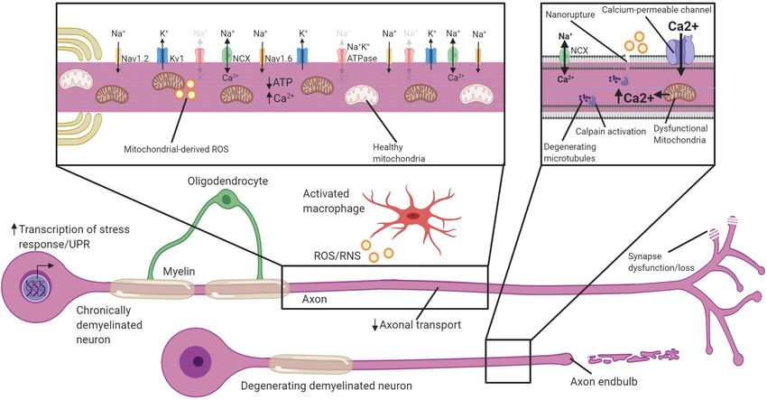
Duncan et al.                                                                                         Mechanisms of Oligodendroglial Axonal Support

the integrity of myelinated neurons is dependent on                       van Roermund et al., 2011; Wiesinger et al., 2013). As a
oligodendrocyte support. We focus on recent advances in                   consequence, there is an accumulation of VLCFA in cells
our understanding of the cellular mechanisms by which                     leading to increased oxidative stress culminating in myelin
oligodendrocytes support axonal and neuronal integrity, how               loss as well as progressive axonal degeneration (Powers et al.,
neurons adapt to demyelination, and the intra-axonal cascades             2000; Fourcade et al., 2008). Whether the progressive axonal
contributing to their degeneration.                                       degeneration occurring in ALD is a primary or secondary to
                                                                          myelin degeneration has not been fully determined. However,
                                                                          peroxisome impairment via the deletion of Pex5 selectively
DEMYELINATION AND NEURONAL                                                from oligodendrocyte lineage cells results in the accumulation
DAMAGE; BEYOND CLASSICAL                                                  of VLCFA and axonal degeneration, demonstrating that axonal
DEMYELINATING DISEASES                                                    degeneration can, in principal, be secondary to oligodendrocytic
                                                                          peroxisomal dysfunction (Kassmann et al., 2007). Pelizaeus-
Disruption of oligodendrocyte-axon contact and demyelination              Merzbacher disease (PMD) is an X-linked hypomyelinating
causes dysfunction in a wide-range of neurological pathologies.           leukodystrophy caused by mutation, deletion or duplication of
MS is considered the prototypical CNS demyelinating disease and           the PLP1 gene. PLP1, which encodes proteolipid protein (PLP)
features myelin loss in lesions throughout the gray and white             and its alternative splicing variant DM20, are the most abundant
matter. Clinically, MS typically presents as a relapsing-remitting        myelin proteins (Jahn et al., 2020). PMD patients typically have
(RRMS) course of neurologic dysfunction, though occasionally              a global developmental delay (motor and cognitive) as well
people with MS experience a progressive accumulation of                   as hypotonia, spasticity, and ataxia (Inoue, 2019). PLP1 gene
neurologic disability with few or no relapses (primary progressive        duplication is the most common cause of PMD (Mimault et al.,
MS, PPMS). Demyelination is not the only sequalae in MS;                  1999). Rodent models of homozygous Plp1 gene duplication
there is also considerable brain atrophy (De Stefano et al.,              result in premature arrest of myelination and oligodendrocyte
2010), which is reflective of ongoing axonal damage and                   apoptosis likely as a result of PLP and cholesterol accumulation
neurodegeneration. Acute demyelinating lesions have the highest           leading to endoplasmic reticulum (ER) stress (Kagawa et al., 1994;
rate of axonal damage and the degree of damage is correlated              Readhead et al., 1994; Simons et al., 2002; Karim et al., 2007;
with inflammation (Ferguson et al., 1997; Bitsch et al., 2000;            Elitt et al., 2018). Mice hemizygous for the PLP gene duplications
Kornek et al., 2000; Kuhlmann et al., 2002). Cytotoxic T-cells            myelinate normally before developing significant demyelination,
found within these lesions can directly damage neurons (Medana            inflammation and axonal degeneration (Anderson et al., 1998;
et al., 2001; Yan et al., 2003; Shriver and Dittel, 2006) and             Ip et al., 2006). Ultrastructural examination of clinical gene
drugs targeting the activation and infiltration of adaptive immune        duplications also reveal considerable axonal damage and
cells in the CNS are effective at reducing relapses (Derfuss              degeneration (Laukka et al., 2016). The examples of PMD and
et al., 2020). However, these drugs typically fail to prevent the         ALD highlight that neuronal integrity is often impaired in genetic
accumulation of progressive disability during chronic phases of           demyelinating pathologies.
the disease (secondary progressive, SPMS), when acute T-cell                 Myelin loss and axonal damage are also features of a number
mediated lesions wane. Instead, it is the widespread loss of              of neurologic conditions less commonly thought of as myelin-
axons and neurons during the progressive phase of the disease             related disorders. In Alzheimer’s disease (AD), white matter
that drives permanent disability (Fu et al., 1998; Bjartmar               damage is one of the earliest pre-clincal pathologic changes
et al., 2000; De Stefano et al., 2001). Crucially, remyelination          (Hoy et al., 2017; Nasrabady et al., 2018). Single-cell and spatial
fails to regenerate myelin along the majority of demyelinated             transcriptomic analyses reveal upregulation of genes involved
axons leaving them chronically demyelinated (Patrikios et al.,            in remyelination in AD brains (Grubman et al., 2019; Mathys
2006; Goldschmidt et al., 2009). These chronically demyelinated           et al., 2019; Agarwal et al., 2020; Chen et al., 2020). However,
inactive lesions constitute the bulk of lesions in MS (Frischer           late term senescent plaques may be inhibitory for remyelination
et al., 2015) and display signs of ongoing axonal damage (Kornek          and potentially co-op oligodendrocyte progenitor cells (OPCs)
et al., 2000). Axonal damage in these chronically demyelinated            into a pro-inflammatory role (Zhang et al., 2019). These human
lesions is, therefore, likely the central contributor to persistent       and rodent studies highlight an underappreciated involvement
disability in MS.                                                         of oligodendrocyte lineage cells in the pathophysiology of
    The susceptibility of axons to damage following myelin                AD. Likewise, the neurodegenerative disorder amyotrophic
and oligodendrocyte loss is observed in other demyelinating               lateral sclerosis (ALS) also features myelin damage. Decreased
pathologies as well. Leukodystrophies are a heterogeneous                 expression of myelin basic protein (MBP) in the motor cortex and
group of genetic disorders characterized by abnormalities in              spinal cord is observed in ALS (Kang et al., 2013). Animal models
the development or maintenance of CNS myelin (Kohler                      with mutant Sod1, which recapitulate the motor neuron loss
et al., 2018; van der Knaap et al., 2019; Wolf et al., 2020).             observed in ALS, have increased oligodendrocyte loss coupled
Adrenoleukodystrophy (ALD) is an X-linked demyelinating                   with a failure of new oligodendrocytes to mature (Kang et al.,
leukodystrophy caused by mutation in the ABCD1 gene (Mosser               2013; Philips et al., 2013). In zebrafish models this precedes motor
et al., 1993), which encodes an ATP-binding cassette transporter          neuron degeneration, suggesting myelin and oligodendrocyte
necessary for very long-chain fatty acid (VLCFA) transport                dysfunction is an early pathology in ALS (Kim et al., 2019).
into the peroxisome for degradation (McGuinness et al., 2003;             Knockout of mutant Sod1 from OPCs delays motor decline and

Frontiers in Cell and Developmental Biology | www.frontiersin.org     2                                      March 2021 | Volume 9 | Article 653101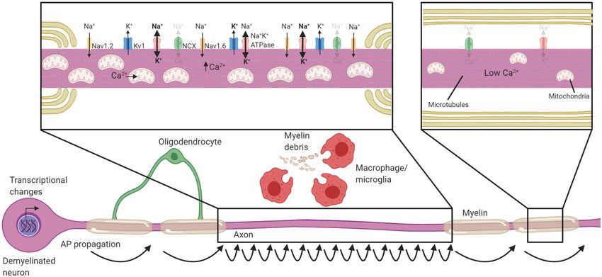
Duncan et al. Mechanisms of Oligodendroglial Axonal Support
increases survival time, possibly by restoring oligodendrocyte can be divided into subdomains; the internode (corresponding
support to the neuron (Lee et al., 2012; Kang et al., 2013). to the compacted region of myelin), the paranodes (where
Beyond ALS and AD, traumatic injuries to both the brain and the outer loops of the myelin contact the axon), the node of
spinal cord exhibit conduction deficits and acute demyelination Ranvier (the ∼1 µm gap between adjacent myelin internodes)
(Kakulas, 2004; Guest et al., 2005; James et al., 2011; Marion and the juxtaparanode (the interface between the paranode
et al., 2018) which may also strip axons of oligodendrocyte and compact myelin, rich in potassium channels) (Figure 1).
support and leave them vulnerable to degeneration. However, During development, the clustering of voltage-gated sodium and
few axons following spinal cord injury remain chronically potassium channels to these domains coincides with the process
demyelinated (Kakulas, 2004; Lasiene et al., 2008). Whether this of myelination, and is disrupted in dysmyelinating mutants
is due to subsequent degeneration of demyelinated axons or (Rasband et al., 1999; Mathis et al., 2001; Arroyo et al., 2002).
efficient remyelination remains unclear (Duncan et al., 2020). Several partially redundant mechanisms act in parallel to drive
Demyelination and oligodendrocyte loss is also observed in the clustering of nodal proteins during myelination (Susuki
ischemic stroke (Rosenzweig and Carmichael, 2013; Sozmen et al., 2013). Firstly, glial secreted proteins such as Contactin
et al., 2016), and improved myelin regeneration is associated 1, Phosphocan, and Tenascin-R help cluster axonal neurofascin
with enhanced functional recovery (Sozmen et al., 2016). This 186 and voltage-gated sodium channel subunits to sites of future
suggests a functional role of demyelination in the deficit nodes (Kaplan et al., 1997; Susuki et al., 2013; Freeman et al.,
following ischemia, at least in rodent models. The breakdown 2015; Dubessy et al., 2019; Thetiot et al., 2020). Subsequently,
of oligodendroglial support to neurons and demyelination may oligodendroglial neurofascin 155 interacts with axonal Contactin
be common to a wide range of disorders. For these reasons, 1 and Caspr at the paranodes, establishing septate-like junctions
it is crucial to understand how oligodendrocytes shape axonal that form a barrier between the axon and the myelin loops that
function and support their long-term survival. prevents the lateral diffusion of nodal proteins (Rasband et al.,
1999; Charles et al., 2002; Susuki et al., 2013). Finally, the nodal
and paranodal complexes are stabilized through interactions with
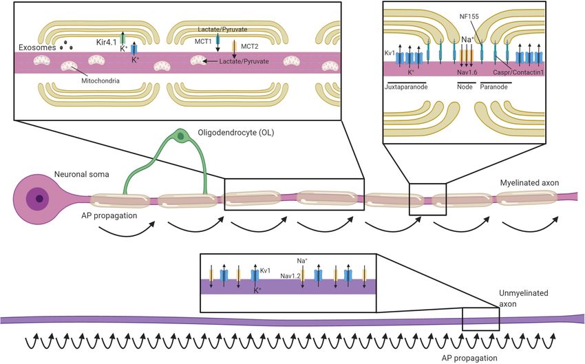
MYELINATING GLIA ORGANIZE THE the axonal cytoskeleton. These interactions are dependent on
AXON proteins such as AnkyrinG and protein 4.1B, which tether nodal
and paranodal proteins to axonal spectrins, respectively (Komada
Oligodendrocyte progenitor cells differentiate to form mature and Soriano, 2002; Susuki et al., 2013; Brivio et al., 2017).
oligodendrocytes which extend multiple processes that Voltage-gated potassium channels are predominantly restricted
ensheath nearby axons with concentric layers of membrane to the juxtaparanodal regions through interactions between glial
(for comprehensive reviews on the development and structure of Contactin 2 and axonal Caspr2 (Poliak et al., 2003; Traka
CNS myelin see Aggarwal et al., 2011; Rasband and Peles, 2015; et al., 2003). This localization of voltage-gated ion channels
Simons and Nave, 2016; Stassart et al., 2018; Stadelmann et al., essentially restricts the regeneration of action potentials to the
2019). Depending on the CNS region, each oligodendrocyte will node of Ranvier, with current then flowing longitudinally along
myelinate somewhere between 20 and 60 axons, with myelin the myelinated segments of the axon (Hartline and Colman,
internodes being from 20 to 200 µm in length (Chong et al., 2007; Figure 1). This process, termed saltatory conduction,
2012). During myelination, the leading edge of the developing dramatically increases conduction velocity relative to axonal
myelin sheath circles repetitively around the axon, remaining size (Waxman and Bennett, 1972). Although most work has
closely associated with the axon to pass under previous myelin focused the longitudinal current flow along the interior of the
wraps with each revolution. Meanwhile, the outer wraps extend axon, a recent study indicates that conduction velocity is also
laterally, with the terminal edges attaching to the axon in a influenced by current flow through the periaxonal space between
series of loops that ultimately form the paranode. Over time the innermost myelin wrap and the axon (Cohen et al., 2020).
the cytoplasm is excluded from most regions of the myelin, This may prove to be highly significant in light of findings that the
producing compact myelin (Snaidero et al., 2014). Some areas width of this space is regulated by neuronal activity and during
of non-compact myelin remain; the paranodal loops and the learning (Cullen et al., 2021).
innermost “tongue” of myelin adjacent to the axon remain
uncompacted, providing an area of oligodendrocyte cytoplasm
closely opposed to that of the axon. In addition, cytoplasmic OLIGODENDROCYTE SUPPORT OF
channels extend through the myelin sheath and provide a
AXONAL HEALTH
connection between the oligodendrocyte cell body and the
inner myelin layer (Velumian et al., 2011; Snaidero et al.,
2014). Although most prominent during development, these
Oligodendrocytic Shuttling of
cytoplasmic channels remain in the adult (Snaidero et al., Monocarboxylates and Glucose to the
2017) and likely act as an important conduit for organelles and Axon
molecules to support the myelin sheath. Although the best understood roles of myelin are to allow
An important role of myelin is to establish distinct axonal for rapid saltatory conduction by electrically insulating the
domains (recently reviewed in detail in Lubetzki et al., 2020a; axon and establishing distinct axonal domains, myelination
Rasband and Peles, 2021). The myelinated regions of axons also has profound implications for axonal energy demand
Frontiers in Cell and Developmental Biology | www.frontiersin.org 3 March 2021 | Volume 9 | Article 653101Duncan et al. Mechanisms of Oligodendroglial Axonal Support FIGURE 1 | Oligodendrocytes regulate axonal structure, conduction and support their survival. Schematic of an axon myelinated by an oligodendrocyte and an unmyelinated axon. Myelin permits saltatory conductance where action potentials are generated at the nodes of Ranvier. The high membrane resistance and low capacitance generated by the myelin sheath promotes rapid current flow along the myelinated section of the axon to the next node, greatly increasing conduction velocities relative to unmyelinated axons. Oligodendrocytes contact axons at the paranode (via NF155-Caspr/Contactin1) and are crucial for the organization, clustering and maintenance of sodium channels (primarily Nav1.6) at the nodes, as well as Kv1 potassium channels at the juxtaparanodes. In contrast, unmyelinated axons have potassium and sodium (primarily Nav1.2 channels) channels dispersed along the axon and are not confined to discrete excitatory clusters. Oligodendrocytes secrete exosomes that can support neuronal health and buffer potassium via the expression of Kir4.1. Oligodendrocytes provide glycolysis byproducts via monocarboxylate transporters (MCTs), which enter the Krebs cycle and are critical to support axonal metabolism. and metabolism. Myelination reduces axonal capacitance and from the neuronal cell body, myelinated axons may rely on substantially decreases the amount of energy required to metabolic support from the myelinating glia to meet their energy restore the resting ion concentrations after each depolarization requirements. Oligodendrocytes express the monocarboxylate (Harris and Attwell, 2012). However, myelin, with its concentric transporter MCT-1 and can transfer glycolysis products such membranes, also requires considerable resources to produce and as lactate to axons (Figure 1), where it can be converted to maintain. The act of myelination can be therefore be seen as ATP (Funfschilling et al., 2012; Lee et al., 2012). MCT-1 is shifting some of the metabolic load of neuronal firing from axons expressed both within the myelin sheath and along the adaxonal onto oligodendrocytes. Even though it has been estimated that it surface, ideally placing it to directly supply myelinated axons may take 15–23 million action potentials for the relative decrease with energy sources they would otherwise be isolated from in energy expenditure by a myelinated axon to offset the energetic Rinholm et al. (2011); Lee et al. (2012). Within the corpus cost of making the myelin sheath (Harris and Attwell, 2012), callosum, oligodendrocytes support axonal metabolic function by this investment may become beneficial at times of high activity providing glucose as well as glycolysis products (Meyer et al., where an unmyelinated axon may otherwise struggle to support 2018). Whether the relative oligodendroglial supply of glucose successive rounds of repolarization through the activity of the verses the supply of lactate to axons varies across different regions Na+/K+-ATPase. Nevertheless, as noted in several reviews on of the CNS is yet to be systematically investigated. Intriguingly, the subject (Mierzwa et al., 2010; Nave, 2010; Stassart et al., 2018), oligodendrocyte expression of the GLUT1 glucose transporter the presence of myelin comes with the liability of impeding the is regulated by NMDA receptor activity (Saab et al., 2016). axon’s ability to take up glucose and other metabolites directly Given oligodendrocytes do not store glycogen, this suggests a from the extracellular space, as myelin restricts diffusion of most mechanism by which this oligodendroglial uptake of glucose molecules to gaps at the paranodal junctions. Combined with and subsequent supply of glycolysis products might be matched the substantial logistical difficulty of transporting energy sources to levels of activity in the myelinated axons (Micu et al., 2016), Frontiers in Cell and Developmental Biology | www.frontiersin.org 4 March 2021 | Volume 9 | Article 653101
Duncan et al.                                                                                         Mechanisms of Oligodendroglial Axonal Support

supporting axons during times of heightened metabolic load                 allow for preferential uptake of oligodendrocyte-released EVs
(Micu et al., 2017). This feature of supporting axons through              by the myelinated axon, allowing for a relatively targeted and
provision of glycolysis products would mirror the role of                  activity-regulated transfer of supportive metabolites or signaling
astrocytes and invertebrate glial cells (Pellerin and Magistretti,         molecules. Such a transfer has been demonstrated to occur
1994; Tekkok et al., 2005; Volkenhoff et al., 2015).                       between ensheathing glia and the giant squid axon (Buchheit and
    Global heterozygosity of the Slc16a1 gene (which encodes the           Tytell, 1992), and likely represents an evolutionarily conserved
MCT-1 transporter) in mice causes axonal pathology by eight                relationship between glia and axons.
months of age, including axonal swellings, degeneration, and                   A large number of details remain to be determined about
enlarged axonal mitochondria (Lee et al., 2012). In contrast,              the role of oligodendrocyte exosomes, including their cargo.
a more recent study found that conditionally ablating Slc16a1              Oligodendrocyte-derived exosomes are known to be enriched
within mature oligodendrocytes (using MOG-Cre) resulted in a               in chaperone proteins and enzymes mediating protection
more modest and delayed axonopathy, becoming apparent from                 against oxidative stress (Kramer-Albers et al., 2007), but thus
postnatal day 750 (Philips et al., 2021). This suggests that some          far individual components has received little experimental
of the neurodegeneration in the global heterozygous mice is                attention. One notable exception is ferritin heavy chain, which
likely secondary to expression of monocarboxylate transporters             is secreted with oligodendrocytes EVs and protects neurons
in cell types other than myelinating oligodendrocytes, such                against ferroptotic cell death in vitro (Mukherjee et al., 2020).
as astrocytes. Nevertheless, the late-onset axonal pathology               Oligodendrocyte conditional ablation of the Fth1 gene in mice
seen in oligodendrocyte conditional knockouts of Slc16a1                   resulted in neuronal loss and oxidative damage, indicating that at
clearly indicates that oligodendrocyte provision of metabolites is         least some of the neurodegeneration seen in the oligodendrocyte
required for axonal integrity, at least in the aging CNS (Philips          Rab35 conditional knockouts may be secondary to loss of ferritin
et al., 2021). Given (Lee et al., 2012) found reduced expression           heavy chain secretion (Mukherjee et al., 2020). It should be
of MCT1 in the cortex of ALS patients and oligodendrocytes                 noted that oligodendroglial expression of ferritin heavy chain
of SOD1 mutants it is tempting to speculate that enhancing                 is also required for early postnatal myelination (Wan et al.,
oligodendrocyte provision of glycolysis products to axons could            2020), which raises the potential confounder that some of the
be neuroprotective in disease contexts. A recent study seeking to          axonal degeneration in the Fth1 condition knockout mice could
virally overexpress MCT1 in oligodendrocytes in the model failed           be secondary to myelin defects. Nevertheless, with conditional
to find any therapeutic benefit, however (Eykens et al., 2021). This       ablation of the Fth1 gene in adulthood the neuronal loss
highlights the need for a better understanding of the situations           occurred in the absence of detectable disruption to myelin
in which oligodendrocyte provision of glycolysis products is a             (Mukherjee et al., 2020), arguing against this interpretation.
limiting factor for axonal survival.                                       Reduced EV release is seen in oligodendrocytes derived from
                                                                           Plp1 and Cnp1 null mice, both of which display progressive
Extracellular Vesicle Transfer to Axons                                    axonal degeneration (Frühbeis et al., 2020). Together, these
Extracellular vesicles (EVs) are lipid bilayer-bound structures            results identify the secretion of EVs from oligodendrocytes
that can carry a variety of cargos including metabolites,                  as a potentially important mechanism for axonal support by
proteins, lipids, mRNAs, and miRNAs. Based on size and                     oligodendrocytes.
release mechanisms, they are classed as exosomes [30–100 nm,
released from multivesicular bodies (MVBs)] or microvesicles
(100–1,000 nm, released by budding of the plasma membrane).                Potassium Buffering and the Glial
Once secreted, EVs can be taken up by other cells, where                   Syncytium
they modulate cellular phenotypes and gene expression (Holm                Action potentials rely on differential concentrations of Na+ and
et al., 2018). Increasing evidence indicates that oligodendrocyte-         K+ ions across the neuronal membrane, with the extracellular
derived EVs support neuronal integrity. Oligodendrocyte MVBs,              space being relatively high in Na+ and low in K+ . The
the precursor to released exosomes, are concentrated in regions of         repolarization phase of each action potential releases K+ into
non-compact myelin and at the adaxonal loop (Hsu et al., 2010;             to the extracellular space, which needs to be removed by a
Fruhbeis et al., 2013). Following secretion, oligodendrocyte EV’s          network of glial cells to enable subsequent action potentials
are taken up by neurons (Kramer-Albers et al., 2007; Fruhbeis              (Rash, 2010). Astrocytes fulfill this function throughout much
et al., 2013; Mukherjee et al., 2020), and promote their survival,         of the CNS. However in myelinated fibers, voltage-gated
at least in culture (Fruhbeis et al., 2013). Conversely, mice              K+ channels are primarily localized to the juxtaparanodal
with conditional oligodendrocyte ablation of Rab35 (required               region where they can release K+ into the periaxonal space
for secretion of exosomes from oligodendrocyte MVBs) display               underlying the myelin (Wang et al., 1993; Poliak et al.,
progressive oxidative damage and neuronal loss (Hsu et al.,                2003). Oligodendrocytes express the inward-rectifying potassium
2010; Mukherjee et al., 2020). Somewhat like the expression                channel Kir4.1 (coded by the Kcnj10 gene) at the perinodal
of glucose transporters (see section “Oligodendrocytic Shuttling           area and along the inner myelin tongue, where it would
of Monocarboxylates and Glucose to the Axon”), the release                 be well placed to clear K+ from the periaxonal space
of EVs from oligodendrocytes is stimulated by activation of                (Schirmer et al., 2018; Figure 1). Indeed, oligodendrocyte Kcnj10
oligodendroglial glutamate receptors (Fruhbeis et al., 2013). The          conditional knockouts display delayed K+ clearance from the
release of EVs along the periaxonal space would presumably                 white matter, deficits in high-frequency axonal firing, and

Frontiers in Cell and Developmental Biology | www.frontiersin.org      5                                     March 2021 | Volume 9 | Article 653101
Duncan et al. Mechanisms of Oligodendroglial Axonal Support
seizures (Larson et al., 2018; Schirmer et al., 2018). Interestingly, These strategies differ dramatically in the extent of myelin and
the failure of oligodendrocytes to clear potassium from the axonal damage as well as the aspects of MS modeled. A key shared
periaxonal space is also crucial for the long-term health of axons. characteristic of these models is the fairly rapid and effective
Kcnj10 conditional knockouts display axonal mitochondrial remyelination. At this time no model accurately mimics the
swelling and degeneration in long white matter tracts such as stresses placed on the axon by long-term remyelination failure
the spinal cord and optic nerve and loss of retinal ganglion like that seen in MS. Nevertheless, these models have revealed
cells (RGCs; Schirmer et al., 2018). Oligodendrocytes may rely much about the extent to which oligodendrocytes contribute to
heavily on gap junctions to siphon potassium away from the neuronal integrity.
inner myelin tongue and into the broader glial syncytium.
Heterotypic gap junctions form between oligodendrocytes and EAE
astrocytes, predominantly through either oligodendrocyte Cx47 Experimental autoimmune encephalitis (EAE) is a family of
and astrocyte Cx43 or oligodendrocyte Cx32 and astrocyte Cx30 models in which the immune system is activated to target the
(Kamasawa et al., 2005; Orthmann-Murphy et al., 2007; Magnotti myelin sheath for degradation. Typically this is achieved by
et al., 2011). These heterotypic junctions could mediate the the transfer of myelin-reactive T cells or the administration
directional shuttling of potassium from the oligodendrocytes into of myelin peptides alongside adjuvants to drive the immune
the astrocytes (Fasciani et al., 2018), though oligodendrocyte- response (Kipp et al., 2012; Ransohoff, 2012; Lassmann and
oligodendrocyte gap junctions may also help disperse potassium Bradl, 2017). In recent years, the most common model of
(Kettenmann et al., 1983; Battefeld et al., 2016). EAE involves immunization with the 35–55 peptide of myelin
It is likely that the roles of connexin-based channels between oligodendrocyte glycoprotein (MOG35−55 ), a myelin protein
glia could extend well beyond potassium buffering. For example, found in the outermost lamellae (Mendel et al., 1995), along
in vitro studies demonstrate that labeled glucose analogs can be with complete Freund’s adjuvant and pertussis toxin. In C57BL/6
trafficked between oligodendrocytes and astrocytes, raising the mice this reliably causes inflammatory demyelinating lesions,
possibility that gap junctions could mediate similar trafficking mostly within the spinal cord, which are characterized by
of glucose between astrocytes and oligodendrocytes in vivo (Niu CD4+T-cell infiltration (Soulika et al., 2009; Berard et al.,
et al., 2016). Consistent with this idea, genetic disruption of Cx47, 2010). Significant axonal damage and subsequent transection
necessary to fully connect oligodendrocytes to astrocytes, blocks occur within the spinal cord (Kim et al., 2006; Aharoni et al.,
the ability of glucose-loaded corpus callosum oligodendrocytes 2011), and are correlated with persistent decline (Wujek et al.,
to support axonal firing in conditions of oxygen and glucose 2002; Papadopoulos et al., 2006). Both transport deficits and
deprivation (Meyer et al., 2018). Similarly, loading thalamic swellings are observed prior to demyelination and the extent
astrocytes with glucose or lactate supports postsynaptic activity of axonal damage is closely correlated with, and driven by
during oxygen and glucose deprivation, a protective effect that is inflammatory infiltrate (Soulika et al., 2009; Nikic et al., 2011;
blocked by disruption of Cx32 and Cx47 (Philippot et al., 2021). Sorbara et al., 2014). Nevertheless, genetic manipulations of
Together, these data indicate gap junctions may serve to link glial the oligodendrocyte lineage to improve remyelination also
networks and distribute metabolites that are ultimately shuttled enhance axonal preservation following EAE (Mei et al., 2016),
to axons through oligodendrocytes. Recent findings indicate indicating oligodendroglial and myelin damage likely contributes
that astrocytes and oligodendrocytes may also act in concert to to axon loss in this model. However, the stochastic nature of
regulate the breakdown of glutamate and redistribution of its demyelinating lesions in EAE and challenges in uncoupling
metabolites, with subsets of oligodendrocytes in the spinal cord immunomodulatory effects from remyelination make it difficult
and midbrain expressing glutamine synthetase (Philippot et al., to use this model to make mechanistic insights about how
2021). Nevertheless, the metabolites that are trafficked between oligodendrocytes support axons.
astrocytes and oligodendrocytes through gap junctions in more
physiological contexts have been challenging to experimentally Toxic Models
determine, especially given the profound myelin deficits seen in Toxin-induced models of demyelination, such as dietary
mice lacking Cx47 and Cx32 (Menichella et al., 2003). The exact cuprizone or focal injections of lysolecithin (or less commonly
role of these glial gap junctions in supporting axonal health is ethidium bromide) offer strict spatial and temporal control
likely to be an ongoing area of important work. over demyelination and subsequent remyelination. Lysolecithin
is typically injected into white matter tracts where it acts as
a detergent to disrupt membranes and induce demyelination
RELATIONSHIP BETWEEN (Plemel et al., 2018). This is followed by rapid remyelination
DE/DYSMYELINATION AND AXONAL of remaining axons (Woodruff and Franklin, 1999). While this
LOSS IN RODENT MODELS model has been an enormously important tool to understand
the mechanisms of remyelination, lysolecithin does not act
Demyelinating and dysmyelinating animal models have offered selectively on myelin membranes, but disrupts all membranes
key insights into how oligodendrocytes support neurons. often killing astrocytes and triggering calcium accumulation and
Demyelination in rodents is typically produced in one of three subsequent degeneration of axons (Zhao et al., 2015; Plemel
ways; via autoimmune attack against myelin, administration of et al., 2018). The combination of very rapid remyelination,
demyelinating toxins, or genetic depletion of oligodendrocytes. small lesion size and axon damage through the direct action
Frontiers in Cell and Developmental Biology | www.frontiersin.org 6 March 2021 | Volume 9 | Article 653101Duncan et al. Mechanisms of Oligodendroglial Axonal Support
of the toxin makes lysolecithin-mediated demyelination an axons, particularly if models with delayed oligodendrogenesis
unsuitable model for delving in to how oligodendrocytes support and remyelination can be developed.
neuronal integrity. Conversely, cuprizone can cause prolonged
demyelination throughout the corpus callosum, hippocampus, Long-Term Breakdown of
cortex and the cerebellum for as long as the animals are
fed cuprizone (Matsushima and Morell, 2001; Gudi et al.,
Oligodendrocyte Support in Myelin Gene
2014; Bai et al., 2016; Zhan et al., 2020). Cuprizone is a Knockouts Leaves Neurons Vulnerable
copper chelator, but the precise mechanism by which cuprizone to Damage
induces demyelination is unclear, though oxidative damage Substantial demyelination in animal models does not invariably
to oligodendrocyte is seen within days of administration of lead to neurodegeneration, conversely, germline knockout of
cuprizone followed by both apoptotic and non-apoptotic forms several genes expressed solely within the oligodendrocyte lineage
of cell death (Buschmann et al., 2012; Skripuletz et al., 2013; go on to develop axonal degeneration despite forming normal
Jhelum et al., 2020). At higher doses, cuprizone is widely toxic levels of myelin. Plp1 null mice develop compact myelin but
to cells, inducing hepatoxicity and spongiform encephalopathy lack stable intermembrane bonding resulting in separation of
(Suzuki, 1969; Suzuki and Kikkawa, 1969), though damage is the myelin lamellae (Boison and Stoffel, 1994; Rosenbluth et al.,
most prominent in oligodendrocytes at the doses typically given 1996; Coetzee et al., 1999). However, outright demyelination
to induce demyelination. Axon degeneration can be severe with remains rare even with aging (Klugmann et al., 1997; Luders et al.,
between 20% and 50% of the axons lost in the corpus callosum 2019). These mice develop profound axonal transport defects
depending on the length and dose of cuprizone administered and progressive axonal loss, particularly in long and thin axons
(Irvine and Blakemore, 2006; Manrique-Hoyos et al., 2012). (Griffiths et al., 1998; Garbern et al., 2002; Edgar et al., 2004).
Interestingly, following withdrawal of cuprizone from the diet Axonal spheroids, indicative of axonal damage, are observed
there is ongoing damage to axons despite remyelination (Lindner by four months of age in germline knockouts of Plp (Griffiths
et al., 2009) ultimately culminating in reduced axon number et al., 1998), and four months following tamoxifen administration
and motor coordination (Manrique-Hoyos et al., 2012). This in inducible oligodendrocyte-specific knockouts (Luders et al.,
indicates that even with successful remyelination ongoing axonal 2019). Axonal spheroids precede T-cell mediated infiltration
injury can occur. Whether the failure to fully protect axons though are coincident with astrogliosis and microglial activation
after remyelination is due to an inherent inability of new, (Luders et al., 2017, 2019). Similarly, mice lacking the Cnp1
remyelinating oligodendrocytes to adequately support axons, an gene have progressive axonal loss culminating in considerable
ongoing cytotoxic inflammatory state induced by cuprizone, or axonal degeneration in the brain and shortened lifespan (Lappe-
persistent toxicity remains unclear. Siefke et al., 2003; Edgar et al., 2009). Comparable to the Plp
null mice, axon loss begins by about four months of age (Lappe-
Genetic Models Siefke et al., 2003; Edgar et al., 2009). CNP is expressed in
Demyelination can be induced by the genetic targeting of the inner, non-compact tongue of myelin (Braun et al., 1988;
the oligodendrocyte lineage, which avoids the direct action of Trapp et al., 1988). Its knockout swells the inner tongue, but
inflammation or toxins on neurons. This is typically achieved via compact myelin thickness is normal and in early adulthood
the inducible expression of “suicide” genes in oligodendrocytes (P60) has equivalent numbers of myelinated axons (Edgar et al.,
such as diphtheria toxin subunit A (DTA; Traka et al., 2010; Pohl 2009). So, what explains this apparent discrepancy – substantial
et al., 2011), DTA receptor in conjunction with diphtheria toxin demyelination and oligodendrocyte apoptosis throughout the
(Oluich et al., 2012), activated caspases (Caprariello et al., 2012), CNS does not necessarily lead to axon degeneration, yet a number
or via the deletion of a key gene for myelin or oligodendrocyte of single gene knockouts in oligodendrocytes do? In the case
maintenance like Myrf (Koenning et al., 2012; Hartley et al., of toxin and genetic models of demyelination oligodendrocyte
2019). Axon swellings are observed prior to and during outright support is usually rapidly restored through oligodendrogenesis
demyelination in the DTA models (Pohl et al., 2011; Oluich and remyelination with the bulk of remyelination occurring
et al., 2012) but axonal loss is not observed, at least within within two weeks in focal chemical models of demyelination
the visual system (Traka et al., 2010). Following demyelination, (Duncan G. J. et al., 2017) and within two months in cuprizone
the number of oligodendrocytes recovers and the mice rapidly demyelination (Sachs et al., 2014) and oligodendrocyte depletion
remyelinate in DTA models (Traka et al., 2010; Pohl et al., 2011). (Traka et al., 2010). In contrast, within the Plp knockouts
Similarly, genetic mutants that congenitally lack compact myelin and Cnp1 null mice oligodendrocyte-axon interactions may be
such as the Mbp mutant shiverer mice or les rats, do not have impaired throughout lifespan with axon loss taking at least four
progressive axonal loss, and retain ensheathing oligodendrocytes months to accrue. In both Plp and Cnp knockout mice there
(Rosenbluth, 1980; Griffiths et al., 1998; Smith et al., 2013). is considerable evidence oligodendrocyte-axon interactions are
These studies illustrate a critical point – loss of myelin per se impeded. Plp-null oligodendrocytes are outcompeted for axons
does not invariably lead to the degeneration of the underlying by wildtype oligodendrocytes (Yool et al., 2001). This is especially
axon when oligodendroglial support is restored rapidly or true for small diameter axons, which are preferentially vulnerable
retained (as in the case of Mbp mutants). Additionally, the cell- in Plp null mice (Yool et al., 2001; Nave and Trapp, 2008).
specificity of these genetic models will likely be highly beneficial PLP may aide in the extension of processes and ensheathment
to elucidate mechanisms by which oligodendrocytes support of axons, which is necessary for the long-term stability of
Frontiers in Cell and Developmental Biology | www.frontiersin.org 7 March 2021 | Volume 9 | Article 653101Duncan et al. Mechanisms of Oligodendroglial Axonal Support
oligodendrocytes following differentiation (Hughes et al., 2018) a failure to remyelinate increases axonal damage, X-irradiation
and likely indicates a diminishment of oligodendrocyte-axon was applied during cuprizone demyelination to deplete OPCs
support in Plp null mice. Likewise, CNP is known to maintain necessary for remyelination (Blakemore and Patterson, 1978;
the opening of cytoplasmic channels in myelin (Snaidero et al., Irvine and Blakemore, 2007). Increased axonal damage, and
2017). If these channels are compromised in the Cnp1 null fewer axons with highly phosphorylated neurofilaments were
mice, oligodendrocyte support functions such as delivery of EVs observed in the corpus callosum, both of which were rescued
(Fruhbeis et al., 2020) or lactate (Funfschilling et al., 2012; Lee by transplantation of OPCs capable of remyelinating (Irvine
et al., 2012) could be disrupted, leaving the myelinated portions and Blakemore, 2008). However, irradiation-induced changes
of the axons stressed and vulnerable to loss. These studies in both astrogliosis and inflammation confounded whether
indicate that, a long-term breakdown of normal oligodendroglial remyelination failure is the specific causative agent that increased
support, even if the myelin sheath is broadly maintained, can axonal damage. A more cell-specific gain of function approach
trigger axon loss. was recently undertaken to determine the role of remyelination
in axonal integrity by deleting the muscarinic receptor 1
from oligodendrocyte lineage cells (Mei et al., 2016). This
Does Remyelination Restore Neuronal resulted in more rapid remyelination and a greater number of
Health and Function? neurofilament-positive axons following EAE, providing evidence
With remyelinating therapies entering clinical trials (Green of a neuroprotective role of remyelination. However, whether
et al., 2017; Plemel et al., 2017; Stangel et al., 2017; Cadavid a failure to remyelinate, like that seen in MS lesions, triggers
et al., 2019; Lubetzki et al., 2020b) it will be important to worsened axonal loss in the absence of autoimmune T-cell
establish whether new oligodendrocytes are capable of supporting infiltration remains untested. Inducible cell-specific knockouts
axonal integrity and function to a similar degree as those have been used to block de novo oligodendrogenesis in adulthood
formed during development. Experimentally, there is evidence (McKenzie et al., 2014; Schneider et al., 2016; Xiao et al., 2016; Pan
remyelinating oligodendrocytes may not confer the same level et al., 2020) and during repair (Duncan G. J. et al., 2017; Duncan
of support to neurons. In the cuprizone model of demyelination, et al., 2018) and may provide a selective approach to determine to
axon loss can continue despite accomplished remyelination and what extent remyelination protects axons.
cessation of cuprizone administration (Manrique-Hoyos et al., Remyelinated axons typically have shorter internodes and
2012). Remyelinated axons have higher mitochondrial content, thinner myelin. This begs the question to what extent is
suggesting that metabolic support may not be fully restored remyelination capable of restoring conduction and behavior?
and a greater share of the energetic burden remains on the Computer simulations indicate the sudden loss of a single
neuron (Zambonin et al., 2011). The myelin of adult-born myelinated internode along an axon may be sufficient to
oligodendrocytes is often thinner and internodes are typically temporarily block conduction (Koles and Rasminsky, 1972;
shorter than those derived during development (Lasiene et al., Waxman and Brill, 1978). In agreement with these simulations,
2009; Young et al., 2013; Duncan I. D. et al., 2017). This may conduction is highly diminished through focal lesions in the days
contribute to the persistent motor deficits in genetic models after an injection of lysolecithin or ethidium bromide (Smith
of demyelination despite considerable levels of remyelination et al., 1979; Black et al., 1991). Endogenous remyelination was
(Hartley et al., 2019). One approach to determine how effective shown decades ago to improve conduction and increase the
remyelination is in protecting axons and restoring function has speed of propagation relative to demyelinated axons (Smith
been to use the relative heterogeneity in remyelination in MS as a et al., 1979), a finding that identified remyelination as a truly
natural experiment. The level of axon damage between the subset regenerative process. Remyelination restores sodium channel
of lesions capable of remyelination in MS – so called shadow clustering to the nodes and Kv1.1 and Kv1.2 channels to the
plaques – is less than those that remain chronically demyelinated juxtaparanode (Rasband et al., 1998; Coman et al., 2006).
(Kornek et al., 2000; Kuhlmann et al., 2002). Likewise, to examine Nodal structure is critical for action potential propagation and
remyelination’s role in recovery in MS, a recent study used its restoration likely helps reestablish conduction (Arancibia-
positron emission tomography with a compound sensitive to Carcamo et al., 2017). Perhaps one of the most compelling
myelin changes (Pittsburgh compound B) to characterize the instances by which remyelination promotes functional recovery
extent of remyelination relative to disability (Bodini et al., 2016). is following high-dose radiation injury to rats. High doses of
Those with greater levels of remyelination had lower levels of radiation depletes OPCs, induces oligodendrocyte death and
disability (Bodini et al., 2016). It is important to note that these causes demyelination (Kurita et al., 2001). Following brain-
studies, while informative, represent only correlative evidence wide demyelination induced by radiation, injection of human
that remyelination is protective and restores function, not OPCs resulted in considerable remyelination and fully restored
causal data. It remains possible that axons less-damaged during performance on a novel object recognition task or the rotarod
inflammatory demyelination are more receptive to remyelination depending if OPCs are injected into either the corpus callosum
in MS, which could also explain these associations. (for novel objection recognition) or cerebellum (for rotarod)
Experimentally, only a few animal studies have attempted to (Piao et al., 2015). Remyelination is less critical for recovery
determine if remyelination directly improves axonal health. This in instances when axonal damage is high, or the spread of
is in large part due to difficulty in decoupling inflammatory and demyelination is low like traumatic injury to the spinal cord
degenerative processes from that of remyelination. To assess if (Duncan et al., 2018). In such cases axons are likely able to
Frontiers in Cell and Developmental Biology | www.frontiersin.org 8 March 2021 | Volume 9 | Article 653101Duncan et al. Mechanisms of Oligodendroglial Axonal Support
restore conduction through small areas of demyelination (up to instigates sodium channels to spread along the axolemma (Rios
several millimeters in length) (Felts et al., 1997). Remyelination, et al., 2003; Dupree et al., 2004). There is also increased expression
therefore, seems more likely to propel recovery when the extent of sodium channels (England et al., 1991) including the Nav 1.2
of demyelination is high, axon damage is low and the area of sodium channel following demyelination (Figure 2; Craner et al.,
remyelination is large. 2003, 2004; Coman et al., 2006), which is normally restricted
It is now clear that remyelination is broadly effective at to unmyelinated axons (Caldwell et al., 2000; Boiko et al.,
improving nodal structure, conduction, and function at least 2001; Lubetzki et al., 2020a). The upregulation and increased
relative to demyelinated axons. However, two recent publications expression of sodium channels likely has the benefit of restoring
(Bacmeister et al., 2020; Orthmann-Murphy et al., 2020) have conduction through demyelinated segments stretching several
made an interesting observation; the pattern of remyelination millimeters (Felts et al., 1997), and might be crucial to recovery
in gray matter diverges significantly from that of the initial during the relapsing-remitting phase of MS and in rodent
myelination. Following cuprizone diet administration, the vast models of inflammatory demyelination (Trapp and Nave, 2008).
majority of myelinated internodes are lost in the upper cortical However, demyelinated axons have notably slower conduction,
layers, where axons usually display an intermittent pattern and are more susceptible to generating ectopic action potentials
of myelination (Bacmeister et al., 2020; Orthmann-Murphy (Smith and McDonald, 1982; Hamada and Kole, 2015). Further,
et al., 2020). Following cessation of cuprizone administration, heightened sodium channel expression results in increased
many axons were remyelinated at near-identical locations to axoplasmic Na+ accumulation. Sodium must then be removed
the original internodes (Orthmann-Murphy et al., 2020). Axons from the axon for repolarization via the increased operation
which had a higher initial degree of myelination were more of Na+ K+ ATPase, a highly energetic process (Trapp and Stys,
likely to be precisely remyelinated, suggesting the preference for 2009; Harris and Attwell, 2012). Changes to excitatory domains
myelinating particular neuronal populations can be maintained following demyelination may alleviate conduction block, but it
(Orthmann-Murphy et al., 2020). However, ∼32% of denuded culminates in slow, discordant, energy-intensive propagation of
myelin sheaths were not replaced after 8 weeks, with a large APs in the absence of oligodendrocyte myelination.
number of new myelin sheaths instead being formed along
previously unmyelinated axonal segments (Orthmann-Murphy
et al., 2020). Even relatively small changes in de novo myelination Increased Mitochondrial Content in
by new oligodendrocytes have significant impacts on motor Demyelinated Axons
learning (McKenzie et al., 2014; Xiao et al., 2016), spatial learning It is perhaps not surprising that to support the increased
(Steadman et al., 2019), and fear conditioning (Pan et al., 2020). energetic burden on the axon after demyelination, mitochondrial
This suggests that the altered pattern of cortical myelination content is increased within demyelinated axons (Figure 2).
seen following remyelination has the potential to disrupt An increase in the number of mitochondria was first detected
higher-order brain function and synchrony between neuronal in experimental demyelinated lesions (Mutsaers and Carroll,
circuits. Interestingly, in the motor cortex forelimb reach 1998; Sathornsumetee et al., 2000), then observed in MS
training increased the proportion of demyelinated internodes (Sathornsumetee et al., 2000; Zambonin et al., 2011). Increased
that received myelin, suggesting selective activity paradigms may mitochondrial content and activation of respiratory chains is
be necessary to reconstitute myelin patterns fully (Bacmeister likely a generic change axons undergo when they lack myelin,
et al., 2020). Training and rehabilitation might therefore need as it is also observed in transgenic models like the compact
to be coupled with remyelinating therapeutics to not only myelin-deficient shiverer (Andrews et al., 2006; Joshi et al., 2015).
increase the quantity but to target myelin to appropriate axons There are both motile and stationary pools of mitochondria in
in demyelinating disease. the axon, and stationary mitochondria tend to accumulate in
areas of high metabolic demand (Misgeld et al., 2007; Misgeld
and Schwarz, 2017; Mandal and Drerup, 2019). A plausible
signal for immobilization of mitochondria in demyelinated axons
NEURONAL ADAPTATIONS TO
is the activity of Na+ K+ ATPase (Zhang et al., 2010; Ohno
DEMYELINATION et al., 2011), linking metabolic requirements to mitochondrial
trafficking. Indeed, demyelination increases the number of
Demyelination-Induces Changes to stationary mitochondria and this likely aids in meeting local
Axonal Ion Channels metabolic burden of demyelinated axons (Kiryu-Seo et al., 2010).
The disruption of oligodendrocyte-axon interactions during The axonal mitochondrial anchoring protein syntaphilin (Kang
demyelination fundamentally reshapes the organization of et al., 2008) increases in expression following demyelination and
excitatory domains along the axon. Breakdown of paranodal is necessary for the immobilization of mitochondria within the
contact between the oligodendrocyte and the axon occurs at a axon (Ohno et al., 2014). Following cuprizone demyelination,
very early stage during demyelination in both MS (Wolswijk and deletion of syntaphilin results in potentiated axon damage
Balesar, 2003; Coman et al., 2006; Howell et al., 2006) and in demonstrating the importance of mitochondrial anchoring to
EAE (Fu et al., 2011). The paranode acts as a diffusion barrier areas of high demand (Ohno et al., 2014). Energetic failure within
necessary to maintain the sodium channels at the node (Rios the axon is the likely cause of increased damage in syntaphilin
et al., 2003; Dupree et al., 2004; Zhang et al., 2020), and its loss knockouts, as the blockade of sodium channels, and therefore
Frontiers in Cell and Developmental Biology | www.frontiersin.org 9 March 2021 | Volume 9 | Article 653101Duncan et al. Mechanisms of Oligodendroglial Axonal Support
FIGURE 2 | Neuronal adaptations to acute demyelination. Schematic of a partially demyelinated neuron early after demyelination. Conduction is reestablished
through the demyelinated segment by the increased expression of sodium channels along the axolemma, but it is notably slower. Demyelinated axons require
greater Na+ entry to depolarize the axon, necessitating increased activity of the Na+ K+ ATPase. Mitochondria increase in number and size within demyelinated
axons to meet the higher demand for ATP and also uptake Ca2+ . If Na+ K+ ATPase has sufficient ATP, the NCX is rarely activated in the reverse direction (faded
arrows). Transcriptional changes occur within the neuron in response to demyelination and could be critical to these adaptions. Faded text and indicates low activity
or levels, bolded text or thick arrows indicates increased levels following demyelination.
reduced activation of Na+ K+ ATPase, ameliorates the axonal found in. Additionally, loss of specific types of cells may
damage (Ohno et al., 2014). Neuronal mitochondrogenesis and bias the differential expression data. Single-cell RNAseq offers
immobilization of mitochondria to sites of demyelination is an unbiased approach to examine the heterogeneity in gene
therefore necessary to meet the increased energy burden placed expression between different cell types or can be used to
on the axon following demyelination. determine if the cell-type constituents are changing. Single nuclei
RNAseq was undertaken on the cortical and adjacent subcorticial
Neuronal Transcriptional Responses to white matter lesions from people with MS who did not receive
Demyelination immunomodulatory treatments (Schirmer et al., 2019). There
Transcriptional changes often underlie differences in cellular is selective vulnerability of L2/L3 cortical neurons that were
function, and gene expression profiling has been undertaken Cux2+, and these neurons demonstrated enhanced activation
in experimental chemical demyelinating lesions (Lovas et al., of cell-stress pathways and protein folding response (Schirmer
2010) as well as in MS to examine neuronal gene changes et al., 2019). At this point, gene-expression studies comparing
(Dutta et al., 2007, 2011; Schirmer et al., 2019). Dutta et al. demyelinated versus myelinated neurons have uncovered wide-
(2011) took advantage of the variable degree of demyelination ranging expression changes in axonal transport, synaptic stability,
within individual MS hippocampi to assess the influence inhibitory neurotransmission and the activation of cell stress
of demyelination on gene expression. There is a significant pathways, which together reveals that virtually all aspects of their
reduction in the expression of genes regulating axonal transport cellular function are altered following demyelination. Future
and synaptic structure in the demyelinated hippocampi relative studies should assess whether these transcriptional changes are
to myelinated hippocampi in MS and healthy controls (Dutta induced by the inflammatory milieu of MS lesions or are a general
and Trapp, 2011). Subsequent studies have confirmed that consequence of demyelination. In addition, it will be important
synapse loss is a robust and early event in demyelinating to functionally determine which of the transcriptional changes
disease (Jurgens et al., 2016; Werneburg et al., 2020) and axon in demyelinated neurons are adaptive and which represent
transport is highly impaired by demyelination (Sorbara et al., maladaptive or pathological changes.
2014). There is also a shift toward inhibitory neurotransmission
with genes involved in glutamatergic signaling downregulated
and increased expression of genes involved in GABAergic MECHANISMS OF AXONAL
neurotransmission following demyelination in the hippocampus DEGENERATION FOLLOWING
(Dutta et al., 2011, 2013). These findings highlight how DEMYELINATION
transcriptional changes can be used to identify physiological
changes. One disadvantage of whole-tissue approaches is that Neurons undergo swift changes in response to demyelination
they obscure which specific cell types expression changes are by altering their transcription, distribution of their excitatory
Frontiers in Cell and Developmental Biology | www.frontiersin.org 10 March 2021 | Volume 9 | Article 653101You can also read