Development and characterization of a chronic implant mouse model for vagus nerve stimulation - eLife
←
→
Page content transcription
If your browser does not render page correctly, please read the page content below
TOOLS AND RESOURCES

                                    Development and characterization of a
                                    chronic implant mouse model for vagus
                                    nerve stimulation
                                    Ibrahim T Mughrabi1†, Jordan Hickman2,3†, Naveen Jayaprakash1,
                                    Dane Thompson1,4, Umair Ahmed1, Eleni S Papadoyannis5,6,7,8, Yao-Chuan Chang1,
                                    Adam Abbas1, Timir Datta-Chaudhuri1, Eric H Chang1, Theodoros P Zanos1,
                                    Sunhee C Lee9, Robert C Froemke5,6,7,8, Kevin J Tracey1, Cristin Welle2,3*,
                                    Yousef Al-Abed1, Stavros Zanos1*
                                    1
                                      Institute of Bioelectronic Medicine, The Feinstein Institutes for Medical Research,
                                    Northwell Health, Manhasset, United States; 2Departments of Neurosurgery,
                                    University of Colorado Anschutz Medical Campus, Aurora, United States;
                                    3
                                      Department of Physiology and Biophysics, University of Colorado Anschutz
                                    Medical Campus, Aurora, United States; 4The Elmezzi Graduate School of Molecular
                                    Medicine, Manhasset, United States; 5Skirball Institute for Biomolecular Medicine,
                                    New York University School of Medicine, New York University, New York, United
                                    States; 6Department of Neuroscience and Physiology, Neuroscience Institute,
                                    Center for Neural Science, New York University School of Medicine, New York
                                    University, New York, United States; 7Department of Otolaryngology, New York
                                    University School of Medicine, New York University, New York, United States;
                                    8
                                      Howard Hughes Medical Institute Faculty Scholar, New York University School of
                                    Medicine, New York University, New York, United States; 9Institute of Molecular
                                    Medicine, The Feinstein Institutes for Medical Research, Northwell Health,
*For correspondence:                Manhasset, United States
cristin.welle@cuanschutz.edu
(CW);
szanos@northwell.edu (SZ)
†
 These authors contributed          Abstract Vagus nerve stimulation (VNS) suppresses inflammation and autoimmune diseases in
equally to this work                preclinical and clinical studies. The underlying molecular, neurological, and anatomical mechanisms
                                    have been well characterized using acute electrophysiological stimulation of the vagus. However,
Competing interest: See
page 18                             there are several unanswered mechanistic questions about the effects of chronic VNS, which
                                    require solving numerous technical challenges for a long-term interface with the vagus in mice.
Funding: See page 18
                                    Here, we describe a scalable model for long-term VNS in mice developed and validated in four
Received: 20 July 2020              research laboratories. We observed significant heart rate responses for at least 4 weeks in 60–90%
Accepted: 02 April 2021             of animals. Device implantation did not impair vagus-mediated reflexes. VNS using this implant
Published: 06 April 2021            significantly suppressed TNF levels in endotoxemia. Histological examination of implanted nerves
Reviewing editor: Isaac M Chiu,     revealed fibrotic encapsulation without axonal pathology. This model may be useful to study the
Harvard Medical School, United      physiology of the vagus and provides a tool to systematically investigate long-term VNS as therapy
States                              for chronic diseases modeled in mice.
   Copyright Mughrabi et al. This
article is distributed under the
terms of the Creative Commons
Attribution License, which
                                    Introduction
permits unrestricted use and        The vagus nerve (VN), the principal nerve of the parasympathetic nervous system, occupies a crucial
redistribution provided that the    role in the reflex control of physiological homeostasis (Berthoud and Neuhuber, 2000). The roles of
original author and source are      VN reflexes in controlling the cardiovascular, pulmonary, and gastrointestinal systems have been
credited.                           studied for more than 100 years, but only recently has the role of VN reflexes been studied in

Mughrabi, Hickman, et al. eLife 2021;10:e61270. DOI: https://doi.org/10.7554/eLife.61270                                         1 of 24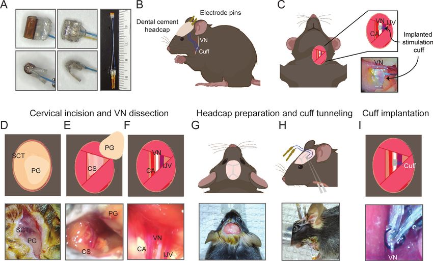
Tools and resources                                                                    Immunology and Inflammation Neuroscience

                                   controlling inflammation (Pavlov et al., 2020; Pavlov et al., 2018; Tracey, 2009; Tracey, 2002). We
                                   and others have defined VN mechanisms, termed ‘the inflammatory reflex (IR),’ which inhibit cyto-
                                   kine production in the spleen, and attenuates inflammation in preclinical and clinical studies
                                   (Borovikova et al., 2000; Rosas-Ballina et al., 2008; Rosas-Ballina et al., 2011). Clinical VN stimu-
                                   lating devices are used in the treatment of epilepsy (The Vagus Nerve Stimulation Study Group,
                                   1995) and depression (Rush et al., 2000), and are being studied in the treatment of brain disorders
                                   such as tinnitus (Tyler et al., 2017; Vanneste et al., 2017), stroke (Engineer et al., 2019;
                                   Kimberley et al., 2018), and Alzheimer’s disease (Merrill et al., 2006; Sjögren et al., 2002), as well
                                   as peripheral organ and systemic diseases, such as heart failure (De Ferrari et al., 2017), cardiac
                                   arrhythmias (Huang et al., 2015; Nasi-Er et al., 2019; Stavrakis et al., 2015; Yamaguchi et al.,
                                   2018), pulmonary hypertension (Ntiloudi et al., 2019; Yoshida et al., 2018), rheumatoid arthritis
                                   (Koopman et al., 2016), Crohn’s disease (Bonaz et al., 2016), and lupus (Aranow, 2018). Addi-
                                   tional possible indications for vagus nerve stimulation (VNS) include common disorders in which
                                   inflammation is implicated, such as type 2 diabetes, obesity, and atherosclerosis (Couzin-
                                   Frankel, 2010; Furman et al., 2019; Slavich, 2015; Strowig et al., 2012).
                                       Optimal preclinical studies of VNS in models of chronic disease require long-term implantation of
                                   a VNS device optimized for small animals, but to date the majority of chronically implanted VN devi-
                                   ces have been limited to neurological and cardiovascular diseases in rats, pigs, dogs, and other large
                                   animals (Nasi-Er et al., 2019; Yamaguchi et al., 2018; Yoshida et al., 2018; Annoni et al., 2019;
                                   Beaumont et al., 2016; Chinda et al., 2016; Farrand et al., 2017; Li et al., 2004; Meyers et al.,
                                   2018; Nuntaphum et al., 2018; Yamakawa et al., 2015; Ganzer et al., 2018). The mouse is cur-
                                   rently the species of choice in the study of disease pathophysiology, genetic mechanisms, and drug
                                   screening (Bryda, 2013; Perlman, 2016). However, VNS research in mice has been limited to acute
                                   stimulation (Bansal et al., 2012; Caravaca et al., 2019; de Lucas-Cerrillo et al., 2011;
                                   Huffman et al., 2019; Huston et al., 2006; Meneses et al., 2016; Saeed et al., 2005; Shukla et al.,
                                   2015; The et al., 2007), primarily because of significant surgical and technical challenges that
                                   accompany a mechanically (Cuoco and Durand, 2000; Prasad et al., 2014; Rydevik et al., 1981)
                                   and electrochemically (McCreery et al., 1992; Negi et al., 2010) stable long-term interface with the
                                   microscopic anatomy of the mouse VN. As a consequence, the therapeutic role of VNS in chronic
                                   inflammatory diseases and the long-term effects on neural mechanisms are largely unexplored. A
                                   functional and reliable long-term VNS implant in the mouse is necessary to broaden the translational
                                   potential of VNS in chronic diseases and provides an experimental tool to probe the long-term
                                   effects of VN neuromodulation on autonomic and neuroimmune circuits.
                                       Here, we describe a surgical technique to permanently implant a micro-cuff electrode onto the
                                   mouse cervical vagus for long-term VNS—developed, refined, and validated through a collaboration
                                   between four research labs (two labs at Feinstein Institutes, University of Colorado, and New York
                                   University). This produced a standardized, long-lasting, functionally consistent implant with expected
                                   stimulus-elicited physiological responses that persist for at least 4 weeks. Because the implant does
                                   not interfere with physiological VN-mediated reflexes, including baroreflex, lung stretch reflex, and
                                   feeding reflexes, and successfully inhibits serum TNF levels in acute endotoxemia, this method may
                                   be useful in facilitating mechanistic studies of long-term VN neuromodulation.

                                   Materials and methods
Key resources table
Reagent type (species) or resource     Designation                     Source or reference        Identifiers         Additional information
Commercial assay, kit                  Quick adhesive                  Parkell                    Cat. # S380
                                       cement
                                       system (Metabond)
Chemical compound, drug                Phenylephrine                   West-Ward Pharmaceutical   NDC 0641-6229-01
Other                                  Lipopolysaccharide (LPS) from   Sigma-Aldrich              Cat. # L-4130
                                       Escherichia coli 0111:B4
Commercial assay, kit                  TNF-a mouse                     Invitrogen                 Cat. # 88-7324-88
                                       ELISA kit
Continued on next page

Mughrabi, Hickman, et al. eLife 2021;10:e61270. DOI: https://doi.org/10.7554/eLife.61270                                             2 of 24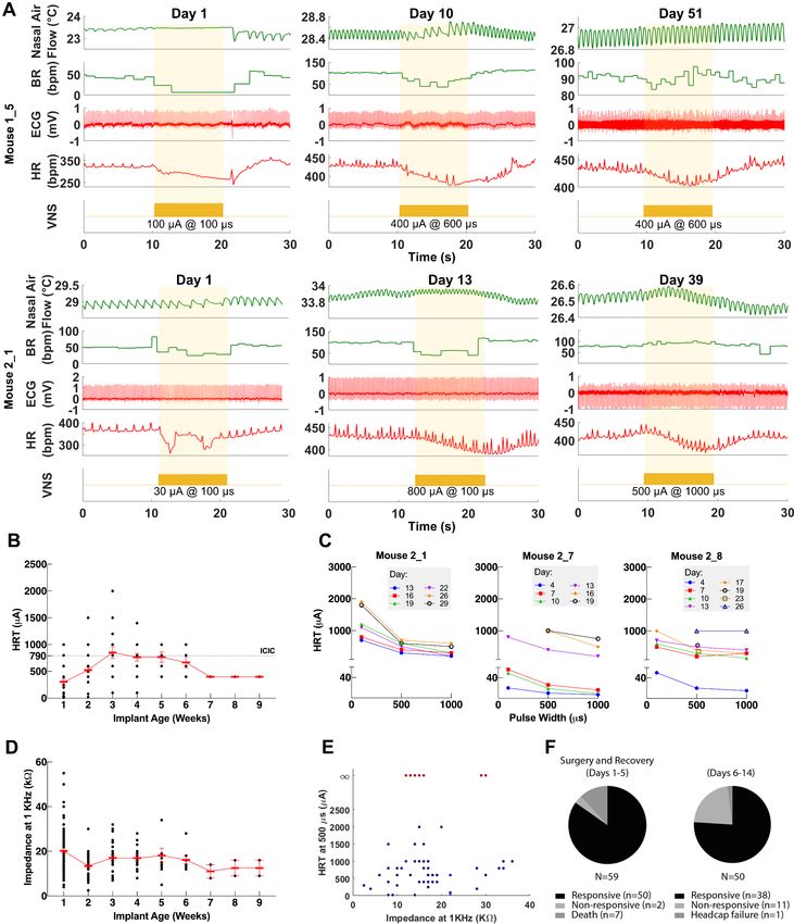
Tools and resources                                                                              Immunology and Inflammation Neuroscience

Continued

Reagent type (species) or resource        Designation                        Source or reference             Identifiers            Additional information
Antibody                                  Rabbit anti-                       Abcam                           Cat. # ab8135          1:500
                                          neurofilament                                                      RRID:AB_306298
                                          heavy polypeptide
Antibody                                  Goat anti-rabbit                   Invitrogen                      Cat. # A-11008         1:500
                                          Alexa 488                                                          RRID:AB_143165
                                          secondary antibody
Commercial assay, kit                     Trichrome stain kit                Abcam                           Cat. # ab150686

                                      Electrode preparation
                                      150 mm MicroLeads cuff electrodes (MicroLeads Neuro, Gaithersburg, MD) and 100 mm CorTec
                                      microsling cuffs (CorTec, Freiburg, Germany) were commercially fabricated and used for cuff prepa-
                                      ration (Figure 1A). MicroLeads cuffs are constructed with medical-grade assembly techniques using
                                      biocompatible materials, largely silicone, polyimide, and platinum iridium, and utilize a self-closing
                                      mechanism. CorTec microsling cuffs are also constructed with biocompatible materials including a
                                      silicone base, platinum iridium contacts, and a parylene C coating, and utilize a buckle-like closing

Figure 1. Surgical procedure for long-term implantation. (A) Lead wires of cuff electrodes produced by MicroLeads or CorTec are cut to a length of
2.5–3.0 cm and soldered to gold pins (right panel, MicroLeads). Front-facing (upper panels) and side (lower panels) view of 150 mm MicroLeads and 100
mm CorTec cuff electrodes. (B, C) Overview of implant, headcap with pins and location of the vagus nerve (VN) cuff. The cuff is implanted on the left
cervical VN. (D) A 1 cm ventral incision is made about 0.5 cm lateral to the sternal notch, exposing subcutaneous tissue (SCT) and the parotid gland
(PG). (E) SCT is bluntly dissected freeing the PG, which is then retracted from view exposing the carotid sheath (CS). (F) The VN is bluntly dissected
away from the carotid artery (CA) and the internal jugular vein (IJV). (G) The scalp is incised to expose lambda and bregma. (H) A subcutaneous tunnel
is created from skull base to cervical incision site, either between the eye and ear (depicted) or directly caudal to the ear. (I) The cuff is tunneled under
the sternomastoid muscle and implanted on the VN. Pins are finally secured to the skull with dental cement. Created with BioRender.com.

Mughrabi, Hickman, et al. eLife 2021;10:e61270. DOI: https://doi.org/10.7554/eLife.61270                                                              3 of 24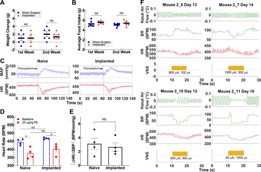
Tools and resources                                                                   Immunology and Inflammation Neuroscience

                                   mechanism. These bipolar platinum-iridium micro-cuff electrodes were soldered to gold sockets
                                   after cutting the lead wires to a length of 2.5–3.0 cm (Figure 1A). Electrical impedance was mea-
                                   sured in saline for each electrode at 1 kHz using MicroProbes Impedance Tester (MicroProbes, Gai-
                                   thersburg, MD). For sterilization, the soldered electrodes were submerged in 0.55% ortho-
                                   phthaladehyde solution (Cidex OPA, Advanced Sterilization Products, Irvine, CA) or 70% ethanol for
                                   15 min, rinsed four times with sterile saline, and if Cidex used sonicated in saline for 5 min as a final
                                   rinse.

                                   Implantation procedure
                                   Male C57BL/6 mice were purchased from Charles River Laboratories (Wilmington, MA) at the age of
                                   8–12 weeks. Animals were housed under 12 hr light/dark cycle with ad libitum access to food and
                                   water. All animal experiments complied with relevant ethical guidelines and were approved by the
                                   Institutional Animal Care and Use Committee of the Feinstein Institutes for Medical Research (proto-
                                   col numbers: 2016-029, 2017-010, and 2019-010) and University of Colorado Anschutz Medical Cam-
                                   pus (protocol number: 00238). The surgery features an implantation of a cuff electrode on the
                                   cervical VN with lead wires that are tunneled to a headcap secured with dental cement to the skull
                                   (Figure 1B, C).
                                      Mice were placed on a heated surgical platform equipped with a dissecting microscope in the
                                   supine position under isoflurane anesthesia at 4% induction and 1.5% maintenance. Hair over the
                                   neck area was removed using a depilatory cream (Nair, Church and Dwight, Ewing, NJ) or with an
                                   electric shaver and skin disinfected using alternating swabs of betadine and 70% ethanol. A 1 cm
                                   vertical incision was made starting at the level of the sternal notch about 0.5 cm left of midline
                                   (Figure 1D). The left lobe of the parotid (salivary) gland (PG) was bluntly dissected away from subcu-
                                   taneous tissue (SCT) (Figure 1D, E). The muscles forming the anterior triangle were exposed provid-
                                   ing a window to view the carotid sheath (CS) housing the carotid artery (CA), VN, and internal
                                   jugular vein (IJV). Next, the surgical field was expanded by retracting the sternomastoid muscle lat-
                                   erally using a magnetic fixator retraction system (Fine Science Tools, Foster City, CA) to ensure ade-
                                   quate access to the sheath. Using blunt dissection, the CS was isolated and the VN carefully
                                   dissected away from the fibrous connective tissue (Figure 1F). The incision site was temporarily
                                   closed, and the mouse was turned to the prone position to access the skull. The head was shaved
                                   and disinfected with alternating swabs of betadine and 70% ethanol. After application of a local
                                   analgesic, a fold of skin over the center of the skull was removed exposing bregma and lambda. The
                                   exposed skull was treated with three alternating rinses of hydrogen peroxide and saline, while gently
                                   scoring the skull surface with a sterile scalpel in between rinses (Figure 1G). After drying the skull
                                   with compressed air, acrylic dental cement (Metabond, Parkell, Edgewood, NY) was applied to the
                                   right half of the exposed area and allowed to set for 5 min. Using blunt dissection at the left edge of
                                   the scalp incision, a subcutaneous tunnel was created between the animal’s eye and ear down to the
                                   ventral neck incision location (Figure 1H). Alternatively, the subcutaneous tunnel can be performed
                                   from caudal to the dorsal ear at the base of the skull, or through a 1 cm midline incision on the dor-
                                   sal neck, to the ventral incision. The preformed subcutaneous tunnel was accessed using blunt dis-
                                   section at the lower edge of the neck incision, and a pair of fine straight forceps were used to pull
                                   the electrode cuff subcutaneously into the neck area (Figure 1H). The cuff was further tunneled
                                   under the sternomastoid muscle and placed next to the VN. The VN was gently lifted with a micro
                                   surgical hook and the cuff passed underneath it with care taken to avoid excessive manipulation.
                                   The nerve was then placed into the cuff close to the nerve’s original anatomic position and the cuff-
                                   specific closing mechanism engaged (Figure 1I). To confirm successful electrode placement, a brief
                                   stimulus was delivered through the externalized electrode leads to measure heart rate
                                   (HR) response. The neck incision was then closed with 6-0 nylon suture, and the animal turned to the
                                   prone position. The externalized connectors were held at an angle against the exposed part of the
                                   skull and acrylic dental cement (Metabond) applied covering the leads and part of the connectors
                                   and allowed to set for 5 min (Figure 1B). To seal the headcap to the skin, liquid surgical adhesive
                                   (Vetbond, 3M, Saint Paul, MN) was applied to the skin-cement junction. Alternatively, a six-channel
                                   pedestal (F12794, P1 Technologies, Roanoke, VA), surrounded by a 2 cm circle of polypropylene
                                   mesh (PPKM404.35, Surgical Mesh, Brookfield, CT), was placed into the dorsal midline neck incision
                                   after forming a subcutaneous pocket. The incision superior and inferior to the pedestal was closed
                                   with absorbable suture and surgical clips. The mice were moved to clean, warmed cages and

Mughrabi, Hickman, et al. eLife 2021;10:e61270. DOI: https://doi.org/10.7554/eLife.61270                                             4 of 24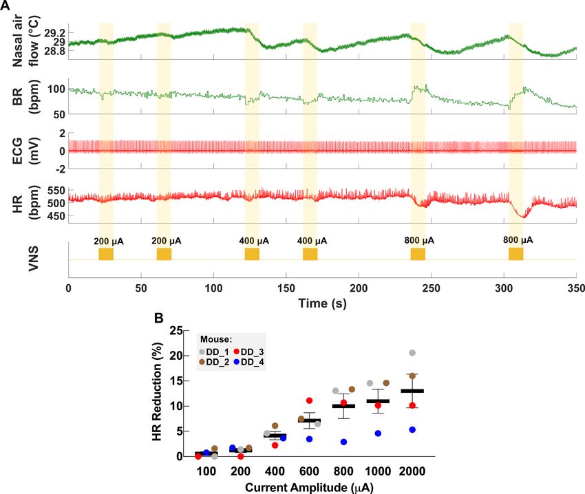
Tools and resources                                                                  Immunology and Inflammation Neuroscience

                                   monitored until conscious and mobile. The surgical procedures were carried out under strict aseptic
                                   conditions, and animals were supplemented with warm saline intra- and postoperatively. Sham sur-
                                   gery animals underwent the same procedures, including nerve isolation and manipulation and subcu-
                                   taneous tunneling, without the creation of a headcap. In some experiments, animals’ body weights
                                   and food intake were recorded daily post-implantation for at least 2 weeks. To monitor food con-
                                   sumption, singly housed animals were provided with a measured amount of laboratory chow (about
                                   13 g) in a Petri dish each day, which was collected and weighed the next day and replaced with a
                                   fresh amount.
                                       For awake stimulation experiments, mice were instrumented with implanted ECG electrodes to
                                   measure heart rate threshold (HRT) in conscious animals. Following the surgical approach described
                                   above, three platinum wires were tunneled subcutaneously along the cuff leads from the skull to the
                                   ventral neck. The left ECG lead was tunneled subcutaneously through a 1 cm incision at the left cos-
                                   tal margin and the exposed part fixed to the underlying muscle with 6-0 nylon suture. The right ECG
                                   lead was tunneled subcutaneously from the neck incision and sutured to the pectoralis muscle. The
                                   ground ECG lead was imbedded in the neck between the right lobe of the salivary gland and the
                                   skin. The ECG and cuff leads were connected to a multi-channel nano-connector (Omnetics Connec-
                                   tor Corporation, Minneapolis, MN) and cemented to the skull as described before.

                                   Nerve stimulation and physiological monitoring
                                   Validation experiments were carried out in several cohorts of mice by three research groups (Zanos
                                   group at Feinstein Institutes, Welle group at University of Colorado Anschutz Medical Campus, and
                                   Tracey group at Feinstein Institutes). Electrode functionality was evaluated by the ability to induce a
                                   decrease in HR during stimulation in anesthetized animals. HRT was defined as the minimum current
                                   intensity required to elicit an ~5–15% reduction in HR using a stimulus train of 300 bi-phasic, charge-
                                   balanced, square pulses at a pulsing frequency of 30 Hz with short (100 ms) or long (500–1000 ms)
                                   pulse widths (PWs). In most cases, HRT was initially determined with short PWs, which was changed
                                   to long PWs (500/600 ms and finally to 1000 ms whenever HRT exceeded 2 mA); in four mice, HRT
                                   was determined with both short and long PWs over several sessions. In one cohort, mice were tested
                                   on 3–7 days during the first week post-implantation, then once or twice weekly thereafter, whereas
                                   another two cohorts were tested less frequently or regularly. During testing sessions, anesthetized
                                   mice were instrumented with ECG electrodes and a nasal temperature sensor (IT-23 microprobe,
                                   Physitemp Instruments, Clifton, NJ) to measure ECG and nasal air flow and calculate HR and breath-
                                   ing rate (BR). The physiological signals were amplified using a biological amplifier (Bio-Amp Octal,
                                   ADInstruments, Colorado Springs, CO) for ECG and Temperature Pod (ADInstruments) for nasal
                                   temperature and digitized using PoweLab 16/35 (ADInstruments). The digital signals were then
                                   streamed to a PC running LabChart v8 (ADInstruments). VNS was delivered by a rack-mounted stim-
                                   ulus generator (STG4008, Multichannel Systems, Reutlingen, BW Germany). In a fourth cohort of ani-
                                   mals, stimulation response was defined as a reduction in HR measured with an infrared paw sensor
                                   (Mouse Stat Jr, Kent Scientific) or respiratory rate (measured visually) in response to a stimulus train
                                   of 0.2–1 mA intensity, short PW, and 30 Hz frequency. Stimulation failure occurred when there was
                                   no response in either HR reduction or BR alterations. Other failure modalities included headcap fail-
                                   ure. Cuff functionality was tested regularly within the first 14 days. Thereafter, a subset of the mice
                                   was selected for additional stimulation testing on a per-needed basis for further experiments.
                                      In awake experiments, animals implanted with ECG leads were gently restrained and connected
                                   to a commutator (P1 Technologies, Roanoke, VA) that interfaced with the stimulus generator and
                                   the bio-amplifier; HRT was determined as described above. Intensity at maximum charge injection
                                   capacity (CIC) was calculated using the average reported value of CIC for platinum iridium (50–150
                                   mC/cm2) (Cogan, 2008; Merrill et al., 2005) applied to the implanted electrode surface area
                                   (0.00474 cm2) for short and long PWs.

                                   Baroreflex assessment
                                   Implanted and naive mice were anesthetized and placed on a warmed surgical platform in the supine
                                   position and instrumented with ECG leads. The right external jugular vein was isolated by blunt dis-
                                   section and two sutures were placed rostrally and caudally. The rostral suture was ligated to prevent
                                   bleeding. After occluding blood flow by pulling on the caudal suture, a small incision was made in

Mughrabi, Hickman, et al. eLife 2021;10:e61270. DOI: https://doi.org/10.7554/eLife.61270                                            5 of 24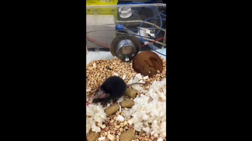
Tools and resources                                                                 Immunology and Inflammation Neuroscience

                                   the jugular vein and a 1 French catheter (Instech Labs, Plymouth Meeting, PA) was carefully
                                   advanced into the vessel after removing the caudal suture. A small amount of saline was injected to
                                   confirm the catheter was functional. The right CS was then exposed. A 1.4 French pressure catheter
                                   (SPR-671, Millar, Houston, TX) was carefully advanced into the artery using the same technique
                                   described for the jugular vein. Once the two catheters were in place and confirmed functional, 100
                                   ml of phenylephrine (25 mg/kg) supplemented with heparin (7 U/ml) in saline was injected into the
                                   jugular vein over 7 s (Fleming et al., 2013) and pressure and HR monitored. Systolic and diastolic
                                   pressure and ECG signals were amplified using Bio-Amp Octal (ADInstruments) as described before.
                                   To calculate the baroreflex sensitivity index, a 10 s window around the peak systolic blood pressure
                                   (BP) was identified to calculate the systolic BP and corresponding HR after phenylephrine injection.
                                   Baseline values were calculated from a 10 s window immediately before the injection.

                                   LPS endotoxemia challenge
                                   Lipopolysaccharide (LPS) from Escherichia coli 0111:B4 (Sigma-Aldrich, St. Louis, MO) was dissolved
                                   in saline and sonicated for 30 min before administration. LPS doses were determined empirically to
                                   produce physiological levels of TNF as described in Caravaca et al., 2019. In one set of experi-
                                   ments, performed by the Tracey group (Feinstein Institutes), 8-week-old mice (n = 12) were
                                   implanted with a left VN cuff. On day 9–17 post-surgery, VNS or sham stimulation was delivered
                                   twice (once in the morning and once in the evening) under light anesthesia using 1 mA intensity at
                                   250 ms PW and 30 Hz frequency for 5 min. On the following day, mice were administered LPS (0.7
                                   mg/kg, i.p.) 5 hr after receiving a third dose of VNS or sham stimulation. In another set of experi-
                                   ments, performed by the Zanos group (Feinstein Institutes), 8-week-old mice were implanted with a
                                   left VN cuff. HRT was determined at least 5 days before endotoxemia to avoid any long-lived VNS
                                   effects. 2–6 weeks post-implantation, animals were anesthetized and received either sham stimula-
                                   tion or VNS at HRT intensity using 250 ms PW and 10 Hz frequency for 5 min. LPS was administered
                                   to mice (0.1 mg/kg, i.p.) 3 hr after stimulation. In both sets of experiments, blood was collected by
                                   cardiac puncture 90 min post-LPS injection and left to clot for 1 hr at room temperature. The blood
                                   samples were then centrifuged at 2000 xg for 10 min and serum collected for TNF determination by
                                   ELISA (Invitrogen, Carlsbad, CA) following the manufacturer’s instructions. Serum samples were
                                   assayed in duplicate for each animal.

                                   Histology and immunohistochemistry
                                   Mice with long-term implants of at least 2 weeks old (n = 4) or naive controls were anesthetized, and
                                   the implant site carefully exposed to locate and isolate the nerve relative to anatomical landmarks.
                                   Mice were then euthanized, and the nerve fixed in place by filling the incision site with 10% buffered
                                   formalin for about 30 min. The nerve, along with the cuff in implanted animals, was then explanted
                                   and kept in 10% formalin overnight. The following day, the cuff electrodes were removed under a
                                   dissecting microscope, and the nerve samples grossed and prepared for paraffin embedding or fro-
                                   zen sectioning. Serial cross-sections of the tissue specimens were obtained at 5 mm thickness using a
                                   microtome and subsequently deparaffinized in preparation for staining. In some experiments, mice
                                   with chronic implants of at least 4 weeks (n = 5) were euthanized and segments of the neck were
                                   excised and fixed in 10% buffered formalin for at least 2 weeks. Fixed segments were then prepared
                                   for frozen sectioning. Serial cross-sections of the tissue specimens were obtained at 50 mm thickness
                                   using a cryostat. Standard immunohistochemical protocols were followed to stain the mounted sec-
                                   tions for neurofilament (Crosby et al., 2016). Briefly, sections were rinsed with 1 Tris-buffered
                                   saline (TBS) then blocked for 1 hr using 1% normal goat serum and Triton X-100 (Sigma Aldrich) in
                                   TBS. Sections were then incubated with a primary antibody against neurofilament (1:500, ab8135,
                                   Abcam, Cambridge, MA) overnight at 4˚C. The following day, sections were rinsed and incubated
                                   with goat anti-rabbit Alexa 488 secondary antibody (1:500, Thermo Fisher, Waltham, MA) for 2 hr at
                                   room temperature. Following incubation, stained slides were rinsed three times with TBS buffer then
                                   mounted with Fluoromount-G (Thermo Fisher). Images of the VN were obtained with 100 magnifi-
                                   cation using a Keyence BZ-X810 fluorescence microscope (Keyence, Osaka, Japan). Hematoxylin and
                                   eosin (H&E) and Masson’s trichrome (Trichrome Stain Kit, Abcam) stains were performed using stan-
                                   dard protocols. In neck block sections, the left VN was identified either within the tissues covering
                                   the upper margin of the cuff or in the most anterior part of the neck adjacent to the cuff.

Mughrabi, Hickman, et al. eLife 2021;10:e61270. DOI: https://doi.org/10.7554/eLife.61270                                          6 of 24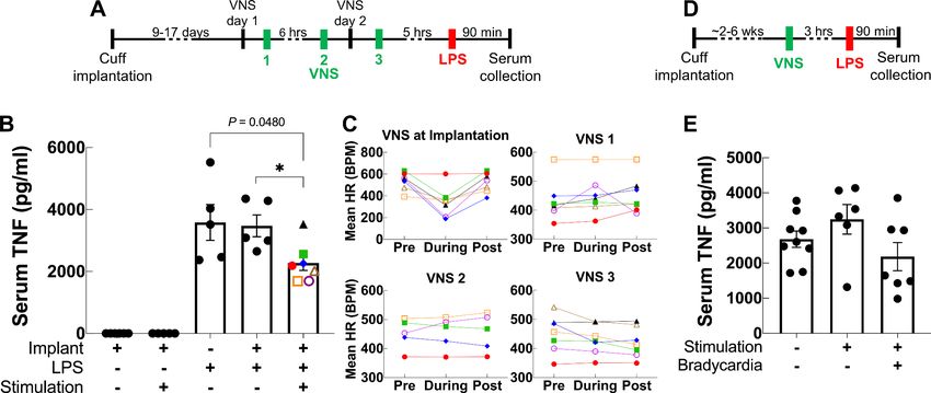
Tools and resources                                                                  Immunology and Inflammation Neuroscience

                                   Statistical analysis
                                   Pearson correlation was used to characterize the relationship between implant age and HRT, and
                                   between implant age and electrical impedance; p-values less than 0.05 were deemed statistically sig-
                                   nificant. Student’s t-test (or Mann–Whitney U for non-Gaussian variables) was used to compare
                                   between two means with Bonferroni correction for multiple comparisons; p-values less than 0.05
                                   were deemed statistically significant.

                                   Results
                                   A surgical procedure to interface with the VN in mice
                                   We first set out to design a surgical process that allows for successful long-term implantation of a
                                   micro-cuff electrode onto the mouse cervical VN. Due to the small size of the nerve (~100 mm in
                                   diameter) and cuff, this required a carefully considered protocol. We first optimized our surgical
                                   approach to isolate the CS with minimal tissue injury by employing blunt dissection using a set of
                                   fine forceps and a surgical hook (Figure 1E). Retracting the sternocleidomastoid muscle and parotid
                                   gland is critical in obtaining an adequate view of the sheath before VN isolation. Following the same
                                   principle, the VN was carefully dissected along its length using fine blunt forceps after identifying
                                   the pulsating internal CA just posterolateral to the trachea (Figure 1F). Electrode tunneling is
                                   another critical step that requires minimizing lead wire travel distance and mechanical strain, while
                                   maintaining stability. We found that subcutaneous tunneling around the neck or directly between
                                   the eye and ear (Figure 1H) both result in equally successful implants. Further, tunneling deep to
                                   the sternocleidomastoid muscle helps align the cuff on the same plane as the nerve and minimize lat-
                                   eral tracking of the cuff by providing a muscular border. We also found that maintaining a front-fac-
                                   ing cuff orientation (Figure 1A) as the cuff is tunneled to the vagus nerve ensures easy placement
                                   and prevents the cuff from pulling at or twisting the nerve. Careful adjustment of the cuff orientation
                                   intraoperatively to achieve minimal anatomic disruption improves surgical outcomes. Potential
                                   mechanical damage is further reduced by using cuffs slightly larger (100–150 mm) than the diameter
                                   of the vagus to prevent compression and by incorporating coiled wires to reduce mechanical strain.
                                   Moreover, construction of a robust headcap contributes to the stability and longevity of the implant
                                   and results in minimal headcap failures (n = 1) (Figure 3F). Careful preparation of the skull, including
                                   complete tissue removal, and adequate scoring and drying helps bind the cement to the skull sur-
                                   face and prevent infections. Also, the silicone construction of the cuff shell as well as including ade-
                                   quate distance between the edges of the cuff and stimulating electrodes reduces current leakage to
                                   surrounding tissues.

                                   VNS through the long-term implant elicits changes in HR and BR
                                   The cervical VN comprises parasympathetic motor and visceral sensory fibers that regulate many
                                   physiological functions including HR and breathing (Berthoud and Neuhuber, 2000;
                                   Agostoni et al., 1957; Chang et al., 2015). To characterize the physiological outcomes of stimula-
                                   tion through the implanted micro-cuff, we stimulated the VN with increasing current intensity while
                                   measuring stimulus-elicited changes in HR and BR in animals under isoflurane anesthesia. VNS pro-
                                   duces decreases in HR as well as changes in BR (Figure 2A). The magnitude of HR reduction is
                                   dependent on current intensity (r = 0.8971, p=0.0062) (Figure 2B), whereas BR shows more variable
                                   responses, including slowing down and acceleration of breathing (Figure 2A, Figure 3A). In con-
                                   scious mice (n = 2), VNS produces comparable dose-dependent HR responses (Video 1, Figure 2—
                                   figure supplement 1). Animals receiving awake VNS do not show any signs of distress or visible
                                   changes in BR.

                                   Longitudinal changes in implant functionality
                                   To assess the longitudinal functionality of each implant, we determined HRT over time, defined as
                                   the minimum current intensity of a stimulus train (300 pulses at 30 Hz) required to elicit an approxi-
                                   mately 5–15% decrease in HR. VNS delivered using the implant elicits drops in HR and changes in
                                   breathing for up to 8 weeks post-implantation (Figure 3A). Initial HRT values determined with short
                                   PW were variable among animals (range = 30–400 mA, mean = 156, SD = 118, n = 20 mice) and
                                   increased over the first week post-implantation (Pearson r = 0.89, p = 0.0064, n = 20 mice)

Mughrabi, Hickman, et al. eLife 2021;10:e61270. DOI: https://doi.org/10.7554/eLife.61270                                            7 of 24
Tools and resources Immunology and Inflammation Neuroscience
Figure 2. Dose-dependent physiological responses to vagus nerve stimulation (VNS). (A) Representative traces from a chronically implanted mouse
showing nasal air flow (top panel) and extracted breathing rate (BR, second panel), and ECG (third panel) and extracted heart rate (HR, fourth panel).
Trains of VNS of increasing intensity from 200 to 800 mA (fifth panel, yellow traces) cause BR and HR responses with increasing magnitudes. (B)
Percentage of HR reduction as a function of VNS intensity in four chronically implanted mice (parameters: short pulse width, frequency 30 Hz, duration 5
s).
The online version of this article includes the following source data and figure supplement(s) for figure 2:
Source data 1. Source data file (.xlsx) containing heart rate (HR) measurements at baseline and during vagus nerve stimulation (VNS) at different intensi-
ties used to create Figure 2B.
Figure supplement 1. Vagus nerve stimulation (VNS) in a conscious mouse.
(Figure 3—figure supplement 1A), whereas HRT values determined with long PWs did not change
significantly with time (r = 0.22, p NS, n = 26 mice) (Figure 3B). HRT values with short PW were
54% greater on average than those with long PW, and that relationship was maintained over time
(Figure 3C). Pre-implantation impedance values did not correlate with initial HRT (r = 0.34, p NS,
n = 15 mice) (Figure 3—figure supplement 1B), and bipolar electrical impedance did not increase
over the period of testing (r = 0.68, p = 0.0415, n = 29) (Figure 3D). Interestingly, there was no
Mughrabi, Hickman, et al. eLife 2021;10:e61270. DOI: https://doi.org/10.7554/eLife.61270 8 of 24Tools and resources Immunology and Inflammation Neuroscience Figure 3. Longitudinal changes in heart rate threshold (HRT) and electrode impedance of long-term implants. (A) Examples of physiological responses to vagus nerve stimulation (VNS) in two mice, showing changes in breathing rate (BR; green trace) and heart rate (HR; red trace) elicited by a train of VNS with HRT intensity, as determined on that day of testing (yellow trace, with stimulus parameters shown). HRT is defined as the stimulation intensity required to produce an ~5–15% decrease in HR. (B) HRT values vs. implant age (n = 26 mice), where multiple measurements were grouped together Figure 3 continued on next page Mughrabi, Hickman, et al. eLife 2021;10:e61270. DOI: https://doi.org/10.7554/eLife.61270 9 of 24
Tools and resources                                                                          Immunology and Inflammation Neuroscience

Figure 3 continued
under the corresponding week. Horizontal gray dotted line indicates intensity corresponding to maximum charge injection capacity (ICIC) with 600 ms
pulse width (PW), as calculated for these electrodes. (C) HRT values determined with VNS trains of 0.1, 0.5, and 1 ms-wide pulses at different implant
ages in three mice. (D) Electrical impedance measured at different implant ages (n = 29 mice). (E) HRT values plotted against electrode impedance
from individual measurements performed during a 40-day period post-implant. ‘Infinity’ HRT values (data points in red) indicate implants that did not
produce a HR response up to 2 mA at 2 ms PW. Pearson correlation was 0.05 (p NS). (F) Surgical success rates for one of the tested cohorts of animals.
The online version of this article includes the following source data and figure supplement(s) for figure 3:
Source data 1. Source data file (.xlsx) containing pre-implantation and longitudinal impedance and heart rate threshold (HRT) values from cohorts 1–3.
Figure supplement 1. Dependence of heart rate threshold (HRT) on electrode impedance and implant age.

                                    correlation between changes in electrical impedance and HRT values (r = 0.05, p NS); in some mice,
                                    nonfunctional cuffs continued to have relatively low impedance values despite their inability to
                                    induce a physiological response (Figure 3E). Implant failure occurred more frequently in earlier com-
                                    pared to later cohorts: percentage of mice with functional implants at 4 weeks post-implantation
                                    increased from 40% in cohort 1 to 90% in cohort 3 (Table 1). In a separate group of animals,
                                    implants were 96% functional during the first 5 days after surgery (50/52 mice, excluding deaths dur-
                                    ing surgery); functional implants were tested again in days 6–14, with a success rate of 76% (38/50)
                                    (Table 1 and Figure 3F). In a random subset of those implants, 17/18 and 11/13 were functional in
                                    the 15–30 and 30+ days period, respectively (Table 1). Overall, electrode failures occurred during
                                    the first 2 weeks post-implantation and implant functionality stabilized thereafter (Table 1).

                                    Long-term implantation does not impact vagally mediated reflexes
                                    The VN modulates several vital bodily functions via reflexes, including appetite, BP, and respiration
                                    (Paintal, 1973). To demonstrate that long-term cuffing of the VN does not affect these reflexes, we
                                    evaluated the implanted animals’ weight change and food intake, baroreflex, and breathing reflexes.
                                    The change in body weight of implanted animals during the first and second weeks post-implanta-
                                    tion is not different than sham surgery controls (Figure 4A, Figure 4—figure supplement 1A). Fur-
                                    ther, the average food intake during the first 2 weeks is similar in both groups (Figure 4B,
                                    Figure 4—figure supplement 1B) and within the range of reported daily average intake in healthy
                                    animals (Bachmanov et al., 2002). Implanted mice do not exhibit elevated levels of serum TNF 2–3
                                    weeks post-implantation (Figure 4—figure supplement 1C). To evaluate the vagal component of
                                    the baroreflex, we injected implanted and naive animals with phenylephrine, a vasopressor that
                                    increases BP, and recorded reflexive changes in HR (Figure 4C). Both implanted and naive animals
                                    have similar HR at baseline, with a similarly significant decrease upon phenylephrine injection
                                    (Figure 4C, D). Further, the baroreflex sensitivity index, expressed as the ratio of heart rate change
                                    (DHR) to systolic blood pressure change (DSBP),
                                    is not significantly different between the two
                                    groups (Figure 4E). We also evaluated whether
                                    long-term cuffing affected vagally mediated
                                    breathing reflexes (e.g., Herring–Breuer reflex;
                                    Chang et al., 2015) by evoking breathing
                                    changes with electrical stimulation. We found
                                    that mice with long-term implants exhibit
                                    changes in breathing (Figure 4F, Figure 4—fig-
                                    ure supplement 1D) similar to those induced in
                                    acute VNS experiments (Figure 4—figure sup-
                                    plement 1E).
                                                                                               Video 1. Vagus nerve stimulation (VNS) in a conscious
                                                                                               mouse. Video clip showing a conscious mouse with a
                                    VNS using the long-term implant                            long-term VN implant and ECG leads, connected to a
                                    inhibits TNF release in                                    commutator and receiving VNS on post-implant day 15.
                                    endotoxemia                                                The screen shows heart rate (HR; green trace) and a
                                    Acute VNS decreases serum TNF levels in acute              stimulation event (purple trace). VNS occurs at the 23 s
                                    inflammation by modulating the immune                      time point.
                                    response via a neuroimmune mechanism termed                https://elifesciences.org/articles/61270#video1

Mughrabi, Hickman, et al. eLife 2021;10:e61270. DOI: https://doi.org/10.7554/eLife.61270                                                      10 of 24
Tools and resources Immunology and Inflammation Neuroscience
Table 1. Functional implants across time in several animal cohorts.
Implants were tested in four cohorts. In cohorts 1–3, implant functionality was determined based on
heart rate threshold, and implant failure was defined as the absence of a physiological response
upon stimulation with 3 mA or higher on three consecutive testing sessions. In cohort 4, functionality
was determined based on a reduction in heart rate or breathing rate and failure was defined as
absence of response with 1 mA on one occasion.
Functional cuffs
Days post-implantation: 1–5 6–14 15–29 30+
Cohort 1 (n = 10) 8/10 (80%) 6/10 (60%) 4/10 (40%) 4/10 (40%)
Cohort 2 (n = 10) 10/10 (100%) 7/10 (70%) 7/10 (70%) 6/10 (60%)
Cohort 3 (n = 9) 9/9 (100%) 8/9 (90%) 8/9 (90%) –
Cohort 4 (n = 52) 50/52 (96%) 38/50 (76%) 17/18* 11/13*
*
Group is a randomly selected subset of the (6–14 days) functional implants (n = 38).
the IR (Borovikova et al., 2000). To test whether our long-term implant can produce a similar effect,
we used it to deliver VNS in an LPS endotoxemia model of acute inflammation using a set of param-
eters that have been shown to inhibit TNF release in acute experiments in mice (Caravaca et al.,
2019). Mice with 9- to 17-day-old implants received three VNS doses (1 mA intensity, 250 ms PW, 30
Hz frequency, 5 min duration): two doses administered 1 day prior to LPS administration and one
dose on the following day, 5 hr before LPS or vehicle (saline) administration (Figure 5A). We found
that VNS significantly decreases serum TNF in stimulated animals compared to sham stimulation
(Figure 5B). Notably, these parameters did not usually induce a change in HR during stimulation
(Figure 5C). In another experiment, we tested whether one-time VNS could decrease serum TNF.
Mice with either 6-week-old or 16- to 19-day-old implants received VNS (HRT intensity, 250 ms PW,
10 Hz frequency, and 5 min duration) or sham stimulation, 3 hr before LPS administration
(Figure 5D). Overall, we found that one-time VNS does not produce a significant decrease in TNF
levels. However, out of the 14 stimulated mice, VNS produced a decrease in HR in seven animals, of
which four exhibited more than ~40% decrease in serum TNF compared to sham-stimulated controls
and animals with no HR response (Figure 5E). Mice that lacked a physiological response had TNF
levels comparable to sham-stimulated controls.
Long-term implantation does not induce significant nerve damage
Long-term efficacy of peripheral nerve implants could deteriorate due to direct nerve damage or
reaction of surrounding tissue to the electrode (Anderson et al., 2008; Tyler and Durand, 2003).
To determine the impact of these processes in our long-term implants, we collected cuffed and non-
cuffed left VNs from implanted mice at ~2–6 weeks post-implantation for gross and histological anal-
ysis; naive mice were used as controls. The implant site appeared healed and exhibited moderate
tissue growth encompassing the lead wires and cuff surfaces in animals with both functional and non-
functional implants (example from 12 days post-implantation shown in Figure 6A). Histological anal-
ysis of explanted cuffed left nerves revealed preserved nerve fibers compared with non-cuffed left
nerves from naive controls with no obvious axonal pathology or inflammation (Figure 6B, Figure 6—
figure supplement 1A, B). The explanted nerves were surrounded by increased amounts of fibrotic
tissue (Figure 6C) or exhibited thickened perineurium (Figure 6B, Figure 6—figure supplement
1A). In another group of animals (n = 6) in which we examined cross-sections of whole neck blocks
just above the cuff margin 6 weeks post-implantation, histological analysis revealed similar preserved
nerves (Figure 6—figure supplement 1C).
Discussion
VNS is an emerging bioelectronic therapy with possible applications in many chronic diseases. How-
ever, its translational potential is hindered by the lack of a reliable long-term VNS implant in mice—
the preferred species in the preclinical study of human diseases (Vandenbergh, 2008). Development
of a simple, well-characterized, and reliable long-term VNS interface in mice will allow for
Mughrabi, Hickman, et al. eLife 2021;10:e61270. DOI: https://doi.org/10.7554/eLife.61270 11 of 24Tools and resources Immunology and Inflammation Neuroscience Figure 4. Vagally mediated reflexes in animals with long-term implants. (A) Weight change at first and second week post-surgery in mice subjected to sham surgery (n = 6) and in implanted mice (n = 6). (B) Average food intake during the first and second week post-surgery in sham surgery and in implanted mice (n = 6 in each group). (C) Example of baroreflex-mediated changes in heart rate (HR) in response to phenylephrine (PE)-elicited increase in blood pressure in a naive animal (left) and in an animal with a long-term implant (right). Traces showing mean arterial blood pressure (MAP; green trace) and HR (red trace); vertical line indicates time of PE injection. (D) HR in naive and implanted animals before and after PE injection. (E) Baroreflex sensitivity index in naive and implanted animals (n = 4 in each group). Index is calculated as the absolute value of the change in HR (DHR) over the change in systolic blood pressure (DSBP), before and after PE injection. (F) Examples of HR (red trace) and BR (green trace) changes in four mice with long-term implants showing responses to vagus nerve stimulation (VNS); BR responses include rapid, shallow breathing (upper left), and slowing down (upper right, lower left), or cessation of breathing (lower right) during VNS. Data is presented as mean ± SEM; NS = not significant, *p
Tools and resources                                                                          Immunology and Inflammation Neuroscience

Figure 5. Vagus nerve stimulation (VNS) using the long-term implant inhibits TNF release in endotoxemia. (A) Mice with 9- to 17-day-old implants
received three doses of VNS over 2 days using previously reported parameters (intensity 1 mA, pulse width [PW] 250 ms, frequency 30 Hz).
Lipopolysaccharide (LPS) was administered 5 hr after the last VNS dose, and blood was collected 90 min post-LPS injection. (B) Serum TNF levels from
implanted mice that received no LPS (saline) with sham or VNS (first and second bars), sham surgery mice that received LPS (third bar), and implanted
mice that received LPS with sham or VNS (fourth and fifth bars). Data shown as mean ± SEM. *p = 0.0177, by Mann–Whitney with Bonferroni correction
for multiple comparisons. (C) Mean heart rate (HR) before, during, and after each stimulation event for endotoxemic mice that received VNS. Each line
corresponds to a subject with matching shape and color in fifth bar in (B). (D) In a separate experiment, mice with 2- to 6-week-old implants (n = 22)
received a single dose of VNS, or sham VNS, for 5 min (intensity at heart rate threshold [HRT], PW 250 ms, frequency 10 Hz). LPS was administered 3 hr
after VNS and blood was collected 90 min post-LPS injection. (E) Serum TNF levels from mice that received sham stimulation (left bar), mice that
received VNS without a HR response (middle bar), and mice that received VNS that elicited a HR response (right bar). Data shown as mean ± SEM.
p NS (VNS with bradycardia vs. sham, and VNS with vs. without bradycardia) by Mann–Whitney with Bonferroni correction for multiple comparisons.

                                        To maximize the applicability of our tool in various research programs carried out by teams with
                                    different areas of expertise, we tried to accomplish two goals: ease of assembly and use, and repro-
                                    ducibility. Assembly of the implant makes use of only off-the-shelf supplies and materials, including
                                    commercially available micro-cuffs and common physiological sensors. For example, determining
                                    HRT requires the use of a simple rodent heart monitor. The surgical technique, which reflects the
                                    aggregate experience of three research groups, was refined and simplified over the course of sev-
                                    eral animal cohorts. Finally, validation of the longitudinal performance of this implant by three
                                    research groups supports the reproducibility of this method when exercised by different
                                    investigators.
                                        Previous research using VNS in mouse models of disease has been limited to acute, single-event,
                                    stimulation (Huffman et al., 2019; The et al., 2007; Ji et al., 2014). These studies, although of sig-
                                    nificant translational value, provide less insight into the possible role of VNS in the treatment of
                                    chronic conditions. Moreover, acute stimulation studies are carried out under anesthesia, which con-
                                    founds the results due to the effects of some anesthetics, such as isoflurane, on decreasing vagal
                                    tone (Marano et al., 1996; Picker et al., 2001) and suppressing the immune response (Cruz et al.,
                                    2017). For these reasons, a long-term interface to deliver stimulation to the VN is needed. However,
                                    to date, reported attempts at a stable and functional interface with the VN in mice either focused on
                                    long-term neural recordings with no longitudinal stimulation data (Caravaca et al., 2017;
                                    Falcone et al., 2020), or provided insufficient evidence of long-term implant functionality and effec-
                                    tiveness (Ten Hove et al., 2020). For example, while Caravaca et al., 2017 reported longitudinal
                                    neural recordings, VNS was delivered to a separate subset of animals 3 hr post-implantation in a ter-
                                    minal model of endotoxemia, essentially an acute procedure. Falcone et al., 2020 used their long-
                                    term microwire interface to collect neural signals but did not provide longitudinal stimulus-elicited

Mughrabi, Hickman, et al. eLife 2021;10:e61270. DOI: https://doi.org/10.7554/eLife.61270                                                      13 of 24
Tools and resources Immunology and Inflammation Neuroscience
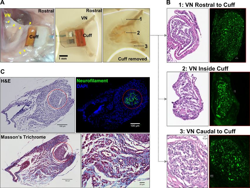
Figure 6. Gross anatomy and histology of cuffed nerve and surrounding tissues. (A) 3-week-old micro-cuff electrode upon explantation (right panel).
The cuff was carefully removed with intact rostral and caudal portions (middle panel) and nerve sectioned at three levels (dotted lines, left panel). (B)
Cross-sections of cuffed vagus nerve at corresponding levels shown in (A) stained with hematoxylin and eosin (H&E) (left panels) and for axons
(neurofilament, green; right panels). (C) Explanted cuffed vagus nerve (red dotted circle, upper left panel, H&E) stained for axons (upper right,
neurofilament, green; DAPI nuclear stain, blue), and collagen (lower panel, Masson’s Trichrome).
The online version of this article includes the following figure supplement(s) for figure 6:
Figure supplement 1. Histology examples of cuffed and non-cuffed VNs.
physiological responses to demonstrate continued stimulation functionality. In a recent study by
Ten Hove et al., 2020, VNS was delivered in mice for 12 days via a commercial cuff electrode to
study its effects on ACE2 expression in intestinal cells. Although the authors provided histological
evidence for an intact nerve structure, but without showing viable nerve fibers, they relied on visual
observation of stimulus-elicited breathing and movement changes to confirm electrode functionality,
an unreliable method for assessing nerve fiber engagement, as pain or irritation could produce a
similar behavior. They also used electrical impedance to determine implant functionality, which, as
shown in this report, is not well correlated to functional performance. Therefore, the reported lack
of effect may be related to nonfunctioning implants that went undetected, given their method of
testing. In this study, we established the feasibility of a long-term vagus interface in mice capable of
producing stimulus-elicited responses for at least 4 weeks, a period that allows for therapeutic
assessment in many models of chronic disease. Importantly, almost all animals achieved a full
Mughrabi, Hickman, et al. eLife 2021;10:e61270. DOI: https://doi.org/10.7554/eLife.61270 14 of 24Tools and resources Immunology and Inflammation Neuroscience
recovery from the implantation procedure during the first week and maintained a stable implant
after the second week. This is evident from the slow attrition of functional cuffs over time seen in
randomly selected subsets of animals (Figure 3F). We also showed that this approach can be used
to deliver dose-controlled VNS in conscious animals without repeated exposure to anesthetics, while
ensuring consistent stimulation longitudinally and across different animals (Video 1). VNS in two con-
scious animals produced comparable changes in HR as those seen in anesthetized animals and
implant longevity in those two animals ranged between 2 and 3 weeks. To our knowledge, this
report is the first demonstration of chronic VNS in the mouse that provides longitudinal evidence of
stimulus-elicited physiological responses.
VNS causes reduction in HR, mainly through activation of efferent cardioinhibitory fibers
(Buschman et al., 2006). In a functional neural interface, increasing stimulation intensity leads to
recruitment of more fibers, and hence a larger stimulus-evoked response (Chang et al., 2020;
Zaaimi et al., 2008). This is consistent with our observation wherein changes in stimulation intensity
resulted in a dose-dependent decrease in HR (Figure 2B). Additionally, higher stimulation intensities
lead to the ordered recruitment of different fiber types according to size: from large A, to intermedi-
ate-size B, to small C fibers (Blair and Erlanger, 1933). The variable changes in BR we observed at
different stimulation intensities (Figure 2A, Figure 3A) can be explained by activation of either A or
C fibers, which differentially affect breathing in mice (Chang et al., 2015) and in rats (Chang et al.,
2020). In addition, a functional nerve interface exhibits a characteristic relationship between intensity
and PW (strength-duration): to produce a response, lower stimulation intensities are required at lon-
ger PWs, and vice versa (Geddes and Bourland, 1985). Our long-term implants produced HR
responses at lower intensities with longer PWs, a relationship that remains consistent with time
(Figure 3C). These findings indicate a robust electrode-tissue interface with the VN.
An effective approach to delivering long-term VNS must include standardized methods for verify-
ing implant functionality and controlling stimulation dose over the course of treatment. To evaluate
electrode performance across time, we used HRT as a quick and accessible measure of fiber recruit-
ment in real time (Yoo et al., 2016). Initial threshold values were variable among animals and
increased over the first week post-implantation in almost all mice (Figure 3—figure supplement
1A). The variability in baseline thresholds at the time of implantation is not explained by differences
in pre-implantation impedance and is likely due to variability in electrode placement, which affects
fiber engagement. The gradual increase in HRT over time can be attributed to fibrotic encapsulation
of electrode surfaces, a process that evolves over days to weeks and is known to reduce the efficacy
of implanted neural interfaces by increasing tissue resistivity and the distance between the nerve
and the electrode contact surface (Anderson et al., 2008; Grill and Mortimer, 1994; Farah et al.,
2019; Vasudevan et al., 2017). We observed increased tissue growth within the cuff, which, upon
histological examination, comprised fibrotic tissue around the nerve (Figure 6). This may explain
why longer PWs were more effective in eliciting HRT in aged implants: the effect of distance on acti-
vation threshold is weaker for long pulses than for short pulses (Grill and Mortimer, 1995). In addi-
tion to assessing performance, HRT was used as a method to estimate individualized stimulation
doses. Since animals exhibit variable threshold values at baseline and across time, employing fixed
parameters would result in variable fiber recruitment, inconsistent therapeutic dosing, and, possibly,
undesirable off-target effects. This is of particular importance when VNS is delivered therapeutically,
wherein standardized doses are desirable within and in between animals, and across time. Previous
reports from rat and large animal models implemented a similar approach to adjust stimulation
intensity using respiratory twitching (Yoshida et al., 2018; Nishizaki et al., 2016), and HR
(Huffman et al., 2019; Chapleau et al., 2016; Yu et al., 2014) to standardize stimulation protocols,
but not on a dose-by-dose basis. Notably, electrical impedance, which is commonly used as a mea-
sure of electrode integrity and performance (Straka et al., 2018), did not always correlate with
changes in HRT on an individual implant basis as implants aged (Figure 3E). In fact, impedance
tended to decrease over the period of implantation as HRT increased (Figure 3B, D). Moreover, sev-
eral nonfunctional cuffs had relatively low impedance values (Figure 3E). For these reasons, we rely
on HRT values as a reliable indicator of electrode integrity and a method to estimate stimulation
dosage (Qing et al., 2018).
Implant longevity can be influenced by abiotic factors (Prasad et al., 2014; Vasudevan et al.,
2017), such as lead breakage and electrode degradation. We observed three cases of lead wire
breakage out of nine nonfunctional implants in three groups of animals. In all three cases, breakage
Mughrabi, Hickman, et al. eLife 2021;10:e61270. DOI: https://doi.org/10.7554/eLife.61270 15 of 24Tools and resources Immunology and Inflammation Neuroscience
occurred at the junction between the lead wire and electrode, exposing a mechanically weak point
that should be reinforced during manufacturing. Neural electrodes can degrade with long-term use
(Negi et al., 2010). This process is accelerated at stimulation intensities that exceed the electrode’s
ability to transfer charge without undergoing irreversible damage (CIC) (Negi et al., 2010;
Merrill et al., 2005). The damage imparted by exceeding this threshold is not limited to the elec-
trode but can affect the nerve as well (Cogan et al., 2016). Although we did not examine explanted
electrodes for morphological or electrochemical changes, we did take note of platinum-iridium’s
maximum charge capacity (Cogan, 2008), which was not exceeded in most animals (Figure 3B, Fig-
ure 3—figure supplement 1A). On the few occasions when ICIC was exceeded, it likely did not
cause significant damage to the electrode as the stimulation events were brief (10 s) and limited in
number. This becomes important when VNS is administered therapeutically in chronic models,
wherein stimulation protocols may be employed on a daily basis for up to several weeks. In such set-
tings, care should be taken not to exceed ICIC. Standardizing stimulation dose could prevent
exceeding CIC while still delivering therapeutic stimulation. Mechanical forces generated by chronic
cuffing can also damage nerves (Agnew and McCreery, 1990; Larsen et al., 1998; Somann et al.,
2018). Our histological analysis revealed viable nerve tissue with no obvious fiber loss or axonal frag-
mentation. Therefore, damage to the nerve is unlikely to be a contributing factor to increased HRT
over time. However, we interpret this preliminary analysis with caution, in part, due to the lack of
small-fiber detail in these relatively thick sections (5 and 50 mm). Future studies will rely on higher
resolution images with resin-embedding and electron microscopic analysis, which is the gold stan-
dard of peripheral nerve examination. Apart from electrode- and tissue-related factors, we found
that surgical proficiency contributed greatly to implant success, which is evident from the increase in
successful implantations over successive groups of animals (Table 1, cohorts 1–3, from earlier to
later) completed over several months by an initially inexperienced surgeon. Over time, fine adjust-
ments to electrode placement, surgical approach, and post-surgical care lead to higher success
rates.
The VN maintains homeostasis by controlling many bodily functions, including appetite, respira-
tion, and BP (Paintal, 1973). Chronic cuffing of the vagus could, in principle, cause damage to the
nerve, thereby affecting these functions. For example, severing the VN causes weight loss in mice
(Kral and Görtz, 1981) by affecting appetite and gastric transit times among other factors
(Dezfuli et al., 2018; Khound et al., 2017). We did not observe weight loss or changes in food
intake in our implanted animals compared with controls, which suggests that those vagal pathways
were not affected (Figure 4A, B). The VN also mediates the afferent and the efferent-parasympa-
thetic component of the baroreflex, a homeostatic mechanism that maintains BP within normal range
by modulating HR (Glick and Braunwald, 1965). In an intact system, an increase in BP causes a
reflexive decrease in HR by increasing vagal activity, which is consistent with our observations in
implanted animals. The responsiveness of this system can be quantified using the baroreflex sensitiv-
ity index or the ratio of change in HR to change in BP, which we found in implanted mice to be com-
parable to naive controls (Figure 4C, D). The vagus also carries vital sensory information from the
lung to regulate breathing (Carr and Undem, 2003). Recent evidence has shown that this is medi-
ated by large, myelinated A fibers that cause slowing down of breathing and small, unmyelinated C
fibers that cause rapid shallow breathing (Chang et al., 2015). These sensory pathways control respi-
ration by reflexive activity that changes BR, such as the Herring–Breuer inflation and deflation
reflexes (Schelegle and Green, 2001; Siniaia et al., 2000; Yu, 2016). In our studies, we were able
to evoke similar changes in BR using VNS in anesthetized animals (Figure 4F). We, however, did not
observe visible changes to BR during awake VNS. Even though there may still exist small alterations
in BR that went unnoticed visually, changes in BR in awake animals may be more difficult to visually
inspect due to movement, higher baseline BR compared to anesthetized mice, and compensatory
mechanisms that are suppressed during anesthesia. Patients receiving VNS for epilepsy do not show
changes in tidal volume or BR during wakefulness (Banzett et al., 1999), yet exhibit decreases in
air flow when VNS is activated during sleep (Murray et al., 2001). Taken together, these data
strongly suggest that the long-term implant does not cause damage to the nerve to the degree that
would affect vital bodily functions. Importantly, the fact that these preserved reflexes are mediated
by large, myelinated fibers further supports the viability of the nerve after chronic cuffing as large
fibers are the most sensitive to chronic compression injury (Dahlin et al., 1989).
Mughrabi, Hickman, et al. eLife 2021;10:e61270. DOI: https://doi.org/10.7554/eLife.61270 16 of 24You can also read


