Applying fecal microbiota transplantation (FMT) to treat recurrent Clostridium difficile infections (rCDI) in children - PeerJ
←
→
Page content transcription
If your browser does not render page correctly, please read the page content below
Applying fecal microbiota transplantation
                                   (FMT) to treat recurrent Clostridium
                                   difficile infections (rCDI) in children
                                   Shaaz Fareed1 ,4 ,* , Neha Sarode2 ,* , Frank J. Stewart3 , Aneeq Malik4 ,
                                   Elham Laghaie4 , Saadia Khizer5 , Fengxia Yan6 , Zoe Pratte3 , Jeffery Lewis7 and
                                   Lilly Cheng Immergluck1 ,4
                               1
                                 Department of Microbiology/Biochemistry/Immunology, Morehouse School of Medicine, Atlanta, GA,
                                 United States of America
                               2
                                 Department of Organismic & Evolutionary Biology, Harvard University, Boston, MA,
                                 United States of America
                               3
                                 School of Biological Sciences, Georgia Institute of Technology, Atlanta, GA, United States of America
                               4
                                 Clinical Research Center, Morehouse School of Medicine, Atlanta, GA, United States of America
                               5
                                 Clinical Research, Children’s Healthcare of Atlanta, Atlanta, GA, United States of America
                               6
                                 Department of Community Health & Preventive Medicine, Morehouse School of Medicine, Atlanta, GA,
                                 United States of America
                               7
                                 Pediatric Gastroenterology, Children’s Center for Digestive Health Care, LLC, Atlanta, GA,
                                 United States of America
                               *
                                 These authors contributed equally to this work.

                                       ABSTRACT
                                       Background. Fecal Microbiota Transplantation (FMT) is an innovative means of
                                       treating recurrent Clostridium difficile infection (rCDI), through restoration of gut
                                       floral balance. However, there is a lack of data concerning the efficacy of FMT and its
                                       impact on the gut microbiome among pediatric patients. This study analyzes clinical
                                       outcomes and microbial community composition among 15 pediatric patients treated
                                       for rCDI via FMT.
                                       Methods. This is a prospective, observational, pilot study of 15 children ≤18 years, who
Submitted 10 December 2017
Accepted 2 April 2018
                                       presented for rCDI and who met inclusion criteria for FMT at a pediatric hospital and
Published 30 May 2018                  pediatric gastroenterology clinic. Past medical history and demographics were recorded
Corresponding authors
                                       at enrollment and subsequent follow-up. Specimens of the donors’ and the patients’
Neha Sarode,                           pre-FMT and post-FMT fecal specimen were collected and used to assess microbiome
nehasarode@fas.harvard.edu             composition via 16S rRNA gene sequencing.
Lilly Cheng Immergluck,                Results. FMT successfully prevented rCDI episodes for minimum of 3 months post-
limmergluck@msm.edu
                                       FMT in all patients, with no major adverse effects. Three patients reported continued
Academic editor                        GI bleeding; however, all three also had underlying Inflammatory Bowel Disease
Keith Crandall
                                       (IBD). Our analyses confirm a significant difference between pre-and post-FMT gut
Additional Information and             microbiome profiles (Shannon diversity index), whereas no significant difference was
Declarations can be found on
page 14                                observed between post-FMT and donor microbiome profiles. At the phyla level, post-
                                       FMT profiles showed significantly increased levels of Bacteroidetes and significantly
DOI 10.7717/peerj.4663
                                       decreased levels of Proteobacteria. Subjects with underlying IBD showed no difference
    Copyright                          in their pre-and post-FMT profiles.
2018 Fareed et al.                     Conclusion. The low rate of recurrence or re-infection by C. difficile, coupled with
Distributed under                      minimal adverse effects post-FMT, suggests that FMT is a viable therapeutic means to
Creative Commons CC-BY 4.0             treat pediatric rCDI. Post-FMT microbiomes are different from pre-FMT microbiomes,
OPEN ACCESS

                                   How to cite this article Fareed et al. (2018), Applying fecal microbiota transplantation (FMT) to treat recurrent Clostridium difficile in-
                                   fections (rCDI) in children. PeerJ 6:e4663; DOI 10.7717/peerj.4663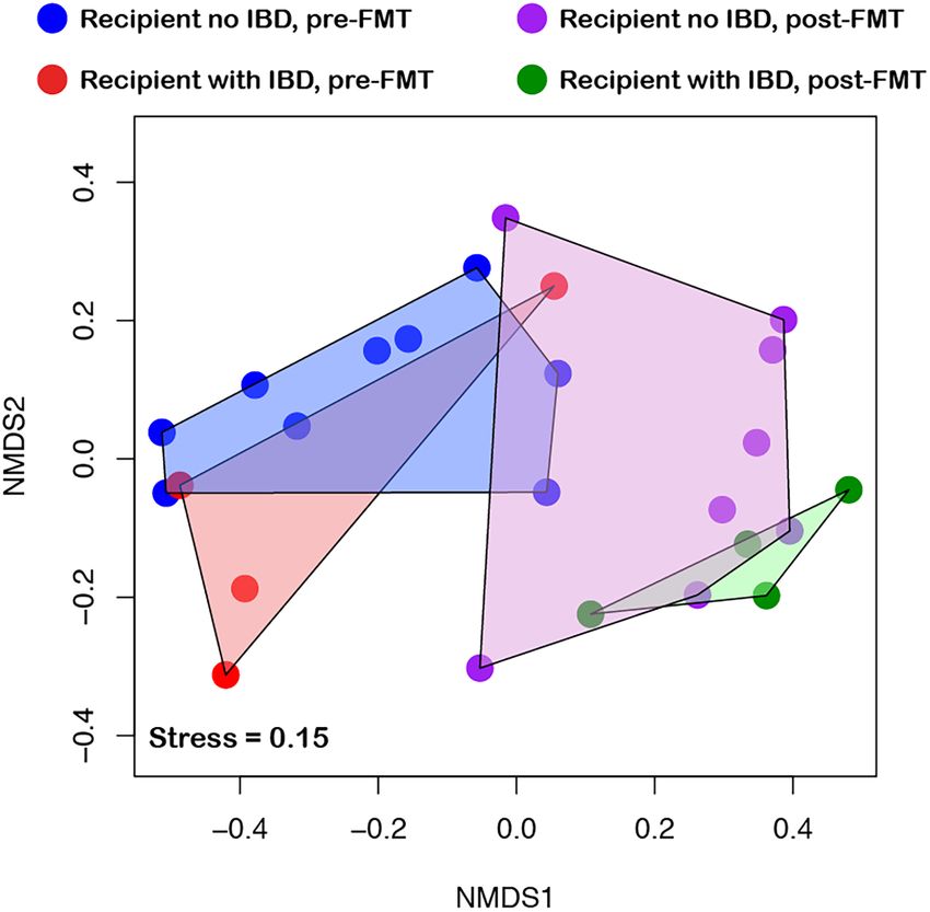
and similar to those of healthy donors, suggesting successful establishment of a healthier
                                      microbiome.

                                   Subjects Microbiology, Gastroenterology and Hepatology, Infectious Diseases, Pediatrics,
                                   Translational Medicine
                                   Keywords Fecal microbiota transplantation, Pediatrics, Clostridium difficile infection, Micro-
                                   biome

                                   INTRODUCTION
                                   Childhood infections by the anaerobic gram-positive bacterium Clostridium difficile pose a
                                   significant health challenge, with limited viable treatment options for recurrent infections.
                                   Approximately half a million Clostridium difficile infections (CDI) occur annually in the
                                   United States and are associated with approximately 29,000 deaths (Lessa et al., 2015).
                                   Although an estimated 17,000 CDI cases involve children ages 1 to 17 years (Center for
                                   Disease Control and Prevention, 2015), the majority of studies exploring treatment efficacy
                                   focus on adults (Lees et al., 2016). Even though clinical disease is not seen frequently among
                                   infants and very young children, presumably due in part to the lack of toxin-binding
                                   receptors in this age group coupled with protective maternal antibodies, the incidence
                                   among children has continued to increase over the past two decades (Nylund et al., 2011;
                                   Zilberberg, Tillotson & McDonald, 2010). Risks associated with CDI include prior antibiotic
                                   use (Schutze et al., 2013) and gut motility dysfunction, with higher incidence observed
                                   in children with inflammatory bowel disease (IBD) (Pascarella et al., 2009). Recurrence
                                   of disease also has increased, with rates estimated to be as high as 25% (Aslam, Hamill
                                   & Musher, 2005). Currently, the preferred first line treatment for recurrent CDI (rCDI)
                                   is oral vancomycin (Leong & Zelenitsky, 2013; Sandora et al., 2011). However, up to 25%
                                   of patients undergoing oral vancomycin therapy for rCDI relapse within one month
                                   of treatment (Louie et al., 2011; Vincent & Manges, 2015). Furthermore, dysbiosis in gut
                                   commensal bacteria, caused initially by C. difficile infection, is further exacerbated by
                                   antibiotic use (Isaac et al., 2017).
                                       The human gut microbiome is a topic of extensive interest given overwhelming evidence
                                   for the microbiome’s role in shaping health and disease (Dore & Blottiere, 2015; Estrela,
                                   Whiteley & Brown, 2015; Lozupone et al., 2012). Bacteroidetes and Firmicutes are the major
                                   gut phyla of interest: they compose >90% of the healthy human gut microbiome, and as
                                   commensal bacteria, function as a barrier in preventing the C. difficile overgrowth (Bien et
                                   al., 2013; Malys, Campbell & Malys, 2015). Studies profiling rCDI gut microbiomes show
                                   overall decreased species richness, decreased ratios of Bacteroidetes to Firmicutes, and
                                   increased Proteobacteria, (Chang et al., 2008; Theriot & Young, 2015) suggesting attempts
                                   to correct microbiome dysbiosis as a potential treatment strategy for rCDI.
                                       For children, the repeated bouts of diarrhea, abdominal pain, and medical treatment
                                   associated with rCDI incur costs and interfere with normal function. One innovative
                                   approach to re-establish the gut microbiome is through fecal microbiota transplantation
                                   (FMT). In a review of 317 adult rCDI patients (average age of 53 years) treated via

Fareed et al. (2018), PeerJ, DOI 10.7717/peerj.4663                                                                                 2/19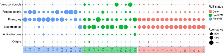
FMT, FMT alleviated rCDI symptoms in 92% of cases (Gough, Shaikh & Manges, 2011).
                                   Furthermore, FMT has been shown to successfully address dysbiosis by improving the
                                   ratio of Bacteroidetes and Firmicutes, thus producing a post-FMT gut bacterial community
                                   similar to that of the donor (Seekatz et al., 2014). In general, the pediatric microbiomes
                                   not only differ in the proportions of major gut phyla (Hollister et al., 2015), but also
                                   exhibit significantly higher interpersonal variation and lower diversity compared to adult
                                   microbiomes (Yatsunenko et al., 2012). Though FMT has showed promising results in
                                   adults (Rao & Safdar, 2016), this treatment option has been applied sparingly in children
                                   (Russell et al., 2010), and even fewer studies have analyzed the efficacy of FMT to treat
                                   pediatric rCDI and the impact of the procedure on pediatric gut microbiomes (Hourigan
                                   & Oliva-Hemker, 2016).
                                      The primary goal of this prospective, observational and epidemiological pilot study was
                                   to assess the practicality, safety and efficacy of FMT as a treatment option for pediatric
                                   patients with rCDI. A secondary aim was to observe the gut microbiota changes elicited
                                   by FMT within pediatric rCDI patients. There have been 45 cases using FMT in children
                                   with rCDI and the largest series involved 10 children, three of whom had IBD (Kronman
                                   et al., 2015). To our knowledge, ours is the largest FMT study to be conducted among
                                   pediatric patients with rCDI, which involves children both with and without underlying
                                   inflammatory bowel disease.

                                   MATERIALS & METHODS
                                   Overall study design
                                   This is an observational epidemiology study to look at children who failed multiple courses
                                   of standard antibiotic therapy for rCDI and elected to undergo FMT. Prior to sampling, this
                                   study was approved by the institutional review board of Children’s Healthcare of Atlanta
                                   and Morehouse School of Medicine.
                                       Study participants (n = 15 pediatric patients) were screened and recruited from a
                                   pediatric hospital (Children’s Healthcare of Atlanta Hospital at Scottish Rite) and a private
                                   pediatric gastroenterology clinic (Children’s Center for Digestive Health Care, LLC) in
                                   Atlanta, Georgia. All study participants were children (age range 21 months to 18 years)
                                   who presented with symptoms consistent with rCDI, as determined from examination by
                                   the same pediatric gastroenterologist. Patients were eligible for the FMT procedure if they
                                   fit the study definition of rCDI: ongoing diarrhea, a positive C. difficile fecal specimen test
                                   (polymerase chain reaction assay to detect C. difficile toxin genes), and history of failed CDI
                                   treatment with antibiotics for a minimum of two prior CDI episodes, of which, at least one
                                   episode was treated with a course of oral vancomycin within the previous eight weeks (Cohen
                                   et al., 2010). All FMT procedures were performed via either colonoscopy or nasojejunal tube
                                   by the same pediatric gastroenterologist. Previous episodes of CDI were recorded if patient
                                   had a documented positive C. difficile toxin PCR test and a diagnosis of CDI in his/her
                                   medical records. Informed consent (or assent when age appropriate) was obtained from a
                                   patient’s parent or legal guardian prior to the FMT procedure. A survey questionnaire was
                                   administered in a personal interview from study staff to record participant demographic

Fareed et al. (2018), PeerJ, DOI 10.7717/peerj.4663                                                                          3/19
information (age, gender, race/ethnicity), underlying chronic health conditions, prior
antibiotic use within 12 months of the time of enrollment, and CDI related symptoms.
The following information was also collected from each participant’s medical records:
nature of prior episodes of CDI, dates of positive PCR C. difficile toxin tests, and provider
notes documenting history of infections that required antibiotics and specific symptoms
consistent with CDI. Five donor samples were ordered from OpenBiomeTM (Medford,
MA, USA) to be used for the FMT procedure. All donors were non-family related and
underwent laboratory screening by OpenbiomeTM (Osman et al., 2016; Osman et al., 2016)
for infectious risk factors (including Multi-drug resistant organisms (MDRO’s), serologic
testing, and fecal specimen testing) and potential microbiome-mediated conditions.
FMT procedure
FMT procedure was performed on subjects via colonoscopy (n = 14) and nasojejunal tube
(n = 1). Prior to FMT, all patients undergoing a colonoscopy completed a standard bowel
prep using Polyethylene Glycol 3350. Vancomycin was discontinued at least 48 h prior
to the FMT. Colonoscopies were performed under general anesthesia or under conscious
sedation. Up to 240 ml of donor fecal specimen was instilled into the terminal ileum
and cecum during colonoscopy in 14 of 15 patients. In one patient, who had undergone
colonoscopy due to an underlying disease, 60 cc of donor fecal specimen was instilled via
nasojejunal tube into the proximal jejunum.
Follow-up
Follow-up telephone calls were made by research staff after 24 h and one-week post-FMT
procedure. Survey questionnaires were administered by research staff at one month and
≥3 months post-FMT time points, alongside scheduled evaluation by single pediatric
gastroenterologist. Responses to questions about study participant’s symptoms, including
fever, vomiting, abdominal pain and distention, evidence of allergic reaction or bloody
fecal specimens were recorded in a relational database. If patients were not available to
respond at the three-month follow-up visit, attempts to collect a response were performed
for up to 15 months after the procedure. (Although fecal samples were collected outside
of the three-month study period, patients were not clinically re-evaluated for evidence of
rCDI beyond three months of the date of fecal microbiota transplantation.)
Fecal specimen collection
Three fecal specimen samples were collected for each study participant: (1) from the
donor—used for transplant, (2) from the study participant before transplant (pre-FMT),
(3) from the study participant following the FMT procedure (at least 3 months after
FMT procedure), with the latter used to analyze the effect of FMT on gut microbiome
composition. Fecal samples were de-identified and stored in a −80 ◦ C freezer until DNA
extraction.
Fecal specimen DNA extraction
DNA was extracted from fecal specimen samples using the MO BIO PowerSoil DNA
extraction kit (MOBIO, Carlsbad, CA, USA) following manufacturer instructions, with
Fareed et al. (2018), PeerJ, DOI 10.7717/peerj.4663 4/19modifications as recommended by the Human Microbiome Project (HMP) Manual of
Procedures (version 12.0). Briefly, before step 2 of the standard protocol, the PowerBead
tubes containing samples were heated at 65 ◦ C for 10 min, followed by a second incubation
at 95 ◦ C for 10 min to improve cell lysis. The manufacturer protocol was followed for the
remainder of the procedure, except for step 12, during which the tubes were centrifuged for
2 min. DNA was extracted from triplicate subsamples for each fecal specimen sample, with
the extracted DNA then pooled for quantification and processing. DNA concentrations
were measured using the Qubit 2.0 DNA quantification system (ThermoFisher Scientific,
Waltham, MA, USA).
16S rRNA gene PCR and amplicon sequencing
Microbiome taxonomic composition in donor, pre-FMT, and post-FMT fecal specimen was
characterized by Illumina sequencing of PCR amplicons encompassing the V4 hypervariable
region of the 16S rRNA gene. Amplicons were synthesized using Platinum PCR Supermix
(ThermoFisher Scientific, Waltham, MA, USA) with V4-specific primers F515 and R806
(Caporaso et al., 2010), and uniform amounts of DNA template per reaction. Both forward
and reverse primers were barcoded and appended with Illumina-specific adapters, as
per instructions by Kozich et al. (2013). Thermal cycler conditions were as follows: initial
denaturation at 94 ◦ C (3 min), followed by 30 cycles of denaturation at 94 ◦ C (45 s), primer
annealing at 55 ◦ C (45 s) and primer extension at 72 ◦ C (90 s), and a final extension at
72 ◦ C for 10 min. Amplicons were verified on agarose gel electrophoresis for size (∼400 bp)
and purified using Diffinity RapidTip2 pipet tips (Diffinity Genomics, West Chester, PA,
USA). Barcoded and Illumina adaptor-appended amplicons for each sample were pooled at
equimolar concentrations and sequenced on an Illumina MiSeq using a 500 cycle v2 reagent
kit (250 bp paired end) with 15% PhiX genomic library addition to increase read diversity.
Statistical and diversity analyses
Demultiplexed amplicon read pairs were quality trimmed with Trim Galore! (Babraham
Bioinformatics, Cambridge, UK), using a base Phred33 score threshold of Q25 and a
minimum length cutoff of 100 bp. High quality paired reads were then merged using
the software FLASH (Magoč & Salzberg, 2011). Merged reads were analyzed using QIIME
v1.9.0 (Caporaso et al., 2010), according to standard protocols. Briefly, reads were screened
for chimeras using QIIME’s identify_chimeric_seqs.py script with Usearch61 (Edgar, 2010).
Non-chimeric sequences were clustered into Operational Taxonomic Units (OTUs) at 97%
sequence similarity using UCLUST (Edgar, 2010) based on open-reference OTU picking
with the script pick_open_reference_otus.py. Taxonomy was assigned to representative
OTUs from each cluster using the Greengenes database (Aug 2013 release) (DeSantis et
al., 2006).
R (R Core Team, 2017) packages Phyloseq (McMurdie & Holmes, 2013) and Vegan (Jari
Oksanen et al., 2017) were used to measure alpha and beta diversity, respectively. Alpha
diversity was estimated by the number of observed unique OTUs and the Shannon diversity
index at an even sampling depth of 27,700 sequences. OTUs observed across at least 70%
samples were used for identifying shared OTUs between groups. Microbiome compositional
Fareed et al. (2018), PeerJ, DOI 10.7717/peerj.4663 5/19differences among samples were assessed using non-metric multidimensional scaling
(NMDS) based on Bray-Curtis distances at OTU level.
The significance of the effect of group status (donor, pre-FMT, post-FMT) on
microbiome composition was assessed using the envfit function in Vegan, with 999
permutations. Wilcoxon signed rank test with Benjamini–Hochberg correction for multiple
hypothesis testing was used to compare the taxonomic differences between pre- and post-
FMT, pre- FMT and donor as well as post-FMT and donor. The differences between
IBD and non-IBD were compared using Wilcoxon two sample test for demographic
information (age, gender, race/ethnicity), underlying chronic health conditions, prior
antibiotic use within 12 months of the time of enrollment, and CDI related symptoms
and participant’s medical records: Nature of prior episodes of CDI, dates of positive PCR,
C. difficile toxins, and provider notes documenting history of infections which required
antibiotics and specific symptoms consistent with CDI. R and SAS 9.4 were used to perform
all the statistical tests and adjusted p-value < 0.05 (after multiple hypothesis correction)
was reported as statistically significant.
RESULTS
Patient enrollment/population characteristics
Fifteen pediatric study participants (eight female, seven male) were enrolled in this study.
Of the potentially 45 specimens, only 40 fecal bio specimens were collected from these 15
patients. (Three post-FMT fecal samples were lost to follow up (LTFU), and two Pre-FMT
samples did not yield enough DNA for analysis.) The median age of patients was eight years
(range: 21 months to 18 years) (Table 1). Patients had an average of three episodes of CDI
prior to FMT (range: two to five CDI episodes.). On average, post-FMT fecal samples were
collected 6 months after the FMT procedure (range: three to 14 months) (Fig. 1).
Clinical outcomes
All 15 patients tolerated the FMT procedure without complications immediately post
procedure. For the 12 patients who completed their three-month follow-up appointments,
all experienced clinical resolution of CDI and had no recurrent episodes within the
duration of the follow-up period (three months). Abdominal pain was reported by five
patients (none had underlying IBD) at the three-month visit; however, the treating
pediatric gastroenterologist attributed the pain to other causes, including functional pain
and irritable bowel syndrome not related to CDI. All six patients with underlying ulcerative
colitis (n = 5) or Crohn’s disease (n = 1) reported hematochezia and ongoing abdominal
distension beyond three months of their FMT date.
Microbiome analysis—overall bacterial community diversity and
richness
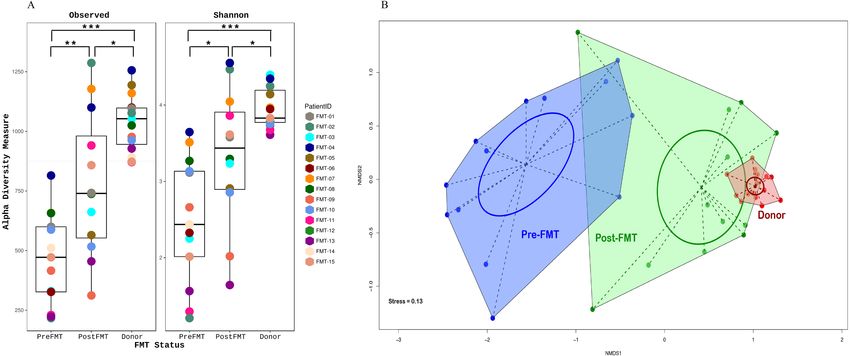
FMT significantly altered gut microbiome composition in all the participants. Alpha
diversity (within sample diversity), measured as both observed OTU richness and Shannon
diversity, differed between all compared FMT groups, with diversity in the donor sample
higher compared to the matched pre- and post-FMT samples collected from recipients;
Fareed et al. (2018), PeerJ, DOI 10.7717/peerj.4663 6/19Table 1 Patient characteristics. Summary of patient profiles for all subjects enrolled in this study. Included are demographic information, delivery
route of fecal microbiota transplantation (FMT), underlying inflammatory bowel disease (IBD), and recorded number of prior Clostridium difficile
infections (CDI) episodes.
ID Sex Race FMT delivery Age Time FMT Number of Underlying IBD Number of FMT
method (years) and post- antibiotic CDI episodes donor
at FMT FMT stool courses prior prior to FMT ID
collection to FMTc
(Months)
1 F Biracial Colonoscopy 2 3 4 No 4 05
2 F Black Colonoscopy 7 9 1 Ulcerative Colitis 3 05
3 M White Colonoscopy 16 4 4 Ulcerative Colitis 4 05
4 F Biracial Colonoscopy 8 14 3 No 3 05
5 M Black Colonoscopy 10 7 4 Ulcerative Colitis 3 05
6 F Black Nasojejunal 8 LTFUa 8 Ulcerative Colitis 3 05
Tube
7 M White Colonoscopy 1 6 4 No 4 37
8 F White Colonoscopy 7 4 2 Crohn’s Disease 3 37
9 F White Colonoscopy 18 4 2 No 2 37
10 F White Colonoscopy 2 6 2 No 3 37
11 F White Colonoscopy 8 6 2 No 5 37
12 M White Colonoscopy 2 LTFU 2 No 2 12
13 M Biracialb Colonoscopy 5 3 3 No 3 77
14 F White Colonoscopy 15 LTFU 7 No 3 66
15 M White Colonoscopy 10 2 1 No 3 77
Notes.
a
LTFU = Sample Lost to Follow Up.
b
Biracial = Participant self-identified as Black/White.
c
Number of antibiotic courses only within the period of 12 months prior to FMT was included.
All patients had up to date vaccinations for routine immunizations, including Diphtheria-Tetanus-acellular Pertussis, Haemophilus influenzae Type B, Influenza, Pneumococcal
Conjugate Vaccine, with seven or 13 Serogroups, Inactivated Polio Virus, Hepatitis A and B; Rotavirus; and Varicella.
pre-FMT fecal samples had the lowest diversity. There was no significant difference
in observed OTU richness between the five donor samples (Observed OTU p-value =
0.07894; Shannon diversity p-value 0.04306). Post-FMT microbiomes, although more
diverse compared to pre-FMT microbiomes, were less diverse than donor microbiomes
(Fig. 2A). A significant difference (Kruskal-Wallis test, p-value < 0.001) was also observed
in shared OTU’s between all three groups (donor, recipient pre-FMT, and recipient
post-FMT), where within each comparison, donor vs. post-FMT profiles showed the
smallest variance (calculated using Wilcoxon signed rank test), while pre- vs post-FMT
profiles had the greatest variance.
FMT status (donor, recipient pre-FMT, and recipient post-FMT) also significantly
influenced microbiome beta diversity (between sample diversity), as measured by Bray-
Curtis distance-based NMDS ordinations (r2 = 0.65, p-value < 0.001; envfit function in
Vegan), with post-FMT microbiome composition being more like that of donor than that
of the pre-FMT group (Fig. 2B). Together, these results indicate a positive shift in microbial
community composition in response to FMT, suggesting a restructuring to a non-disease
state akin to the donor.
Fareed et al. (2018), PeerJ, DOI 10.7717/peerj.4663 7/197
6
6
5
4
Frequency
3
3
2
2
1
1
0
0
3 4-6 7-9 10-12 13-15
Months Post-FMT
Figure 1 Time of stool collection post-fecal microbiota transplantation (FMT). Displayed is a his-
togram detailing how many patients submitted stool at the displayed time periods (in months) Post-FMT.
Full-size DOI: 10.7717/peerj.4663/fig-1
Microbiome analysis—taxon level changes
The relative representation of major bacterial phyla of interest varied substantially in
response to FMT (Fig. 3). Notably, the average ratio of Bacteroidetes to Firmicutes (B:F),
the predominant phylum-level indicator of a ‘normal/healthy’ gut microbiome, increased
over 7-fold from pre-FMT (average B:F of 0.15) to post-FMT microbiomes (average B:F:
1.12), with the recipient post-FMT B:F approaching that of donor communities (average
B:F: 1.49) (Table 2). The shift in this ratio was driven largely by changes in the proportional
representation of Bacteroidetes, which comprised an average of only 7% relative abundance
across all pre-FMT samples (excluding study participant 15, who was an outlier), but
showed a relative abundance of 41% and 57% in recipient post-FMT and donor samples,
respectively (Table 2). Much of the observed variation within the Bacteroidetes was due
to proportional shifts involving a single genus, Bacteroides. In addition to Bacteroidetes,
the phyla Proteobacteria and Verrucomicrobia were significantly enriched in donor and
post-FMT compared to pre-FMT fecal samples (Fig. 3), whereas these three phyla did not
differ in abundance between donor and post-FMT fecal samples, highlighting a shift in the
microbiome profile, likely caused by FMT, towards that of the donor.
Fareed et al. (2018), PeerJ, DOI 10.7717/peerj.4663 8/19Figure 2 Alpha and beta diversity of samples. (A) Alpha diversity is a measure of species richness
within a sample which is quantitatively expressed here as a box-plot of the number of observed unique
taxa/OTU’s and Shannon diversity index on y-axis. Individual samples are colored according to their
patient ID grouped by their FMT status on the x-axis. (B) Beta diversity is a measure of taxonomic
composition diversity between sample that is represented as Non-metric multidimensional scaling
(NMDS) ordination of samples based on Bray-Curtis distance matrix. The color and shape of samples
are according to their FMT status identity (RED, donor; BLUE, pre-FMT or GREEN, post-FMT),
with samples belonging to the same FMT group connected to form polygons. The more similar the
groups are to each other (donor and post-FMT), the closer their polygon clusters are going to be on
the ordination plot and vice versa (pre-FMT compared to both donor and post-FMT). The dotted lines
connect individual samples to the group centroid while the ellipse gives an estimate of standard deviation
of the scores. (FMT, Fecal Microbiota Transplantation).
Full-size DOI: 10.7717/peerj.4663/fig-2
Figure 3 Distribution of Major Phyla between Donor, Recipient Pre- and Post-FMT Microbiomes.
The bubble plot above displays the proportions of phyla observed in each sample. Red indicates donor
samples, blue indicates pre-FMT samples, and green indicates post-FMT samples. An increase in Bac-
teroidetes can be observed from pre to post-FMT samples, as well as a decrease in Proteobacteria. Post-FMT
phyla proportions are more similar to donor profiles. (FMT, Fecal Microbiota Transplantation).
Full-size DOI: 10.7717/peerj.4663/fig-3
Microbiome analysis—bacterial profile by genus
The value of examining the proportional representation of microbial phyla with regards to
gut microbiome health has been questioned (Jandhyala et al., 2015). When examining the
changes that occurred instead at the genus level, the most noticeable shifts were reflective of
the changes that occurred at the phylum level (Table 2). Specifically, the genus Bacteroides
showed a considerable and significant increase in recipients’ pre- to post-FMT (from
Fareed et al. (2018), PeerJ, DOI 10.7717/peerj.4663 9/19Table 2 Microbiome analyses- taxon level changes between Bacteroidetes, Firmicutes, and Proteobac-
teria. Abundance of major microbial phyla in stool microbiomes, expressed for each sample type as the
percentage of total sequences classifiable to the phylum level, averaged across all samples.
Phylum Donor Pre-FMT Post-FMT
Bacteroidetes 57% 7% 41%
Firmicutes (F) 38% 50% 36%
Proteobacteria (B) 4% 41% 5%
Average B:F Ratio 1.49 0.15 1.12
average 6.5 to 39.2 %). The average relative abundance of the genus Clostridium in the
donor was 0.02% (min = 0.001%, max = 0.08%), consistent with a non-disease state. In
response to FMT, the average relative abundance of Clostridium in FMT recipients fell
roughly 80%, from an average of 1% pre-FMT (min = 0%, max = 4.9%) to 0.16% post-
FMT (min = 0%, max = 1.58%). Three of the FMT recipient participants had Clostridium
relative abundance of 0%, four had very low but detectable percentages (≤0.01%), while
6 had levels ≥0.5%. Genera identified to be altered significantly in relative abundance per
FMT status are listed in Table S1.
Effect of underlying IBD
Of the 15 patients enrolled, 10 had no underlying inflammatory bowel disease (IBD),
whereas five had been diagnosed with IBD (four ulcerative colitis and one Crohn’s disease
with one patient having had total colectomy) (Table 3). Community taxonomic profiles
clustered according to pre- versus post-FMT status, rather than IBD status (Fig. 4),
suggesting that underlying IBD had little to no effect on the microbial shift observed
because of FMT. There was a marginal effect in the Proteobacteria abundance on separation
of community with and without IBD (r 2 = 0.54, p = 0.018).
Furthermore, no discernable differences in post-procedural symptoms such as
abdominal pain, vomiting, or bloody fecal specimens were observed between patients
with or without underlying IBD.
DISCUSSION
Fecal microbiota transplantation (FMT) is a novel approach to treating rCDI by restoration
of a healthy gut microbiome (Bakken et al., 2011). FMT has become an acceptable
therapeutic consideration for adults, particularly after recurrence of CDI (Surawicz et
al., 2013). However, few data are available from pediatric studies beyond case reports or
small case series for children (Hourigan & Oliva-Hemker, 2016). Our findings are consistent
with what has been reported in adults and children who have undergone FMT using non-
familial, non-autologous donors. Specifically, in our study of 15 children, we show that this
procedure is well tolerated by children, irrespective of underlying gastrointestinal disorders
(ulcerative colitis and Crohn’s) and that the taxonomic shift in the gut microbiome to
resemble the donor’s microbiome is similar regardless of compositional variation in
pre-FMT profiles among patients. Moreover, resolution of CDI symptoms seemed to last
from at least three months to as long as 14 months.
Fareed et al. (2018), PeerJ, DOI 10.7717/peerj.4663 10/19Table 3 Comparison of symptoms post fecal microbiota transplantation (FMT) for patients with and without underlying inflammatory bowel
diseases (IBD). Gastrointestinal (GI) symptoms include one or more of the following: abdominal pain, fever, abdominal distension, or bloody
stool. (The categorical variables were compared using Fisher’s Exact test due to the small sample and the continuous variables were evaluated using
Wilcoxon two independent sample test.)
Variable No underlying IBD Underlying IBD p-value
(n = 10, 66%) (n = 5, 33%)
Age, median(IQR) 6.55(8.01) 8.02(2.61) 0.4053
Gender male% 5(50) 2(40) 1.0000
GI symptoms prior to FMT (yes, %) 9(90) 5(100) 1.0000
GI symptoms at 24-hour post FMT 0.5055
No 3(30) 0(0)
Yes 7(70) 4(100)
GI symptoms at 1-week post FMT 0.0667
No 1(12.5) 3(75)
Yes 7(87.5) 1(25)
GI symptoms at 1-month post FMT 0.2657
No 3(37.5) 4(80)
Yes 5(62.5) 1(20)
GI symptoms at 3 months post FMT 1.0000
No 2(28.6) 1(20)
Yes 5(71.4) 4(80)
Route of FMT 0.3333
Colonoscopy 10(100) 4(80)
NJ Tube 0 1(20)
Number of antibiotic courses prior to FMT, median(IQR) 2.5(2) 4(2) 0.6217
Number of ED visits within a year of FMT, median(IQR) 1.5(3) 4(3) 0.0468
Number of CDI episodes, median(IQR) 3(1) 3(0) 0.9456
B:F Ratio, median(IQR) 0.0021(0.0068) 0.0030(0.0027) 0.8209
Clostridia Distribution, median (IQR) 0.0055(0.0127) 0.00001(0.0109) 0.4102
Very young children with rCDI represent a challenging population, given that the
microbial gut community has not ‘matured’ and the diversity of dominant phyla is
shifting. In our study, almost 25% of participants were less than two years of age (one of
the four patients had ulcerative colitis and received FMT at 21 months of age). However,
we did not identify any significant differences in the distribution of dominant phyla across
the different ages. There are limited data on how FMT (usually transplantation is with adult
donors, who have ‘mature’ microbial profiles) might affect those as young as 2 years of
age (Kahn, Gorawara-Bhat & Rubin, 2012; Kronman et al., 2015; Russell et al., 2014; Walia
et al., 2014; Wang et al., 2015).
Similar to what has been observed in larger adult studies (Seekatz et al., 2014) we
also found that for most of our pediatric patients, FMT seemed to induce a post FMT
microbiome that was significantly different from the pre-FMT microbiome profile, and
more similar to the healthy donor profile. It would be interesting to explore serial fecal
biospecimens for up to one year post FMT and compare the trends of the microbial
Fareed et al. (2018), PeerJ, DOI 10.7717/peerj.4663 11/19Figure 4 Effect of inflammatory bowel disease (IBD) on community diversity. NMDS ordination of
samples considering their IBD status showed that, underlying IBD condition had no significant effect on
community dynamics. Samples are colored based on the combination of their FMT and IBD status, with
samples belonging to the same group connected with polygon for visual clarity. (FMT, Fecal Microbiota
Transplantation).
Full-size DOI: 10.7717/peerj.4663/fig-4
community composition, particularly as it relates to the Bacteroidetes: Firmicutes ratio for
both patients with and without underlying gastrointestinal disorders.
Overall, pre-FMT profiles showed high abundance of Proteobacteria, with low amounts
of Bacteroidetes. Post-FMT profiles showed an increase in Bacteroidetes, with a large
decrease in Proteobacteria. These findings are in agreement with results from past
studies of adult patients (Seekatz et al., 2014), De’Argenio and Salvatore, for example,
found that a high abundance of Proteobacteria pre-FMT is associated with underlying
conditions, such as ulcerative colitis and Crohn’s disease (D’Argenio & Salvatore, 2015).
Furthermore, Proteobacteria is often selected over Bacteroidetes and Firmicutes when colitis
is present (Bradley & Pollard, 2017). It is possible that these factors were responsible for
the distributions of Proteobacteria observed in this study.
Fareed et al. (2018), PeerJ, DOI 10.7717/peerj.4663 12/19With regards to major gut phyla, two patients showed a decrease in the relative abundance
of Bacteroidetes from pre- to post-FMT. Although this was not expected, as all other
profiles showed an often, large increase in Bacteroidetes abundance, these two patients
also demonstrated significant decreases in the abundances of Proteobacteria, thus still
appearing more similar to their respective ‘healthy’ donor profiles. It is likely that an
increase in Bacteroidetes was not observed since the pre-FMT profiles of these patients
already displayed higher abundances of Bacteroidetes in comparison to the other subjects.
Interestingly, underlying gastrointestinal conditions did not adversely affect tolerance
of the procedure or preponderance of post procedural adverse effects, nor were there
any significant differences observed in pre-FMT profiles or distribution of Bacteroidetes,
Firmicutes, or Proteobacteria; the shift in major gut phyla post-FMT was similar across all
patients.
Limitations/future directions
Because this was a prospective observational pilot study, microbiome results and statistical
implications are limited by a low sample size (n = 15 patients). Not all follow-up phone
calls were completed within the time frame set in the study design, and 5 post-FMT samples
were not obtained. Additional variables that could impact the microbiome, such as diet,
exercise, environmental factors, puberty, etc., were not collected, although the list of such
potential factors could be in practice rather exhaustive. Fecal samples were ultimately
analyzed three to 12 months’ post-procedure due to inconsistencies in patient compliance.
Our covariate analysis is also limited by the fact that study surveys were self-reported.
We recognize the risks associated with FMT, and although this study shows promising
results, we suggest that FMT should only be used once current guidelines of vancomycin
therapy have failed. Future randomized control trials of vancomycin taper versus FMT
would be beneficial in determining which option is most effective for treating recurrence.
Trials with larger sample sizes need to be performed to establish the effect that FMT
has on the microbiome. Furthermore, longitudinal studies of pediatric patients would
establish long-term efficacy of this treatment option. Given the small number of pediatric
studies, establishing a national FMT database would be useful in analyzing the impact
and side effects of FMT. Since this study was an observational study, we did not collect
information on participants’ diet or environmental factors which may play a role in the
gut microbiome. However, in future studies these determinants of health are important to
consider in patients who undergo FMT and also in characterizing factors which affect the
gut microbiome.
CONCLUSIONS
In our limited prospective study of children with rCDI, with and without IBD, FMT is
efficacious, with minimal adverse effects, and improves the intestinal microbiome in favor
of higher proportion of Bacteroidetes.
Fareed et al. (2018), PeerJ, DOI 10.7717/peerj.4663 13/19ACKNOWLEDGEMENTS
We would like to thank: Dr. Sangita Ganesh (Georgia Institute of Technology) for her
help with sample processing; Ms. Jonelle McKay, Dr. Robert C. Jerris, Dr. Mark Gonzales
(Children’s Healthcare of Atlanta’s Clinical Microbiology Laboratory), for their assistance
in the processing and proper storage of all biosamples used in the molecular analyses;
Program managers Ms. Anaam Mohammed (Pediatric Emergency Medicine Associates,
LLC) and Ms. Victoria Churchill (Morehouse School of Medicine) for co-ordinating the
project across institutions; and finally, our heartfelt gratitude to the nurses and providers
at Children’s Center for Digestive Health Care.
ADDITIONAL INFORMATION AND DECLARATIONS
Funding
This work was supported by the Children’s Healthcare of Atlanta, Friends’ Fund; National
Center for Advancing Translational Sciences of the National Institutes of Health under
Award Number UL1TR002378; K-08 Agency Healthcare Research Quality HS024338-
01; Centers for Disease Control and Prevention Emerging Infections Program Grants
U01C10000307-05 and U01000312. The funders had no role in study design, data collection
and analysis, decision to publish, or preparation of the manuscript.
Grant Disclosures
The following grant information was disclosed by the authors:
Children’s Healthcare of Atlanta, Friends’ Fund.
National Center for Advancing Translational Sciences of the National Institutes of Health:
UL1TR002378.
K-08 Agency Healthcare Research Quality: HS024338-01.
Centers for Disease Control and Prevention Emerging Infections Program Grants:
U01C10000307-05, U01000312.
Competing Interests
The authors declare there are no competing interests.
Author Contributions
• Shaaz Fareed performed the experiments, analyzed the data, prepared figures and/or
tables, authored or reviewed drafts of the paper, approved the final draft.
• Neha Sarode performed the experiments, analyzed the data, contributed reagents/ma-
terials/analysis tools, prepared figures and/or tables, authored or reviewed drafts of the
paper, approved the final draft.
• Frank J. Stewart conceived and designed the experiments, performed the experiments,
analyzed the data, contributed reagents/materials/analysis tools, authored or reviewed
drafts of the paper, approved the final draft.
• Aneeq Malik and Elham Laghaie performed the experiments, analyzed the data, authored
or reviewed drafts of the paper, approved the final draft.
Fareed et al. (2018), PeerJ, DOI 10.7717/peerj.4663 14/19• Saadia Khizer authored or reviewed drafts of the paper, approved the final draft.
• Fengxia Yan analyzed the data, prepared figures and/or tables, authored or reviewed
drafts of the paper, approved the final draft.
• Zoe Pratte performed the experiments, approved the final draft.
• Jeffery Lewis conceived and designed the experiments, performed the experiments,
analyzed the data, contributed reagents/materials/analysis tools, authored or reviewed
drafts of the paper, approved the final draft, identified the patients with underlying GI
condition; performed all the FMT.
• Lilly Cheng Immergluck conceived and designed the experiments, performed the
experiments, analyzed the data, contributed reagents/materials/analysis tools, prepared
figures and/or tables, authored or reviewed drafts of the paper, approved the final draft.
Human Ethics
The following information was supplied relating to ethical approvals (i.e., approving body
and any reference numbers):
The following Institutional Review Boards provided approval for the conduct of this
study:
Children’s Healthcare of Atlanta; Morehouse School of Medicine; Georgia Institute of
Technology.
Data Availability
The following information was supplied regarding data availability:
The raw data, de-identified, has been included as a Supplemental file.
Supplemental Information
Supplemental information for this article can be found online at http://dx.doi.org/10.7717/
peerj.4663#supplemental-information.
REFERENCES
Aslam S, Hamill RJ, Musher DM. 2005. Treatment of Clostridium difficile-associated
disease: old therapies and new strategies. The Lancet Infectious Diseases 5:549–557
DOI 10.1016/S1473-3099(05)70215-2.
Bakken JS, Borody T, Brandt LJ, Brill JV, Demarco DC, Franzos MA, Kelly C, Khoruts
A, Louie T, Martinelli LP, Moore TA, Russell G, Surawicz C, Fecal Microbiota
Transplantation Workgroup. 2011. Treating Clostridium difficile infection with fecal
microbiota transplantation. Clinical Gastroenterology and Hepatology 9:1044–1049
DOI 10.1016/j.cgh.2011.08.014.
Bien J, Bozko M, PM N, Bozko P. 2013. Editorial: gut microbiota and gastrointestinal
diseases: to treat or not to treat. Current Pharmaceutical Design 20:4533–4534.
Bradley PH, Pollard KS. 2017. Proteobacteria explain significant functional variability in
the human gut microbiome. Microbiome 5:1–23 DOI 10.1186/s40168-017-0244-z.
Caporaso JG, Kuczynski J, Stombaugh J, Bittinger K, Bushman FD, Costello EK, Fierer
N, Peña AG, Goodrich JK, Gordon JI, Huttley GA, Kelley ST, Knights D, Koenig JE,
Fareed et al. (2018), PeerJ, DOI 10.7717/peerj.4663 15/19Ley RE, Lozupone CA, McDonald D, Muegge BD, Pirrung M, Reeder J, Sevinsky
JR, Turnbaugh PJ, Walters WA, Widmann J, Yatsunenko T, Zaneveld J, Knight
R. 2010. QIIME allows analysis of high-throughput community sequencing data.
Nature Methods 7:335–336 DOI 10.1038/nmeth.f.303.
Center for Disease Control and Prevention. 2015. Nearly half a million Americans
suffered from Clostridium difficile infections in a single year. CDC Newsroom.
Available at https:// www.cdc.gov/ media/ releases/ 2015/ p0225-clostridium-difficile.
html.
Chang JY, Antonopoulos DA, Kalra A, Tonelli A, Khalife WT, Schmidt TM, Young
VB. 2008. Decreased diversity of the fecal microbiome in recurrent Clostridium
difficile—associated diarrhea. The Journal of Infectious Diseases 197:435–438
DOI 10.1086/525047.
Cohen SH, Gerding DN, Johnson S, Kelly CP, Loo VG, McDonald LC, Pepin J, Wilcox
MH, Society for Healthcare Epidemiology of America, Infectious Diseases Society
of America. 2010. Clinical practice guidelines for Clostridium difficile infection in
adults: 2010 update by the society for healthcare epidemiology of America (SHEA)
and the infectious diseases society of America (IDSA). Infection Control and Hospital
Epidemiology 31:431–455 DOI 10.1086/651706.
D’Argenio V, Salvatore F. 2015. The role of the gut microbiome in the healthy adult
status. Clinica Chimica Acta 451:97–102 DOI 10.1016/j.cca.2015.01.003.
DeSantis TZ, Hugenholtz P, Larsen N, Rojas M, Brodie EL, Keller K, Huber T, Dalevi
D, Hu P, Andersen GL. 2006. Greengenes, a Chimera-Checked 16S rRNA gene
database and workbench compatible with ARB. Applied and Environmental Micro-
biology 72:5069–5072 DOI 10.1128/AEM.03006-05.
Dore J, Blottiere H. 2015. The influence of diet on the gut microbiota and its conse-
quences for health. Current Opinion in Biotechnology 32:195–199
DOI 10.1016/j.copbio.2015.01.002.
Edgar RC. 2010. Search and clustering orders of magnitude faster than BLAST. Bioinfor-
matics 26:2460–2461 DOI 10.1093/bioinformatics/btq461.
Estrela S, Whiteley M, Brown SP. 2015. The demographic determinants of human mi-
crobiome health. Trends in Microbiology 23:134–141 DOI 10.1016/j.tim.2014.11.005.
Gough E, Shaikh H, Manges AR. 2011. Systematic review of intestinal microbiota
transplantation (Fecal Bacteriotherapy) for recurrent Clostridium difficile infection.
Clinical Infectious Diseases 53:994–1002 DOI 10.1093/cid/cir632.
Hollister EB, Riehle K, Luna RA, Weidler EM, Rubio-Gonzales M, Mistretta T-A, Raza
S, Doddapaneni HV, Metcalf GA, Muzny DM, Gibbs RA, Petrosino JF, Shulman
RJ, Versalovic J. 2015. Structure and function of the healthy pre-adolescent pediatric
gut microbiome. Microbiome 3:1–13 DOI 10.1186/s40168-015-0101-x.
Hourigan SK, Oliva-Hemker M. 2016. Fecal microbiota transplantation in children: a
brief review. Pediatric Research 80:2–6 DOI 10.1038/pr.2016.48.
Fareed et al. (2018), PeerJ, DOI 10.7717/peerj.4663 16/19Isaac S, Scher JU, Djukovic A, Jimenez N, Littman DR, Abramson SB, Pamer EG,
Ubeda C. 2017. Short- and long-term effects of oral vancomycin on the hu-
man intestinal microbiota. Journal of Antimicrobial Chemotherapy 72:128–136
DOI 10.1093/jac/dkw383.
Jandhyala SM, Talukdar R, Subramanyam C, Vuyyuru H, Sasikala M, Nageshwar
RD. 2015. Role of normal gut microbiota. World Journal of Gastroenterology
21:8787–8803 DOI 10.3748/wkg/v21.i29.8787.
Jari Oksanen FGB, Friendly M, Kindt R, Legendre P, McGlinn D, Minchin PR, O’Hara
RB, Simpson GL, Solymos P, Henry M, Stevens H, Szoecs E, Wagner H. 2017.
vegan: Community Ecology Package. R package version 2.4-6. Available at https:
// CRAN.R-project.org/ package=vegan.
Kahn SA, Gorawara-Bhat R, Rubin DT. 2012. Fecal bacteriotherapy for ulcerative
colitis: patients are ready, are we? Inflammatory Bowel Diseases 18:676–684
DOI 10.1002/ibd.21775.
Kozich JJ, Westcott SL, Baxter NT, Highlander SK, Schloss PD. 2013. Development of
a dual-index sequencing strategy and curation pipeline for analyzing amplicon se-
quence data on the MiSeq Illumina sequencing platform. Applied and Environmental
Microbiology 79:5112–5120 DOI 10.1128/AEM.01043-13.
Kronman MP, Nielson HJ, Adler AL, Giefer MJ, Wahbeh G, Singh N, Zerr DM, Suskind
DL. 2015. Fecal microbiota transplantation via nasogastric tube for recurrent
Clostridium difficile infection in pediatric patients. Journal of Pediatrics Gastroenterol-
ogy and Nutrition 60:23–26 DOI 10.1097/MPG.0000000000000545.
Lees EA, Miyajima F, Pirmohamed M, Carrol ED. 2016. The role of Clostridium difficile
in the paediatric and neonatal gut—a narrative review. European Journal of Clinical
Microbiology & Infectious Diseases 35:1047–1057 DOI 10.1007/s10096-016-2639-3.
Leong C, Zelenitsky S. 2013. Treatment strategies for recurrent Clostridium difficile
infection. The Canadian Journal of Hospital Pharmacy 66:361–368.
Lessa FC, Mu Y, Bamberg WM, Beldavs ZG, Dumyati GK, Dunn JR, Farley MM,
Holzbauer SM, Meek JI, Phipps EC, Wilson LE, Winston LG, Cohen JA, Limbago
BM, Fridkin SK, Gerding DN, McDonald LC. 2015. Burden of Clostridium difficile
infection in the United States. New England Journal of Medicine 372:825–834
DOI 10.1056/NEJMoa1408913.
Louie TJ, Miller MA, Mullane KM, Weiss K, Lentnek A, Golan Y, Gorbach S, Sears P,
Shue Y-K. 2011. Fidaxomicin versus Vancomycin for Clostridium difficile infection.
New England Journal of Medicine 364:422–431 DOI 10.1056/NEJMoa0910812.
Lozupone CA, Stombaugh JI, Gordon JI, Jansson JK, Knight R. 2012. Diversity,
stability and resilience of the human gut microbiota. Nature 489:220–230
DOI 10.1038/nature11550.
Magoč T, Salzberg SL. 2011. FLASH: fast length adjustment of short reads to improve
genome assemblies. Bioinformatics 27:2957–2963 DOI 10.1093/bioinformatics/btr507.
Malys MK, Campbell L, Malys N. 2015. Symbiotic and antibiotic interactions between
gut commensal microbiota and host immune system. Medicina 51:69–75.
Fareed et al. (2018), PeerJ, DOI 10.7717/peerj.4663 17/19McMurdie PJ, Holmes S. 2013. phyloseq: an R package for reproducible interac-
tive analysis and graphics of microbiome census data. PLOS ONE 8:e61217
DOI 10.1371/journal.pone.0061217.
Nylund CM, Goudie A, Garza JM, Fairbrother G, Cohen MB. 2011. Clostridium difficile
infection in hospitalized children in the United States. Archives of Pediatrics and
Adolescent Medicine 165:451–457 DOI 10.1001/archpediatrics.2010.282.
Osman M, O’Brien K, Stoltzner Z, Ling K, Koelsch E, Dubois N, Khoiri A, Amaratunga
K, Smith M, Kassam Z. 2016. Safety and efficacy of fecal microbiota transplantation
for recurrent Clostridium difficile infection from an international public stool bank:
results from a 2050-patient multicenter cohort. Open Forum Infectious Diseases
3:2120–2120 DOI 10.1093/ofid/ofw172.1668.
Osman M, Stoltzner Z, O’Brien K, Ling K, Koelsch E, Dubois N, Amaratunga K, Smith
M, Kassam Z. 2016. Donor efficacy in fecal microbiota transplantation for recurrent
Clostridium difficile: evidence from a 1,999 -Patient Cohort. Open Forum Infectious
Diseases 3(Suppl_1):841 DOI 10.1093/ofid/ofw194.48.
Pascarella F, Martinelli M, Miele E, Del Pezzo M, Roscetto E, Staiano A. 2009. Impact
of Clostridium difficile infection on pediatric inflammatory bowel disease. Jornal de
Pediatria 154:854–858 DOI 10.1016/j.jpeds.2008.12.039.
Rao K, Safdar N. 2016. Fecal microbiota transplantation for the treatment of Clostridium
difficile infection. Journal of Hospital Medicine 11:56–61 DOI 10.1002/jhm.2449.
R Core Team. 2017. R: a language and environment for statistical computing. Vienna: R
Foundation for Statistical Computing. Available at https:// www.R-project.org/ .
Russell G, Kaplan J, Ferraro M, Michelow IC. 2010. Fecal bacteriotherapy for relapsing
Clostridium difficile infection in a child: a proposed treatment protocol. Pediatrics
126:e239–242 DOI 10.1542/peds.2009-3363.
Russell GH, Kaplan JL, Youngster I, Baril-Dore M, Schindelar L, Hohmann E, Winter
HS. 2014. Fecal transplant for recurrent Clostridium difficile infection in children
with and without inflammatory bowel disease. Journal of Pediatrics Gastroenterology
and Nutrition 58:588–592.
Sandora TJFM, Flaherty K, Helsing L, Scanlon P, Potter-Bynoe G, Gidengil CA, Lee
GM. 2011. Epidemiology and risk factors for Clostridium difficile infection in chil-
dren. Pediatric Infectious Disease Journal 7:580–584
DOI 10.1097/INF.0b013e31820bfb29.
Schutze GE, Willoughby RE, Brady MT, Byington CL, Davies HD, Edwards KM, Glode
MP, Jackson MA, Keyserling HL, Maldonado YA, Murray DL, Orenstein WA,
Zaoutis TE. 2013. Clostridium difficile infection in infants and children. Pediatrics
131:196–200 DOI 10.1542/peds.2012-2992.
Seekatz AM, Aas J, Gessert CE, Rubin TA, Saman DM, Bakken JS, Young VB. 2014.
Recovery of the gut microbiome following fecal microbiota transplantation. mBio
5:e00893–00814 DOI 10.1128/mBio.00893-14.
Surawicz CM, Brandt LJ, Binion DG, Ananthakrishnan AN, Curry SR, Gilligan PH,
McFarland LV, Mellow M, Zuckerbraun BS. 2013. Guidelines for diagnosis,
Fareed et al. (2018), PeerJ, DOI 10.7717/peerj.4663 18/19treatment, and prevention of Clostridium difficile infections. American Journal of
Gastroenterology 108:478–498 DOI 10.1038/ajg.2013.4.
Theriot CM, Young VB. 2015. Interactions between the gastrointestinal micro-
biome and Clostridium difficile. Annual Review of Microbiology 69:445–461
DOI 10.1146/annurev-micro-091014-104115.
Vincent C, Manges AR. 2015. Antimicrobial use, human gut microbiota and Clostridium
difficile colonization and infection. Antibiotics 4:230–253
DOI 10.3390/antibiotics4030230.
Walia R, Garg S, Song Y, Girotra M, Cuffari C, Fricke WF, Dutta SK. 2014. Efficacy of
fecal microbiota transplantation in 2 children with recurrent Clostridium difficile
infection and its impact on their growth and gut microbiome. Journal of Pediatrics
Gastroenterology and Nutrition 59:565–570 DOI 10.1097/MPG.0000000000000495.
Wang J, Xiao Y, Lin K, Song F, Ge T, Zhang T. 2015. Pediatric severe pseudomem-
branous enteritis treated with fecal microbiota transplantation in a 13-month-old
infant. Biomedical Reports 3:173–175 DOI 10.3892/br.2014.403.
Yatsunenko T, Rey FE, Manary MJ, Trehan I, Dominguez-Bello MG, Contreras M,
Magris M, Hidalgo G, Baldassano RN, Anokhin AP, Heath AC, Warner B, Reeder
J, Kuczynski J, Caporaso JG, Lozupone CA, Lauber C, Clemente JC, Knights
D, Knight R, Gordon JI. 2012. Human gut microbiome viewed across age and
geography. Nature 486:222–227.
Zilberberg MD, Tillotson GS, McDonald C. 2010. Clostridium difficile infections
among hospitalized children, United States, 1997–2006. Emerging Infectious Diseases
16:604–609 DOI 10.3201/eid1604.090680.
Fareed et al. (2018), PeerJ, DOI 10.7717/peerj.4663 19/19You can also read