HYPERMOBILITY AND EHLERS-DANLOS - NZORD
←
→
Page content transcription
If your browser does not render page correctly, please read the page content below
HYPERMOBILITY AND
             EHLERS-DANLOS
            SYNDROMES (EDS) –
    NEW ZEALAND GUIDELINE
            2019

     Includes Generalised Joint
Hypermobility (GJH) and Hypermobility
     Spectrum Disorders (HSD)

New Zealand Hypermobility and Ehlers-Danlos Syndromes Guideline 2019
                                                                                  Version 1
                                                                       8/1/19   Page 1 of 24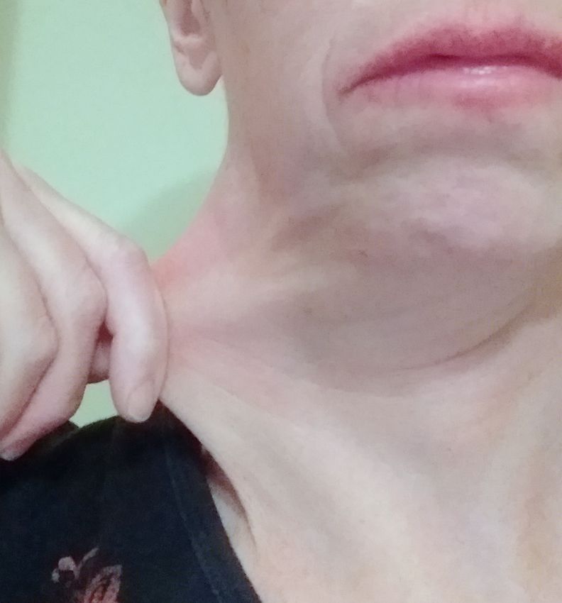
This guideline was written by members of the Hypermobility and Ehlers-
Danlos Syndromes NZ Working Group, convened by the NZ Organisation
for Rare Disorders 2017-2018.
WORKING GROUP MEMBERS (Alphabetical)

NAME                      ORGANISATION                   ROLE
BURLING, Fraser           Rheumatologist Auckland        Rheumatologist
CALLEAR, Rachel           Hutt Valley DHB                Paediatric Rheumatologist Physiotherapist
CAMPBELL-STOKES,          Hutt Valley DHB                Paediatric Rheumatologist Hutt Valley DHB
Priscilla
BROMHEAD, Collette        NZORD                          Chief Executive, Relationship Manager
CRAWFORD, Lisa
PRESTON, Matthew          BOPDHB; Eastbay Radiology      Medical - Radiologist
ROBERTSON, Stephen        Department of Women’s and      Curekids Professor of Paediatric Genetics
                          Children’s Health
                          Dunedin School of Medicine
                          University of Otago
TAYLOR, Will              Hutt Valley DHB                Consultant Rheumatologist and
                          Wakefield Specialist Medical   Rehabilitation Physician, Hutt Valley DHB,
                          Centre                         Assoc Prof

New Zealand Hypermobility and Ehlers-Danlos Syndromes Guideline 2019
                                                                                             Version 1
                                                                       8/1/19              Page 2 of 24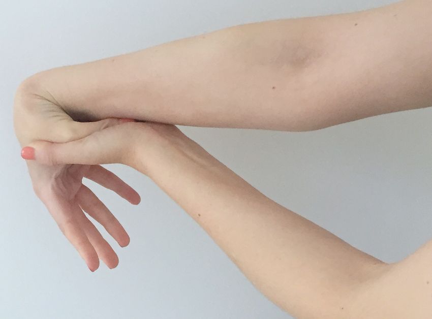
KEY POINTS
About this document
This Guideline is intended to assist clinicians and allied healthcare providers to recognise joint
hypermobility, consider alternative diagnoses and develop an initial management plan when they see
patients with one or more of the following:

    •   Joint hypermobility (double-jointed), especially when associated with musculoskeletal pain
    •   Recurrent joint subluxations/dislocations
    •   Skin fragility or hyperextensible skin in isolation or together with
    •   Unusual bruising or bleeding

Hypermobility spectrum disorders (HSD) are a group of conditions related to Joint Hypermobility (JH).
HSD are diagnosed only after other possible conditions have been excluded, such as Ehlers Danlos
Syndrome(s) (EDS) including Hypermobile EDS (hEDS) and the rarer EDS forms. Individuals with Joint
Hypermobility in 5 or more joints (who do not meet the criteria for EDS) are described as having
Generalised Joint Hypermobility (GJH) and may still have significant effects on their health.

The spectrum of Hypermobility disorders ranges from GJH through to hEDS and the other EDS types and
there may be overlaps, as illustrated in Figure 1. They are considered together here for simplicity and
because of potential diagnostic overlap at first sight. This is based on New York International Guidelines (1)

Figure 1.The spectrum of Hypermobility disorders encompasses Generalised Joint Hypermobility (GJH) through
Hypermobility Spectrum Disorder (HSD) and into the Ehlers Danlos Syndromes. Also included in this group are other
disorders caused by trauma, neurological deficit, muscle disorders and heritable connective tissue disorders such as
Marfan’s Syndrome.

New Zealand Hypermobility and Ehlers-Danlos Syndromes Guideline 2019
                                                                                                          Version 1
                                                                                  8/1/19                Page 3 of 24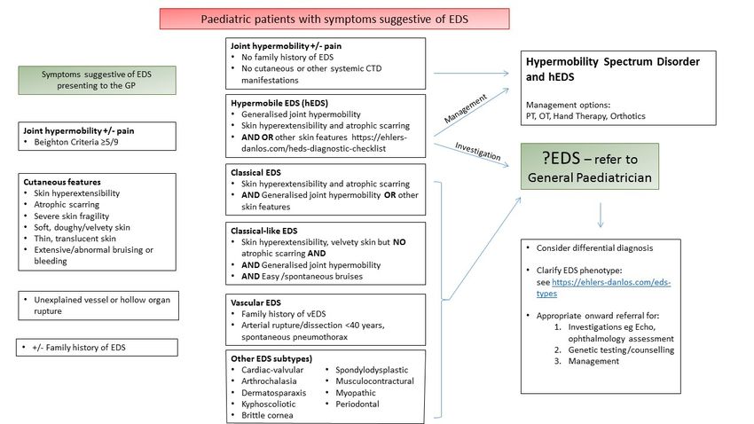
RED FLAGS

1. vEDS – VASCULAR EDS (2–4)

    • Arterial rupture or unusual bleeding in a child or young adult. Aorta, other large vessels and small
      vessels can be involved
    • See How to Save a Life (2,3) and vEDS reference article (4)

2. All EDS – ANAESTHETICS AND SURGERY

    • The use of intranasal DDAVP (desmopressin) will dramatically reduce the risk of bleeding both pre-
      operatively and in the presence of an acute bleed/haemorrhage (regardless of whether the patient is
      clinically vascular EDS or not)
    • Local anaesthetic – local, regional or epidural has less effect and slower onset in EDS (5)
           o May need more anaesthetic and longer wait before beginning procedure
    • Surgical issues
           o Prone to bleeding
           o Problems with tourniquet – compartment syndrome
           o Potential for tissue damage and subluxation with positioning issues

          Clinicians (including dentists) planning surgery or anaesthesia in a patient with
         EDS should carefully read Wiesmann et al: Recommendations for anaesthesia and
             perioperative management in patients with Ehlers-Danlos Syndrome(s)(6)

3. CAUTION: PHYSIOTHERAPISTS

Traditional physiotherapy techniques can cause injury to the EDS population and often need adapting-
patient recovery is generally much slower than in non-EDS patients.

New Zealand Hypermobility and Ehlers-Danlos Syndromes Guideline 2019
                                                                                                   Version 1
                                                                            8/1/19               Page 4 of 24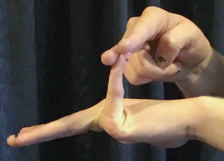
Epidemiology classification
Some degree of joint hypermobility is common in children especially in children under the age of 3. It is
usually "benign" in that there are no apparent detrimental effects. It is important to differentiate these from
the person who is borderline symptomatic or overtly part of a more significant clinical picture. The diagnosis
of EDS or one of its subtypes is usually deferred until the child is older or has a strong family history and/or
other defining symptom.

                                    THE HYPERMOBILITY SPECTRUM
       Phenotype (Clinical)        Beighton Score           Musculoskeletal        Systemic Features
                                                               Features            (historic presence
                                                                                         of JH)
       Asymptomatic GJH                 Present                  Absent                   Absent
       HSD (G-HSD)                      Present                  Present                  Absent
       hEDS                             Present                  Present                  Present

GENERALISED JOINT HYPERMOBILITY (GJH)

There is a spectrum of GJH

   1. Asymptomatic (non-syndromic) GJH – hypermobility without other symptoms. Other causes, e.g.
      neurological deficit must be excluded
   2. Symptomatic GJH that doesn’t meet criteria for hEDS – Hypermobility Spectrum Disorder
   3. A well-defined syndrome - hEDS

The essential difference between HSD and hEDS lies in the stricter criteria for diagnosing hEDS compared
to HSD. There is currently no reliable genetic marker available for hEDS.

Injury in patients with joint hypermobility can lead to micro- and/or macro-trauma, which in the long run can
be a leading cause of pain and in adulthood degenerative changes. This can range from hyperextension
injury to mild subluxation through to frank dislocation. Micro-trauma may not be obvious to see but leads to
pain and joint degeneration. Often the patient knows the joint is "out" - subluxed - but this is not seen
clinically or on x-ray. Macro-trauma is seen as an actual visible subluxation/dislocation.

HSD – HYPERMOBILITY SPECTRUM DISORDER (7,8)

   •   Relatively common
   •   Hypermobility and significant additional symptoms are typically limited to the musculoskeletal
       system
   •   Pain may be significant and debilitating
   •   Types
          o Generalised HSD – G-HSD
                   § GJH
                   § Musculoskeletal manifestations – Similar to hEDS. Usually generalised pain.
                   § No clinical features outside the musculoskeletal system
          o Peripheral HSD – P-HSD
                   § Hypermobility in hands and feet only
                   § Musculoskeletal manifestations – Similar to hEDS
          o Localised HSD – L-HSD
New Zealand Hypermobility and Ehlers Danlos Syndromes Guideline 2019
                                                                                                      Version 1
                                                                               8/1/19               Page 5 of 24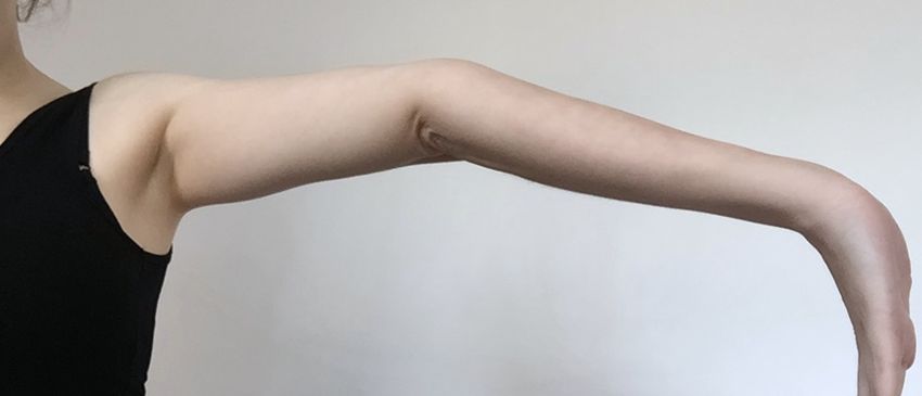
§ Hypermobility in a single joint
                     § Musculoskeletal manifestations – Similar to hEDS
            o    Historical – history of one of the above usually with 5-Point Questionnaire

EDS – EHLERS-DANLOS SYNDROMES

EDS (Ehlers-Danlos Syndromes) are a group of inherited disorders characterised by defects in collagen
mainly affecting the ligaments and soft tissues.

   •   Relatively rare – incidence is approximately 1:5000. F>M
   •   Basis in some is abnormal collagen – different types of collagen in each of the EDS types
   •   The commonest (>80-90%) is hEDS – genetic basis currently not known
   •   cEDS and vEDS are uncommon – have a known genetic basis (see Table below)
           o All the rest are very rare but genetic basis is clear

There are 13 types of EDS with significant overlap in features (7,9) with more subtypes being described as
research progresses.

                               Hypermobile - hEDS               Classical - cEDS
                                (Most common)                     (Common)
                                                       EDS
                                 Vascular - vEDS                10 Other types
                                (Rare - Most risk)                (Very rare)

                                   CLINICAL CLASSIFICATION OF EDS
EDS SUBTYPE            ABBREV                INHERITANCE           GENETICS              COLLAGEN
Classical              cEDS                  AD                    COL5A1, COL5A2        Type 5
Vascular               vEDS                  AD                    COL3A1                Type 3
Hypermobile            hEDS                  AD                    Unknown               Unknown
10 other types         See Ref (1,9)

New Zealand Hypermobility and Ehlers-Danlos Syndromes Guideline 2019
                                                                                                     Version 1
                                                                               8/1/19              Page 6 of 24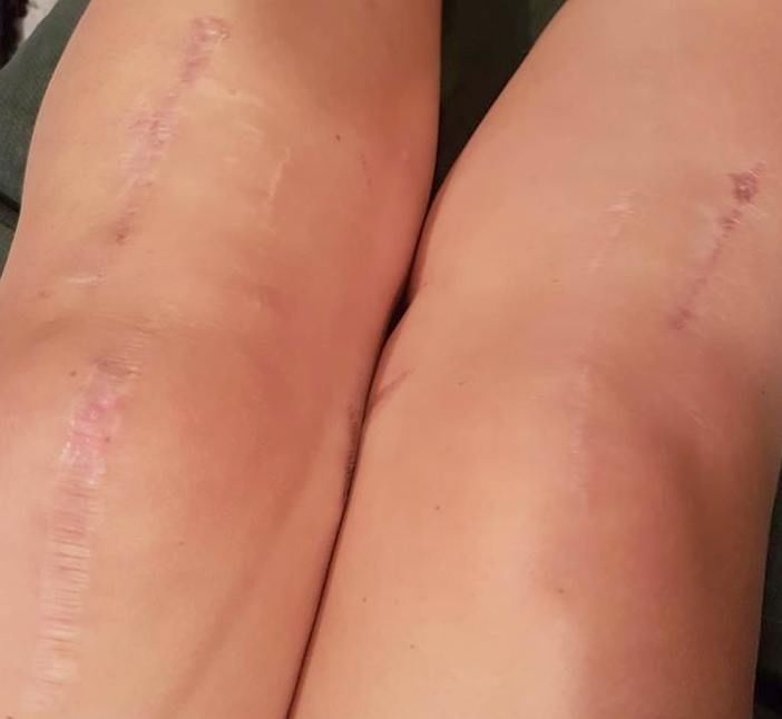
hEDS – HYPERMOBILE EDS (1,10,11)

   •   Most common of the Ehlers Danlos Syndromes
   •   No genetic basis yet identified therefore diagnosis is phenotypic
   •   See hEDS Diagnostic Checklist for details and how to apply criteria - includes a downloadable pdf
       (10)
           o Criterion 1 - Main feature (must be present)
                 § Generalised Joint Hypermobility (Beighton Score ≥ 4/9 over age 50; ≥ 5/9 in young
                     adults; ≥ 6/9 in children and adolescents) (12)
           o Criterion 2 (at least 2 features)
                 § Feature A – Generalised connective tissue disorder (at least 5/12 present)
                         • Soft, velvety skin
                         • Mild skin hyperextensibility (not as much as cEDS)
                         • Unexplained striae
                         • Piezogenic papules (nodules on side of feet)
                         • Recurrent/multiple hernias
                         • Atrophic scarring
                         • Arachnodactyly
                         • Arm span-Height ratio: ≥ 1.05
                         • Pelvic floor prolapse
                         • Dental crowding
                         • Mitral valve prolapse
                         • Aortic root dilatation
                 § Feature B
                         • Family history (vertical transmission down generations but some individuals
                              can be non-penetrant)
                 § Feature C (at least 1) - see details in checklist
                         • Musculoskeletal pain
                         • Chronic widespread pain ≥ 3 months
                         • Recurrent joint dislocations/instability in absence of significant trauma (may
                              also occur with trauma)
           o Criterion 3 – All must be present
                 § No skin fragility
                 § Reasonable exclusion of other connective tissue disorders e.g. Marfan syndrome
                 § Exclusion of other disorders that could cause GJH

   To diagnose hEDS all 3 criteria must be satisfied.

   •   Other features that can occur (not an exhaustive list)
          o Sleep disturbance, chronic fatigue, Postural orthostatic tachycardia syndrome (POTS),
              functional GIT disorders, unusual hernias, internal hernias, dysautonomia, Raynaud’s,
              MCAS, some cardiac features, osteoarthritis secondary to joint instability, headaches, TMJ
              dysfunction, increased gynaecological presentations, pelvic floor dysfunction, anxiety,
              depression. Multiple other features that affect quality of life may be part of the spectrum.
   •   Overall effect on life may range from severe (bed-ridden) to relatively minor

New Zealand Hypermobility and Ehlers-Danlos Syndromes Guideline 2019
                                                                                                  Version 1
                                                                            8/1/19              Page 7 of 24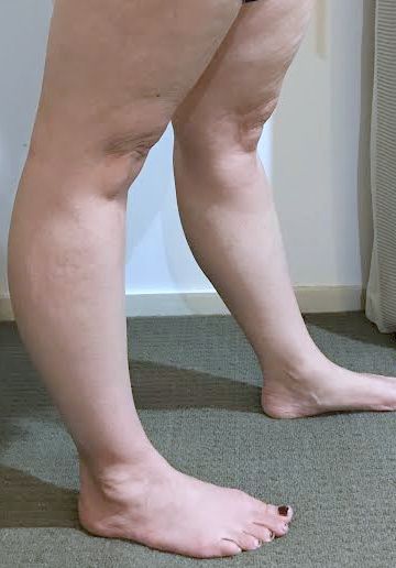
cEDS – CLASSICAL EDS (1,13)

   •    Relatively common
   •    Major criteria
           o Skin features - hyperextensible skin, atrophic scarring (esp. knees & elbows)
           o Generalised Joint Hypermobility
   •    Minor criteria
           o Easy bruising
           o Soft, doughy skin
           o Skin fragility
           o Molluscoid pseudotumours
           o Subcutaneous spheroids
           o Hernia or history of
           o Epicanthal folds
           o Complications of GJH
           o Family history of 1st degree relative

To diagnose cEDS:

                •   Criterion 1 – Skin features

                Plus

                •   Criterion 2 – GJH &/or at least 3 minor criteria

                Diagnostic confirmation with genetic testing is possible

vEDS – VASCULAR EDS (1,4)

   •    Rare and dangerous

        Major criteria                                        Minor criteria
        •   Family history proven vEDS                        •   Bruising not related to trauma or in
        •   Arterial rupture at young age                         unusual sites
        •   Spontaneous colon perforation in                  •   Thin, translucent skin with easily visible
            absence of other disease                              veins
        •   Uterine rupture without predisposing              •   Characteristic facial appearance
            cause                                             •   Spontaneous pneumothorax
        •   Carotid-cavernous sinus fistula without           •   Acrogeria
            trauma                                            •   Talipes equinovarus
                                                              •   Congenital hip dislocation
                                                              •   Hypermobility of small joints
                                                              •   Tendon and muscle rupture
                                                              •   Keratoconus
                                                              •   Gingival recession and fragility
                                                              •   Early onset varicose veins

        To diagnose need a family history with arterial rupture or dissection younger than 40 or any of the
        other major features.

       Genetic testing is important to corroborate a clinical diagnosis.

  New Zealand Hypermobility and Ehlers-Danlos Syndromes Guideline
                                                                                                       Version 1
                                     2019                                      8/1/19                Page 8 of 24
ALL OTHER TYPES (1)
UNUSUAL SYMPTOMS THAT CAN BE ASSOCIATED WITH EDS (esp. hEDS)
• Dysautonomia (autonomic dysfunction) such as POTS (postural orthostatic tachycardia syndrome)
• MCAS (mast cell activation syndrome) may masquerade as unusual allergies or bladder symptoms
• GIT symptoms including constipation
• Raynaud’s Syndrome
Neurological + Spinal Manifestations of EDS (14)
The connective tissue problems seen in EDS can result in diverse nervous system issues from nerves
being trapped, deformed or otherwise damaged. Better guidelines based on stronger evidence are needed
for improved diagnosis and treatment of this specialty area. Neurological and spinal manifestations of EDS
can include the following conditions:
• Idiopathic Intracranial Hypertension (Pseudotumor Cerebri): Idiopathic intracranial hypertension
(IIH) is a poorly understood condition but involves increased intracranial pressure, headaches,
visual disturbances, light sensitivity, and sometimes tinnitus, nausea, and vomiting
• Chiari I Malformation (CMI): CMI is a disorder affecting the tissue around the brainstem, in which a
lack of space causes obstruction of the normal fluid movement around the brain. Obstruction of fluid
circulation may flatten the pituitary gland, leading to hormone changes. Chiari malformation Type I
(CMI) has been reported as a condition that can occur with hypermobile EDS (hEDS)
• Atlantoaxial Instability: Atlantoaxial instability (AAI) is a potential complication of all forms of EDS.
Slow development of motor skills, headache, and limb weakness have all been attributed to loose
ligaments and overly moveable joints connecting the head and neck
• Craniocervical Instability: Craniocervical instability (CCI) is a type of loose ligament condition in
EDS that results in injury to the nervous system. CCI occurs when ligaments from the skull to the
spine don’t restrict unsafe movement. Nervous damage from loose ligaments may explain the slow
development of motor skills, poor coordination, learning difficulties, headaches, and clumsiness in
the EDS population
• Segmental Kyphosis and Instability
• Tethered Cord Syndrome: Tethered cord syndrome (TCS) is a condition that can be present in
EDS, where the spinal cord is attached to surrounding tissue in a way that creates elongation and
tension of the nervous tissue, leading to low back pain, loss of bladder control, lower body
weakness, and loss of sensation
• Movement Disorders: Disorders of too much movement such as uncontrolled muscle contraction,
tremor, fidgeting/dancing movements, twitches, and jerking are reported from EDS patients. Pain
and injury are frequent components of EDS, and there is evidence suggesting movement disorders
may cause these injuries
• Tarlov Cyst Syndrome: Tarlov cysts are fluid-filled sacs that can develop near the spinal cord and
can put pressure on adjacent neural structures. These abnormalities can be without symptoms, but
significant issues can arise such as pain, and bowel/bladder control problems. Of patients
undergoing destruction of Tarlov cysts, success is reported in 80–88% of patients, with few
complications.
New Zealand Hypermobility and Ehlers-Danlos Syndromes Guideline 2019
Version 1
8/1/19 Page 9 of 24DIAGNOSIS
HISTORY
• Personal or family history of hypermobility (double-jointed) – now or historically at any stage of life
• Hyperextensible (stretchy) skin
• Musculoskeletal pain (joint and muscle pain)
• One or more of these:
o Family history – all EDS has a genetic component
o Repeated dislocations (often of more than one joint) - often “spontaneous” with no trauma or
less than usual trauma
o Easy bruising – especially in children where the history is of no trauma but there are obvious
bruises. Is in the differential for non-accidental injury.
o Vessel rupture or unusual internal bleeding in a young person
o Unusual or severe prolapses and hernias or bowel perforation in younger people
o Associated symptoms that may be present (but are not diagnostic) include (but not limited
to): fatigue, headaches, sleep disturbance, autonomic dysfunction (especially POTS –
Postural Orthostatic Tachycardia Syndrome), irritable bowel, and Mast Cell Activation
Syndrome
GENETICS
Of the three most common forms of EDS, only vEDS and cEDS have an established genetic basis.
Determining the exact molecular basis for these conditions can be useful for diagnosis, management and
reproductive decision-making. It is, however, not mandatory that every person with a clinical diagnosis of
EDS undergoes molecular analysis. For those who meet the minimal clinical requirements for an EDS
subtype—but who have no access to molecular confirmation; or whose genetic testing shows one (or more)
gene variants of uncertain significance in the genes identified for one of the EDS subtypes; or in whom no
causative variants are identified in any of the EDS-subtype-specific genes—a “provisional clinical
diagnosis” of an EDS subtype can be made.
Primary care referrers should note that there is substantial symptom overlap between the EDS subtypes
and the other connective tissue disorders including hypermobility spectrum disorders, as well as a lot of
variability. Where there are definite clinical pointers towards vEDS, they remain key to considering this as a
diagnosis rather than the presence or absence of hypermobility as a discriminative feature. Such symptoms
should be brought to the attention of genetic services when requesting assessment, as triage is applied to
identify those who will most benefit from a test.
To arrange for an assessment of the genetic basis for a person with a vEDS or cEDS phenotype, a referral
can be made to Genetic Health Services NZ through any one of their three hubs centered in Christchurch,
Wellington or Auckland. See the GHSNZ website for details on how to lodge a referral and request for
assessment at www.genetichealthservice.org.nz. Rarer forms of EDS can also be assessed in a similar
manner on a case-by-case basis.
For hEDS (by far the most common), although there are clear familial components underpinning the
disorder, a defined molecular basis has not been discovered. Note that genetic services criteria for
acceptance of a referral are such that assessment is not guaranteed, especially for hEDS.
New Zealand Hypermobility and Ehlers-Danlos Syndromes Guideline 2019
Version 1
8/1/19 Page 10 of 24EXAMINATION
See Aids to Diagnosis
• Look for other EDS features to differentiate between Generalised Joint Hypermobility (GJH),
Hypermobility Spectrum Disorder (HSD) and one of the EDS types - usually Hypermobile EDS
(hEDS)
• In adults, if too stiff or painful to do Beighton Score, use the “Five Point Questionnaire” (see Aids to
Diagnosis section below) to assess historical hypermobility
• Look for other causes of hypermobility e.g. neurological, trauma etc
• Check skin for hyperextensibility – especially in cEDS.
o 3 cm stretch at neck, elbow, and knee. 1.5 cm forearm. 1 cm palm (thenar eminence).
• Check skin for atrophic scarring (See Figure B in Aids to Diagnosis)
• Assess joints and tender areas – typical inflammatory signs absent in face of significant pain
Investigations
• There are no specific or suggestive laboratory findings
• There are specific known genetic mutations/abnormalities for Classic EDS (cEDS) and Vascular
EDS (vEDS) and most of the rare types but not for hEDS. Clinical diagnosis is made first using
criteria outlined in (1) followed by genetic confirmation wherever possible and practical
DIFFERENTIAL DIAGNOSIS
Other causes of hypermobility - constitutional, Marfan’s Syndrome, Trisomy 21, previous injury, neuropathic
joints, osteogenesis imperfecta
• Vessel rupture – all other congenital and acquired causes including trauma
• Joint and muscle pain
o Other arthropathies usually are typically inflammatory in nature, have more swelling and
often have suggestive radiology or laboratory
o Fibromyalgia
• Bruising (especially in children) – other vasculopathies and clotting defects, e.g. von Willerbrands
• Stretchy skin - there may be other causes but in the context of hypermobility think of cEDS
Ignorance of EDS often leads to a misdiagnosis, which may lead to treatment that exacerbates symptoms,
as well as poor management causing a great deal of pain and suffering (12).
Indications for transfer or initial specialised assessment
• Acute vascular rupture is a medical emergency – consult vascular surgeons
• All others – a clinician with an interest in EDS if available
Practical Tip:
When hypermobility or EDS is suspected during the initial consultation do two things:
• Ask the 5-Point Questionnaire (see Aids to Diagnosis)
• Perform a Beighton Score (see Aids to Diagnosis)
If Beighton seems ≥4 and if they answer Yes to ≥2 of the Questions, make a follow-up appointment to go
through things in more detail.
New Zealand Hypermobility and Ehlers-Danlos Syndromes Guideline 2019
Version 1
8/1/19 Page 11 of 24PAEDIATRIC CONSIDERATIONS FOR EDS AND HSD
• Hypermobility and musculoskeletal pain are both very commonly seen in children
• Studies show over 60% of adolescent girls and 35% boys have a Beighton score ≥ 4/9, 26% and
11.5% when defined as ≥ 6/9
• Often hypermobility causes no functional problems or pain and can be advantageous for certain
activities e.g. sport or music
• Studies show over 30% of school age children complain of regular musculoskeletal pain
• The clinical challenge is to distinguish between those children within the normal spectrum of
hypermobility and those with suspected EDS
• Secondary referral should be made if hypermobility and pain present with features of EDS (see EDS
diagnostic criteria) or other features suggestive of a connective tissue disorder (marfanoid habitus,
easy bruising, blue sclera)
• Referral to orthotics/physiotherapy should be considered for all hypermobile children with on-going
musculoskeletal pain or impaired function (see Physiotherapy management guidelines)
New Zealand Hypermobility and Ehlers-Danlos Syndromes Guideline 2019
Version 1
8/1/19 Page 12 of 24AIDS TO DIAGNOSIS
Figure A: BEIGHTON SCORE – Assessment tool for hypermobility
1 2 3 4 5
1 point for each side for 1-4 and 1 point for 5. Total 9. If ≥ 4/9, hypermobility is present(1).
See reference (12) for a video on how to complete the Beighton Score Assessment accurately.
1 point for each side for 1-4 and 1 point for 5. Total 9. If ≥ 4/9, hypermobility is present(1).
See reference (12) for a video on how to complete the Beighton Score Assessment accurately.
*Photos are licenced under a Creative Commons Attribution – NonCommercial-No Derivatives 4.0 International License.
THE FIVE-POINT QUESTIONNAIRE
Use in cases where it is not possible to do Beighton Score or where superimposed pain and stiffness in an
adult will give a falsely low Beighton Score (1)
1. Can you now (or could you ever) place your hands flat on the floor without bending your knees?
2. Can you now (or could you ever) bend your thumb to touch your forearm?
3. As a child, did you amuse your friends by contorting your body into strange shapes or could you do
the splits?
4. As a child or teenager, did your shoulder or kneecap dislocate on more than one occasion?
5. Do you consider yourself “double-jointed”?
A “yes” answer to ≥ 2/5 questions suggests joint hypermobility with 80–85% specificity
New Zealand Hypermobility and Ehlers-Danlos Syndromes Guideline 2019
Version 1
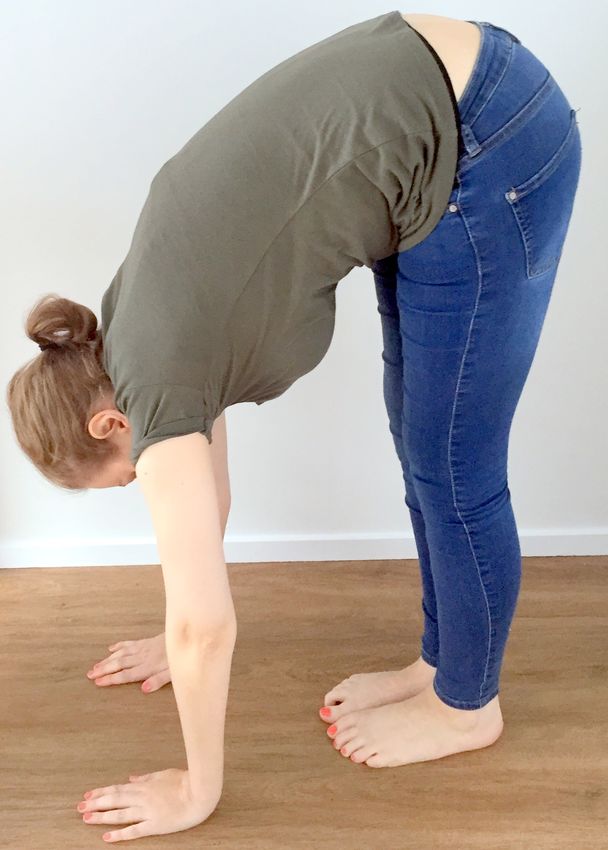
8/1/19 Page 13 of 24Figure B:
SKIN - HYPEREXTENSIBILITY SKIN - SCARRING
*Photos are licenced under a Creative Commons Attribution – NonCommercial-No Derivatives 4.0 International License.
New Zealand Hypermobility and Ehlers-Danlos Syndromes Guideline 2019
Version 1
8/1/19 Page 14 of 24Management
ACUTE EMERGENCIES
• Vascular rupture – appropriate vascular surgery or interventional radiology referral
• Dislocations – appropriate orthopaedic referral
• Acute pain – usual principles
• Bleeding - DDAVP (Desmopressin) intra-nasally is recommended for acute haemorrhage to help
stop bleeding
PAIN – INITIAL (ACUTE)
Pain has no proven pathology in EDS (15), rather pain is believed to be largely caused by repeated cycles
of differing types of trauma injuries. Undiagnosed and untreated injuries lead to further musculoskeletal
dysfunction and pain as poor compensating strategies are employed to cope with the acute unresolved
injury pain. Usually the pain an EDS patient is suffering is not benign and the underlying cause should be
diagnosed and treated where possible (15–17).
• Usual principles for initial management of acute or chronic pain
o Usually start with paracetamol and work up as needed
o There are no specific analgesics proven to have significant advantage in Hypermobility
Spectrum Disorder/EDS
• Bracing or splinting can be helpful in the short term
• Hot or cold packs, warm bath, etc.
INJURIES – INSTABILITY, SUBLUXATIONS, DISLOCATIONS, TENDON & LIGAMENT
INJURIES – INITIAL EMERGENCY AND ACUTE (SHORT TERM) STRATEGIES
• Each individual injury episode (persisting subluxation or actual dislocation, tendon or ligament strain
or tear) should be treated as a new trauma on its own merits and not just passed off as “part of your
condition”
• Dislocations cause pain and overstretching of the muscles which in turn cause muscle spasms.
Immediate assessment and treatment of a dislocation is important because once muscle spasm
occurs, it is very difficult to reduce the joint without anaesthesia (18). Immediate diagnosis and
intervention is important to improve patient outcomes (19)
• If the joint does not spontaneously reduce, reductions should only be attempted by clinicians
experienced in reduction techniques (19). Treatment is usually closed reduction, as soon as
possible, to decrease potential complications, which may include soft tissue injury, articular surface
injury, and neurovascular compromise (18)
• Dislocation - initial reduction
o Because those with EDS may have severe pain, instability or injury elsewhere in the same
region, some typical traction and twisting movements used for reduction may need to be
modified to prevent injury elsewhere from the procedure itself, e.g. with shoulder dislocation
pulling from the wrist or forearm may injure wrist or elbow. May need to modify hand position
and grip. The force needed to reduce may be less than that needed for non-EDS. Some
patients can “spontaneously reduce”
o Protect skin – it may be fragile. Use padding if needed.
New Zealand Hypermobility and Ehlers-Danlos Syndromes Guideline 2019
Version 1
8/1/19 Page 15 of 24o Ligament and tendon injuries may take longer to heal and may recur after relatively less
trauma after the first episode
• Subluxations may respond well to gentle manipulation rather than actual reduction
• Splinting and bracing are important after reduction (see Physiotherapy Management section)
JOINT INSTABILITY AND PAIN – LONG TERM STRATEGIES TO STABILISE
After an initial injury some of the joints may become prone to repeat injury from relatively minor trauma
• Physiotherapy – see Management Summary
• External bracing – see below
• If the dislocation/subluxation leads to joint instability and/or recurrent dislocation, either prolotherapy
or surgery will likely be required (20) International literature states that due to largely unsuccessful
surgical outcomes, prolotherapy should be considered before surgery (21,22). Sclerosant
prolotherapy is a non-surgical approach to joint stabilisation that may be appropriate in selected
circumstances in adults. Materials (chemical/sclerosant) injected into ligaments are thought to
induce healing with scarring and shortening which in turn increases joint stability and thereby
reduces pain and subluxation
• Whilst surgery may have a sub-optimal outcome it is important in critical areas, e.g. craniocervical
instability. When referring to or discussing with an orthopaedic surgeon, neurosurgeon, etc., be
clear that the patient has EDS where issues may include slow healing, early recurrence and poor
response to local anaesthetic agents
SURGERY AND ANAESTHESIA (5,23,24)
Clinicians planning surgery or anaesthesia in a patient with EDS: Wiesmann et al:
Recommendations for anaesthesia and perioperative management in patients with Ehlers-Danlos
Syndrome(s)26
• Surgical complications may be increased due to slow healing and potential for bleeding. Appropriate
strategies should be planned and discussed in EDS context
• DDAVP (Desmopressin) intra-nasally pre-operatively will reduce the risk of a life-threatening
haemorrhage
• Recurrence of prolapse, hernias, etc. after surgery may occur because of the inherently abnormal
ligaments
• Some issues with anaesthesia:
o Unstable neck may be an issue with positioning
o Slow and suboptimal response to local anaesthetic including epidurals
o Tourniquet can cause bruising and compartment syndrome
o Positioning can cause unexpected subluxations including temporomandibular joint during
anaesthesia
PATIENT SUPPORT
• Assure the patient you don’t think it’s all in their head – this is often what they have been told for a
long time
• Offer patient information on Ehlers-Danlos Society website https://www.ehlers-danlos.com/ and to
look for support groups
• Support groups offer advice on how to cope with day-to-day living with a painful chronic disease that
may significantly disrupt life
New Zealand Hypermobility and Ehlers-Danlos Syndromes Guideline 2019
Version 1
8/1/19 Page 16 of 24REFERRAL
• If available refer to a clinician with experience in EDS – geneticist, rheumatologist, etc.
• A multidisciplinary team is best placed to lead patient care
• Chronic pain teams can help to design an overall pain management strategy focused on pain
blocks, nerve blocks and drug pain management. It is important not to dismiss EDS patients’ pain
as being only of a psychological origin
• However, in some cases where physical pain and loss of independent function are seriously
impacting on quality of life it can be helpful to refer to a clinical psychologist. There are numerous
techniques, which can help individuals to cope better, including effective pain management
strategies, strategies to reduce stress, anxiety and worry, improving sleep as well as relaxation
techniques and mindfulness, many find this support invaluable (15,25). Patients may question such
a referral with an assumption you think their pain is all “in their head”. It is important to explain that
chronic pain is often a mix of nociceptive pain (due to tissue injury) as well as neuropathic pain
(from the peripheral or central nervous system) and that the best results come from addressing both
(17).
New Zealand Hypermobility and Ehlers-Danlos Syndromes Guideline 2019
Version 1
8/1/19 Page 17 of 24Physiotherapy Management Summary
Physiotherapists play an important role in the multidisciplinary approach to rehabilitation that is
recommended for hEDS and HSD patients. It is important to provide patients with both biopsychosocial
support and education on connective tissue disorders, as well as a tailored course of treatment aimed at
empowering the patient toward self-management (15,26,27). Whilst there are currently no formal routes to
receive additional training for Physiotherapists in hEDS and HSD in New Zealand, the references provided
throughout this section, and the following websites provide excellent information:
www.ehlers-danlos.org
www.ehlers-danlos.com
GRADUATED EXERCISE PROGRAMME
Although there have been very few treatment intervention studies undertaken to date, authors have
reported that tailored exercise brings improvements in joint stability, pain and proprioception, as do
graduated exercise programmes combined with education, behavioural and lifestyle advice (15). The
program chosen should be individualised and applied carefully to avoid exacerbation of pain (16).
It is recommended to start with static exercises within the hypermobile range, especially for those in
significant pain, then progress onto dynamic work (28). The exercises may be progressed from non-weight
bearing to weight bearing (15). Improving core stability is the most appropriate starting point (15). Muscle
patterning problems should be addressed initially (e.g. hamstring dominance) otherwise general
strengthening will be of no benefit and may make instability worse.
The evidence seems mixed as to whether resistance exercises should be advised or not. This will probably
depend on the severity of the patient’s symptoms. Bluestein states; “Low-resistance exercises with a
gradual increase in repetition are often recommended. Determining the zone in which patients are “safe but
sore” requires practice and an appropriate level of vigilance. Although resistance bands can be beneficial,
caution should be used with such devices.” (28).
Stretching exercises should be gentle to avoid subluxation and dislocation.
An approach called The Muldowney Exercise Protocol was discussed during the New York international
symposium in May 2016 (pers. com. Dr Fraser Burling). This book, which can be purchased online, offers
guidelines and resources for treatment, particularly for those physiotherapists with limited EDS
patient experience. The full protocol may take >1 year to work through (6-9 months in the younger adult)
and therefore may need to be followed by the patient at home with guidance and oversight from the treating
physiotherapist. As always, therapists must perform an appropriate assessment with any treatment tailored
to the individual patient’s needs.
GRADUATED ACTIVITY PROGRAMME
Slow progressive increases in activity and avoiding end range postures may be useful when trying to
increase function and reduce disability. Pain and muscular weakness often cause hEDS and HSD patients
to become more sedentary, exacerbating their symptoms. It is therefore important to incorporate some
aerobic work into the rehab programme (15–17) .
• Exercising in water is ideal as there is less strain on joints
• Hydrotherapy is useful, (28) however local pools are recommended for swimming as
hydrotherapy pools may be too warm (15)
• Tai chi and pilates can facilitate balance and control (15)
New Zealand Hypermobility and Ehlers-Danlos Syndromes Guideline 2019
Version 1
8/1/19 Page 18 of 24• Bicycling can be good for aerobic work and again does not over-stress the joints (15)
• Nordic pole walking can be particularly effective as it is a functional activity (15), but should
be avoided if any shoulder injury is present (pers. commun. Dr F. Burling).
The key to success with any of these activities is tailoring the program to the needs and abilities of your
patient, as what works for someone with a milder generalised joint hypermobility may be detrimental to a
patient with more severe hEDS.
Pacing is a key area of education for patients with hEDS and HSD. As with other chronic pain conditions, a
cycle of “boom and bust” often can occur which leads to a flare in pain with over activity, followed by
prolonged rest and further deconditioning. These cycles can be lessened by teaching pacing skills and can
assist in increasing function and activity. See reference (28,30) for patient information on pacing.
For individuals with a higher level of fitness advice should be given to keep active and consider the sport
depending on their joint issues, e.g. avoid long distance running for those with anterior knee pain. Long
term enjoyment is the key to long term management.
PROPRIOCEPTION TRAINING
Proprioception and balance problems are common issues in hEDS and EDS. Proprioception exercises are
therefore important to incorporate into an exercise programme. See reference(15) for details of appropriate
balance exercises.
POSTURE AWARENESS AND CORRECTION
Assessment of posture regularly finds sub-optimal posture, both static and dynamic in EDS and hEDS
patients. Posture re-education, often starting with the trunk can help improve quality of life and reduce pain
(15). Use of biofeedback can be used to assist postural awareness (17).
MANUAL THERAPY
Clinical experts recommend the use of manual therapy in the management of EDS. Techniques can help
alleviate pain associated with muscle spasm as well as being helpful for stiff joints (15,16,31) The following
techniques have all been recommended:
• Muscle energy techniques
• Myofascial release
• Joint mobilisation techniques
• Mulligan techniques: mobilisations with movement
SUPPORTS, TAPING AND COMPRESSION CLOTHING
The use of aids and supports are not encouraged by all experts as they are reported to reduce muscle
activity and promote dependency (15,16). However, using a support will often help prevent injuries, aid post
injury recovery, improve proprioception and reduce pain which is beneficial in improving function in many
instances (15).
Tapes and Bandages
• When the purpose is to restrict undesired motion, adhesive, non-stretch (rigid) sports tape is
generally the most appropriate as it can help support vulnerable joints for a few days at a time
• Tape may also help improve proprioception and posture
New Zealand Hypermobility and Ehlers-Danlos Syndromes Guideline 2019
Version 1
8/1/19 Page 19 of 24• If more rigid support is needed, any tape may be applied in layers
• Care must be taken as allergic reactions to the adhesive may occur and there is a risk of damage
to fragile skin as the tape is removed
• Cohesive bandages may also be helpful and are easily transported for emergency self-help
Neoprene or Elastic Supports
Lightly shaped, stretchy sleeves (e.g. Tubigrip) or Neoprene supports can be used for wrists, elbows,
shoulders, knees and ankles. These offer light support and may help reduce some of the stresses exerted
on hypermobile joints when exercising or to ease pain for daily living. Available in sports shops, online and
may be available via PT/Occupational Therapy (OT) department. Alternate supports e.g. Omotrain for
shoulders can be valuable for shoulder rehabilitation.
Compressive Clothing (Support tights, sportswear)
These are usually made from tight, stretchy material that is thinner than neoprene clothing. Compression
provides good proprioceptive feedback, can improve low blood pressure, joint stability, proprioception and
spatial awareness. Spio is a company that provides garments for children. Currently funding for these
garments is at the discretion of the local DHB Orthotics service.
Splints
Splints work in a similar way to neoprene supports but may have plastic/metal stays that limit or stop
movements in the wrong direction. These splints are good for short-term use, e.g. for immobilising a
painful wrist/hand after a dislocation/subluxation, or for continuing support during exercise to help regain
muscle control.
Ring splints may be helpful in preventing excessive finger joint strain. Local DHB PT or OT services may
provide finger splints.
ORTHOTICS
Evidence based guidelines suggest that children with flexible flat feet presenting with pain or impaired
function, such as that commonly seen in hEDS/HSD should use orthotics and/or sensible footwear (16).
Adults should be advised to wear supportive shoes with cushioned soles and supportive fastenings (e.g.
sports shoe). If this is insufficient, studies have shown tailored orthotics rather than over the counter items
to be of most benefit to adult EDS patients (16). Patients should also be evaluated for a leg length
discrepancy, and if this is found, tailored orthotics are indeed essential (31).
PAIN MANAGEMENT
Physiotherapists can advise on and apply pain management strategies as an adjunct to exercise. Although
little clinical evidence exists, general consensus is that the following may be beneficial:
• Heat
• Cold
• Manual therapy:
o Gentle soft tissue massage
o Gentle trigger point massage
o Gentle myofascial release
• TENS
• Mindfulness
New Zealand Hypermobility and Ehlers-Danlos Syndromes Guideline 2019
Version 1
8/1/19 Page 20 of 24• Relaxation techniques:
o Imagery
o Breathing techniques
• Sleep hygiene advice
Referral to a multi-disciplinary pain management programme is recommended for those living with chronic
pain, fatigue and disability.
POSTURAL ORTHOSTATIC TACHYCARDIA SYNDROME (POTS)
POTS can be a life-altering and debilitating chronic health condition. Simply standing up can be a
challenge for people with POTS as their body is unable to adjust to gravity.
POTS is characterised by orthostatic intolerance (the development of symptoms when upright that are
relieved by lying down). Symptoms include:
• headaches
• fatigue
• palpitations
• sweating
• nausea
• fainting
• dizziness
• an increase in heart rate from the lying to upright position of greater than 30 beats per minute,
or a heart rate of greater than 120 beats per minute within 10 minutes of standing.
Light to moderate exercise can be helpful for POTS symptoms. For further information and management
strategies please see the webpages below:
• http://www.potsuk.org
• www.dysautonomiainternational.org
Please note: This summary is written using current literature available and is subject to change as further
research becomes available.
Acknowledgements
This document was made possible by the generous support offered by the working group members and
input from associated experts; NZORD would like to extend sincere thanks to them all.
NZORD are also grateful to Madeleine Pook (Chartered Physiotherapist UK) for assistance on the
Physiotherapy summary section along with Neil Challenger, Physiotherapist CCDHB.
New Zealand Hypermobility and Ehlers-Danlos Syndromes Guideline 2019
Version 1
8/1/19 Page 21 of 24REFERENCES
1. Malfait F, Francomano C, Byers P, Belmont J, Berglund B, Black J, et al. The 2017 international classification of
the Ehlers-Danlos syndromes. Am J Med Genet C Semin Med Genet. 2017 Mar;175(1):8–26.
2. Urgent – EDS Today [Internet]. [cited 2018 May 28]. Available from: http://edstoday.org/urgent/
3. vEDS_cdrombooklet.pdf [Internet]. [cited 2018 May 28]. Available from: http://edstoday.org/wp-
content/uploads/2016/09/vEDS_cdrombooklet.pdf
4. Byers PH, Belmont J, Black J, Backer JD, Frank M, Jeunemaitre X, et al. Diagnosis, natural history, and
management in vascular Ehlers–Danlos syndrome. Am J Med Genet C Semin Med Genet. 175(1):40–7.
5. Our Printable Materials [Internet]. The Ehlers Danlos Society. [cited 2018 May 29]. Available from:
https://www.ehlers-danlos.com/brochures/
6. Wiesmann et al. - 2014 - Recommendations for anesthesia and perioperative m.pdf [Internet]. [cited 2018 May
29]. Available from: https://ehlers-danlos.com/wp-content/uploads/recommendations-for-anesthesia.pdf
7. Castori M, Tinkle B, Levy H, Grahame R, Malfait F, Hakim A. A framework for the classification of joint
hypermobility and related conditions. Am J Med Genet C Semin Med Genet. 2017;175(1):148–57.
8. What are the hypermobility spectrum disorders? [Internet]. The Ehlers Danlos Society. [cited 2018 May 29].
Available from: https://www.ehlers-danlos.com/what-is-hsd/
9. The Types of EDS [Internet]. The Ehlers Danlos Society. [cited 2018 May 29]. Available from: https://www.ehlers-
danlos.com/eds-types/
10. hEDS Diagnostic Checklist [Internet]. The Ehlers Danlos Society. [cited 2018 May 29]. Available from:
https://www.ehlers-danlos.com/heds-diagnostic-checklist/
11. Tinkle B, Castori M, Berglund B, Cohen H, Grahame R, Kazkaz H, et al. Hypermobile Ehlers–Danlos syndrome
(a.k.a. Ehlers–Danlos syndrome Type III and Ehlers–Danlos syndrome hypermobility type): Clinical description
and natural history. Am J Med Genet C Semin Med Genet. 175(1):48–69.
12. Assessing Joint Hypermobility [Internet]. The Ehlers Danlos Society. [cited 2018 May 29]. Available from:
https://www.ehlers-danlos.com/assessing-joint-hypermobility/
13. Bowen JM, Sobey GJ, Burrows NP, Colombi M, Lavallee ME, Malfait F, et al. Ehlers–Danlos syndrome, classical
type. Am J Med Genet C Semin Med Genet. 175(1):27–39.
14. Henderson FC, Austin C, Benzel E, Bolognese P, Ellenbogen R, Francomano CA, et al. Neurological and spinal
manifestations of the Ehlers–Danlos syndromes. Am J Med Genet C Semin Med Genet. 175(1):195–211.
15. Physical therapy for hypermobility – The Ehlers-Danlos Support UK [Internet]. [cited 2018 May 29]. Available
from: https://www.ehlers-danlos.org/information/physical-therapy-for-hypermobility/
16. Engelbert RHH, Juul-Kristensen B, Pacey V, Wandele I de, Smeenk S, Woinarosky N, et al. The evidence-based
rationale for physical therapy treatment of children, adolescents, and adults diagnosed with joint hypermobility
syndrome/hypermobile Ehlers Danlos syndrome. Am J Med Genet C Semin Med Genet. 175(1):158–67.
17. Chopra P, Tinkle B, Hamonet C, Brock I, Gompel A, Bulbena A, et al. Pain management in the Ehlers–Danlos
syndromes. Am J Med Genet C Semin Med Genet. 175(1):212–9.
New Zealand Hypermobility and Ehlers-Danlos Syndromes Guideline 2019
Version 1
8/1/19 Page 22 of 2418. Joint dislocation - Symptoms, diagnosis and treatment | BMJ Best Practice [Internet]. [cited 2018 May 29].
Available from: https://bestpractice.bmj.com/topics/en-us/583
19. In-game Management of Common Joint Dislocations [Internet]. [cited 2018 May 29]. Available from:
https://www.ncbi.nlm.nih.gov/pmc/articles/PMC4000468/
20. Ericson WB, Wolman R. Orthopaedic management of the Ehlers–Danlos syndromes. Am J Med Genet C Semin
Med Genet. 175(1):188–94.
21. Hakala RV. Prolotherapy (proliferation therapy) in the treatment of TMD. Cranio J Craniomandib Pract. 2005
Oct;23(4):283–8.
22. Castori M. Pain in Ehlers-Danlos Syndromes: Manifestations, Therapeutic Strategies and Future Perspectives.
Expert Opin Orphan Drugs. 2016 Sep 20;4.
23. Wiesmann T, Castori M, Malfait F, Wulf H. Recommendations for anesthesia and perioperative management in
patients with Ehlers-Danlos syndrome(s). Orphanet J Rare Dis [Internet]. 2014 Dec [cited 2018 May 29];9(1).
Available from: http://ojrd.biomedcentral.com/articles/10.1186/s13023-014-0109-5
24. local-anesthetic-failure.pdf [Internet]. [cited 2018 May 29]. Available from: https://ehlers-danlos.com/wp-
content/uploads/local-anesthetic-failure.pdf
25. Ehlers Danlos Syndromes Toolkit [Internet]. [cited 2018 Jul 9]. Available from: http://www.rcgp.org.uk/eds
26. Billings SE, Deane JA, Bartholomew JEM, Simmonds JV. Knowledge and perceptions of Joint Hypermobility and
Joint Hypermobility Syndrome amongst paediatric physiotherapists. Physiother Pract Res. 2015 Jan 1;36(1):33–
41.
27. Terry RH, Palmer ST, Rimes KA, Clark CJ, Simmonds JV, Horwood JP. Living with joint hypermobility syndrome:
patient experiences of diagnosis, referral and self-care. Fam Pract. 2015 Jun;32(3):354–8.
28. Bluestein LS. Pain Management in Patients With Hypermobility Disorders: Frequently Missed Causes of Chronic
Pain. Top Pain Manag. 2017 Jul;32(12):1.
29. Oh TWIST Home - Discover what could be making you so tired too! [Internet]. Oh TWIST. [cited 2018 May 29].
Available from: http://ohtwist.com/
30. Managing fatigue, sleeping problems and brain fog – The Ehlers-Danlos Support UK [Internet]. [cited 2018 Jul
31]. Available from: https://www.ehlers-danlos.org/information/managing-fatigue-sleeping-problems-and-
brain-fog/
31. Living Life to the Fullest With Ehlers-Danlos Syndrome [Internet]. Muldowney Physical Therapy. [cited 2018 May
28]. Available from: http://www.muldowneypt.com/living-life-to-the-fullest-with-ehlers-danlos-syndrome/
For Patients and Doctors Seeking Advice
1. Ehlers-Danlos Society – Patient Support https//ehlers-danlos.com/patient-support/
2. Loosely speaking – New Zealand support Facebook page – type “Loosely speaking into the Facebook search
box, is a closed group so ask to join
https://www.facebook.com/groups/LooselySpeakingNZ
3. International EDS support group. Type “EDS - Zebras need Zebras” into the Facebook search to access this
very supportive and large patient group, is a closed group so ask to join.
New Zealand Hypermobility and Ehlers-Danlos Syndromes Guideline 2019
Version 1
8/1/19 Page 23 of 244. This is a group for people who consider themselves a part of the LGBTQ+ community and have Ehlers-Danlos
Syndrome. This is an inclusive group. Check “Rainbow Zebras” on Facebook search box.
5. NZORD – New Zealand Organisation for Rare Disorders at http://www.nzord.org.nz – includes Specialist
directory
6. Myths and facts – for patients – at http://edstoday.org/myths-and-facts/
New Zealand Hypermobility and Ehlers-Danlos Syndromes Guideline 2019
Version 1
8/1/19 Page 24 of 24You can also read