Clinical and genetic basis of congenital myasthenic syndromes - SciELO
←
→
Page content transcription
If your browser does not render page correctly, please read the page content below
DOI: 10.1590/0004-282X20160106
 VIEW AND REVIEW

  Clinical and genetic basis of congenital
  myasthenic syndromes
  Bases clínicas e genéticas das síndromes miastênicas congênitas
  Paulo Victor Sgobbi de Souza1, Gabriel Novaes de Rezende Batistella1, Valéria Cavalcante Lino1,
  Wladimir Bocca Vieira de Rezende Pinto1, Marcelo Annes1, Acary Souza Bulle Oliveira1

  ABSTRACT
  Neuromuscular junction disorders represent a wide group of neurological diseases characterized by weakness, fatigability and
  variable degrees of appendicular, ocular and bulbar musculature involvement. Its main group of disorders includes autoimmune
  conditions, such as autoimmune acquired myasthenia gravis and Lambert-Eaton syndrome. However, an important group of diseases
  include congenital myasthenic syndromes with a genetic and sometimes hereditary basis that resemble and mimick many of the
  classic myasthenia neurological manifestations, but also have different presentations, which makes them a complex clinical,
  therapeutic and diagnostic challenge for most clinicians. We conducted a wide review of congenital myasthenic syndromes in their
  clinical, genetic and therapeutic aspects.
  Keywords: myasthenia gravis; congenital myasthenic syndromes, genetics.

  RESUMO
  Distúrbios da junção neuromuscular representam um grupo amplo de doenças neruológicas caracterizadas por fraqueza, fadigabilidade
  e graus variados de envolvimento das musculaturas apendicular, ocular e bulbar. Os principais grupos de doenças deste grupo incluem
  condições auto-imunes, como a miastenia gravis auto-imune adquirida e a síndrome de Lambert-Eaton. Entretanto, um outro grupo
  importante de doenças incluem as sindromes miastênicas congênitas com uma base genética e eventualmente hereditária que lembra e
  mimetiza muitas das manifestações neurológicas clássicas das miastenias, mas também se apresentam de diferentes formas tornando um
  desafio clínico, terapêutico e diagnóstico complexo para a maioria dos clínicos. Realizamos ampla revisão sobre as síndromes miastênicas
  congênitas em seus aspectos clínicos, genéticos e terapêuticos.
  Palavras-chave: miastenia gravis; síndromes miastênicas congênitas, genética.

       In 1895, Friedrich Jolly using Greek prefixes for muscle                Although most commonly considered in the differential diag-
  and weakness created the term “myasthenia” and used the                      nosis of early-onset hypotonia in the infant and neonate,
  Latin word “gravis” to describe the severity of weakness that                CMS must be considered in a wide group of clinical and
  can often lead patients to death1. Myasthenia is described as                electroneuromyographic scenarios, even including cases with
  a clinical group of conditions affecting neuromuscular trans-                juvenile and adult-onset symptomatology2,3. It is essential to
  mission and can occur in the context of acquired and heredi-                 consider CMS as an important differential diagnosis, as many
  tary congenital conditions.                                                  presentations represent well-responsive therapeutic forms
       Congenital myasthenic syndromes (CMS) comprise a het-                   and different inheritance patterns are involved, emphasizing
  erogeneous group of rare inherited diseases in which the neu-                the crucial role of genetic counseling.
  romuscular transmission in the motor plate is compromised
  by one or more genetic pathophysiological specific mecha-
  nisms2. Congenital myasthenic syndromes can be classified                    DIAGNOSIS AND CLINICAL FEATURES
  according to the pattern of inheritance, based on the altered
  protein involved in the motor plate, or by taking into account                   At present there are no well-defined diagnostic criteria for
  the site at the neuromuscular junction (pre-synaptic, synap-                 CMS. Congenital myasthenic syndromes should be suspected
  tic, or postsynaptic) involved with the dysfunction (Table 1)3.              in cases of: (i) early-onset fatigable muscle weakness mainly

  Universidade Federal de São Paulo, Departamento de Neurologia e Neurocicurgia, Divisão de Doenças Neuromusculares, São Paulo SP, Brasil.
  1

  Correspondence: Wladimir Bocca Vieira de Rezende Pinto; Departamento de Neurologia e Neurocicurgia, Universidade Federal de São Paulo (UNIFESP);
  Rua Estado de Israel, 899; 04022-002 São Paulo SP, Brasil. E-mail: wladimirbvrpinto@gmail.com
  Conflict of interest: There is no conflict of interest to declare.
  Received 09 September 2015; Received in final form 05 March 2016; Accepted 03 May 2016.

750
Table 1. Classification of congenital myasthenic syndromes (CMS) related to pattern of inheritance and molecular targets at the
neuromuscular junction.
I. Pattern of inheritance
Autosomal dominant (gain-of-function) Slow-channel syndrome, SNAP25B*, SYT2*
Autosomal recessive (loss-of-function) All other subtypes
II. Site of defect and molecular targets at the neuromuscular junction
Presynaptic defects ChAT deficiency, SNAP25B deficiency, synaptotagmin-2 deficiency
Primary deficiency, slow-channel syndrome (CHRNA1, CHRNB, CHRND, CHRNE, CHRNG),
Acetylcholine receptor defects
fast-channel syndrome (CHRNA, CHRND, CHRNE)
Synaptic basal lamina defects Acetylcholinesterase deficiency (ColQ), β2-laminin deficiency
Endplate development and maintenance Agrin deficiency, MuSK deficiency, LRP4 deficiency, Dok-7 deficiency, rapsyn deficiency,
congenital defects COL13A1 mutations
Metabolic and mitochondrial disorders Congenital disorders of glycosylation, SLC25A1 gene mutations
Congenital myopathies with secondary neuromuscular transmission compromise (MTM1,
Others
RYR1, DNM2, TPM3, BIN1); PREPL deletion; plectin deficiency
*extremely rare presentations.
involving ocular, bulbar and proximal limb musculature established, and most of the current knowledge has been
(generally varying from birth to late childhood); (ii) a positive obtained by reports of isolated case reports4.
family history of a specific disorder or sometimes only the A study in the UK estimated that the prevalence of CMS
history of a hypotonic infant; (iii) clinical and neurophysi- with a defined genetic diagnosis is approximately 9.2 cases
ological myasthenic findings with a negative antibody test- per million children under 18 years old in the population5.
ing profile; (iv) electromyography (EMG) studies showing There are also some ethnic variations in the genetic diag-
decremental responses of 10% or more in the amplitude or nosis of CMS with some mutations being more common
in the quarter of the area from the first evoked compound in some populations, such as the 1293insG mutation in the
motor action potential (CMAP), or single-fiber EMG studies gene CHRNE coding the epsilon subunit of the acetylcho-
compatible with a neuromuscular junction dysfunction; and line receptor, more frequently found in families from north
(v) the presence of a specific clinical syndromic phenotype Africa6, and the 1267delG mutation also in the CHRNE in
(i.e. Escobar syndrome, Pierson syndrome)2,3,4. gypsy families from southeast Europe7.
However, in some congenital myasthenic syndromes, the An important epidemiological profile on CMS was
onset of clinical manifestations may be late with involve- obtained in a study performed at the Mayo Clinic. Most cases
ment in adolescence or adulthood, other family members occured as a consequence of postsynaptic defects (68%), and
may not have been affected by the disease and electromy- basal lamina defects (13.7%), development and maintenance
ography changes may not be present in all muscles or occur of the end plate defects (12.5%), pure presynaptic defects
intermittently4. Therefore, although some clinical, therapeu- (5.9-8%) and congenital myopathies with secondary neuro-
tic and neurophysiological clues allow a high clinical suspi- muscular junction transmission defects (0.3%) also represent
cion for each CMS subtype (Tables 2 and 3), in many cases of other rare congenital myasthenic syndromes. Thus, post-
rapsyn deficiency, primary deficiency of endplate acetylcho- synaptic forms represent up to 75-80% of all CMS cases3,4,8.
line receptor and fast-channel syndrome, there are frequently In southern Brazil, in the state of Paraná, a minimum preva-
no specific hallmarks in presentations2,3,4. lence of 0.18/100.000 of CMS is estimated9.
The list of differential diagnoses of CMS is huge and includes
some of the following conditions: congenital myopathies, con- Pathophysiology
genital muscular dystrophy, limb-girdle muscular dystrophy, To understand the pathophysiological mechanisms
mitochondrial myopathies, facioscapulohumeral dystrophy, involved in CMS, it is essential to recognize key aspects of the
myotonic dystrophy and autoimmune myasthenia gravis, neuromuscular junction structure and function. The neuro-
motor neuron disease and peripheral neuropathies (Table 4)2. muscular junction has three basic components: (i) the presyn-
aptic nerve terminal, where there is the biosynthesis process,
Epidemiology storage and release of acetylcholine, the main neurotransmitter
The prevalence of CMS is very difficult to estimate due to involved in primary muscular contraction process; (ii) the syn-
the clinical variability of cases and the fact that many cases aptic space or synaptic cleft, where acetylcholine is released by
have no specific etiologic diagnosis or are undiagnosed. Since the presynaptic nerve terminal and where there is a complex
they are rare medical conditions in which definite diagnosis network of local proteins responsible for maintenance of the
rests on clinical, electromyography and specific genetic test- structure of the neuromuscular junction; and (iii) the postsyn-
ing, few data are available. Furthermore, there are few series aptic muscle membrane, where there are acetylcholine recep-
of patients where this complete diagnostic profile has been tors responsible for the action potential deflagration, endplate
Souza PVSS et al. Congenital myasthenic syndromes 751Table 2. Main diagnostic clues to guide differential diagnosis of congenital myasthenic syndromes (CMS) subtypes.2,4
I. Clinical clues of CMS subtypes
ChAT deficiency, synaptotagmin-2 deficiency, acetylcholinesterase, slow-channel
Early-onset phenotypes (neonatal, lactancy, syndrome, fast-channel syndrome, agrin deficiency, LRP4 deficiency, MuSK deficiency,
early infancy) Dok-7 deficiency, rapsyn deficiency, GFPT1, ALG2, ALG14, GMPPB, PREPL deficiency,
plectin deficiency, SCN4A, COL13A1
Late-onset phenotypes (late infancy, Acetylcholinesterase deficiency (variants), slow-channel syndrome,
adolescence, juvenile, adult) agrin deficiency, SCN4A
LGMD-like phenotype GFPT1, LRP4 deficiency, plectin deficiency
Apneic episodes ChAT deficiency, SCN4A
Laryngeal stridor and vocal cord palsy Dok-7 deficiency
Epilepsy with intellectual disability DPAGT1 deficiency
Cerebellar ataxia SNAP25B
Pupillary defects ColQ mutations
Primary AChR deficiency, Dok-7 deficiency, SNAP25B, agrin deficiency, MuSK
Ptosis
deficiency, rapsyn deficiency, PREPL, congenital myopathies, COL13A1
Rapsyn deficiency, primary AChR deficiency, MuSK deficiency, Dok-7 deficiency,
Ophthalmoparesis (± strabismus)
congenital myopathies
Agrin deficiency, MuSK deficiency, Dok-7 deficiency, rapsyn deficiency, PREPL,
Facial palsy
congenital myopathies
Cervical weakness Slow-channel syndrome, Dok-7 deficiency, COL13A1
Arthrogryposis multiplex congenital-like Rapsyn deficiency, ChAT deficiency, AChR deficiency (CHRND, CHRNG), SNAP25B,
phenotype and early-onset joint contractures synaptotagmin-2
Dysmorphic features Rapsyn deficiency, β2-laminin deficiency, COL13A1
Pierson syndrome (β2-laminin: ocular malformation, congenital nephrosis); rapsyn
Congenital malformations deficiency; GFPT1 deficiency; SLC25A1 (optic nerve hypoplasia, corpus callosum
agenesis)
Anoxic encephalopathy-like phenotype Rapsyn deficiency, SCN4A
Epidermolysis bullosa simplex Plectin deficiency
II. Histopathological clues of CMS subtypes
Tubular aggregates in the sarcoplasmic
GFPT1, DPAGT1 and ALG2-related CMS subtypes
reticulum
Vacuolar autophagic myopathy GFPT1 and DPAGT1-related subtypes
LMGD: limb-girdle muscular dystrophy.
potential and the acetylcholinesterase enzyme, involved with (whole-exome sequencing). This has clarified the patho-
the breakdown of acetylcholine and subsequent restoration physiological mechanisms of this spectrum of disorders,
of the resting potential of the membrane postsynaptic poten- not restricted to the neuromuscular junction structures
tial8,10,11,12. Mutations in the genes encoding proteins related to and pointing to new opportunities and approaches for
any one of these three components of the neuromuscular junc- future therapeutic modalities4,13,14.
tion could give rise to different CMS phenotypes.
Currently, major defects involved in the etiology of CMS
occur2,4: (A) in the presynaptic terminal; (B) associated with PRE-SYNAPTIC SYNDROMES
the synaptic basal lamina membrane; (C) the acetylcholine
receptor; (D) shortcomings in the maintenance and develop- Deficiency of choline acetyltransferase (ChAT)
ment of the neuromuscular junction; (E) congenital defects (OMIM #254210)
in glycosylation; and (F) other sites and mechanisms (Figure). Presynaptic defects represent less than 6% of all CMS
cases. The enzyme choline acetyltransferase (encoded by
Clinical and genetic basis of congenital myasthenic the CHAT gene, 10q11.23), located in the presynaptic ter-
syndromes minal region of the axon of the lower motor neuron, is
Currently, more than 20 different genes are involved involved in the biosynthesis of the neurotransmitter ace-
as monogenic causes of CMS, with an increasing num- tylcholine from acetyl-coenzyme A (acetyl-CoA) and cho-
ber of new discoveries every year driven by next-gen- line2,4. The severity of ChAT deficiency depends on the type
eration sequencing with complete exome sequencing and site of mutation affecting the enzyme structure, the
752 Arq Neuropsiquiatr 2016;74(9):750-760Table 3. Therapeutic clues in the differential diagnosis of congenital myasthenic syndromes (CMS) subtypes2,4.
Quinidine-responsive phenotype Slow-channel syndrome
Fluoxetine-responsive phenotype Slow-channel syndrome
Acetazolamide-responsive phenotype Sodium channel
Amifampridine-responsive phenotype Primary AChR deficiency, SNAP25, fast channel syndrome, MuSK, rapsyn,
(3,4-diaminopyridine) ALG2, ALG14, GFPT1, DPAGT1, COL13A1
Dok-7, Primary AChR deficiency, MuSK (partial), acetylcholinesterase
Albuterol-responsive phenotype
deficiency
Rapsyn, acetylcholinesterase, primary AChR deficiency, MuSK, DPAGT1
Salbutamol-responsive phenotype
(partial), COL13A1
Ephedrine-responsive phenotype Laminin-β2 deficiency, agrin, acetylcholinesterase, Dok-7
GFPT1, sodium channel (SCN4A), centronuclear congenital myopathy, ChAT
deficiency, fast-channel syndrome, AChR deficiency, paucity of synaptic
Pyridostigmine-responsive phenotype
vesicles and reduced quantal release (partial), congenital myopathies
(partial: centronuclear myopathy), PREPL (transient)
Acetylcholinesterase deficiency, Laminin-β2 deficiency, slow-channel
Pyridostigmine-avoidance phenotype (clinical worsen)
syndrome, LRP4, Dok-7 and MuSK deficiencies; agrin deficiency
Amifampridine-avoidance phenotype (clinical worsen) Acetylcholinesterase deficiency, slow channel syndrome, LRP4 mutations
Fluoxetine-avoidance phenotype (clinical worsen) Fast channel syndrome
Quinidine-avoidance phenotype (clinical worsen) Fast channel syndrome
Plectin deficiency (pyridostigmine, amifampridine), laminin-β2, plectin,
Clinical refractory phenotype
acetylcolinesterase (pyridostigmine), DPAGT1 (partial), GMPPB
Table 4. Main differential diagnosis of congenital myasthenic syndromes (CMS) of early- and late-onset presentations.
I. Early-onset (neonatal, infant)
Motor neuron disease: spinal muscular atrophy (types I-III)
Congenital muscular dystrophy, infantile myotonic dystrophy type I, congenital myopathies (central core disease, centronuclear,
nemaline, myotubular, congenital fiber-type disproportion)
Neonatal myasthenia gravis, seropositive and seronegative acquired autoimmune myasthenia gravis, infantile botulism
Mitochondrial myopathies
Toxic exogenous poisoning
Miscellaneous: Möbius syndrome, brainstem anomalies, congenital fibrosis of extraocular muscles
II. Late-onset (teenager, young adult)
Motor neuron Disease
Metabolic myopathies: mitochondrial myopathies, muscle lipidosis
LGMDs, FSHD, congenital myopathies
Seropositive and seronegative acquired autoimmune myasthenia gravis
Toxic exogenous poisoning
Chronic fatigue syndrome
FSHD: facioscapulohumeral muscular dystrophy; LGMD: limb-girdle muscular dystrophy2,4.
most severe clinical manifestations being found in muta- is common. Some patients may present with apnea and
tions that affect the catalytic domain of the enzyme or the marked hypotonia at birth with difficulty in maintaining
binding site to the substrate2,4,8. spontaneous breathing and they can also develop sec-
Mutations in the CHAT gene produce a classical ondary brain atrophy due to hypoxemia in the neonatal
CMS phenotype with early onset in the neonatal period period, while other patients are normal at birth and rarely
and associated with serious and threatening episodes of begin to manifest apneic episodes during childhood or
apnea2,3,4. Episodes of apnea may have abrupt onset and can early adolescence. Recurrent acute attacks of respiratory
be triggered after episodes of physical or emotional stress failure lasting for weeks have also been described. Severe
or illnesses in patients with few or no prior myasthenic presentations with arthrogryposis multiplex-like pheno-
symptoms 2,15,16. Variable ptosis without ophthalmoparesis type have also been described2,15,16.
Souza PVSS et al. Congenital myasthenic syndromes 753Rapsyn Laminin-β2
Dok-7 Nerve MASC
MuSK terminal
ErbBR
Lrp4
Na* channels
Sc (Nav1:4)
hw
AchR 5 an SNAP25B,
nc
ell synaptotagmin-2
Agrin Muscle
Presynaptic
ColQ cystoskeleton
1 vesicles
ChAT
AChE
Rough endoplasmic
8 reticulum & ribosomes
Plectin 1
Perlecan
2
Muscle 4 3
Fiber
SARCOMERE 4 4 6 7
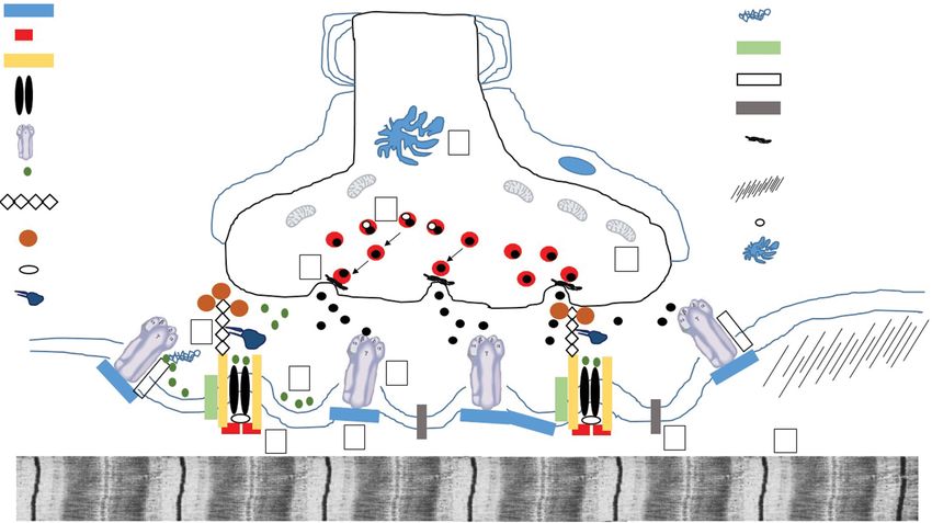
Figure. Representation of the main pathophysiological mechanisms involved in different forms of CMS: (1) acetylcholine
biosynthesis defects and vesicular transport and fusion defects; (2) acetylcholinesterase deficiency; (3) acetylcholine receptor
defects; (4) agrin deficiency; (5) congenital disorders of glycosylation; (6) channelopathies; (7) congenital myopathies with
secondary neuromuscular transmission defects; and (8) mitochondrial dysfunction. Legends: ChAT: choline acetyltransferase;
ErbBR: epidermal growth factor receptor; MASC: muscle-associated specificity component; AChE: acetylcholinesterase; Lrp4:
low-density lipoprotein receptor-related protein 4; ColQ: collagen-like tail subunit or acetylcholinesterase-associated collagen;
MuSK: muscle-specific tyrosine kinase receptor; Dok-7: downstream of tyrosine kinase 72,3,4.
An indicator to consider CMS by mutations in the CHAT Deficiency of SNAP25B was reported with an autoso-
gene is the presence of a decrease in the amplitude of the mal dominant pattern of CMS presenting in an 11-year-old
CMAP associated with a potential endplate 50% below the African-American female patient with a history of intrauterine
baseline (normal decrease is less than 30%) followed by a hypotonia, cyanosis at birth, delayed psychomotor develop-
very slow recovery 5-10 minutes after subtetanic stimula- ment, multiple joint contractures, muscle weakness, fatiga-
tion2. CMAP amplitude decrease is not a pathognomonic ble ptosis, associated with ataxia and early childhood epi-
finding of ChAT deficiency, being present in other CMS. lepsy, whose biomolecular studies revealed a decrease in the
However, the recovery is usually complete in less than five amount of acetylcholine released from the presynaptic nerve
minutes.2,4 In such cases, the amplitudes of the CMAP, end- terminal from each motor nerve impulse4,18.
plate potential and miniature endplate potentials are normal
at rest, decreasing after a repetitive stimulation test at 10 Hz Deficiency of synaptotagmin-2 (OMIM #616040)
for five minutes and marked reduction of CMAPs with slow Synaptotagmin-2 protein is a synaptic vesicle protein
recovery to the basal line in 10 to 15 minutes3. that acts as a calcium sensor during the neuromuscular
transmission process.19 Mutations have been described in
Deficiency of SNAP25B (OMIM #616330) two families with a motor neuropathy phenotype charac-
The proteins of the SNARE class (soluble N-ethylmaleimide- terized by hollow feet and hammer toes with diminished or
sensitive factor attachment protein receptors) constitute abolished reflexes associated with a fatigability of proximal
the core of the intracellular machinery of fusion of synaptic and distal muscles with weakness. Electromyography tests
vesicles17. SNAP25 (synaptosomal-associated protein 25-Kd, demonstrated low amplitude CMAP with a marked increase
encoded by the SNAP25 gene, 20p12.2) and the protein syn- in the amplitude of the CMAP after performing a fast exer-
taxin bind to target cell membranes and are termed t-SNAREs, cise, mimicking the findings of Lambert-Eaton myasthenic
which have an important role in protein and vesicles anchor- syndrome2,20. This presentation has been linked to autosomal
ing, initiation of intracellular vesicles and the rapid triggering dominant inheritance presentations of CMS linked to SYT2
the exocytosis process17. gene (1q32.1).
754 Arq Neuropsiquiatr 2016;74(9):750-760SYNDROMES ASSOCIATED WITH THE SYNAPTIC Defects of the acetylcholine receptor
BASAL LAMINA The nicotinic acetylcholine receptor is a pentameric
complex comprising four transmembrane subunits, com-
Deficiency of endplate acetylcholinesterase by posed of α2βδε in the adult neuromuscular plate and α2βδγ
mutations in COLQ (OMIM #603034) in the fetal neuromuscular plate, as well in extrajunctional
The acetylcholinesterase in the neuromuscular plate is an regions2,4. The genes encoding the α (CHRNA1 gene, 2q31.1),
asymmetrical enzyme which may be constituted by one, two δ (CHRND gene, 2q37.1) and γ (CHRNG gene, 2q37.1) chains
or three catalytic subunits tetramers that are anchored in the are located on the long arm of chromosome 2 at different
basal membrane with a triple-stranded collagen tail called genetic loci, while the genes encoding the β chain (CHRNB1
ColQ (acetylcholinesterase-associated collagen). Mutations gene) and ε (CHRNE gene) are located in different loci on the
in the COLQ gene (3p25.1) promote a prolongation of the res- short arm of chromosome 17p13.22,4. In southern Brazil, as a
idence time of acetylcholine in the neuromuscular junction, consequence of founder effect mutations of Hispanic settlers
producing a desensitization of muscle acetylcholine receptor origin, CHRNE gene recessive mutations represent the main
and a secondary myasthenic syndrome21,22. This presentation cause of CMS9.
corresponds to 13,4% of all CMS cases3. Mutations in genes encoding the acetylcholine receptor
The clinical picture begins at birth or in early child- subunits can produce CMS by means of three mechanisms:
hood as an autosomal recessive CMS, characterized by (1) reducing the number of acetylcholine receptors in the
progressive muscle weakness affecting all voluntary mus- postsynaptic membrane; (2) extended opening called the
cles and can spare the extrinsic ocular muscles. There is a slow-channel syndrome; (3) short opening of the receptors
direct correlation of site of mutations and clinical presen- known as the fast-channel syndrome.2,4
tations and course. Most cases associated with C-terminal
mutations have a light clinical course and late-onset of Primary deficiency of the acetylcholine receptor
symptoms. Most cases related to N-terminal mutations Primary deficiency of the acetylcholine receptor repre-
or rod-domain of ColQ have early-onset severe presenta- sents the most common form of CMS (34 to 50% of all cases)
tions. Diagnosis is essential as genetic counseling repre- and results from mutations in genes coding any of the sub-
sents an important clinical step and as some cases present units of the acetylcholine receptor, with those related to the
good clinical responses to the use of albuterol or ephed- ε subunit being the most common and severe2,4. The clinical
rin21,22. Electrophysiological studies typically confirm the picture is characterized by ptosis, refractory marked ophthal-
presence of a decremental response pattern of the repeti- moparesis and severe muscle weakness of the limbs. There
tive CMAPs greater than 10%23. is generally a partially responsive pattern to pyridostigmine,
amifampridine and albuterol use in clinical practice2,4,25.
Deficiency of laminin-β2 (OMIM #609049) Mutations in both alleles of CHRNA1, CHRNB or CHRND
Laminin-β2 protein is encoded by the LAMB2 gene genes are usually incompatible with life, resulting in death in
(3p21.31) and expressed in the basal lamina of the end- the fetal period2,4,25.
plate terminal of the neuromuscular junction in the eyes
and kidneys participating in the differentiation process of Slow-channel syndrome
the pre-synaptic region as well as the alignment of the pre- Slow-channel syndrome is part of the kinetic defects
synaptic motor terminal with the post-synaptic region in of the acetylcholine receptor due to dominant mutations
the muscle membrane3. Only one case has been reported in the domains or pore receptor ligands of the receptor-
of autosomal recessive congenital myasthenic syndrome binding portion. It resembles, in many aspects, the ace-
resulting from deficiency of laminin-β2 (less than 0,5% of tylcholinesterase deficiency. This typically presents with
all CMS cases) in a 20-year-old female patient with eye and symptoms in the first decade of life with prominent and
kidney malformations, compatible with Pierson syndrome severe involvement of the scapular, cervical and dor-
or microcoria-congenital nephrotic syndrome. Classically sal forearm (wrist and finger extensors) muscle groups,
it represents a pyridostigmine-refractory form of CMS sparing extrinsic ocular muscles and only exceptionally
with predominant proximal limb weakness, where ocu- presenting with mild asymmetric ptosis2,26,27. Atypical
lar abnormalities (including nonreactive fixed narrowing late-onset presentations have also been described as
of the pupils, hypoplasia of the iris and the ciliary body, mimicking generalized autoimmune myasthenia gravis
and lenticonus posterior) and congenital kidney disease with associated mild late-onset myopathic changes and
(including neonatal-onset congenital nephrotic syndrome, rarely with mild to moderately raised serum creatine
early-onset end-stage renal disease and diffuse mesangial kinase levels. However, the good therapeutic response
sclerosis) represent major cardinal signs. Another allelic profile to fluoxetine and quinidine and unresponsiveness
presentation may be as hereditary nephrotic syndrome to amifampridine and pyridostigmine distinguishes this
type 5 (MIM #614199)2,24. condition from acquired forms26,27,28.
Souza PVSS et al. Congenital myasthenic syndromes 755Fast-channel syndrome pelvic and shoulder girdle musculature. The clinical phe-
Fast-channel syndrome arises from autosomal recessive notype results from missense mutations Glu1233Lys and
mutations in different domains of the acetylcholine recep- Arg1277His. Allelic conditions include sclerosteosis type 2
tor subunits and clinically mimicks a typical autoimmune and Cenani-Lenz syndactyly syndrome2,32.
acquired myasthenia gravis starting in the first decade of life
with good clinical response to treatment with pyridostig- MuSK deficiency (OMIM #616325)
mine and amiframpidine. Fluoxetin and quinidine use must The MuSK deficiency is extremely rare, results from
be avoided. Allelic conditions include the multiple pterygium mutations of the MUSK gene (9q31.3) and manifests through
syndrome (lethal type)2,3,29. respiratory failure with neonatal ptosis or in the infant with
proximal appendiceal involvement, facial and ocular and
Defects in the development and maintenance of bulbar variable33. MuSK is involved in end-plate maturation
the endplate and maintenance processes, and aids rapsyn proper function
Congenital myasthenic syndromes can arise by the and its complex interactions with AChR in the postsynaptic
effect of mutations in genes related to the development membrane. The allelic condition is the fetal akinesia defor-
and maintenance of the endplate, including AGRN (agrin; mation sequence2,33.
1p36.33), MUSK (9q31.3), LRP4 (11p11.2), DOK7 (4p16.3),
RAPSN (11p11.2) and COL13A1 (10q22.1) genes. The agrin Dok-7 deficiency (OMIM #254300)
protein is secreted in the terminal nerve synaptic cleft Dok-7 deficiency represents less than 10% of all CMS
and binds to the LRP4 (low density lipoprotein receptor- cases. It represents the second most common cause of
related protein 4) protein of the postsynaptic membrane, CMS in southern Brazil9. Different mutations have been
originating a secondary complex that activates a MuSK described in the DOK7 gene (4p16.3), the most common
(muscle skeletal receptor tyrosine kinase) receptor with being the c.1124_1127dupTGCC mutation related to auto-
tyrosine kinase activity, allowing the phosphorylation of somal recessive CMS. The clinical spectrum is very wide,
Dok-7 (downstream of tyrosine kinase 7) and rapsyn activa- occurring with wrist weakness and lower cervical and
tion (Figure). Thus, modulatory effects occur and the con- facial involvement and some forms with bulbar and oph-
centration of acetycholine in the postsynaptic membrane thalmoparesis involvement. It represents an important
receptor alters the expression of nuclear genes related to differential diagnosis of limb-girdle muscular dystrophies
the differentiation of the endplate and promoting the main- (LGMD) and initiates predominance of type 1 fibers, atro-
tenance of the postsynaptic membrane1,2. phy of type 2 fibers, and “target” fiber formations34,35. An
LGMD-like pattern of weakness, ptosis and mild facial
Agrin deficiency (OMIM #615120) weakness with moderate to severe bulbar symptoms and
Agrin is a proteoglycan secreted in the synaptic basal laryngeal stridor and vocal cord palsy occurs in mutations
laminae by the terminal nerve, phosphorylating and acti- of the last codon. Other cases present as lower limb dom-
vating MuSK by the LRP4 receptor. Mutations of the AGRN inant progressive amyotrophy. Muscle biopsy discloses
(1p36.33) gene, coding agrin, originates a typical late-onset generally unspecific findings. Clinical worsening is com-
CMS phenotype starting between the fourth and fifth mon after pyridostigmine use and clinical improvement
decades of life and an early-onset variant in infants and neo- frequently observed after ephedrine, salbutamol and alb-
nates. The typical form originates ptosis and mild weakness uterol use34,35,36.
in the face and proximal limbs weakness, starting from child-
hood and resulting from homozygous missense mutations Rapsyn deficiency (OMIM #616326)
(Gly1709Arg) and other types with more severe phenotypes Rapsyn (receptor-associated protein of the synapse,
(Gln353X and Val1725Phe). The subtype of early-onset CMS 43-Kd) deficiency results from homozygous mutations in the
relates to generalized amyotrophy in lower limbs and slowly RAPSN gene (11p11.2) and represents around 15% of all CMS
progressive weakness with lipossubstitution of the poste- cases. This autosomal recessive CMS manifests most com-
rior compartment of the leg, sparing cranial nerves and axial monly in infants during the first year of life (rarely in scholars
muscles and without ophthalmoparesis (despite ptosis being and young adults), the most common pattern in one third of
frequently found). There is a good response to the use of patients being an arthrogryposis phenotype at birth, multi-
ephedrine and generally failure to the use of amifampridine ple congenital contractures and other congenital malforma-
and pyridostigmine3,30,31. tions, frequently in the context of patients with post-anoxic
encephalopathy after myasthenic crisis due to infectious
LRP4 deficiency (OMIM #616304) complications2,3,4. It is remarkable that normal adduction and
The LRP4 deficiency was described in a 14-year-old abduction of the eye with strabismus and ophthalmoparesis is
patient with neonatal respiratory failure, delayed motor found in less than 25% of cases. There is some regional segre-
development and muscle weakness and fatigability of gation of mutations in India and Europe (Asn88Lys mutation).
756 Arq Neuropsiquiatr 2016;74(9):750-760Another important finding relates to clinical-genetic correla- components for processes of O-linked and N-linked gly-
tions: the c.38A>G mutation presents with a milder pheno- cosylation of basic proteins. Mutations in the GFPT1
type with ptosis, prognathism, facial and masticatory muscle gene (2p13.3) represents less than 3% of all CMS cases.
weakness and nasal voice; the N88K mutation represents most Typically, disability originates slowly progressive muscle
neonatal and early-infancy-onset cases; A38G mutation com- weakness with an LGMD-like phenotype responsive to
monly presents with ptosis, hypernasal speech, facial weak- pyridostigmine use and tubular aggregates of sarcoplas-
ness and prominent masticatory dysfunction37. The allelic con- mic reticulum in muscle biopsy. Some variants showed
dition leads to to fetal akinesia deformation sequence. Thus, multiple arthrogryposis at birth, slow progression until
the presence of an early-onset phenotype mimicking chonic eight years old with severe dysphagia and severe myopa-
nonprogressive hypoxic-ischaemic clinical findings with neu- thy with autophagic vacuoles in muscle specimens. Ptosis
romuscular junction dysfunction signs should raise the possi- and respiratory insufficiency are rarely found. Raised
bility of rapsyn deficiency. serum CK is found in around 25% of cases2,3,40.
COL13A1-related CMS (OMIM #616720) Deficiency of DPAGT1 (OMIM #614750)
A new autosomal recessive very early-onset CMS has TheenzymeDPAGT1(dolichyl-phosphateN-acetylglucosamine
been recently described resulting from homozygous muta- phosphotransferase) catalyzes the first step in glycosylation of
tions in the COL13A1 gene (10q22.1), coding the alpha-1 N-linked proteins. The resulting clinical spectrum of mutations in
chain of the nonfibrillar transmembrane collagen type the DPAGT1 (11q23.3) gene initiates moderate or severe muscle
XIII, involved in autocrine control of the development and weakness and cognitive impairment, with a therapeutic profile
maturation of the neuromuscular junction38. Clinical pre- unresponsive to pyridostigmine and amifampridine, and partially
sentation is broad and includes ptosis, proximal and dis- responsive to salbutamol. The most common mutations involve
tal limb hypotonia, delayed motor milestones, poor head His375Tyr and Val264Met. Allelic conditions include CDG type Ij.
control, feeding difficulties, childhood-onset spinal rigid- Muscle biopsy shows small tubular aggregates, fiber type dispropor-
ity, respiratory insufficiency, dyspnea on exertion with tion with small caliber type 1 fibers and autophagic vacuolar myop-
exercise intolerance, dysphagia, gastroesophageal reflux athy, resembling similar patterns to STIM1-related myopathies3,41,42.
and recurrent lower respiratory tract infections. Mild
dysmorphic features have also been reported, including ALG2 and ALG14 deficiencies (OMIM #616228;
pes cavus without early joint contractures, low-set ears, OMIM #616227)
micrognathia, high-arched palate and pectus carinatum. The ALG2 (asparagine-linked glycosylation 2) enzyme
Neurophysiological studies show significant decremental participates directly in the second and third steps of
response on repetitive nerve stimulation testing38. N-glycosylation. ALG14 forms a complex with the multiglu-
cosyltransferase ALG13. DPAGT1 participates indirectly in
Congenital disorders of glycosylation the first step of N-glycosylation. Both syndromes arising from
Congenital disorders of glycosylation (CDGs) relate to ALG2 (9q22.33) and ALG14 (1p21.3) genes mutations have
different systemic and neurometabolic syndromes with dif- an early-onset LGMD-like CMS starting in the preschooler
ferent congenital malformations, subtypes of congenital with typical myasthenic syndrome complaints. ALG14 defi-
muscular dystrophy and congenital myasthenic syndromes ciency presents with a similar pattern to ALG2-related CMS,
by mutations in the genes encoding the enzymes GFPT1 despite the absence of tubular aggregates in muscle biopsy.
(glutamine:fructose-6-phosphate amidotransferase 1, 2p13.3), Pyridostigmine may benefit some patients. Allelic conditions
DPAGT1 (dolichyl-phosphate UDP-N-acetylglucosamine include CDG type Ii. Raised serum CK levels can be observed
N-acetylglucosamine-1-phosphotransferase, 11q23.3) ALG2 in both cases2,43.
(alpha-1,3-mannosyltransferase, 9q22.33) and ALG14 (UDP-N-
acetylglucosaminyltransferase, 1p21.3). The histopathological Deficiency of GMPPB
presence of muscle tubular aggregates in the setting of char- GMPPB gene (3p21.31) mutations have recently been
acteristic neurophysiological findings in pre- and postsynap- described as a cause of autosomal recessive CMS. Cases
tic impaired neuromuscular junction must indicate genetic present with a notably exclusive appendicular (mainly
evaluation for CDG-related CMS2,3. Recently, a fifth glycosyl- proximal) weakness phenotype without facial and eye
ation gene called GMPPB (GDP-mannose pyrophosphorylase muscle involvement. They also present with typical myo-
B, 3p21.31) has also been linked to new CMS phenotypes39. pathic changes in different laboratory evaluations, includ-
ing high serum creatine kinase levels and unspecific
GFPT1 deficiency (OMIM #610542) myopathic changes in muscle biopsy and muscle MRI
The GFPT1 (glutamine:fructose-6-phosphate ami- studies39. Mutations in this same gene also give rise to a
dotransferase-1) enzyme regulates glucose entering in broad clinical spectrum with congenital muscular dystro-
the hexosamine pathway for the formation of precursor phies from the dystroglycanopathy family, including allelic
Souza PVSS et al. Congenital myasthenic syndromes 757conditions such as autosomal recessive congenital muscu- reports in the literature of congenital myopathies with
lar dystrophies from the dystroglycanopathy group (types mutations that can lead to secondary impairment of the
A, B and C). Despite its rarity, GMPPB-related myasthenic neuromuscular junction from school age to young adult,
syndromes should be considered in the setting of a nearly notably MTM1 (coding myotubularin, Xq28), RYR1 (cod-
proximal appendicular myopathic patient in whom ther- ing ryanodine receptor 1, 19q13.2), DNM2 (coding dyna-
apeutic strategies have failed or clinical outcomes have min 2, 19p13.2), TPM3 (coding tropomyosin 3, 1q21.3)
unexpectedly worsened39. and BIN1 (coding bridging integrator 1 or amphiphy-
sin II, 2q14.3). This results in variable combinations of
Other myasthenic syndromes myasthenic syndrome (with ptosis, ophthalmoparesis
and facial paresis), exercise intolerance, variable partial
PREPL Deletion Syndrome (OMIM #606407)
responsiveness to piridostigmin or corticosteroids (espe-
The PREPL (prolyl endopeptidase-like) protein, coded
cially in centronuclear myopathies) and variable decre-
by the PREPL gene (2p21), is an essential activator of clath-
mental responses (19-35% from baseline) or non-existent
rin adapter protein associated with type 1 (AP1), involved in
response in neurophysiological studies. Muscle biopsy in
vesicular transport and filling with acetylcholine and other
such cases usually shows atrophy of type 1 fibers with
neurotransmitters. The hypotonia-cystinuria syndrome is
occasional loss of mitochondria and central myofibril-
caused by deletions in the SLC3A1 gene and recessive muta-
tions in the PREPL gene, originating cystinuria type A, defi- lar degeneration 2,3,47. It is essential to highlight that most
ciency of growth hormone, ptosis, and limb, facial and bul- genes related to these phenotypes are involved with
bar weakness, and neonatal hypotonia. The symptoms are different neuromuscular phenotypes, including nema-
moderate and transiently responsive to pyridostigmine dur- line myopathy, centronuclear myopathy, CAP myopa-
ing childhood2,4,44. thy, fiber-type disproportion congenital myopathy, lethal
congenital contracture syndrome type 5 and Charcot-
Deficiency of plectin Marie-Tooth disease spectrum.
Mutations in the PLEC gene (8q24.3), coding plectin 1,
initiate a rare complex spectrum of clinical manifestations, Mutations in the SLC25A1 gene
involving epidermolysis bullosa simplex with nail dystro- The SLC25A1 (solute carrier family 25, mitocondrial car-
phy, limb-girdle muscular dystrophy type 2Q and congeni- rier; citrate transporter, member 1) protein mediates mito-
tal myasthenic syndrome. The neuromuscular phenotypes chondrial citrate/isocitrate shuttle promoting cytosolic
develop in patients with epidermolysis bullosa in childhood malate re-entry into the citric acid cycle and the move-
that, in the course of life, evolves into a progressive proximal ment of citrate across the mitochondrial inner membrane.
myopathic phenotype refractory to pyridostigmine and dec- Mutations in the SLC25A1 gene (22q11.21) initiate cogni-
remental response pattern in neurophysiological studies2,45. tive impairment, myasthenic symptoms, hypoplasia of the
optic nerve, corpus callosum agenesis and eventually sec-
Defects in sodium channels (SCN4A) (OMIM #614198) ondary 2-hydroxyglutaric aciduria.2,48 An allelic condition
An extremely rare form of CMS (less than 0,5% of all includes the autosomal recessive combined D-2- and L-2-
cases) was originally associated with a 20-year-old female hydroxyglutaric aciduria (OMIM #615182). Thus, a CMS
patient with brief and abrupt attacks of muscle weakness
complex phenotype linked with different central nervous
(including bulbar palsy) and respiratory failure from birth
system malformations should make clinicians aware of the
that led to hypoxic-ischemic encephalopathy resulting from
possibility of SLC25A1 gene mutations.
Val1442Glu mutation in the SCN4A gene (17q23.3), resulting
In conclusion, CMS represents a genetically hetero-
in dysfunction of neuromuscular transmission in the unex-
geneous and important differential diagnosis group of
citability rest state3,45. Each acute attack typically lasts from
early-onset peripheral hypotonic disorders. As a poten-
three to 30 minutes, similar to choline acetyltransferase
deficiency. Allelic conditions include hyperkalemic periodic tially treatable group of neuromuscular diseases, clini-
paralysis type 2, hypokalemic periodic paralysis type 2, atypi- cians should be aware of this possibility when facing a
cal acetazolamide-responsive myotonia congenita and para- hypotonic baby or infant with ophthalmoparesis, eyelid
myotonia congenita4,46 . ptosis, facial palsy, recurrent unprovoked apneic episodes,
dysphagia, dysphonia and early-onset distal joint contrac-
Congenital myopathies with secondary defects of tures, sometimes mimicking congenital arthrogryposis
neuromuscular transmission multiplex. Late-onset presentations of CMS may also be
Congenital myopathies represent an extremely rare considered, especially in cases of refractory seronegative
cause of congenital myasthenic syndrome-like phe- myasthenia gravis, highly suggestive phenotypes or posi-
notype (less than 0,5% of all causes). There are several tive family history of myasthenia.
758 Arq Neuropsiquiatr 2016;74(9):750-760References
1. Berrih-Aknin S, Le Panse R. Myasthenia gravis: a comprehensive intellectual disability. Neurology. 2014;83(24):2247-55.
review of immune dysregulation and etiological mechanisms. J doi:10.1212/WNL.0000000000001079
Autoimmun. 2014;52:90-100. doi:10.1016/j.jaut.2013.12.011 19. Herrmann DN, Horvath R, Sowden JE, Gonzalez M, Sanchez-
2. Engel AG, Shen XM, Selcen D, Sine SM. Congenital myasthenic Mejias A, Guan Z et al. Synaptotagmin 2 mutations cause
syndromes: pathogenesis, diagnosis, and treatment. Lancet Neurol. an autosomal-dominant form of lambert-eaton myasthenic
2015;14(4):420-34. doi:10.1016/S1474-4422(14)70201-7 syndrome and nonprogressive motor neuropathy. Am J Hum Genet.
2014;95(3):332-9. doi:10.1016/j.ajhg.2014.08.007
3. Engel AG. Congenital myasthenic syndromes. In: Katirji B, Kaminski
HJ, Ruff RL, editors. Neuromuscular disorders in clinical practice. 20. Baker K, Gordon SL, Grozeva D, Kogelenberg M, Roberts NY, Pike
New York: Springer; 2014. p. 1073-90. M et al. Identification of a human synaptotagmin-1 mutation that
perturbs synaptic vesicle cycling. J Clin Invest. 2015;125(4):1670-8.
4. Rodríguez Cruz PM, Palace J, Beeson D. Inherited disorders of the
doi:10.1172/JCI79765
neuromuscular junction: an update. J Neurol. 2014;261(11):2234-43.
doi:10.1007/s00415-014-7520-7 21. Ohno K, Brengman J, Tsujino A, Engel AG. Human endplate
acetylcholinesterase deficiency caused by mutations in the
5. Parr JR, Andrew MJ, Finnis M, Beeson D, Vincent A, Jayawant S. How
collagen-like tail subunit (ColQ) of the asymmetric enzyme. Proc Natl
common is childhood myasthenia? The UK incidence and prevalence
Acad Sci USA. 1998;95(16):9654-9. doi:10.1073/pnas.95.16.9654
of autoimmune and congenital myasthenia. Arch Dis Child.
2014;99(6):539-42. doi:10.1136/archdischild-2013-304788 22. Mihaylova V, Müller JS, Vilchez JJ, Salih MA, Kabiraj MM, D’Amico
A et al. Clinical and molecular genetic findings in COLQ-mutant
6. Richard P, Gaudon K, Haddad H, Ammar AB, Genin E, Bauché
congenital myasthenic syndromes. Brain. 2008;131(3):747-59.
S et al. The CHRNE 1293insG founder mutation is a frequent
doi:10.1093/brain/awm325
cause of congenital myasthenia in North Africa. Neurology.
2008;71(24):1967-72. doi:10.1212/01.wnl.0000336921.51639.0b 23. Lorenzoni PJ, Scola RH, Gervini BL, Kay CSK, Werneck
LC. Electrophysiological study in synaptic congenital
7. Abicht A, Stucka R, Karcagi V, Herczegfalvi A, Horváth R, Mortier W et
myasthenic syndrome: end-plate acetylcholinesterase
al. A common mutation (epsilon1267delG) in congenital myasthenic deficiency. Arq Neuropsiquiatr. 2009;67(2B):502-4.
patients of Gypsy ethnic origin. Neurology 1999;53(7):1564-9. doi:10.1590/S0004-282X2009000300024
doi:10.1212/WNL.53.7.1564
24. Maselli RA, Ng JJ, Anderson JA, Cagney O, Arredondo J, Williams
8. Lorenzoni PJ, Scola RH, Kay CS, Werneck LC. Congenital myasthenic C et al. Mutations in LAMB2 causing a severe form of synaptic
syndrome: a brief review. Pediatr Neurol. 2012;46(3):141-8. congenital myasthenic syndrome. J Med Genet. 2009;46(3):203-8.
doi:10.1016/j.pediatrneurol.2011.12.001 doi:10.1136/jmg.2008.063693
9. Mihaylova V, Scola RH, Gervini B, Lorenzoni PJ, Kay CK, Werneck LC et 25. Ohno K, Quiram P, Milone M, Wang HL, Harper MC, Pruitt JN 2nd et al.
al. Molecular characterisation of congenital myasthenic syndromes Congenital myasthenic syndromes due to heteroallelic nonsense/
in Southern Brazil. J Neurol Neurosurg Psychiatry. 2010;81(9):973-7. missense mutations in the acetylcholine receptor ε subunit gene:
doi:10.1136/jnnp.2009.177816 identification and functional characterization of six new mutations.
10. Conti-Fine BM, Milani M, Kaminski HJ. Myasthenia gravis: Hum Mol Genet. 1997;6(5):753-66. doi:10.1093/hmg/6.5.753
past, present and future. J Clin Invest. 2006;116(11):2843-54. 26. Ohno K, Hutchinson DO, Milone M, Brengman JM, Bouzat C, Sine
doi:10.1172/JCI29894 SM et al. Congenital myasthenic syndrome caused by prolonged
11. Meriggioli MN, Sanders DB. Autoimmune myasthenia gravis: acetylcholine receptor channel openings due to a mutation in the M2
emerging clinical and biological heterogeneity. Lancet Neurol. domain of the ε subunit. Proc Natl Acad Sci USA. 1995;92(3):758-62.
2009;8(5):475-90. doi:10.1016/S1474-4422(09)70063-8 doi:10.1073/pnas.92.3.758
12. Pinto WBVR, Souza PVS, Oliveira ASB. Normal muscle structure, 27. Ohno K, Wang HL, Milone M, Bren N, Brengman JM, Nakano
growth, development and regeneration. Curr Rev Musculoskelet Med. S et al. Congenital myasthenic syndrome caused by
2015;8(2):176-81. doi:10.1007/s12178-015-9267-x decreased agonist binding affinity due to a mutation in the
acetylcholine receptor ε subunit. Neuron. 1996;17(1):157-70.
13. Das AS, Agamanolis DP, Cohen BH. Use of next-generation
doi:10.1016/S0896-6273(00)80289-5
sequencing as a diagnostic tool for congenital
myasthenic syndrome. Pediatr Neurol. 2014;51(5):717-20. 28. Chaouch A, Müller JS, Guergueltcheva V, Dusl M, Schara U,
doi:10.1016/j.pediatrneurol.2014.07.032 Rakocević-Stojanović V et al. A retrospective clinical study of the
treatment of slow-channel congenital myasthenic syndrome. J
14. Tei S, Ishii HT, Mitsuhashi H, Ishiura S. Antisense oligonucleotide-
Neurol. 2012;259(3):474-81. doi:10.1007/s00415-011-6204-9
mediated exon skipping of CHRNA1 pre-mRNA as potential therapy
29. Shen XM, Brengman JM, Edvardson S, Sine SM, Engel AG. Highly
for Congenital Myasthenic Syndromes. Biochem Biophys Res
fatal fast-channel syndrome caused by AChR ε subunit mutation
Commun. 2015;461(3):481-6. doi:10.1016/j.bbrc.2015.04.035
at the agonist binding site. Neurology. 2012;79(5):449-54.
15. Kraner S, Laufenberg I, Strassburg HM, Sieb JP, Steinlein OK. doi:10.1212/WNL.0b013e31825b5bda
Congenital myasthenic syndrome with episodic apnea in patients
30. Nicole S, Chaouch A, Torbergsen T, Bauché S, de Bruyckere E,
homozygous for a CHAT missense mutation. Arch Neurol.
Fontenille MJ et al. Agrin mutations lead to a congenital myasthenic
2003;60(5):761-3. doi:10.1001/archneur.60.5.761
syndrome with distal muscle weakness and atrophy. Brain.
16. Ohno K, Tsujino A, Brengman JM, Harper CM, Bajzer Z, Udd B et al. 2014;137(9):2429-43. doi:10.1093/brain/awu160
Choline acetyltransferase mutations cause myasthenic syndrome
31. Huzé C, Bauché S, Richard P, Chevessier F, Goillot E, Gaudon K
associated with episodic apnea in humans. Proc Natl Acad Sci USA.
et al. Identification of an agrin mutation that causes congenital
2001;98(4):2017-22. doi:10.1073/pnas.98.4.2017
myasthenia and affects synapse function. Am J Hum Genet.
17. Mohrmann R, de Wit H, Connell E, Pinheiro PS, Leese C, Bruns D et al. 2009;85(2):155-62. doi:10.1016/j.ajhg.2009.06.015
Synaptotagmin interaction with SNAP-25 governs vesicle docking,
32. Ohkawara B, Cabrera-Serrano M, Nakata T, Milone M, Asai N, Ito K et
priming, and fusion triggering. J Neurosci. 2013;33(36):14417-30.
al. LRP4 third β-propeller domain mutations cause novel congenital
doi:10.1523/JNEUROSCI.1236-13.2013
myasthenia by compromising agrin-mediated MuSK signaling in
18. Shen XM, Selcen D, Brengman J, Engel AG. Mutant SNAP25B a position-specific manner. Hum Mol Genet. 2014;23(7):1856-68.
causes myasthenia, cortical hyperexcitability, ataxia, and doi:10.1093/hmg/ddt578
Souza PVSS et al. Congenital myasthenic syndromes 75933. Mihaylova V, Salih MA, Mukhtar MM, Abuzeid HA, El-Sadig SM, 41. Belaya K, Finlayson S, Slater CR, Cossins J, Liu WW, Maxwell S et al.
Hagen M et al. Refinement of the clinical phenotype in musk-related Mutations in DPAGT1 cause a limb-girdle congenital myasthenic
congenital myasthenic syndromes. Neurology. 2009;73(22):1926-8. syndrome with tubular aggregates. Am J Hum Genet. 2012;91(1):193-
doi:10.1212/WNL.0b013e3181c3fce9 201. doi:10.1016/j.ajhg.2012.05.022
34. Müller JS, Herczegfalvi A, Vilchez JJ, Colomer J, Bachinski LL, 42. Selcen D, Shen XM, Brengman J, Li Y, Stans AA, Wieben E et
Mihaylova V et al. Phenotypical spectrum of DOK7 mutations in al. DPAGT1 myasthenia and myopathy: genetic, phenotypic,
congenital myasthenic syndromes. Brain. 2007;130(6):1497-506. and expression studies. Neurology. 2014;82(20):1822-30.
doi:10.1093/brain/awm068 doi:10.1212/WNL.0000000000000435
35. Anderson JA, Ng JJ, Bowe C, McDonald C, Richman DP, Wollmann 43. Cossins J, Belaya K, Hicks D, Salih MA, Finlayson S,
RL et al. Variable phenotypes associated with mutations in DOK7. Carboni N et al. Congenital myasthenic syndromes due to
Muscle Nerve. 2008;37(4):448-56. doi:10.1002/mus.20944 mutations in ALG2 and ALG14. Brain. 2013;136(3):944-56.
36. Lorenzoni PJ, Scola RH, Kay CS, Filla L, Miranda AP, Pinheiro JM doi:10.1093/brain/awt010
et al. Salbutamol therapy in congenital myasthenic syndrome 44. Régal L, Shen XM, Selcen D, Verhille C, Meulemans S, Creemers
due to DOK7 mutation. J Neurol Sci. 2013;331(1-2):155-7. JW et al. PREPL deficiency with or without cystinuria causes a
doi:10.1016/j.jns.2013.05.017 novel myasthenic syndrome. Neurology. 2014;82(14):1254-60.
37. Milone M, Shen XM, Selcen D, Ohno K, Brengman J, Iannaccone ST doi:10.1212/WNL.0000000000000295
et al. Myasthenic syndrome due to defects in rapsyn: clinical and 45. Selcen D, Juel VC, Hobson-Webb LD, Smith EC, Stickler DE, Bite AV
molecular findings in 39 patients. Neurology. 2009;73(3):228-35. et al. Myasthenic syndrome caused by plectinopathy. Neurology.
doi:10.1212/WNL.0b013e3181ae7cbc 2011;76(4):327-36. doi:10.1212/WNL.0b013e31820882bd
38. Logan CV, Cossins J, Rodríguez Cruz PM, Parry DA, Maxwell S, 46. Tsujino A, Maertens C, Ohno K, Shen XM, Fukuda T, Harper CM
Martínez-Martínez P et al. Congenital myasthenic syndrome type et al. Myasthenic syndrome caused by mutation of the SCN4A
19 is caused by mutations in COL13A1, encoding the atypical sodium channel. Proc Natl Acad Sci USA. 2003;100(12):7377-82.
non-fibrillar collagen type XIII alpha-1 chain. Am J Hum Genet. doi:10.1073/pnas.1230273100
2015;97(6):878-85. doi:10.1016/j.ajhg.2015.10.017 47. Rodríguez Cruz PM, Sewry C, Beeson D, Jayawant S, Squier
39. Belaya K, Rodríguez Cruz PM, Liu WW, Maxwell S, McGowan W, McWilliam R et al. Congenital myopathies with secondary
S, Farrugia ME et al. Mutations in GMPPB cause congenital neuromuscular transmission defects: a case report and review
myasthenic syndrome and bridge myasthenic disorders of the literature. Neuromuscul Disord. 2014;24(12):1103-10.
with dystroglycanopathies. Brain. 2015;138(9):2493-504. doi:10.1016/j.nmd.2014.07.005
doi:10.1093/brain/awv185 48. Edvardson S, Porcelli V, Jalas C, Soiferman D, Kellner Y,
40. Selcen D, Shen XM, Milone M, Brengman J, Ohno K, Shaag A et al. Agenesis of corpus callosum and optic nerve
Deymeer F et al. GFPT1-myasthenia: clinical, structural, and hypoplasia due to mutations in SLC25A1 encoding the
electrophysiologic heterogeneity. Neurology. 2013;81(4):370-8. mitochondrial citrate transporter. J Med Genet. 2013;50(4):240-5.
doi:10.1212/WNL.0b013e31829c5e9c doi:10.1136/jmedgenet-2012-101485
760 Arq Neuropsiquiatr 2016;74(9):750-760You can also read