Linkage mechanics and power amplification of the mantis shrimp's strike
←
→
Page content transcription
If your browser does not render page correctly, please read the page content below
3677
The Journal of Experimental Biology 210, 3677-3688
Published by The Company of Biologists 2007
doi:10.1242/jeb.006486

          Linkage mechanics and power amplification of the mantis shrimp’s strike
                  S. N. Patek1,*, B. N. Nowroozi2, J. E. Baio1, R. L. Caldwell1 and A. P. Summers2
      1
       Department of Integrative Biology, University of California, Berkeley, CA 94720-3140, USA and 2Ecology and
                  Evolutionary Biology, University of California–Irvine, Irvine, CA 92697-2525, USA
                                                     *Author for correspondence (e-mail: patek@berkeley.edu)

                                                                   Accepted 6 August 2007

                                                         Summary
   Mantis shrimp (Stomatopoda) generate extremely rapid      transmission is lower than predicted by the four-bar model.
and forceful predatory strikes through a suite of structural The results of the morphological, kinematic and
modifications of their raptorial appendages. Here we         mechanical analyses suggest a multi-faceted mechanical
examine the key morphological and kinematic components       system that integrates latches, linkages and lever arms and
of the raptorial strike that amplify the power output of the is powered by multiple sites of cuticular energy storage.
underlying muscle contractions. Morphological analyses of    Through reorganization of joint architecture and
joint mechanics are integrated with CT scans of              asymmetric distribution of mineralized cuticle, the mantis
mineralization patterns and kinematic analyses toward the    shrimp’s raptorial appendage offers a remarkable example
goal of understanding the mechanical basis of linkage        of how structural and mechanical modifications can yield
dynamics and strike performance. We test whether a four-     power amplification sufficient to produce speeds and forces
bar linkage mechanism amplifies rotation in this system      at the outer known limits of biological systems.
and find that the rotational amplification is approximately
two times the input rotation, thereby amplifying the         Key words: power amplification, predation, movement, feeding, speed,
velocity and acceleration of the strike. The four-bar model  acceleration, Crustacea, kinematic transmission, four-bar linkage
is generally supported, although the observed kinematic      model.

                          Introduction                                            ultimately a latch releases the string, which in turn drives
   All animals face an overriding constraint on their ability to                  forward the arrow (‘release phase’). In the load phase, muscle
produce fast movements – muscles contract slowly and over                         contractions load elastic elements and thereby store potential
small distances. Repeatedly over evolutionary history, animals                    energy. In the release phase, fast movement is actuated through
have overcome this limitation through the use of power                            the rapid release of stored potential energy. It is important to
amplification mechanisms. These mechanisms decrease the                           note that in the release phase, muscle activity plays a minimal
duration of movement and thereby increase speed and                               role or no role at all in actuating the fast movement; the release
acceleration (Alexander, 1983; Alexander and Bennet-Clark,                        of elastic potential energy occurs at far shorter timescales than
1977; Gronenberg, 1996a). Power-amplified animal movements                        muscle contractions. With this mechanism, the arrow
are truly diverse, ranging from the elastic tendons and springy                   accelerates and flies through the air at far greater speeds than
legs of kangaroo and locust jumps, to snapping shrimp                             would have been possible by simply throwing the arrow.
appendages and spring-loaded vertebrate ballistic tongues                            The mysteries of the crossbow – where is energy stored, how
(Alexander, 1990; Alexander and Bennet-Clark, 1977; de Groot                      release is triggered, and the mechanics behind the loading or
and van Leeuwen, 2004; Deban et al., 1997; Heitler, 1974;                         unloading of the bow – are the same principal questions we ask
Lappin et al., 2006; Nishikawa, 1999; Ritzmann, 1973; Versluis                    of a biological energy storage system. The speed and power of
et al., 2000).                                                                    the killing strike of the second thoracic appendages (the
   In all of these systems, relatively slow muscle contractions                   ‘raptorial appendages’) of mantis shrimp (Stomatopoda) are
precede rapid movement. As muscles contract to provide the                        clear evidence of a power amplification system at work (Fig.·1)
necessary work for the movement, elastic potential energy is                      (Burrows, 1969; Patek and Caldwell, 2005; Patek et al., 2004).
typically stored in structural elements (e.g. kangaroo tendons)                   The entire strike occurs over several milliseconds and can reach
while latches and/or antagonistic muscle contractions prevent                     peak speeds of 10–24·m·s–1 (Burrows, 1969; Burrows and
movement until the animal is ready to jump or strike                              Hoyle, 1972; Patek et al., 2004). Peacock mantis shrimp
(Alexander, 1983; Gronenberg, 1996a). A good analogy for                          Odontodactylus scyllarus can directly deliver impact forces of
these biological principles is found in the crossbow: slow                        over 1000·N (thousands of times its body weight) with an equal
muscle contractions of a human arm gradually load (‘load                          or greater force secondarily caused by cavitation bubble
phase’) and store elastic potential energy in the crossbow and                    collapse (Patek and Caldwell, 2005).

                                                       THE JOURNAL OF EXPERIMENTAL BIOLOGY
3678 S. N. Patek and others
                                                                         possibility that there is a leverage system, such as a four-bar
                                                                         linkage, underlying the rapid rotation of the dactyl. While the
                                                                         storage and release of cuticular elastic energy during the release
                                                                         phase is often observed in arthropods, e.g. locust jumping legs
                                                                         (Heitler, 1974), linkage mechanisms in arthropod power
                                                                         amplification mechanisms are not well studied. These jointed
                                                                         leverage systems amplify rotational motion and are typically
                                                                         characterized in terms of kinematic transmission (KT; angular
                                                                         output of the linkage mechanism divided by angular input)
                                                                         (Barel et al., 1977; Westneat, 1994) (Fig.·2). Thus, KT provides
                                                                         a heuristic measure of speed- versus force-modification of the
                                                                         linkage system, such that a high KT system delivers a large
                                                                         angular output (e.g. angular velocity) for a small angular input
                                                                         and can therefore be considered ‘angular velocity-modified’ (in
                                                                         the same sense that the mechanical advantage provided by a
                                                                         long output lever relative to input lever is speed-modified).
                                                                         Linkage models of fish jaws have proved to be powerful tools
                                                                         for examining the evolution and performance in force- versus
                                                                         speed-modified feeding mechanisms within and across species
                                                                         (Alfaro et al., 2004; Collar et al., 2005; Hulsey and Wainwright,
Fig.·1. Odontodactylus scyllarus raptorial appendage. (A) A resting
peacock mantis shrimp with the raptorial appendage circled. Raptorial
                                                                         2002; Muller, 1996; Westneat, 1991; Westneat, 1995; Westneat
appendages are used either for stabbing (dactyl open and extended) or    et al., 1993).
for hammering (dactyl folded in and bulbous heel exposed, as shown          Previous studies have examined the functional morphology
here). (B) Lateral view of an isosurface rendering of segmented CT       of the stomatopod’s raptorial appendage (Burrows, 1969;
scan data of the left raptorial appendage. Each skeletal element has     Kunze, 1981), muscle anatomy and activity patterns during the
been pseudocolored to increase contrast. The isosurface threshold has    strike (Burrows, 1969; Burrows and Hoyle, 1972; McNeill et
been optimized for each element to illustrate the morphology and         al., 1972), and a proposed linkage system and energy storage
spatial relationships. (C) Ventral view of a shadowless volume           mechanism (Patek et al., 2004). Here, we build on these
rendering of left merus (m). Shading corresponds to degree of radio-     previous studies by examining the raptorial morphology and
opacity (mineralization), with lighter colors corresponding to greater   mechanics of peacock mantis shrimp Odontodactylus scyllarus
mineralization and darker to poorly mineralized areas. Note the
                                                                         from several new perspectives, including the use of CT scan
unmineralized region adjacent to the highly mineralized ventral bar
(vb) extending proximally from the base of the meral-V. This             technology to characterize cuticular mineralization patterns and
unmineralized region may permit dorso-proximal flexion of the meral-     functional morphology of the latches as well as high-speed
V (v). (D) Lateral view of the merus using the same rendering            video analysis to measure changing conformations of the
technique as in C. s, saddle; c, carpus; p, propodus; d, dactyl. Scale   appendage segments and strike kinematics. In addition, we
bars, 4·mm.                                                              quantitatively test the previously proposed linkage mechanism
                                                                         (Patek et al., 2004) and assess whether the proposed elastic
                                                                         energy storage mechanism could function given the
   Mantis shrimp, like all crustaceans, control movement with            mineralization patterns of the merus.
antagonistic pairs of muscles that alternately abduct and adduct            The goals of the present study were to examine the anatomy
their appendages. However, in the load phase of a power-                 of the raptorial appendage and the kinematics of the release
amplified strike, mantis shrimp simultaneously activate the              phase of the strike mechanism from these functional
antagonistic muscles connecting the carpus and merus                     perspectives. (1) Energy storage: what is the distribution of
segments in the raptorial appendage as they prepare for a high-          mineralization in the merus and how does this mineralization
powered strike (Fig.·1). Specifically, they contract large, slow         pattern contribute to the elasticity and stabilization of the
extensor muscles in the merus while contracted flexor muscles            appendage? (2) Latching mechanism and pre-strike
in the merus brace a pair of sclerites to prevent movement               stabilization: what are the shapes and orientations of the
(Burrows, 1969; Burrows and Hoyle, 1972; McNeill et al.,                 sclerites and how might they control the preparation for and
1972). When the extensor muscles have fully contracted and               release of the strike? (3) Kinematic transmission: does a four-
the animal is ready to strike, the flexor muscles turn off,              bar exoskeletal linkage system mediate the storage and release
releasing the sclerites, and the appendage rapidly rotates               of potential energy in this system?
outward toward its target (Burrows, 1969; Burrows and Hoyle,
1972; McNeill et al., 1972). It has been proposed that energy                                Materials and methods
is stored in a saddle-shaped portion of the merus exoskeleton               Odontodactylus scyllarus L. (Crustacea, Stomatopoda,
(Patek et al., 2004) and in the connective tissue of merus,              Gonodactyloidea, Odontodactylidae) specimens (11.5–14.8·cm
specifically the extensor muscles and apodemes (Burrows,                 body length) were purchased from aquarist companies and
1969).                                                                   housed in re-circulating saltwater tanks (25–30°C). They were
   The morphological complexity and evolutionary diversity of            fed a diet of fresh snails, frozen shrimp and vitamin-fortified
the mechanical system that drives the raptorial strike raises the        freeze-dried shrimp.

                                              THE JOURNAL OF EXPERIMENTAL BIOLOGY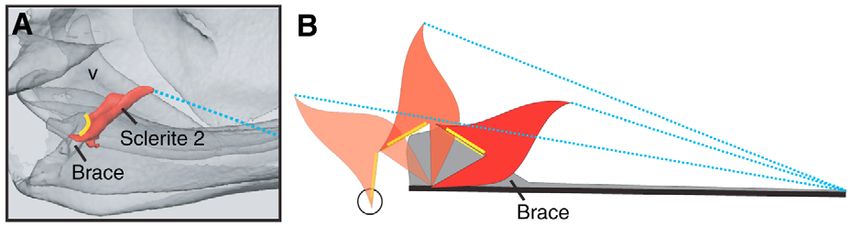
Mantis shrimp mechanics 3679
B individual was micro-CT scanned at the University of Texas
A C
with a slice thickness of 0.0585·mm. Slice images were
s θout reconstructed at a resolution of 1024⫻1024·pixels over a
B s C
c D 4 50·mm field of view. Voxels were 0.0488⫻0.0488⫻0.0585 and
v 3 D bit depth was either 8 or 16, depending on the need for
m B
A visualizing soft tissue. The elements of the raptorial appendage
2 were segmented out of the CT scans and separate images of each
θin 1
were created. Isosurface renderings were utilized to show the
Pp A outer surfaces of each structure and the articulations between
d these structures. These surface renderings were also useful in
identifying areas of reduced mineralization. In addition, volume
renderings of each structure were created to further visualize
areas of greater and lesser mineralization. In volume rendered
C C D images, brighter structures are more highly mineralized. From
c the surface renderings a VRML file was used as input to a rapid
s θout prototype 3-dimensional printer (Z-Corp 310, Burlington, MA,
B D C
3 4 USA) to produce large-scale models of each structure. These
v
m B D
models were helpful in deciphering the articulations between the
A different structures of the raptorial appendage.
2
1 Transmission: kinematics and linkage mechanics
θin
A
We analyzed high-speed images of raptorial strikes and noted
p the changing configurations of the merus in order to characterize
d the dynamics of the flexible elements and linkages of the
raptorial appendage. Animals regularly struck objects coated
with shrimp paste and most animals were willing to strike
Fig.·2. A four-bar linkage model and the associated points in the objects under bright video lights after a period of training. A
proposed mantis shrimp four-bar linkage model. Red circles indicate high-speed imaging system (5000·frames·s–1, 35·s shutter
pivot points; numbers indicate links. Pivots A and D are fixed in space speed; 640⫻480·pixel resolution; HG100K Redlake Systems,
and form Link 1. Link 2 is the input link formed by pivots A and B. San Diego, CA, USA) recorded stomatopods striking a snail
Link 3 is the coupler link formed by pivots B and C. Link 4 is the shell coated in shrimp paste and wired to a stick. The snail shell
follower link formed by pivots C and D. The input angle (in) and was presented to animals within confined burrows and aligned
output angle (out) can be calculated using the law of cosines and the parallel to the glass wall of the aquarium, thereby allowing us
length of the diagonal (green broken line connecting pivots B and D). to film strikes with the animal positioned laterally. Sequences
This particular model configuration is not operational when B, C and in which strikes were directed out of the camera’s plane of view
D are collinear, thereby limiting the input range of in (see Fig.·9 in the
were excluded from the dataset.
Results). (A) A tracing of a high-speed video image of a raptorial
appendage that has completed the load phase and is spring-loaded and The following parameters were measured using high-speed
prepared to strike. The saddle (s) is compressed and the meral-V (v) is imaging: angular velocity, acceleration and strike duration of
rotated proximally. (B) The corresponding linkage model to A. (C) A the dactyl heel (the bulbous structure at the base of the dactyl
raptorial appendage in the release phase. The saddle is hyper-extended segment of the raptorial appendage; Fig.·1) (50–58 strikes; 6
into a flattened shape. The meral-V is fully rotated and open. (D) The individuals; 7–12 strikes per individual), and rotation of the
corresponding linkage configuration to C. m, merus; c, carpus; p, meral-V (a moveable element in the merus segment of the
propodus; d, dactyl. Beige regions in A and C represent arthrodial raptorial appendage; Fig.·1) (24 strikes; 6 individuals; 3–7
membrane; gray regions indicate exoskeleton; yellow area represents strikes per individual) (Matlab v. 6.5 and v. 7.0.4). Meral-V
the saddle. rotation was calculated by measuring the change in angle of the
meral-V relative to horizontal across each video frame. The
acceleration and speed of the dactyl heel were derived from the
Merus mineralization and sclerite functional morphology arc distance traveled by the heel across video image intervals.
Through dissections, computed tomography (CT) scans, Two points were digitized along the propodus/dactyl axis
manipulations and digital image analysis, we examined the formed by the distal two segments of the raptorial appendage,
functional morphology of the sclerites and mineralization which remain folded during a smashing strike (Fig.·1). The
patterns of the merus, and characterized the articulations angular change of this line was calculated across video frames.
connecting the merus and carpus, using a DFC350 FX digital This angle was multiplied by the distance between the
camera and MZ 12.5 microscope (Leica Corp., Germany) and propodus/carpus joint and dactyl/propodus joint, which yielded
custom digital imaging software (Matlab, The Mathworks, the arc distance moved by the heel of the dactyl.
Natick, MA, USA). Speed and acceleration were calculated as the first and second
Mineralization patterns were visualized using 3-D derivatives of distance, respectively. A drawback to computing
reconstructions of CT scans (Amira software, v. 3.1.1, Mercury derivatives from kinematic data is that they only provide average
Computer Systems, Berlin, Germany). A freshly frozen kinematic estimates. Even with curve-fitting and spline methods
THE JOURNAL OF EXPERIMENTAL BIOLOGY3680 S. N. Patek and others
(e.g. Walker, 1998), the transient and non-sinusoidal movement determine the operational range of the input linkage and
of the mantis shrimp’s strike caused the filtered and smoothed compared it to the input range actually used by the mantis
data to fail to track the displacement of the appendage. shrimp. The input range used by the mantis shrimp yielded an
Specifically, the movement of the limb follows a gradual path output of the four-bar model that was effectively approximated
interrupted by a sudden impact and reverse in direction. The as a line (see Results) with a slope equivalent to the predicted
smoothing and spline algorithms applied to these data failed to KT of the system.
track this transient movement and instead continued to follow We statistically evaluated the fit between the predicted four-
the initial path of the appendage. Nonetheless, the animals bar model behavior and the measured kinematics of the raptorial
typically struck the target when the appendage was moving the appendage. Given that the carpus, propodus and dactyl are
greatest distance in the arc and the frame rate of 5000·frames·s–1 tightly coupled once the dactyl begins to sweep toward its target,
under-sampled the movement. Thus, the distances measured we assumed that these three segments share the same pivot point
were underestimates and the resulting velocities and and rotate an equivalent number of degrees during the sweeping
accelerations should also underestimate the rate of movement. phase of the strike. This allowed us to measure the rotation of
Given the uncertainty of deriving accelerations from these data, the propodus as the output angle equivalent to the rotation of
we report acceleration in orders of magnitude. The relative Link 3 (carpus); the propodus is larger, visible in a greater
movement across frames was converted to SI units by measuring proportion of video sequences and can be more accurately
the pixel distance of known structures on the raptorial appendage digitized than the carpus. We tested whether the slope of the
in each frame and converting pixels to meters using the relationship between the input (Link 2) and output angles (Link
calibrated distance. We estimated digitizing measurement error 3=propodus rotation) measured from the high-speed videos was
by digitizing 5 sequences, 10 times each. significantly different than the slope predicted by the four-bar
Four-bar linkage pivot points were identified based on high- model [modified t-test, see p. 32, Grafen and Hails (Grafen and
speed videos, functional morphological observations and Hails, 2002)]; incorporating individual effects and treating
manual manipulations of the specimens. Using a standard four- video sequences nested within individuals as random effects
bar linkage configuration (Uicker et al., 2003; Westneat, 1990), using Residual Maximum Likelihood method in JMP statistical
we identified four pivot points defining four ‘links’: a fixed link, software (v. 5.0.1).
input link, follower link and coupler link (Fig.·2, also see
Results). We measured link lengths using photographs of Results
raptorial appendages at rest (13 individuals) and in digital video Sclerite functional morphology
images when all the pivot points were visible (images from 21 Stomatopods have two sclerites, sclerite 1 and sclerite 2,
video sequences of strikes performed by 4 individuals). Using which serve as ‘latches’ to adduct and hold the carpus, propodus
t-tests (JMP v. 5.0.1), we tested whether specimen and dactyl against the merus while the extensor muscles contract
measurements were equivalent to video-based measurements. In in preparation for a strike (Figs·3, 4) (Burrows, 1969). In O.
addition, we compared the length of Link 4 between these two scyllarus, sclerite 1 is a small, rod-shaped structure positioned
datasets, given that Link 4 is formed by the contracted extensor medially along the ventral edge of the merus and embedded in
muscle and, therefore, should be longer in the photographs of the thick arthrodial membrane that connects the merus and
relaxed appendages than in the video images of appendages carpus (Figs·3, 4). The medial flexor apodeme attaches just
prepared to strike. below the sclerite’s tip and forms a thin, pinnate layer over the
Based on the above morphological and kinematic analyses, medial surface of the merus.
we tested the hypothesis that a four-bar linkage system Just lateral to sclerite 1, sclerite 2 has a surface that articulates
mechanically couples this system (Fig.·2). With the known with an infolding of the merus and sweeps through an arc both
length of the diagonal bar (Fig.·2), the law of cosines was used into and out of the merus (Figs·3, 4). The lateral flexor muscle
to calculate the angles between any of the links during a given attaches to the proximal edge of the sclerite and extends via a
input bar rotation. The lengths of each of the four links (Ln) and large pinnate muscle to attach to the ventral floor of the merus.
the input angle of between link1 and link2 (input) were entered When this muscle is manually pulled in a proximal direction,
into the following equations to calculate the length of the sclerite 2’s articulating surface slides smoothly over the
diagonal bar (Ldb) (Fig.·2): infolding of the merus such that it is braced in place, but not
latched. When released, the sclerite again slides smoothly along
Ldb = L22 + L21 + 2L1L2cos(input) , (1) the articulating meral surface and permits abduction of the
carpus. When not engaged, sclerite 2 protrudes through the
which was then used to calculate the output angle between link3 ventral meral-carpal arthrodial membrane and is visible from the
and link4 (output): outside of the animal (Fig.·4). Both the meral infolding, which
forms the brace, and the long axis of the sclerite, are oriented
output = arccos[(L23+L24–L2db) (2L3L4)–1]·. (2)
slightly medially. Thus, when sclerite 2 is released, it swings
Depending on the relative lengths of the links, a four-bar dorsally and medially.
linkage system may allow a 360° rotation of the input link, but
a more common case is that the input link ‘jams’ after some Merus mineralization and articulations
amount of rotation. This range of input angles for which there The joint articulations between the merus and carpus range
is movement of the output link is called the ‘operational’ range from robust to nearly absent (Figs·3, 5). The lateral meral-carpal
of the four-bar linkage. We used a mathematical model to articulation is visible externally and is only loosely articulated
THE JOURNAL OF EXPERIMENTAL BIOLOGYMantis shrimp mechanics 3681
Fig.·3. CT scans of the internal anatomy of the raptorial appendage and
the degree of mineralization of the merus exoskeleton (gray is
mineralized; transparent regions represent relatively unmineralized
exoskeleton). (A) Medial view of the raptorial appendage (proximal to
right of page; ventral toward the bottom of the page) showing the large
lateral extensor apodeme (e). Ventrally, the lateral flexor apodeme (f)
attaches to sclerite 2 (orange). Medial to sclerite 2 is the small, rod-
shaped sclerite 1 (green). (B) An internal perspective of the merus
viewed from the distal end (lateral to left). Sclerite 2 (s2) is in its
resting, unlocked position such that it hangs externally between the
merus and carpus. The surface of the distal meral-V (v) articulation,
which loosely articulates with the carpus, contrasts with the large
internal joint, which forms the medial carpus joint articulation (j). (C)
Same view as in B with sclerite 2 and sclerite 1 (s1) in closed and
braced positions. Note that sclerite 1 does not appear to directly
articulate with sclerite 2 when in the closed position and instead folds
medially relative to the edge of sclerite 2. (D) Sclerites in resting
position (medial view; distal to left). (E) Locked positions of the
sclerites with the carpus rotated counter-clockwise in preparation to
strike (medial view). m, merus; c, carpus; s, saddle; p, propodus; b,
ventral infolding of merus. Scale bars, 4·mm.
merus’ cuticle – the meral-V and the saddle (Figs·5, 6). The
meral-V is a thick, triangular-shaped structure that connects to
one of the large ventral buttresses forming the underside of the
merus. It has a flexion point along the ventral-lateral margin of
the merus, such that the meral-V bends at this junction and
rotates proximally (Figs·1, 5, 6). The meral-V is flanked by
arthrodial membrane and the ventral buttress also has a region
of low-mineralization adjacent to it (Fig.·1), allowing this
structure room to flex.
Located on the dorsal surface of the merus, the saddle is
constructed of poorly mineralized cuticle with thin, flexible
arthrodial membrane on its medial and lateral sides (Figs·1, 5,
6). During the load phase, the saddle is compressed and forms
a more concave curve along the proximo-distal axis (Fig.·6).
At the same time, the saddle rotates slightly ventrally around
its proximal connection to the merus and articulates with a
notch on the medial side of the merus (Fig.·5B). In the release
phase, the saddle rotates dorsally and hyper-extends into a
more flattened shape before returning to its initial form
(Figs·6, 7).
The saddle and meral-V are connected by a thickened strip
of exoskeleton, the meral bridge (Figs·5, 6). When the meral-V
is rotated proximally during contraction of the extensor muscles,
(Fig.·5A). Opposing this somewhat unconstrained connection the meral bridge is pushed proximally and the saddle is
on the lateral side are two medial meral-carpal articulations simultaneously compressed and rotated (Fig.·6). There are no
(Fig.·5B,C). The internal medial articulation forms a smooth, muscle attachments to the saddle itself. When the saddle, bridge
channel-like surface with a stop at the end, which both stabilizes and merus are released from their compressed positions, the tip
the carpus to move only along the dorso-ventral axis and also of the meral-V pushes through its sliding articulation with the
prevents the carpus from adducting beyond a particular angle. lateral side of the carpus, such that the carpus, propodus, and
The lateral and medial sides of the merus are distinctly dactyl rotate outward toward the target (Figs·6, 7).
asymmetric in terms of mineralization and flexibility. The
medial side is stiff and robust whereas the lateral side is thin Transmission: kinematics
and flexible (Figs·1, 3, 5). Similarly, the dorsal surface is thin At the onset of a strike, the propodus and dactyl slide distally
and flexible while the ventral surface consists of large, stiff along the merus and then transition to a sweeping movement
buttresses running along the distal-proximal axis. There are also with a large rotational velocity (Figs·7, 8). The duration of the
unmineralized regions on the medial and dorsal surfaces where sweeping movement averaged 1.8±0.4·ms (± s.d.) and the
cuticle is replaced by arthrodial membrane. sliding movement averaged 0.9±0.5·ms (6 individuals, 6–12
There are two independently mobile components of the strikes per individual). The dactyl heel reached peak speeds of
THE JOURNAL OF EXPERIMENTAL BIOLOGY3682 S. N. Patek and others
Fig.·4. Sclerite engagement and orientation. (A)
Sclerite 2 (red, solid fill) is in the engaged
position and braced against the ventral meral
infolding. Yellow highlighting indicates the
area of ventral meral infolding against which
the sclerite is braced. The blue dotted line
shows the approximate attachment point and
orientation of the lateral flexor muscle that
engages the sclerite. Shown from the medial
side, with the meral-V (v) behind the sclerite.
Ventral is toward the bottom of the page and proximal is to the right. (B) A schematic diagram of the engaged and resting positions of sclerite 2.
The darkest sclerite (red) is shown in the engaged position with yellow highlighting the articulating surfaces. When the sclerite is released it rotates
distally (to left), to rest with the articulating surface hanging outside the animal (circled). This portion of the sclerite is visible in mantis shrimp
specimens and hangs between the merus and carpus. Blue dotted lines show the approximate orientation and attachment of the lateral flexor muscle.
14.7–23.5·m·s–1 (mean peak speed: 13.7±3.3·m·s–1; mean F=0.6943; P=0.60) nor for meral-V rotation (one-way
median speed: 3.4±1.7·m·s–1), peak angular speeds of 670– ANOVA; F=1.1424; P=0.37).
990·rad·s–1 (mean peak angular speed: 608.9±147.0·rad·s–1;
mean median angular speed: 155.7±79.6·rad·s–1), and mean Transmission: linkage mechanics
peak accelerations on the order of 104·m·s–2. Digitizing The structural asymmetries of the merus, as described above,
measurement error at maximal speeds was on average ±4%. generate two distinct functional regions of the merus.
The rotation of the dactyl heel and meral-V were variable and Specifically, the robust mineralization and paired meral-carpal
correlated with each other. In all sequences, the dactyl heel articulations on the medial side yield stability and resistance to
struck the prey item while the meral-V was still rotating; thus, flexion. By contrast, on the lateral side of the merus,
during these smashing strikes, the propodus and dactyl did not considerable flexion occurs via the rotating meral-V, which
transition to a ballistic, unpowered phase prior to impact. The allows transmission of forces distally to the carpus and
dactyl heel struck the snail across a range of excursion angles, proximally to the meral bridge and saddle. It is on this lateral
such that in some strikes the dactyl/propodus rotated outward side of the merus that we identified the four-bar linkage system
only 7° whereas in other strikes the dactyl struck the snail with which actuates the spring-loaded raptorial strike during the
a maximum extension of 42° (mean: 25±9°). Similarly, the net release phase (Figs·2, 8).
meral-V rotation ranged widely depending on the excursion of The links comprising the four-bar linkage model are
the dactyl when it struck the snail. The net meral-V rotation was designated as follows (Fig.·2): Link 1, fixed link: proximal
on average 9° (range: 3–17°; ±5° s.d.; 5 individuals, 4–6 strikes merus exoskeleton; Link 2, input link: meral-V; Link 3, coupler
per individual) (Fig.·7). Values were not significantly different link: carpus; and Link 4, follower link: contracted extensor
across individuals for propodus rotation (one-way ANOVA; muscle. Previous work showed that the lateral extensor muscle
Fig.·5. The morphology of the raptorial appendage of the
peacock mantis shrimp. Line drawings are presented adjacent to
photographs of the corresponding areas of the raptorial
appendage. Proximal is to the right of the page. (A) Lateral view
highlights the external, loose articulation between the meral-V
(v) and carpus (c; inset). A thin strip of exoskeleton forms the
bridge (b) between the meral-V and saddle (s). (B) Medial view
shows the internal meral-carpal articulation that functions as a
sliding channel joint (left inset). Also visible is the proximal
saddle notch, into which the saddle is pushed during extensor
muscle contraction in the load phase (right inset). (C) Dorsal
view (medial toward top of page) shows the orientation of the
lateral extensor apodeme (a, pink) extending from the carpus
and running beneath the saddle. The bridge (b) runs dorsally
from the lateral meral-V (visible in A) and across to the distal
horn of the saddle (visible in B). The medial meral-carpal
articulation consists of two adjacent articulations (orange
circles); the internal medial meral-carpal articulation is a robust
sliding channel joint (as shown in B, left inset). (A–C) Orange
circles indicate articulations; gray bars indicate internal
buttressing; beige regions are arthrodial membrane; gray regions
indicate exoskeleton; yellow coloration represents the saddle (s).
m, merus; p, propodus; d, dactyl.
THE JOURNAL OF EXPERIMENTAL BIOLOGYMantis shrimp mechanics 3683
Fig.·6. A resting (solid outline) and loaded (light-blue A
overlay) merus segment (m) of the raptorial appendage.
Proximal is to the right of the page, dorsal is toward the s
top of the page. (A) Lateral view of the raptorial b
m
appendage. When the extensor muscles contract in
preparation for a strike in the load phase, the meral-V (v) C
rotates proximo-medially (clockwise in this image), which v s
simultaneously causes the bridge (b) to move proximally.
When the bridge pushes proximally, it pushes against the vb
saddle (s), which is compressed to form a more concave
curve. A mineralized ventral bar (vb) extends proximally
B
v
from the base of the meral-V. (B) Medial view of the
raptorial appendage showing the proximal movement and s m vb
flexion of the saddle caused by extensor muscle
contraction. When seen from the medial view, the saddle
is pushed into a notch on the merus. (C) A diagram of the
possible areas of elastic energy storage (orange spring
icons) during rotation of the merus and flexion of the 1 cm
saddle in preparation for a strike. Here we propose that the
meral-V functions as a spring by flexing along its base, similar to a tape spring, to form a tighter curve during extensor muscle contraction. A
previous study (Patek et al., 2004) proposed that elastic energy is stored as the saddle compresses into a more concave shape.
(Link 4, Fig.·2) remains contracted throughout the release phase appendage. The mean starting angle between Links 1 and 2 was
(Burrows, 1969); contracted muscles are commonly used in 64±5° (± s.d.; mean median 64°; range 40–77°; 24 video
biological linkage systems as fixed-length links (e.g. Muller, sequences, 6 individuals, 1–6 videos per individual).
1987; Westneat, 1990; Westneat, 1994). Pivot A is a fixed pivot Based on the average link lengths and starting angles
point located at the meral-V articulation and located between measured in the high-speed video sequences, we developed an
Links 1 and 2. Pivot B is not fixed in space and is formed by average four-bar model to generate predictions with which to
the lateral meral-carpal articulation between Links 2 and 3. compare the high-speed video data (Figs·9, 10, 11). This four-
Pivot C also is not fixed in space and is located at the lateral bar model is operational when input angles range from 63–99°
extensor apodeme attachment on the carpus between Links 3 and the input angles used by the mantis shrimp most often range
and 4. Pivot D is a fixed pivot formed by the lateral extensor from 64–73°. Within this limited range of input angles, the
muscle attachment immediately proximal to the saddle between model output can be approximated as a line with a slope of 3.56
Links 4 and 1 (there are no muscle attachments to the saddle (least-squares linear regression, R2=0.9970, P3684 S. N. Patek and others
c
v
p
d
Fig.·8. A tracing of a typical strike sequence from high-speed video images with the links and pivots of a four-bar linkage mechanism overlaid
on the tracings. Shown from left to right, images are 0.4·ms apart with the exception of the final two images, which are 0.2·ms apart. The saddle
is colored orange; v, meral-v; c, carpus; p, propodus; d, dactyl. Insets illustrate schematically the compression and release of a spring (orange)
and the braced and released position of sclerite 2 (red sclerite, gray brace).
confidence interval, 1.5–2.4; general linear model incorporating operational range was from 40° to 75° (mean 63±9°). The input
individual effects, R2=0.8233; rotation, F=73.14, PMantis shrimp mechanics 3685
200 key modifications of the raptorial appendage’s merus segment
A permit localized flexion, elastic energy storage and transmission
180 of stored potential energy via linkages. Specifically, the meral-
V both acts as an elastic energy storage device and one of the
160
Output rotation of Link 3 (degrees)
links in the linkage mechanism. In addition, the asymmetries in
140 mineralization and joint architecture allow flexion of the lateral
side of the merus that is offset by the constrained joint system
120 and stiff exoskeleton of the medial side. A linkage mechanism
is formed by the flexible components of the medial side, while
100 a pair of sclerites controls the release of the strike by sliding
80
against a simple brace formed by the merus exoskeleton. Thus,
through the actuation of the strike by a pair of sclerites and the
60 stabilization provided by the medial articulations and
reinforcement of the merus, these animals can strike with
40 remarkable precision to spear elusive fish or hammer hard-
shelled molluscs (Caldwell and Dingle, 1976; Patek and
20
Caldwell, 2005).
0
40 50 60 70 80 90 100 110 120 130 Stabilization and control: mineralization, articulations and
Input rotation of Link 2 (degrees) sclerites
200 Analysis of the mineralization patterns in the merus and
B functional morphology of the joints provide new insights into
Predicted output rotation of propodus (degrees)
180
the stabilization and articulations of the appendage. Arthropod
160 appendages are usually perceived as a uniform series of hinged
cylinders, yet CT scans (Figs·1, 3, 4) and kinematic analyses
140 (Figs·7–9) revealed complex joints, distinct asymmetries in
mineralization, and flexion on the lateral and medial sides of
120 the merus (Fig.·6). For example, the lateral meral-carpal
articulation couples the rotation of the meral-V to the carpus;
100
in contrast, the medial meral-carpal articulations form a
80 stabilizing channel, which restricts the carpus to dorsal/ventral
movements (Fig.·5). Perhaps most surprising is the presence of
60 a flexion point within the merus segment at the base of the
rotatable meral-V; no equivalent flexion point exists on the
40
opposite side of the merus. Instead, a thickened bar of
20 exoskeleton on the medial side of the merus opposes the lateral
flexion of the meral-V (Fig.·1). Thus, the dynamic linkages
0
40 50 60 70 80 90 100 110 120 130
Input rotation of meral-V (degrees) 200
180
Fig.·9. The four-bar model predictions vary depending on relative link
Output angle (degrees)
160
lengths and starting angles. The linkage mechanism is operational in
140
two regions between input rotations of 0 and 360° (region from 0 to
180° shown; the range from 180–360° is the mirror image of 0 to 180° 120
and is never used by mantis shrimp). The horizontal lines at output 100
rotations of 0° and 180° indicate that a change in input rotation does 80
not yield any output rotation (i.e. the linkage mechanism is non- 60
operational). (A) An input rotation between 40° and 120° yields an 40
output rotation depending on relative link lengths. Green traces show 20
the predicted behavior based on the link lengths of a relaxed raptorial 0
appendage (i.e. Link 4 extensor muscle is not contracted). Blue traces 60 70 80 90 100
show the predicted behavior of the relaxed appendages if Link 4 is Input angle (degrees)
constrained to the average shortened length observed in video images.
Red traces illustrate the range of behaviors given the range of link Fig.·10. Predicted four-bar model behavior based on the average link
lengths measured in loaded appendages from video images. The thick lengths of loaded raptorial appendages. For a given input angle, the
black line provides the linkage model behavior given the average link four-bar linkage mechanism yields an output that varies nonlinearly
lengths measured from the loaded images (red lines; also shown in along the range of input angles. The four-bar model is not operational
Fig.·10). (B) The predicted model behavior of four individuals (each beyond the range of input angles shown here. Kinematic analyses
color represents a different individual) given measured inputs and link showed that mantis shrimp typically generate input angles of the meral-
lengths from high-speed video sequences. V in the range 64–73°, as indicated here (yellow line).
THE JOURNAL OF EXPERIMENTAL BIOLOGY3686 S. N. Patek and others
50 in arthropods (reviewed in Gronenberg, 1996a). For example,
Cumulative propodus A trap-jaw ants generate extreme speeds and accelerations during
40 their mandible strikes and have evolved latch systems multiple
rotation (degrees)
times using various modifications of joints and mouthparts
30 (Gronenberg, 1995a; Gronenberg, 1995b; Gronenberg, 1996b;
Gronenberg et al., 1998; Patek et al., 2006). The flea also uses
20
modifications of the exoskeleton to lock a compressed block of
10 resilin in place prior to a jump (Rothschild et al., 1975;
Rothschild and Schlein, 1975). Similarly, the mantis shrimp’s
0 sclerites appear to be mineralized modifications of the flexor
0 2 4 6 8 10 12 14 16 18
apodemes.
Cumulative meral-V rotation (degrees)
80 Transmission: kinematics and linkage mechanics
B The raptorial strikes follow a characteristic series of
rotation (degrees)
60 movements, beginning with a brief, 0.9·ms ‘slide phase’ when
Net propodus
the propodus slides several millimeters distally along the merus
40 and no movement of external meral structures is visible (Figs·7,
8). Then, the saddle begins to lengthen, the meral-V rotates
20
distally and the propodus, dactyl and carpus transition to a
sweeping rotational movement (Figs·7, 8), which lasts an
average 1.8·ms and brings the dactyl heel to an average speed
0
0 2 4 6 8 10 12 14 16 18 of 14·m·s–1 (609·rad·s–1). The magnitude and timing of the
Net meral-V rotation (degrees) meral-V and propodus rotations are correlated, such that greater
rotations of the propodus are correlated with larger meral-V
Fig.·11. The relationships between input angle rotation (Link 2, meral- rotations. Furthermore, the propodus rotates at least twice the
V) and output angle rotation (Link 3, propodus rotation) measured in meral-V rotation over the course of an entire strike (Fig.·11).
high-speed video sequences. (A) The cumulative change in input and A four-bar linkage mechanism and the mechanical coupling
output rotation across video frames (combined data from 23 strikes, proposed previously (Patek et al., 2004) are generally supported
five individuals). (B) The net input and output rotation (the total
by the transmission of a twofold rotational amplification of the
rotation across the full input range) across each strike recorded in the
same individuals as in A, with each individual represented by a meral-V to the propodus (Figs·9–11). However, the KT of the
different symbol. The predicted output based on the four-bar model empirical data is lower than predicted by the model, raising the
slope (crosses) is shown. question as to whether an alternative model should be
considered or, instead, that the four-bar model is appropriate for
the system and some additional effect is absorbing rotational
present on the lateral side are mirrored by a stiff medial wall input of the merus. For example, the incomplete fit of the model
of the merus, which lacks any flexion points. may be caused by non-planar orientation of linkages and the
The CT scans also permitted visualization of the highly presence of a sliding cam-type joint between the merus and
mineralized sclerites. These images (Figs·3, 4) depicted the carpus (Fig.·5A); this joint could yield shifting force vectors or
sclerites’ orientation in an undisturbed specimen and suggested lever arms during meral-V rotation. Shifting mechanical
a somewhat different orientation and mechanism of action than advantage of the contracted extensor muscle relative to the
previously proposed (Burrows, 1969). Rather than using a catch relaxing flexor muscles during latch release (Burrows, 1969)
to lock the raptorial appendage during the loading phase, sclerite may influence the momentum of the dactyl/propodus/carpus
2 slides smoothly over a bracing surface formed by an infolding unit as it rotates around this point [e.g. in bush crickets (Burrows
of the merus (Figs·3, 4). Sclerite 1 folds above sclerite 2 and and Morris, 2003)]. In addition, Burrows noted that strike
does not have a comparable bracing surface. The use of a speeds were influenced by duration, frequency and timing of
smooth brace, rather than binary latch, explains why previous both flexor and extensor muscle activity (Burrows, 1969). Thus,
electromyographic analyses showed that both the extensor and this variable control of muscle activity could cause a change in
flexor muscles remain contracted when the appendage is in a length of Link 4, resulting in variable meral-V rotation and
loaded state and why mantis shrimp typically hold the cocked saddle-shortening, again influencing the output of the system
position for only a brief time period (Burrows, 1969). This (Fig.·9). All of these potential variations on the model should
arrangement also permits mantis shrimp to disengage the system be addressed in future studies, and, although infrequently
without firing; the extensor and flexor muscles can simply performed, alternative models to this four-bar linkage
slowly relax to release the stored energy over a longer time mechanism should be evaluated (e.g. Hoese and Westneat,
period. At present, it is not clear whether the two sclerites have 1996; Muller, 1996).
distinct functions or whether sclerite 1 simply serves to increase In most systems, a high KT is associated with high speeds,
the mechanical advantage of the larger sclerite 2 (Burrows, whereas a low KT is found in systems with large forces. Perhaps
1969) relative to the considerable force generated by the counter-intuitively, even with a relatively high KT of
opposing extensor muscle contraction. approximately 2, the high-speed system of O. scyllarus can also
These latches may be similar in origin to other latch systems generate large forces. Such extreme accelerations, coupled with
THE JOURNAL OF EXPERIMENTAL BIOLOGYMantis shrimp mechanics 3687
an impact between two hard, massive surfaces, cause the strikes Previous research suggested that elastic energy storage in the
to yield high transient forces that can exceed 1000·N (Patek and mantis shrimp system was provided by the extensor apodeme
Caldwell, 2005). Linkage systems that yield a high angular (Burrows, 1969), saddle (Patek et al., 2004) and unspecified
output rotation relative to input rotation are considered ‘speed- cuticular elements (Currey et al., 1982) (Fig.·6). Apodeme
modified’; however, mantis shrimp produce both high speeds elasticity was calculated to be insufficient to power the extreme
and forces through extreme acceleration. Thus, these high kinematics of these strikes (Patek et al., 2004) and it was
transient forces are due to rotational amplification rather than a suggested that the saddle could provide the additional needed
low KT. power. We propose an additional or alternative energy storage
One strength of evaluating linkage mechanics in an arthropod structure: the meral-V. The poor mineralization of the saddle
system is the ability to use exoskeletal markers during actual (Fig.·1) means that although the saddle is flexible, it is unlikely
strikes, unlike vertebrate linkage systems in which the link that a substantial amount of energy can be stored through
lengths and positions have traditionally been limited to conformational changes of this structure. Instead, elastic
inferences from dissection and external soft markers. potential energy is probably stored via multiple sites of cuticular
Specifically, we were able to measure the effects of varying deformation, most likely concentrated in the meral-V (Fig.·6).
Link 4 (formed by the contracted extensor muscle) as well as Ultimately, to resolve this debate, mechanical and material tests
the range of input angles actually used by the mantis shrimp must be made directly on the system as a whole and on each of
(Fig.·9). Link 4 was 14% shorter in contracted, loaded these structures.
appendages than in relaxed appendages. When entered into the The shape of arthropod cuticle, as well as its composition,
four-bar model, these longer link lengths yielded greater influences the presence and degree of elastic energy storage
predicted input angles that were significantly different than the (Vincent, 1990; Vincent and Wegst, 2004; Wainwright et al.,
observed and predicted input angle range of a contracted Link 1976). While the presence of resilin, the arthropod rubber-like
4 length (Fig.·9). Furthermore, we were able to measure actual protein (Weis-Fogh, 1960), has not yet been determined in this
input angles (Fig.·9) in order to evaluate the mechanical space system, the shape of the meral-V suggests an elastic function.
within the model that is actually used by the mantis shrimp. The meral-V and the ventral bar extending from its lateral
Both of these approaches offered insights into the variability of flexion point resembles the human-engineered tape spring, i.e.
the link lengths and input angles across and within individuals, a thin strip with a bend or fold at which elastic energy is stored
suggesting that rotational amplification is robust across a range (Seffen and Pellegrino, 1999; Vehar et al., 2004; Vincent and
of parameters while, at the same time, yielding slightly different Wegst, 2004). The flexion point at the base of the meral-V is
performance output. similar to the elastic bend in a tape spring and the poorly
The twofold KT found in mantis shrimp is high relative to mineralized area adjacent to this bar should permit flexion
four-bar linkages evaluated across fish which range, for (Fig.·1). Furthermore, when manipulated, the meral-V
example, from 0.5 to 1.29 in labrid fish jaws (Alfaro et al., 2004; strongly resists flexion and springs back into an open position
Hulsey and Wainwright, 2002). In addition, some bony fishes when released. The saddle’s function, given the intriguing
may use a spring-loaded four-bar configuration by storing hyperbolic–paraboloid shape and considerable flexion during
elastic energy in the linkage system and then relying on small the load phase, remains to be determined. Hyperbolic–
shifts in relative position of the links to release the system paraboloid shells often are used in engineered systems to
(Muller, 1987). Surprisingly, we were unable to find any reduce local buckling through the presence of two opposite
published arthropod systems in which a four-bar linkage and transverse curves. Thus, the saddle may provide a flexible,
mechanism has been analyzed. yet strong, region of cuticle that allows the necessary space on
the medial side of the merus equivalent to the amount of
Elastic energy storage shortening occurring when the meral-V closes on the lateral
In a system as small as this one, the definitive determination side of the merus. However, while the meral-V is highly
of where elastic energy is being stored is challenging. Two variable across stomatopods, the saddle is highly conserved,
factors determine storage capacity – the amount of deformation and retains its elegant, hyperbolic–paraboloid form across all
of an element and its stiffness. As in the crossbow, either mantis shrimp (R.L.C. and S.N.P., personal observation), thus
character alone is not sufficient. Energy is not stored to an suggesting an important, and as yet not fully determined,
appreciable extent in the string; although it is bent at an acute function.
angle, string has little flexural stiffness. There is also little The integration of elastic energy storage and force
energy stored in the stiff catch mechanism; it does not deform transmission through specialized joint articulations is a hallmark
substantially. The energy storage is principally in the limbs of of arthropod power amplification systems (Bennet-Clark, 1975;
the bow; this can be shown by determining the mechanical Bennet-Clark, 1976a; Bennet-Clark, 1976b; Bennet-Clark and
properties of these structural elements and measuring their Lucey, 1967; Blickhan and Barth, 1985; Sensenig and Shultz,
deflection when the bow is cocked. For the mantis shrimp strike, 2003). Not only is there a rich diversity of power amplification
some deformations are too small to fully characterize from the systems across arthropods, including fleas, locusts and snapping
video and the extent of mineralization offers a proxy for shrimp, but even within the mantis shrimp there is substantial
stiffness. Here we will propose a principal storage mechanism, morphological diversity of the saddle, meral-V and linkage
but the testing of the mechanism awaits nanoindention studies articulations (S.N.P., personal observation) (Ahyong, 2001).
and finer scale resolution of strain in the various parts of the Integrative analyses of the kinematics, material properties and
merus. conformational changes of these systems will continue to reveal
THE JOURNAL OF EXPERIMENTAL BIOLOGY3688 S. N. Patek and others
new insights into the origins and evolutionary diversification of Hoese, W. J. and Westneat, M. W. (1996). Biomechanics of cranial kinesis
powerful animal movements. in birds: testing linkage models in the white-throated sparrow (Zonotrichia
albicollis). J. Morphol. 227, 305-320.
Hulsey, C. D. and Wainwright, P. C. (2002). Projecting mechanics into
We thank Wyatt Korff, T. J. Kelleher, Sanjay Sane, Bill morphospace: disparity in the feeding system of labrid fishes. Proc. R. Soc.
Kier, Dan Dudek, Matt McHenry, Charles Nunn, Mimi Koehl, Lond. B Biol. Sci. 269, 317-326.
Kunze, J. C. (1981). The functional morphology of stomatopod Crustacea.
the Hebets lab and the UC Berkeley Biomechanics Seminar for Philos. Trans. R. Soc. Lond. B Biol. Sci. 292, 255-328.
insightful comments and assistance. We also greatly appreciate Lappin, A. K., Monroy, J. A., Pilarski, J. Q., Zepnewski, E. D., Pierotti,
the constructive comments from two anonymous reviewers. D. J. and Nishikawa, K. C. (2006). Storage and recovery of elastic
Special thanks to Tim Green, Tom Fitz and the British potential energy powers ballistic prey capture in toads. J. Exp. Biol. 209,
2535-2553.
Broadcasting Corporation (BBC) Natural History Unit for their McNeill, P., Burrows, M. and Hoyle, G. (1972). Fine structures of muscles
assistance with filming. Funding was provided by the BBC and controlling the strike of the mantis shrimp, Hemisquilla. J. Exp. Zool. 179,
the Miller Institute for Basic Research in Science (to S.N.P.). 395-416.
Muller, M. (1987). Optimization principles applied to the mechanism of
neurocranium levation and mouth bottom depression in bony fishes
References (Halecostomi). J. Theor. Biol. 126, 343-368.
Ahyong, S. T. (2001). Revision of the Australian Stomatopod Crustacea. Muller, M. (1996). A novel classification of planar four-bar linkages and its
Sydney: Australian Museum. application to the mechanical analysis of animal systems. Philos. Trans. R.
Alexander, R. M. (1983). Animal Mechanics. Boston: Blackwell Scientific Soc. Lond. B Biol. Sci. 351, 689-720.
Publications. Nishikawa, K. C. (1999). Neuromuscular control of prey capture in frogs.
Alexander, R. M. (1990). Elastic mechanisms in the locomotion of vertebrates. Philos. Trans. R. Soc. Lond. B Biol. Sci. 354, 941-954.
Neth. J. Zool. 40, 93-105. Patek, S. N. and Caldwell, R. L. (2005). Extreme impact and cavitation forces
Alexander, R. M. and Bennet-Clark, H. C. (1977). Storage of elastic strain of a biological hammer: strike forces of the peacock mantis shrimp
energy in muscle and other tissues. Nature 265, 114-117. (Odontodactylus scyllarus). J. Exp. Biol. 208, 3655-3664.
Alfaro, M. E., Bolnick, D. I. and Wainwright, P. C. (2004). Evolutionary Patek, S. N., Korff, W. L. and Caldwell, R. L. (2004). Deadly strike
dynamics of complex biomechanical systems: an example using the four-bar mechanism of a mantis shrimp. Nature 428, 819-820.
mechanism. Evolution 58, 495-503. Patek, S. N., Baio, J. E., Fisher, B. F. and Suarez, A. V. (2006).
Barel, C. D. N., van der Meulen, J. W. and Berkhoudt, H. (1977). Multifunctionality and mechanical origins: ballistic jaw propulsion in trap-
Kinematischer transmissionskoeffizient und vierstangensystem als jaw ants. Proc. Natl. Acad. Sci. USA 103, 12787-12792.
funktionsparameter und formmodel fur maandibulare depressionsapparate Ritzmann, R. (1973). Snapping behavior of the shrimp Alpheus californiensis.
beiteleostiern. Anat. Anz. 142, 21-31. Science 181, 459-460.
Bennet-Clark, H. C. (1975). The energetics of the jump of the locust Rothschild, M. and Schlein, Y. (1975). The jumping mechanism of Xenopsylla
Schistocerca gregaria. J. Exp. Biol. 63, 53-83. cheopis. I. Exoskeletal structures and musculature. Philos. Trans. R. Soc.
Bennet-Clark, H. C. (1976a). Energy Storage in Jumping Animals. Oxford, Lond. B Biol. Sci. 271, 457-490.
New York, Toronto, Sydney, Paris, Braunschweig: Pergamon Press. Rothschild, M., Schlein, J., Parker, K., Neville, C. and Sternberg, S. (1975).
Bennet-Clark, H. C. (1976b). Energy storage in jumping insects. In The Insect The jumping mechanism of Xenopsylla cheopis III. Execution of the jump
Integument (ed. H. R. Hepburn), pp. 421-443. Amsterdam: Elsevier. and activity. Philos. Trans. R. Soc. Lond. B Biol. Sci. 271, 499-515.
Bennet-Clark, H. C. and Lucey, E. C. A. (1967). The jump of the flea: a study Seffen, K. A. and Pellegrino, S. (1999). Deployment dynamics of tape springs.
of the energetics and a model of the mechanism. J. Exp. Biol. 47, 59-76. Proc. R. Soc. Lond. A Math. Phys. Sci. 455, 1003-1048.
Blickhan, R. and Barth, F. G. (1985). Strains in the exoskeleton of spiders. J. Sensenig, A. T. and Shultz, J. W. (2003). Mechanics of cuticular elastic energy
Comp. Physiol. A 157, 115-147. storage in leg joints lacking extensor muscles in arachnids. J. Exp. Biol. 206,
Burrows, M. (1969). The mechanics and neural control of the prey capture 771-784.
strike in the mantid shrimps Squilla and Hemisquilla. Z. Vergl. Physiol. 62, Uicker, J. J., Jr, Pennock, G. R. and Shigley, J. E. (2003). Theory of
361-381. Machines and Mechanisms. New York: Oxford University Press.
Burrows, M. and Hoyle, G. (1972). Neuromuscular physiology of the strike Vehar, C., Kota, S. and Dennis, R. (2004). Closed-loop tape springs as fully
mechanism of the mantis shrimp, Hemisquilla. J. Exp. Zool. 179, 379-394. compliant mechanisms – preliminary investigations. In Proceedings of the
Burrows, M. and Morris, O. (2003). Jumping and kicking in bush crickets. J. DETC 2004, International Design Engineering Technical Conference. Salt
Exp. Biol. 206, 1035-1049. Lake City, Utah.
Caldwell, R. L. and Dingle, H. (1976). Stomatopods. Sci. Am. 1976, 81-89. Versluis, M., Schmitz, B., von der Heydt, A. and Lohse, D. (2000). How
Collar, D. C., Near, T. J. and Wainwright, P. C. (2005). Comparative analysis snapping shrimp snap: through cavitating bubbles. Science 289, 2114-2117.
of morphological diversity: does disparity accumulate at the same rate in two Vincent, J. (1990). Structural Biomaterials. Princeton: Princeton University
lineages of centrarchid fishes. Evolution 59, 1783-1794. Press.
Currey, J. D., Nash, A. and Bonfield, W. (1982). Calcified cuticle in the Vincent, J. F. V. and Wegst, U. G. K. (2004). Design and mechanical
stomatopod smashing limb. J. Mater. Sci. 17, 1939-1944. properties of insect cuticle. Arthropod Struct. Dev. 33, 187-199.
de Groot, J. H. and van Leeuwen, J. L. (2004). Evidence for an elastic Wainwright, S. A., Biggs, W. D., Currey, J. D. and Gosline, J. M. (1976).
projection mechanism in the chameleon tongue. Proc. R. Soc. Lond. B Biol. Mechanical Design in Organisms. Princeton: Princeton University Press.
Sci. 271, 761-770. Walker, J. A. (1998). Estimating velocities and accelerations of animal
Deban, S. M., Wake, D. B. and Roth, G. (1997). Salamander with a ballistic locomotion: a simulation experiment comparing numerical differentiation
tongue. Nature 389, 27-28. algorithms. J. Exp. Biol. 201, 981-995.
Grafen, A. and Hails, R. (2002). Modern Statistics for the Life Sciences. New Weis-Fogh, T. (1960). A rubber-like protein in insect cuticle. J. Exp. Biol. 37,
York: Oxford University Press. 889-907.
Gronenberg, W. (1995a). The fast mandible strike in the trap-jaw ant Westneat, M. W. (1990). Feeding mechanics of teleost fishes (Labridae;
Odontomachus I. Temporal properties and morphological characteristics. J. Perciformes): a test of four-bar linkage models. J. Morphol. 205, 269-295.
Comp. Physiol. A 176, 391-398. Westneat, M. W. (1991). Linkage biomechanics and evolution of the unique
Gronenberg, W. (1995b). The fast mandible strike in the trap-jaw ant feeding mechanism of Epibulus insidiator (Labridae: Teleostei). J. Exp. Biol.
Odontomachus. II. Motor control. J. Comp. Physiol. A 176, 399-408. 159, 165-184.
Gronenberg, W. (1996a). Fast actions in small animals: springs and click Westneat, M. W. (1994). Transmission of force and velocity in the feeding
mechanisms. J. Comp. Physiol. A 178, 727-734. mechanisms of labrid fishes (Teleostei, Perciformes). Zoomorphology 114,
Gronenberg, W. (1996b). The trap-jaw mechanism in the dacetine ants 103-118.
Daceton armigerum and Strumigenys sp. J. Exp. Biol. 199, 2021-2033. Westneat, M. W. (1995). Feeding, function, and phylogeny: analysis of
Gronenberg, W., Brandão, C. R. F., Dietz, B. H. and Just, S. (1998). Trap- historical biomechanics in labrid fishes using comparative methods. Syst.
jaws revisited: the mandible mechanism of the ant Acanthognathus. Physiol. Biol. 44, 361-383.
Entomol. 23, 227-240. Westneat, M. W., Long, J. H., Jr, Hoese, W. and Nowicki, S. (1993).
Heitler, W. J. (1974). The locust jump: specialisations of the metathoracic Kinematics of birdsong: functional correlation of cranial movements and
femoral-tibial joint. J. Comp. Physiol. 89, 93-104. acoustic features in sparrows. J. Exp. Biol. 182, 147-171.
THE JOURNAL OF EXPERIMENTAL BIOLOGYYou can also read