FSHAPE, FSHAPE-ECLIP, AND SHAPE-ECLIP PROBE TRANSCRIPT REGIONS THAT INTERACT WITH SPECIFIC PROTEINS
←
→
Page content transcription
If your browser does not render page correctly, please read the page content below
ll
                                                                                                            OPEN ACCESS

Protocol
fSHAPE, fSHAPE-eCLIP, and SHAPE-eCLIP
probe transcript regions that interact with
specific proteins

                                                                                                  Meredith Corley,
                                                                                                  Ryan A. Flynn,
                                                                                                  Steven M. Blue,
                                                                                                  Brian A. Yee,
                                                                                                  Howard Y. Chang,
                                                                                                  Gene W. Yeo
                                                                                                  meracorley@gmail.com
                                                                                                  (M.C.)
                                                                                                  geneyeo@ucsd.edu
                                                                                                  (G.W.Y.)

                                                                                                  Highlights
                                                                                                  Protocols for probing
                                                                                                  protein-RNA
                                                                                                  hydrogen bonds
                                                                                                  (fSHAPE) and RNA
                                                                                                  structure (SHAPE)

                                                                                                  fSHAPE-eCLIP and
                                                                                                  SHAPE-eCLIP probe
                                                                                                  RNA regions bound by
                                                                                                  immunoprecipitated
                                                                                                  protein

                                                                                                  Samples probed in
                                                                                                  parallel under
                                                                                                  different conditions is
Selective 20 -hydroxyl acylation analyzed by primer extension (SHAPE) structure probing
                                                                                                  the basis of each
techniques characterize the secondary structure of RNA molecules, which influence their
                                                                                                  protocol
functions and interactions. A variation of SHAPE, footprinting SHAPE (fSHAPE), probes RNA in
the presence and absence of protein to identify RNA bases that hydrogen-bond with
                                                                                                  Probed regions yield
protein. SHAPE or fSHAPE coupled with enhanced crosslinking and
                                                                                                  fSHAPE or SHAPE
immunoprecipitation (SHAPE-eCLIP or fSHAPE-eCLIP) pulls down RNAs bound by any protein of
                                                                                                  reactivities at
interest and returns their structure or protein interaction information, respectively. Here, we
                                                                                                  nucleotide resolution
describe detailed protocols for SHAPE-eCLIP and fSHAPE-eCLIP and an analysis protocol for
fSHAPE.

                                                                                                  Corley et al., STAR Protocols 2,
                                                                                                  100762
                                                                                                  September 17, 2021 ª 2021
                                                                                                  https://doi.org/10.1016/
                                                                                                  j.xpro.2021.100762
ll
                                                                                                                                      OPEN ACCESS

Protocol
fSHAPE, fSHAPE-eCLIP, and SHAPE-eCLIP probe
transcript regions that interact with specific proteins
Meredith Corley,1,6,* Ryan A. Flynn,2,3 Steven M. Blue,1 Brian A. Yee,1 Howard Y. Chang,4,5
and Gene W. Yeo1,7,*
1Department   of Cellular and Molecular Medicine, Institute for Genomic Medicine, UCSD Stem Cell Program, University of
California San Diego, La Jolla, CA 92093, USA
2Stem   Cell Program, Boston Children’s Hospital, Boston, MA 02115, USA
3Department    of Stem Cell and Regenerative Biology, Harvard University, Cambridge, MA 02138, USA
4Center   for Personal Dynamic Regulomes, Stanford University School of Medicine, Stanford, CA 94305, USA
5Howard   Hughes Medical Institute, Stanford University, Stanford, CA 94305, USA
6Technical   contact
7Lead   contact
*Correspondence: meracorley@gmail.com (M.C.), geneyeo@ucsd.edu (G.W.Y.)
https://doi.org/10.1016/j.xpro.2021.100762

SUMMARY
Selective 20 -hydroxyl acylation analyzed by primer extension (SHAPE) structure
probing techniques characterize the secondary structure of RNA molecules, which
influence their functions and interactions. A variation of SHAPE, footprinting SHAPE
(fSHAPE), probes RNA in the presence and absence of protein to identify RNA bases
that hydrogen-bond with protein. SHAPE or fSHAPE coupled with enhanced cross-
linking and immunoprecipitation (SHAPE-eCLIP or fSHAPE-eCLIP) pulls down RNAs
bound by any protein of interest and returns their structure or protein interaction
information, respectively. Here, we describe detailed protocols for SHAPE-eCLIP
and fSHAPE-eCLIP and an analysis protocol for fSHAPE.
For complete details on the use and execution of these protocols, please refer to
Corley et al. (2020).

BEFORE YOU BEGIN
fSHAPE, fSHAPE-eCLIP, and SHAPE-eCLIP are all methods that probe RNA in cells with small RNA-
reactive reagents and prepare sequencing libraries from the probed RNA, whose analysis generates
‘‘reactivities’’ at nucleotide resolution. Pairwise analysis comparing the reactivities from samples
probed under different conditions produces values that describe the propensity of each nucleobase
to hydrogen bond with protein (fSHAPE) or its propensity to be base-paired (SHAPE).

fSHAPE:

To perform fSHAPE, follow the published icSHAPE protocol (Flynn et al., 2016) for both ‘‘in vitro’’ and
‘‘in vivo’’ icSHAPE, at least two replicates each, ignoring the steps for the DMSO-treated controls.
Analyze fSHAPE sequencing data according to instructions under the quantification and statistical
analysis section of this manuscript.

fSHAPE-eCLIP and SHAPE-eCLIP:

fSHAPE-eCLIP and SHAPE-eCLIP are very similar in execution, having only a few important differ-
ences. They will be presented here together with specific instructions where they differ and are
referred to jointly as (f)SHAPE-eCLIP.

                                                                                            STAR Protocols 2, 100762, September 17, 2021 ª 2021       1
                                  This is an open access article under the CC BY-NC-ND license (http://creativecommons.org/licenses/by-nc-nd/4.0/).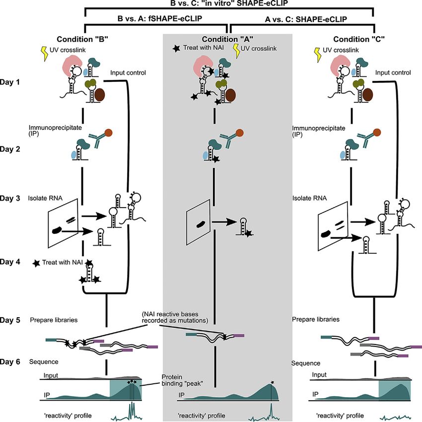
ll
     OPEN ACCESS
                                                                                                                     Protocol

fSHAPE has been tested in K562, HepG2, HeLa, and HEK293T cells. (f)SHAPE-eCLIP has been tested
in K562 cells.

The RNA probing reagent NAI-N3 has been tested with fSHAPE. fSHAPE-eCLIP has been tested with
the reagent NAI. SHAPE-eCLIP has been tested with NAI and dimethyl sulfate (DMS); NAI is
preferred because DMS does not react with all RNA bases.

(f)SHAPE-eCLIP has been performed with antibodies for SLBP (described here) and RBFOX2. These
methods are assumed to be compatible with any of the antibodies successfully employed for eCLIP,
of which there are over 300 (Van Nostrand et al., 2020). It is highly recommended to verify your anti-
body’s immunoprecipitation efficiency before performing this protocol for a full set of samples.

It is recommended to store aliquots of primers at 80 C. Master mixes of enzymatic reactions, minus
the enzymes, may be pre-mixed and aliquoted and stored at 80 C for later use.

Prepare primers

      Timing: 1 h

1. Dilute primers to specified concentrations.
   a. RNA oligo ‘‘InvRiL19’’ for RNA linker ligation: Order 100 nmole, standard desalting. Resus-
      pend at 200 mM; dilute to 40 mM for working stock. Store 5 mL aliquots at 80 C.
   b. DNA oligo ‘‘InvRand3Tr3’’ for cDNA ligation: Order 100 nmole, standard desalting. Resus-
      pend at 200 mM; dilute to 80 mM for working stock. Store 5 mL aliquots at 80 C.
   c. DNA oligo ‘‘InvAR17’’ for reverse transcription: Order 25 nmole, standard desalting. Resus-
      pend at 200 mM; dilute to 5 mM for working stock. Store 5 mL aliquots at 80 C.
   d. PCR_F_D501, PCR_F_D502, PCR_R_D701,PCR_R_D702: Order PAGE purified. Resuspend at
      100 mM; dilute to 20 mM for working stock. Store 5 mL aliquots at 80 C.
      i. Any Illumina standard high-throughput RNA-sequencing primer, such as D503-508, D703-
         712, can substitute for the above.

Cell lines

      Timing: days, depending on cell type

2. Grow K562 cells to 90% confluency. Aim for 40 million cells per sample condition. For 2 sample
   conditions with 2 replicates each at a minimum, this equates to 160 million cells in total.

KEY RESOURCES TABLE

REAGENT or RESOURCE                                              SOURCE                    IDENTIFIER
Antibodies
Anti-stem loop binding protein                                   MBLI                      RRID:AB_10606342; RN045P Lot# 001
Anti-rabbit TrueBlot HRP                                         Rockland                  RRID:AB_2610848; Cat# 18-8816-33
Chemicals, peptides, and recombinant proteins
Dimethylsulfoxide (DMSO)                                         Sigma-Aldrich             Cat# 276855
2-Methylnicotinic acid imidazolide (NAI) in DMSO                 EMD Millipore             Cat# 03-310
Buffer RLT                                                       QIAGEN                    cat # 79216
T4 polynucleotide kinase (PNK), 10 U/mL                          New England Biolabs       Cat# M0201L
5X PNK 6.5 buffer                                                New England Biolabs       Cat# M0201L
1 M DTT                                                          Thermo Scientific         Cat# P2325
100% Ethanol                                                     Thermo Scientific         Cat# T038181000
                                                                                                              (Continued on next page)

2     STAR Protocols 2, 100762, September 17, 2021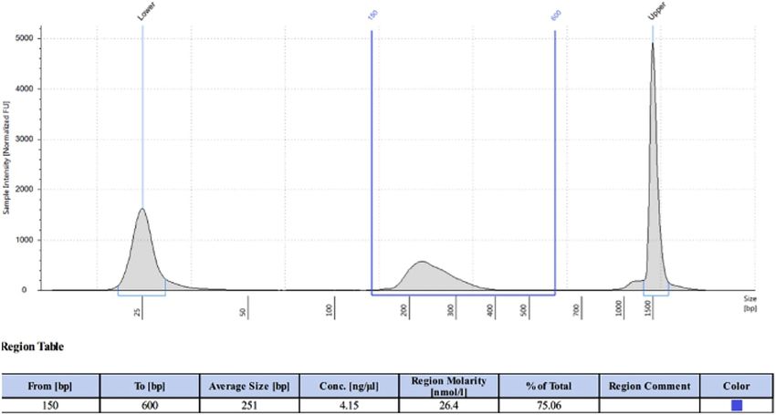
ll
Protocol                                                                                                             OPEN ACCESS

Continued
REAGENT or RESOURCE                                           SOURCE                        IDENTIFIER
100% Methanol                                                 Thermo Scientific             Cat# T001021000
FastAP Thermosensitive Alkaline Phosphatase, 1 U/mL           Thermo Scientific             Ct# EF0651
Proteinase K, 0.8 U/mL                                        New England Biolabs           Cat# P8107S
SuperScript II Reverse Transcriptase, 200 U/mL                Invitrogen                    Cat# 18064014
Turbo DNase, 2 U/mL                                           Life Technologies             Cat# AM2239
T4 RNA ligase 1, high concentration                           New England Biolabs           Cat# M0437M
10 X ligase buffer (with DTT)                                 New England Biolabs           Cat# M0437M
100 mM ATP                                                    New England Biolabs           Cat# M0437M
50% PEG 8000                                                  New England Biolabs           Cat# M0437M
Murine RNase Inhibitor, 40 U/mL                               New England Biolabs           Cat# M0314L
RNase I, 100 U/mL                                             Life Technologies             Cat# AM2295
Q5 PCR Master Mix                                             New England Biolabs           Cat# M0492L
Protease Inhibitor Cocktail III                               EMD Millipore                 Cat# 539134-1SET
ExoSAP-IT                                                     Affymetrix                    Cat# 78201
Tris-HCI buffer, 1 M pH 7.5                                   Invitrogen                    Cat# 15567-027
Tween-20                                                      Sigma-Aldrich                 Cat# P1379
HEPES, 1 M pH 8.0                                             Life Technologies             Cat# 15630-080
Magnesium chloride, 1 M                                       Ambion                        Cat# AM9530G
Sodium chloride, 5 M                                          Ambion                        Cat# AM9759
Potassium chloride, 3M                                        Millipore Sigma               Cat# 60135
Nonidet P40 (NP-40)                                           Roche                         Cat# 11332473001
EDTA, 500 mM                                                  Millipore                     Cat# 324504
HCl, 1.0 N                                                    Sigma-Aldrich                 Cat# H9892
NaOH, 1.0 N                                                   Sigma-Aldrich                 Cat# S2770
Sodium dodecyl sulfate (SDS) 10%                              Sigma-Aldrich                 Cat# 71736
Sodium deoxycholate                                           Sigma-Aldrich                 Cat# 30970
20X MOPS Running Buffer                                       Thermo Fisher                 Cat# NP0001
20X NuPAGE Transfer Buffer                                    Thermo Fisher                 Cat# NP00061
10X TBST Buffer                                               Sigma-Aldrich                 Cat# 91414
10X TBE Buffer                                                Millipore Sigma               Cat# SRE0062
Manganese (II) chloride solution 1M                           Sigma-Aldrich                 Cat# M1787
Glycerol                                                      Thermo Scientific             Cat# 15514011
Power SYBR Green PCR Master Mix                               Applied Biosystems            Cat# 4368577
Dynabeads M-280 Sheep Anti-Rabbit, or anti-mouse,             Life Technologies             Cat# 11204D
10 mg/mL
Dynabeads MyOne Silane, 40 mg/mL                              Life Technologies             Cat# 37002D
Agencourt AMPure XP beads                                     Beckman Coulter               Cat# A63881
ECL Western Blotting Substrate                                Thermo Scientific             Cat# 32106
Broad Range Protein Ladder                                    Thermo Scientific             Cat # 26634
Milk powder                                                   Sigma-Aldrich                 Cat# M7409
SYBR Safe DNA Gel Stain                                       Invitrogen                    Cat# S33102
6X Orange G Buffer                                            New England Biolabs           Cat# B7022S
50 bp DNA Ladder                                              Thermo Scientific             Cat# SM0373
NuSieve GTG Agarose (low melting temperature)                 Lonza                         Cat# 50081
Critical commercial assays
RNA Clean and Concentrator Kit                                Zymo Research                 Cat# R1019
MinElute Gel Extraction Kit                                   QIAGEN                        Cat# 28604
Deposited data
Sequencing data and processed fSHAPE and SHAPE reactivities   (Corley et al., 2020)         GEO: GSE149767
from fSHAPE, fSHAPE-eCLIP, and SHAPE-eCLIP libraries
Experimental models: Cell lines
K562                                                          ATCC                          CCL-243
Oligonucleotides
‘‘InvRiL19’’ (RNA oligo): /5Phos/rArGrArUrCrGrGrArArGrArGr    IDT                           N/A
CrArCrArCrGrUrC/3SpC3/, standard desalting
                                                                                                                (Continued on next page)

                                                                                      STAR Protocols 2, 100762, September 17, 2021    3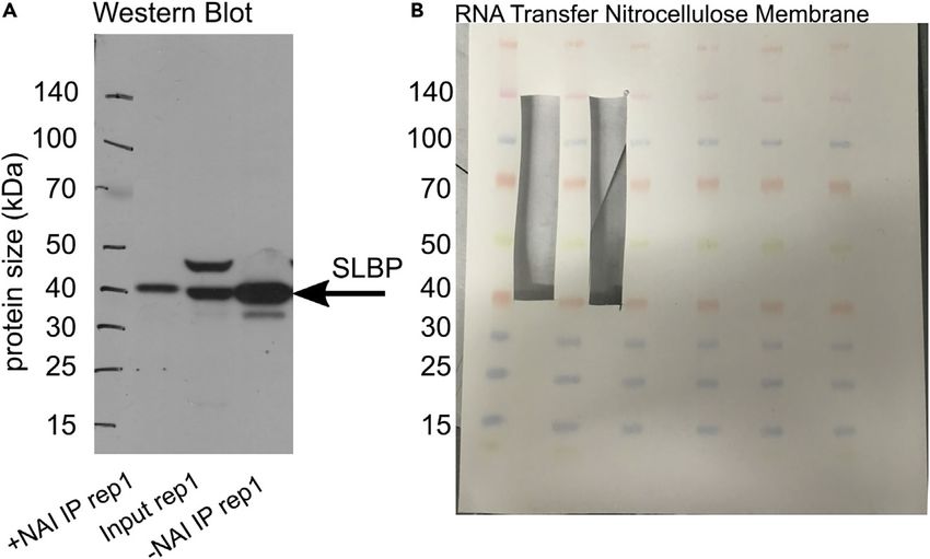
ll
        OPEN ACCESS
                                                                                                                                     Protocol

Continued
REAGENT or RESOURCE                                                          SOURCE                     IDENTIFIER
‘‘InvRand3Tr3’’: /5Phos/NNNNNNNNNNAGATCGGAAGAG                               IDT                        N/A
CGTCGTGT/3SpC3/, standard desalting
‘‘InvAR17’’: CAGACGTGTGCTCTTCCGA, standard desalting                         IDT                        N/A
‘‘PCR_F_D501’’: AATGATACGGCGACCACCGAGATCTACACTATAGC                          IDT                        N/A
CTACACTCTTTCCCTACACGACGCTCTTCCGATCT, PAGE purified
‘‘PCR_F_D502’’: AATGATACGGCGACCACCGAGATCTACACATAGAG                          IDT                        N/A
GCACACTCTTTCCCTACACGACGCTCTTCCGATCT, PAGE purified
‘‘PCR_R_D701’’: CAAGCAGAAGACGGCATACGAGATCGAGTAATGTG                          IDT                        N/A
ACTGGAGTTCAGACGTGTGCTCTTCCGATC, PAGE purified
‘‘PCR_R_D702’’: CAAGCAGAAGACGGCATACGAGATTCTCCGGAGT                           IDT                        N/A
GACTGGAGTTCAGACGTGTGCTCTTCCGATC, PAGE purified
Software and algorithms
Cutadapt 1.14                                                                (Martin, 2011)             https://cutadapt.readthedocs.io/en/stable/
UMItools >= 0.5.0                                                            (Smith et al., 2017)       https://github.com/CGATOxford/UMI-tools
STAR >= 2.4.0i                                                               (Dobin et al., 2013)       https://github.com/alexdobin/STAR
Bedtools >= 2.25.0                                                           (Quinlan and Hall, 2010)   https://bedtools.readthedocs.io/en/latest/
Samtools >= 1.9                                                              (Li et al., 2009)          https://samtools.github.io
Bamtools 2.3.0                                                               (Barnett et al., 2011)     https://github.com/pezmaster31/bamtools
fSHAPE analysis pipeline                                                     This manuscript            https://github.com/meracorley/fSHAPE
fSHAPE- and SHAPE-eCLIP analysis pipelines                                   This manuscript            https://github.com/meracorley/f-SHAPE-
                                                                                                        eCLIP/tree/pipeline
Python >= 3.5.4                                                              N/A                        N/A
Hmmlearn 0.2.1                                                               Scikit-learn               https://hmmlearn.readthedocs.io/en/latest/
Other
Laminar flow hood                                                            Standard lab equipment     N/A
Cell culture incubator                                                       Thermo Scientific          Cat# 51026331
Large centrifuge                                                             Thermo Scientific          Cat# 1189M64
UV crosslinker                                                               Thermo Scientific          Cat# 95034
Small centrifuge                                                             Eppendorf                  Cat# 5406000046
Bioruptor sonicator                                                          Bioruptor                  Cat# B01020001
1.5 mL Tube magnet                                                           Thermo Scientific          Cat# CS15000
0.2 mL Tube magnet                                                           EpiCypher                  Cat# 10-0008
Tube rotator                                                                 Thermo Scientific          Cat# 88881001
Thermomixer                                                                  Eppendorf                  Cat# 5382000023
PAGE gel tank                                                                Invitrogen                 Cat# EI0001
4%–12% Bis-Tris gel, 1.5 mm, 10 well                                         Invitrogen                 Cat# NP0335BOX
Transfer tank                                                                Thermo Scientific          Cat# VEP-2
Power supply for running gels/transfers                                      Invitrogen                 Cat# PS0350
PVDF membrane                                                                Invitrogen                 Cat# PB9320
Nitrocellulose membrane                                                      Invitrogen                 Cat# PB7320
Agarose gel electrophoresis equipment                                        Standard lab equipment     N/A
Thermocycler                                                                 Standard lab equipment     N/A

MATERIALS AND EQUIPMENT

eCLIP Lysis Buffer (step 5), store at 4 C for up to 6 months
Reagent                                                Final concentration                              Amount
1 M Tris-HCl pH 7.4                                    50mM                                             5 mL
5 M NaCl                                               100 mM                                           2 mL
NP-40                                                  1%                                               1 mL
10% SDS                                                0.1%                                             1 mL
Sodium deoxycholate                                    0.5%                                             0.5 g
diH2O                                                  n/a                                              91 mL
Total                                                  n/a                                              100 mL

4       STAR Protocols 2, 100762, September 17, 2021
ll
Protocol OPEN ACCESS
eCLIP High Salt Wash Buffer (step 10), store at 4 C for up to 6 months
Reagent Final concentration Amount
1 M Tris-HCl pH 7.4 50mM 5 mL
5 M NaCl 1M 20 mL
500 mM EDTA 1 mM 200 mL
NP-40 1% 1 mL
SDS 10% 0.1% 1 mL
Sodium deoxycholate 0.5% 0.5 g
diH2O n/a 72.8 mL
Total n/a 100 mL
eCLIP Wash Buffer (step 10), store at 4 C for up to 6 months
Reagent Final concentration Amount
1 M Tris-HCl pH 7.4 20 mM 2 mL
5 M NaCl 5 mM 100 mL
1 M MgCl2 10 mM 1 mL
1% Tween-20 0.2% 20 mL
diH2O n/a 76.9 mL
Total n/a 100 mL
RLTW Buffer (step 38), store at RT for up to 6 months
Reagent Final concentration Amount
RLT Buffer (1X) 1X 9.75 mL
1% Tween-20 0.025% 250 mL
Total n/a 10 mL
PKS Buffer (step 28), store at RT for up to 6 months
Reagent Final concentration Amount
1 M Tris-HCl pH 7.4 100 mM 1 mL
5 M NaCl 50 mM 100 mL
500 mM EDTA 10 mM 200 mL
10% SDS 0.2% 200 mL
diH2O n/a 8.5 mL
Total n/a 10 mL
1X RNA Ligase Buffer (step 15), store at RT for up to 6 months
Reagent Final concentration Amount
1 M Tris-HCl pH 7.5 50 mM 500 mL
1 M MgCl2 10 mM 100 mL
diH2O n/a 9.4 mL
Total n/a 10 mL
3X SHAPE Folding Buffer (step 31), store at RT for up to 6 months
Reagent Final concentration Amount
1 M HEPES pH 8.0 333 mM 333 mL
1 M MgCl2 20 mM 20 mL
5 M NaCl 333 mM 66.6 mL
diH2O n/a 580.4 mL
Total n/a 1 mL
STAR Protocols 2, 100762, September 17, 2021 5ll
OPEN ACCESS
Protocol
SHAPE FS Buffer (step 41), store at RT for up to 6 months
Reagent Final concentration Amount
1 M Tris-HCl pH 8.0 500 mM 500 mL
3 M KCl 750 mM 250 mL
diH2O n/a 250 mL
Total n/a 1 mL
TT Elution Buffer (step 38), store at RT for up to 6 months
Reagent Final concentration Amount
1 M Tris-HCl pH 7.5 10 mM 10 mL
500 mM EDTA 0.1 mM 0.2 mL
1% Tween-20 0.01% Tween-20 10 mL
diH2O n/a 979.8 mL
Total n/a 1 mL
PCR Elution Buffer (step 48), store at RT for up to 6 months
Reagent Final concentration Amount
1 M Tris-HCl pH 7.5 10 mM 10 mL
5 M NaCl 20 mM 4 mL
500 mM EDTA 0.1 mM 0.2 mL
diH2O n/a 985.8 mL
Total n/a 1 mL
Enzymatic reactions:
‘‘Early’’ FastAP treatment (on-bead) (step 13)
Reagent Final concentration Amount
diH2O n/a 38 mL
10X FastAP buffer 1X 5 mL
Murine RNase Inhibitor, 40 U/mL 1.6 U/mL 2 mL
Turbo DNase, 2 U/mL 0.08 U/mL 2 mL
FastAP enzyme, 1 U/mL 0.06 U/mL 3 mL
Total n/a 50 mL
‘‘Early’’ PNK treatment (on-bead) (step14)
Reagent Final concentration Amount
diH2O n/a 116 mL
5X PNK Buffer pH 6.5 1X 30 mL
T4 PNK enzyme, 10 U/mL 0.27 U/mL 4 mL
Total n/a 150 mL
‘‘Early’’ 30 RNA linker ligation (on-bead) (step 16)
Reagent Final concentration Amount
diH2O n/a 8.4 mL
10X Ligase buffer (no DTT; self-made, see buffers above) 1X 3 mL
0.1 M ATP 1 mM 0.3 mL
100% DMSO 3% 0.9 mL
1% Tween-20 0.02% 0.6 mL
50% PEG 8000 15% 9 mL
Murine RNase Inhibitor, 40 U/mL 0.53 U/mL 0.4 mL
RNA ligase high conc., 30 U/mL 2.4 U/mL 2.4 mL
Combine above n/a 25 mL
(Continued on next page)
6 STAR Protocols 2, 100762, September 17, 2021ll
Protocol OPEN ACCESS
Continued
Reagent Final concentration Amount
Beads binding RNA n/a ~2.5 mL
InvRiL19 oligo, 40 mM 3.33 mM 2.5 mL
Total n/a ~30 mL
In vitro structure probing (step 31)
Reagent Final concentration Amount
RNA sample, heat denatured and flash-cooled n/a ~10 mL
diH2O n/a 2.4 mL
3X SHAPE Folding Buffer 1X 6.6 mL
Murine RNase Inhibitor, 40 U/mL 2 U/mL 1.0 mL
Combine above n/a 20 mL
2 M NAI in DMSO 100 mM 1 mL
Total n/a 21 mL
CRITICAL: The reagent NAI should be handled with caution, avoiding contact with skin.
‘‘Late’’ FastAP treatment (step 34)
Reagent Final concentration Amount
diH2O n/a 6 mL
10X FastAP Buffer 1X 2 mL
Murine RNase Inhibitor, 40 U/mL 2 U/mL 1 mL
FastAP enzyme, 1 U/mL 0.1 U/mL 2 mL
Combine above n/a 11 mL
RNA sample n/a ~10 mL
Total n/a 21 mL
‘‘Late’’ PNK treatment (step 35)
Reagent Final concentration Amount
diH2O n/a 45 mL
5X PNK Buffer pH 6.5 1X 20 mL
1 M DTT 5 mM 0.5 mL
Turbo DNase, 2 U/mL 0.02 U/mL 1 mL
T4 PNK enzyme, 10 U/mL 0.4 U/mL 4 mL
Combine above n/a 75 mL
‘‘Late’’ FastAP reaction n/a ~21 mL
Total n/a ~100 mL
‘‘Late’’ 30 RNA linker ligation for Input samples (step 37)
Reagent Final concentration Amount
diH2O n/a 2.8 mL
10X NEB Ligase Buffer (with DTT) 1X 2.0 mL
0.1 M ATP 1 mM 0.2 mL
100% DMSO 3% 0.6 mL
1% Tween-20 0.02% 0.4 mL
50% PEG 8000 15% 6.0 mL
Murine RNase Inhibitor, 40 U/mL 0.6 U/mL 0.3 mL
RNA ligase high conc., 30 U/mL 1.8 U/mL 1.2 mL
Combine above n/a 13.5 mL
RNA sample n/a 5 mL
100% DMSO n/a 1.5 mL
InvRiL19 oligo, 40 mM 1 mM 0.5 mL
Total n/a 20 mL
STAR Protocols 2, 100762, September 17, 2021 7ll
OPEN ACCESS
Protocol
‘‘Late’’ 30 RNA linker ligation for IP samples (step 37)
Reagent Final concentration Amount
diH2O n/a 5.6 mL
10X NEB Ligase buffer (with DTT) 1X 4 mL
0.1 M ATP 1 mM 0.4 mL
100% DMSO 3% 1.2 mL
1% Tween-20 0.02% 0.8 mL
50% PEG 8000 15% 12.0 mL
Murine RNase Inhibitor, 40 U/mL 0.6 U/mL 0.6 mL
RNA ligase high conc., 30 U/mL 1.8 U/mL 2.4 mL
Combine above n/a 27 mL
RNA sample n/a ~10 mL
100% DMSO n/a 2.4 mL
InvRiL19 oligo, 40 mM 1 mM 1.0 mL
Total n/a ~40 mL
SHAPE reverse transcription (step 41)
Reagent Final concentration Amount
10X SHAPE FS Buffer 1X 2.0 mL
Murine RNase Inhibitor, 40 U/mL 0.4 U/mL 0.2 mL
0.1 M DTT 5 mM 1.0 mL
500 mN MnCl2 6 mM 0.24 mL
Superscript II reverse transcriptase, 200 U/mL 10 U/mL 1.0 mL
diH2O n/a 5.36 mL
Combine above n/a 10 mL
RNA sample n/a ~9 mL
5 mM InvAR17 primer 0.25 mM 1 mL
10 mM dNTPs 0.5 mM 1 mL
Total n/a ~20 mL
50 cDNA linker ligation (step 44)
Reagent Final concentration Amount
diH2O n/a 1.4 mL
10X NEB Ligase buffer (with DTT) 1X 4 mL
0.1 M DTT 2 mM 0.2 mL
0.1 M ATP 1 mM 0.1 mL
1% Tween-20 0.02% 0.2 mL
50% PEG 8000 18% 3.6 mL
RNA ligase high conc., 30 U/mL 3.0 U/mL 1.0 mL
Combine above n/a 7.8 mL
RNA sample in dried beads n/a 0 mL
100% DMSO 8% 0.8 mL
InvRand3Tr3 oligo, 80 mM 4.8 mM 0.6 mL
TT Elution Buffer n/a 1.1 mL
Total n/a 10.3 mL
cDNA qPCR quantification (step 46)
Reagent Final concentration Amount
diH2O n/a 3.6 mL
2X PowerSybr master mix 1X 5.0 mL
20 mM D50x primer mixed in equal parts with 20 mM D70x primer 0.4 mM 0.4 mL
1:10 dilution cDNA sample 1:100 1.0 mL
Total n/a 10 mL
8 STAR Protocols 2, 100762, September 17, 2021ll
Protocol OPEN ACCESS
cDNA PCR amplification (step 47)
Reagent Final concentration Amount
2X Q5 PCR master mix 1X 20 mL
20 mM right primer (D50x) 1 mM 2 mL
20 mM left primer (D70x) 1 mM 2 mL
cDNA sample n/a 16 mL
Total n/a 40 mL
Equipment:
Cell culture equipment: laminar flow hood, cell culture incubator
UV crosslinker (254 nm, UV-C)
Bioruptor sonicator, for RNA fragmentation
Centrifuge for 15 mL conicals; Centrifuge for 1.5 mL tubes; minifuge
Magnet for 1.5 mL tubes, 0.2 mL tubes
Tube rotator
Thermomixer
Western blot and RNA transfer equipment: SDS-PAGE gels 4–12% Bis-Tris, 10-well, 1.5 mm (1.0 mm
if 1.5 mm not available), buffer tanks for PAGE and for western blots/RNA transfers, power supply for
gel electrophoresis, PVDF and nitrocellulose membranes, sponges, roller
Thermocycler
Real time qPCR equipment
Agarose gel electrophoresis equipment
STEP-BY-STEP METHOD DETAILS
Treat cells with NAI or DMSO and UV crosslink
Timing: 3 h
Treat cells with the structure probing reagent NAI or mock treat with DMSO, followed by UV cross-
linking to covalently link RNA binding proteins to bound RNA molecules. Complete these steps for
all protocol types: fSHAPE-eCLIP, SHAPE-eCLIP, or ‘‘in vitro’’ SHAPE-eCLIP.
1. Cell sample requirements
a. Grow cells to 30–40 million cells per sample. With two treatment conditions each needing at
least two replicates, this adds up to at least four samples, 120–160 million cells total.
b. fSHAPE-eCLIP and SHAPE-eCLIP both consist of two RNA treatment conditions performed
in parallel. The different conditions are referred to here as ‘‘A’’, ‘‘B’’, or ‘‘C’’ (see Graphical
Abstract):
i. fSHAPE-eCLIP consists of one set of cells treated with NAI (‘‘A’’), i.e., RNA is treated in vivo.
The other set is mock-treated with DMSO, but later treated with NAI once its RNA is in vitro
(‘‘B’’).
STAR Protocols 2, 100762, September 17, 2021 9ll
OPEN ACCESS
Protocol
ii. SHAPE-eCLIP consists of one set of cells treated with NAI (‘‘A’’) and the other mock-treated
with DMSO (‘‘C‘‘). An ‘‘in vitro’’ variant of SHAPE-eCLIP consists of two sets of samples that
are both mock-treated with DMSO at the cell stage, but one set later treats RNA with NAI in
vitro (‘‘B‘‘).
2. Setup
a. Pre-chill on ice 100 mL 1X PBS.
b. Set an incubator to 37 C.
c. Pre-chill on ice metal plate that fits inside crosslinker. Set crosslinker to 400 mJ/cm2 and 2 min.
Crosslinker should emit UV-C light at 254 nm wavelengths.
d. Fetch some dry ice.
3. Treat cells with the structure probing reagent (NAI) or mock-treat with DMSO
a. Split cells into 30–40 million cells per sample, two samples for each treatment condition.In-
structions are for suspension cells; see note below on adherent cells.
b. Reduce each cell sample volume to 2 mL in plain media (no FBS) in 10 cm plates.
c. Per sample pair: For ‘‘A’’ conditions (in fSHAPE-eCLIP or SHAPE-eCLIP), add 170 uL 2 M NAI
structure probing reagent to each sample. For ‘‘B’’ or ‘‘C’’ conditions (in fSHAPE-eCLIP or the
‘‘in vitro’’ variant of SHAPE-eCLIP), add 170 uL DMSO to each sample. Swirl to mix.
d. Incubate plates at 37 C for 10 min.
4. UV crosslink cells and wash
a. Place pre-chilled metal plate into crosslinker and place plates on top. The chilled plate pre-
vents cells from heating up during crosslinking.
b. Remove lids from plates. Run the pre-set crosslinking program.
c. Move cells from plates to 15 mL conicals. Spin down at 500 g for 3 min.
d. Remove supernatant (NAI-treated supernatant should go into its own waste container).
e. Resuspend cells in 5 mL cold PBS. Spin down and remove supernatant as before. Resuspend
again with cold PBS.
f. Spin down cells one more time, remove PBS supernatant, and flash freeze cells in dry ice. Store
at 80 C.
CRITICAL: Removing lids from plates before crosslinking is critical to cells being properly
crosslinked.
Note: Adherent cells should have media removed and replaced with 2 mL plain media (no
FBS) and subsequently treated with 170 uL NAI or DMSO, swirling to mix. Cells should then
be crosslinked as instructed, then scraped off plates with a cell scraper and transferred to
15 mL conicals, following the same washing and centrifugation steps outlined for suspension
cells.
Pause point: Cell pellets can be stored at 80 C for months.
Lyse cells and immunoprecipitate RNA
Timing: 3–16 h
Lyse protein-bound RNA from cells. Pull down RNA bound to the protein of interest with a mono-
clonal antibody coupled to magnetic beads. Complete these steps for all protocol types.
5. Setup
a. Pre-chill eCLIP Lysis Buffer.
b. Thaw Protease Inhibitor.
c. Bioruptor sonicator water tank should be at 4 C (in a cold room).
d. Pre-heat Thermomixer to 37 C.
e. Pre-set 1.5 mL tube centrifuge to 4 C.
10 STAR Protocols 2, 100762, September 17, 2021ll
Protocol OPEN ACCESS
6. Couple antibody to Dynabeads M-280
a. Prepare beads.
i. Use 125 mL beads per sample, 500 mL for two conditions each with two replicates.
ii. Apply bead solution to magnet; remove supernatant.
iii. Wash beads twice in 500 mL cold eCLIP Lysis Buffer.
iv. Resuspend beads in 100 mL cold eCLIP Lysis Buffer for every sample, typically 400 mL.
b. Bind antibody.
i. Add 10 mg antibody per sample to prepared beads.
ii. Rotate at 15 C–25 C for 45 min.
7. Lyse cells, while beads are rotating
a. For two replicates—four samples total—combine 4 mL eCLIP Lysis Buffer with 22 mL Protease
Inhibitor Cocktail III and 44 mL Murine RNase Inhibitor.
b. Retrieve cell pellets from 80 C, keeping on dry ice. Immediately add 1 mL master mix from 7a
to each pellet, pipetting to resuspend. Move each sample to a 1.5 mL tube.
c. Place on ice (not dry ice) and allow to lyse for 5 min.
8. Fragment RNA
a. Sonicate lysed samples in Bioruptor on ‘‘low’’, 4 C, 5 cycles of 30 s on and 30 s off. This assists
cell lysis and shears chromatin, aiding release of RNA-protein complexes.
Note: Alternatively, sonication may be done on ice with a probe sonicator, sonicating each
sample twice at 5 decibels in 10 second bursts. Avoid touching the side of tube with sonicator.
b. Dilute 1 mL RNase I in PBS at 1:50 on ice.
c. Add 10 mL Turbo DNase to each lysed sonicated sample.
d. Add 10 mL diluted RNase I (20 U total) to each sample. Immediately incubate in a Thermomixer
at 37 C, 1200 rpm, 5 min. Place on ice.
e. Centrifuge samples at 15,000 g, 4 C, for 3 min.
f. Meanwhile, proceed with preparing antibody-bound magnetic beads (below).
9. Immunoprecipitate RNA bound by protein of interest
a. Wash antibody beads from 6b two times with 500 mL in cold eCLIP Lysis Buffer.
b. Remove as much supernatant as possible from beads and resuspend once more in 100 mL cold
eCLIP Lysis Buffer per sample, typically 400 mL.
c. Distribute beads very well by pipetting up and down (taking tube off of magnet) and divide
bead solution evenly between separate 1.5 mL tubes, one tube per sample.
d. Only once lysates from 8e are finished centrifuging, apply each bead sample to the magnet
and remove supernatant. Add cleared lysate for each sample from 8e to its own antibody
bead sample.
e. Rotate at 4 C for 2 h, or up to 15 h in cold room.
Note: If doing fSHAPE-eCLIP or ‘‘in vitro’’ variant of SHAPE-eCLIP, opt for 2 hours of rotation
and skip to ‘‘Isolate RNA’’ section.
Ligate 30 RNA adapter
Timing: 3 h
Complete this section only for SHAPE-eCLIP samples at this time in the protocol. Treat RNA and
ligate an adapter, later used for reverse transcription.
10. Setup
a. Verify High Salt Wash Buffer, Wash Buffer, and 1x FastAP Buffers are cold.
b. Pre-heat Thermomixer to 37 C.
STAR Protocols 2, 100762, September 17, 2021 11ll
OPEN ACCESS
Protocol
11. Set aside non-immunoprecipitated sample controls, referred to as ‘‘Input’’ samples
a. Of the sample conditions (‘‘A’’,’’B’’, or ‘‘C’’; see 1b for naming convention), pick one condition
to take sample Input controls from. Typically ‘‘B’’ or ‘‘C’’ conditions. One can carry forward
Input controls for all conditions if one chooses, but this creates more controls than is
necessary.
b. Gently mix samples from 9e well. Take two 20 mL aliquots from each replicate of the chosen
condition, four aliquots total, adding each to separate tubes labeled ‘‘Transfer-Input’’ and
‘‘Western-Input’’ and their replicate number. Set Inputs aside on ice until step 20.
c. The remainder of the samples are referred to as immunoprecipitated (IP) samples.
12. Wash antibody-bound beads
a. Perform the following washes on ice if possible.
b. Apply all IP samples to magnet. Wait for beads to pellet on the magnet, removing the super-
natant when it is clear.
c. Wash bead pellets with 900 mL cold High Salt Wash Buffer by pipetting the buffer onto the
bead pellets. Wait until supernatant is clear (1 min) and remove supernatant by pipetting.
Repeat.
d. Wash beads 2 times as above with 500 mL cold Wash Buffer. During the second wash, move
tubes back and forth on magnet (Methods Video S1). One time is sufficient, such that the
bead pellet is forced to re-form. Wait until beads have reformed pellet until removing super-
natant, 1 min.
e. Resuspend beads with 500 mL 1X FastAP Buffer, pipetting to completely resuspend beads in
buffer.
f. Allow supernatant to clear and remove supernatant. Add 500 mL 1X FastAP Buffer and place
tubes on ice.
13. FastAP treat RNA on beads
a. Prepare ‘‘Early’’ FastAP master mix on ice (see materials and equipment). Mix well.
b. Place IP bead samples on magnet, removing supernatant when clear. Add 50 mL FastAP mas-
ter mix to each IP bead sample.
c. Incubate samples in Thermomixer at 1200 rpm at 37 C for 10 min.
14. PNK treat RNA on beads
a. While FastAP reaction is incubating, prepare ‘‘Early’’ PNK master mix.
b. Mix well and add 150 mL master mix to each sample.
c. Incubate samples in Thermomixer at 1200 rpm at 37 C for 20 min.
15. Wash beads
a. Apply all IP samples to magnet, removing supernatant when clear.
b. Wash bead pellets with 500 mL cold Wash Buffer. Move tubes back and forth on magnet,
allow supernatant to clear and remove. Repeat wash, moving tubes back and forth on mag-
net, followed by removal of clear supernatant.
c. Wash beads 2 times as above with 500 mL cold High Salt Wash Buffer.
d. Wash beads 2 times as above with 500 mL cold Wash Buffer.
e. Wash beads with 300 mL 1X Ligase Buffer (no DTT), moving back and forth on magnet and
removing cleared supernatant.
f. Wash beads 2 times with 300 mL 1X Ligase Buffer (no DTT). Keep beads in buffer on ice until
next step.
16. Ligate 30 RNA adapter
a. Prepare ‘‘Early’’ 30 RNA Ligation master mix. Mix well by pipetting or flicking.
b. Remove supernatant from IP bead samples. Briefly spin samples in minifuge, apply to mag-
net, and use a small pipette tip to remove all residual liquid in tubes.
c. Add 25 mL of master mix to each IP bead sample.
d. Add 2.5 mL 40 mM InvRiL19 to each sample.
e. Incubate at 15 C–25 C for 75 min, flicking tubes every 10 min to mix.
f. Wash beads 2 times with 500 mL cold Wash Buffer.
12 STAR Protocols 2, 100762, September 17, 2021ll
Protocol OPEN ACCESS
Isolate RNA
Timing: 4–24 h
Complete this section for all IP and Input samples and for all protocol types: fSHAPE-eCLIP, SHAPE-
eCLIP, or ‘‘in vitro’’ SHAPE-eCLIP. Verify efficacy of the antibody with a Western blot and isolate IP
and Input RNA with a gel transfer.
17. Setup
a. Pre-heat thermomixer to 70 C.
b. Retrieve gel tank(s) and power supply.
c. Dilute 20X MOPS Running Buffer to 2 L of 1X buffer.
d. Prepare 2 L of cold 1X NuPAGE Transfer Buffer with 10% methanol. Keep at 4 C.
18. For only fSHAPE-eCLIP or ‘‘in vitro’’ SHAPE-eCLIP samples from 9e: set aside non-immunopre-
cipitated samples as controls, referred to as ‘‘Input’’ samples
a. Of the sample conditions (‘‘A’’,’’B’’, or ‘‘C’’; see 1b for naming convention) from step 9e, pick one
condition to take sample Input controls from. Typically ‘‘B’’ or ‘‘C’’ conditions. One can carry forward
Input controls for all conditions if one chooses, but this creates more controls than is necessary.
b. Gently mix samples from 9e well. Take two 20 mL aliquots from each replicate of the chosen
condition, four aliquots total, adding each to separate tubes labeled ‘‘Transfer-Input’’ or
‘‘Western-Input’’ and their replicate number. Set Inputs aside on ice until step 20.
c. The remainder of the samples is referred to as immunoprecipitated (IP) samples.
19. Wash IP bead samples, from step 16f or 18c
a. Apply IP samples to magnet, remove supernatant, and wash two times with 500 mL High Salt
Wash Buffer. Remove cleared supernatant.
b. Resuspend beads in 500 mL cold High Salt Wash Buffer. Add 500 mL cold Wash Buffer. Allow
bead pellets to reform on magnet and remove cleared supernatant.
c. Wash three times with cold Wash Buffer.
20. Prepare IP and Input samples for gel separation
a. Prepare two 4%–12% Bis-Tris SDS-PAGE gels, 12-well, 1.5 mm. One will be used for a West-
ern Blot (‘‘Western’’) and the other for a gel transfer of RNA (‘‘Transfer’’).
b. Remove all supernatant from IP bead samples. Take beads off magnet and resuspend beads
well in 100 mL Wash Buffer.
c. For each resuspended IP bead sample, move 20 mL to a new tube. Label these aliquots with
their treatment type, replicate number, and ‘‘Western.’’ These samples will be used in the
Western Blot.
d. For the remaining 80 mL of IP bead samples, apply to magnet, remove all supernatant, and
resuspend beads in 20 mL Wash Buffer. These samples will be used for the RNA transfer.
e. Prepare a loading buffer master mix of 7.5 mL 4X NuPAGE LDS Buffer with 3 mL 1 M DTT for every
sample (IPs and Inputs for the Western Blot and RNA Transfer; typically 12 samples total).
f. Add 10 mL DTT-Buffer mix to each sample and denature all samples in Thermomixer for
10 min at 70 C, 1200 rpm. Samples are now decoupled from beads.
g. Cool samples on ice for 1 min followed by a brief spin in a minifuge.
h. Place all tubes (including Inputs) on magnet on ice. Desired material is contained in the su-
pernatant.
21. Gel separate IP and Input samples
a. Set up the two Bis-Tris gels. Label wells according to Tables 1 and 2. Fill tanks with 1X MOPS
Running Buffer.
b. Load IP and Input RNA Transfer samples into gel according to map in Table 1. Run at 150 V for
75 min, or until dye front is at bottom.
c. Load half of IP and Input Western Blot samples (15 mL) into gel according to map in Table 2.
Save the remainder of Western Blot samples at 20 C for troubleshooting. Run at 150 V for
75 min, or until dye front is at bottom.
STAR Protocols 2, 100762, September 17, 2021 13ll
OPEN ACCESS
Protocol
Table 1. Gel set up for Transfer samples
Column 1 2 3 4 5 6 7 8 9 10 11 12
Input IP, treatment IP, treatment Input IP, treatment IP, treatment
Sample M rep1 M A/B rep1 M B/C rep1 M rep2 M A/B rep2 M B/C rep2
Volume to Load 5 mL 30 mL 5 mL 30 mL 5 mL 30 mL 5 mL 30 mL 5 mL 30 mL 5 mL 30 mL
% of Sample Represented 2% 80% 80% 2% 80% 80%
M = protein marker (3 uL) in loading buffer (2 uL).
22. Set up membranes while gels are running
a. For the Transfer gel: cut out four Whatman papers, one nitrocellulose membrane, and two
sponges, all sized to fit the gel.
b. For the Western gel: cut out four Whatman papers, one PVDF membrane, and two sponges,
all sized to fit the gel.
c. Fill two separate shallow trays half-way with cold 1X Transfer buffer with 10% methanol. Place
cassettes for Western Blot/RNA Transfer open in trays.
d. Place sponges, Whatman papers, and nitrocellulose/PVDF membranes in trays to soak. Use
rollers to roll bubbles out of sponges if necessary.
23. Western Blot and RNA Transfer
a. Once gels are finished running, carefully remove one side of their casing. Using a straight
edge, remove extraneous portions of the gels: the bottom below the dye front, the wells.
Make sure to note the samples’ orientation in the gels.
b. Set up the RNA Transfer inside open cassette in the shallow tray. Carefully stack layers as fol-
lows, using a small roller carefully roll out bubbles between additions, such that each layer is
flush to the one below.
i. Sponge
ii. 2 Whatman papers
iii. Gel from 21b
iv. Nitrocellulose membrane
v. 2 Whatman papers
vi. Sponge
c. Set up the Western Blot inside open cassette in the shallow tray. Carefully stack layers as fol-
lows, using a small roller carefully roll out bubbles between additions, such that each layer is
flush to the one below.
i. Sponge
ii. 2 Whatman papers
iii. Gel from 21c
iv. PVDF membrane
v. 2 Whatman papers
vi. Sponge
d. Close each cassette. Cassettes should snugly fit the RNA Transfer/Western Blot, holding
everything in place. If loose, add another sponge on top. Note the side of the cassette
that the nitrocellulose/PVDF membrane is closest to.
Table 2. Gel set up for Western Blot
Column 1 2 3 4 5 6 7 9 10
Input IP, treatment IP, treatment Input IP, treatment IP, treatment
Sample M rep1 A/B rep1 B/C rep1 M rep2 A/B rep2 B/C rep2 M
Volume to Load 5 mL 15 mL 15 mL 15 mL 5 mL 15 mL 15 mL 15 mL 5 mL
% of Sample Represented 1% 10% 10% 1% 10% 10%
M = protein marker in loading buffer.
14 STAR Protocols 2, 100762, September 17, 2021ll
Protocol OPEN ACCESS
e. Place cassettes in a wet transfer tank. The side of the cassette that is closest to the nitrocel-
lulose/PVDF membrane should face the positive electrode in the tank, such that material
flows from the gel into the membrane.
f. Fill transfer tank with cold 1X Transfer Buffer with 10% methanol.
g. Run at 30V 10–20 h in cold room (preferred for Transfer), or at 200 mA for 2 h.
Develop the western blot
Timing: 3 h
Develop the Western Blot to confirm that immunoprecipitation was successful.
24. Setup
a. Preheat Thermomixer to 37 C.
b. In the meantime, continue to run the RNA Transfer (recommended). Or, remove the RNA
Transfer nitrocellulose membrane, rinse briefly with sterile 1X PBS, wrap in plastic wrap,
and store at 20 C.
25. Develop western blot
a. Stop Western Blot transfer and remove PVDF membrane from stack.
b. Place a small cut on the membrane for noting its orientation.
c. Incubate membrane in Blocking Buffer (5% milk in 1X TBST Buffer) at 15 C–25 C for 30 min
with gentle mixing.
d. Dilute primary antibody in Blocking Buffer to a final antibody concentration of 0.2–0.5 ug/mL.
Incubate membrane with antibody at 15 C–25 C for 1 h with gentle mixing.
e. Move membrane to fresh container in 1X TBST Buffer. Let incubate for 5 min with gentle mix-
ing. Repeat 2 times.
f. Dilute secondary antibody, rabbit or mouse TruBlot HRP (depending on whether primary
antibody is derived from rabbit or mouse etc), to a final dilution of 1:2000 in Blocking Buffer.
Incubate at 15 C–25 C for 1–3 h with gentle mixing.
g. Move membrane to fresh container in 1X TBST Buffer. Let incubate for 5 min with gentle mix-
ing. Repeat 2 times.
h. Mix equal parts ECL Reagent 1 and 2, 0.5 mL each. Apply to membrane and incubate in place
for 1 min.
i. Remove membrane from solution, touching edges of membrane to paper towels to absorb
excess solution. Encase membrane in protective clear plastic.
j. Expose membrane to film in dark room for 30 s and 5 min time spans. Adjust exposure times
as necessary to render a clear visualization of bands.
k. Verify that protein band appears for IP samples and that it indicates the expected size for the
given protein being immunoprecipitated. See Figure 1A and troubleshooting 1
Note: Bands in the Western Blot may be much darker for the IP samples that were not treated
with NAI in vivo, indicating greater yields for these samples. This is normal and likely due to in
vivo NAI treatment unavoidably resulting in some cellular stress/death.
Pause point: RNA transfer may continue to run at 4 C for 10–20 hours total. Alternatively, the
RNA Transfer nitrocellulose membrane may be rinsed briefly with sterile 1X PBS, wrapped in
plastic wrap, and stored at 20 C for 1 day.
Isolate RNA
Timing: 4 h
Extract RNA from nitrocellulose membrane for all IP and Input samples.
STAR Protocols 2, 100762, September 17, 2021 15ll
OPEN ACCESS
Protocol
Figure 1. RNA isolation steps
(A) Western blot of immunoprecipitated (IP) and Input control (f)SHAPE-eCLIP samples for stem loop binding protein
(SLBP).
(B) (f)SHAPE-eCLIP RNA samples transferred to nitrocellulose membrane, with excised samples.
26. Setup
a. Preheat Thermomixer to 37 C.
b. Fetch sterile razor blades, one per sample (six blades).
c. Clean a glass plate (or other hard surface to cut against) with ethanol and/or RNase-removing
solution.
d. Fetch 1.5 mL tubes, one per sample.
27. Cut RNA Transfer nitrocellulose membrane
a. Fetch nitrocellulose membrane from RNA transfer. Note expected positioning of protein
band within each lane based on the western blot. See Figure 1A.
b. Cut the IP and Input sample lanes using a fresh razor blade for each lane.
i. Cuts should start at the expected size of the immunoprecipitated protein (40 kDa for SLBP)
and extend to 75 kDa above (Figure 1B). (75 kDa correspond to the mass of an RNA mole-
cule that is 230 bases long).
ii. Further cut each excised lane into 1–2 mm pieces. Place pieces into 1.5 mL tubes and label
with sample name (Methods Video S2).
28. Extract RNA from membrane
a. Prepare 150 mL Proteinase K SDS mix per sample, consisting of 130 mL PKS Buffer + 20 mL
Proteinase K.
b. Add Proteinase K mixture to membrane pieces, ensuring all pieces are submerged.
c. Incubate in Thermomixer 37 C for 20 min, 1200 rpm.
d. Further incubate in Thermomixer at 50 C for 20 min, 1200 rpm.
e. Transfer solution in each tube to new 1.5 mL tube.
f. Add 55 mL water to leftover membrane pieces. Transfer water to supernatant above.
29. Purify RNA with RNA Clean and Concentrator Kit column cleanup
a. To each sample add 400 mL (2x starting volume) RNA binding buffer to supernatant from
membrane (step 28 f), pipetting to mix.
b. Add 700 mL (3.5x starting volume) of 100% ethanol and mix well.
c. Transfer 650 mL of each sample to a spin column (the max volume column can accommodate
at once).
d. Centrifuge columns for 30 s at 5000 g at 15 C–25 C. Add flow-through back into columns
and centrifuge again. Discard flow-through.
16 STAR Protocols 2, 100762, September 17, 2021ll
Protocol OPEN ACCESS
e. Load remaining sample to the same column and centrifuge, re-load, centrifuge as before.
Discard flow-through.
f. Wash columns with the following buffers, centrifuging for 30 s at 5000 g, discarding flow-
through each time:
i. 400 mL RNA Prep Buffer
ii. 500 mL RNA Wash Buffer
iii. 500 mL RNA Wash Buffer
iv. 200 mL RNA Wash Buffer. Centrifuge for 1 min at 9000 g.
g. Centrifuge for 2 min at max speed to dry columns.
h. Transfer columns to new 1.5 mL tubes.
i. Add 10 mL water to columns, sit for 1 min, and centrifuge for 30 s at 9000 g.
j. Add flow-through back onto columns and let sit and centrifuge at 9000 g as before. Flow-
through contains purified RNA.
k. Samples may be stored at 80 C.
Pause point: Samples may be kept at 80 C for a few days.
Note: If following SHAPE-eCLIP protocol skip to ‘‘Ligate RNA (late)’’. fSHAPE-eCLIP and the
‘‘in vitro’’ variant of SHAPE-eCLIP proceed to ‘‘In vitro RNA treament.’’
In vitro RNA treatment with NAI
Timing: 1 h
Complete this section only for treatment condition ‘‘B’’ IP samples, either for fSHAPE-eCLIP or the
‘‘in vitro’’ variant of SHAPE-eCLIP (see step 1). Refold IP RNA samples and treat with structure prob-
ing reagent NAI.
30. Setup
a. Samples not undergoing this step should be set aside on ice.
b. Preheat thermocycler or heat block to 95 C.
31. Probe RNA with NAI
a. Heat 10 mL ‘‘B’’ IP samples to 95 C for 2 min, then place on ice for 1 min.
b. Prepare ‘‘in vitro structure probing’’ reaction with heated samples, without NAI (see materials
and equipment).
c. Heat to 37 C for 5 min to re-fold RNA.
d. Add 1 mL 2 M NAI to each sample and continue heating at 37 C for 10 min.
32. Purify RNA with RNA Clean and Concentrator Kit column cleanup
a. To each sample add 42 mL (2x starting volume) RNA binding buffer, pipetting to mix.
b. Add 73.5 mL (3.5x starting volume) of 100% ethanol and mix well.
c. Transfer the entirety of each sample to a spin column.
d. Centrifuge columns for 30 s at 5000 g at 15 C–25 C . Add flow-through back into columns
and centrifuge again. Discard flow-through.
e. Load remaining sample to column and centrifuge, re-load, centrifuge as before. Discard
flow-through.
f. Wash columns with the following buffers, centrifuging for 30 s at 5000 g, discarding flow-
through each time:
i. 400 mL RNA Prep Buffer
ii. 500 mL RNA Wash Buffer
iii. 500 mL RNA Wash Buffer
iv. 200 mL RNA Wash Buffer. Centrifuge for 1 min at 9000 g.
g. Centrifuge for 2 min at max speed to dry columns.
h. Transfer columns to new 1.5 mL tubes.
STAR Protocols 2, 100762, September 17, 2021 17ll
OPEN ACCESS
Protocol
i. Add 10 mL water to columns, sit for 1 min, and centrifuge for 30 s at 9000 g.
j. Add flow-through back onto columns and let sit and centrifuge as before. Flow-through con-
tains purified RNA.
k. Samples may be stored at 80 C.
Pause point: Samples may be kept at 80 C for a few days.
Ligate 30 RNA adapter (late)
Timing: 3 h
For the SHAPE-eCLIP protocol complete this section for Input samples only. For fSHAPE-eCLIP or ‘‘in
vitro’’ SHAPE-eCLIP protocols complete this section for all IP and Input samples. Ligate an adapter,
later used for reverse transcription.
33. Setup
a. Preheat Thermomixer to 37 C.
b. If following SHAPE-eCLIP protocol, set IP samples aside on ice until ‘‘Reverse Transcribe
RNA’’ section. These sample will have already undergone RNA adapter ligation.
34. FastAP treat RNA
a. Prepare ‘‘Late’’ FastAP master mix on ice (see materials and equipment). Mix well.
b. Add 11 mL FastAP master mix to each sample.
c. Incubate samples in Thermomixer at 1200 rpm at 37 C for 10 min.
35. PNK treat RNA
a. While FastAP reaction is incubating, prepare ‘‘Late’’ PNK master mix.
b. Mix well and add 75 mL master mix to each sample.
c. Incubate samples in Thermomixer at 1200 rpm at 37 C for 20 min.
36. Purify RNA with RNA Clean and Concentrator Kit column cleanup
a. To each sample add 200 mL (2x starting volume) RNA binding buffer, pipetting to mix.
b. Add 300 mL (3x starting volume) of 100% ethanol and mix well.
c. Transfer the entirety of each sample to a spin column.
d. Centrifuge columns for 30 s at 5000 g at 15 C–25 C. Add flow-through back into columns
and centrifuge again. Discard flow-through.
e. Load remaining sample to column and centrifuge, re-load, centrifuge as before. Discard
flow-through.
f. Wash columns with the following buffers, centrifuging for 30 s at 5000 g, discarding flow-
through each time:
i. 400 mL RNA Prep Buffer
ii. 500 mL RNA Wash Buffer
iii. 500 mL RNA Wash Buffer
iv. 200 mL RNA Wash Buffer. Centrifuge for 1 min at 9000 g.
g. Centrifuge for 2 min at max speed to dry columns.
h. Transfer columns to new 1.5 mL tubes.
i. Add 10 mL water to columns, sit for 1 min, and centrifuge for 30 s at 9000 g.
j. Add flow-through back onto columns and let sit and centrifuge as before. Flow-through con-
tains purified RNA.
Pause point: Treated RNA may be stored at 80 C.
37. Ligate 30 RNA adapter
a. Preheat Thermomixer or heat block to 65 C.
b. Prepare ‘‘Late’’ 30 RNA Ligation master mix for Inputs and ‘‘Late’’ 30 RNA Ligation mix for IP
samples if treating IP samples at this stage (i.e., for fSHAPE-eCLIP or ‘‘in vitro’’ SHAPE-eCLIP).
18 STAR Protocols 2, 100762, September 17, 2021ll
Protocol OPEN ACCESS
c. For Input samples: remove half of samples (5 mL) and store at 80 C as backups.
d. For IP samples: use entire sample.
e. Incubate samples with InvRiL19 adapter:
i. Add 2 mL of 40 mM InvRiL19 adapter and 1.5 mL DMSO to Input samples.
ii. Add 4 mL of 40 mM InvRiL19 adapter and 2.4 mL DMSO to IP samples (for fSHAPE-eCLIP or
‘‘in vitro’’ SHAPE-eCLIP).
iii. Incubate at 65 C for 2 min.
iv. Snap cool on ice for 1 min.
f. Add 13.5 mL of master mix to Input samples and 27 mL to IP samples. Mix well.
g. Incubate at 15 C–25 C for 60 min, flicking tubes every 15 min to mix.
38. Cleanup samples with Silane magnetic beads
a. Prepare beads:
i. Take 10 mL MyONE Silane Beads per Input sample and 20 mL per sample.
ii. Add 5x volumes RLT Buffer to beads. (500 mL RLT Buffer to 100 mL of beads). Mix well.
iii. Apply to magnet and remove cleared supernatant.
iv. Add 6.3x volumes RLTW Buffer to beads. (630 mL RTLW Buffer to originally 100 mL of
beads). Mix well.
b. Bind RNA samples to beads:
i. Add 61 mL beads in RLTW Buffer to each Input sample, 122 mL to each IP sample. Mix well.
ii. Add 65 mL 100% ethanol to each Input sample, 130 mL to each IP sample.
iii. Pipette mix each sample 10 times and leave pipette tip in tube.
iv. Incubate with beads for 10 min, pipetting to mix every 3–5 min.
c. Wash beads:
i. Prepare 20 mL 80% ethanol.
ii. Apply samples to magnet and discard cleared supernatant.
iii. Add 300 mL 80% ethanol to samples if in 0.2 mL tubes, or 1 mL if in 1.5 mL tubes. Mix well.
iv. Wait 30 s, then apply samples to magnet and discard cleared supernatant.
v. Repeat wash with 80% ethanol. Discard supernatant.
vi. Repeat wash with 100 mL or 750 mL 80% ethanol. Discard supernatant.
vii. Briefly spin tubes, apply to magnet, and remove residual supernatant with a fine tip.
viii. Leave tubes open to let beads dry, 5 min. Beads are dry when they no longer shine or
look wet. (Beads are over-dried if they change color from brown to burnt orange.)
d. Elute RNA from beads:
i. Resuspend beads in 9.5 mL TT Elution Buffer. Sit for 5 min.
ii. Apply samples to magnet and capture supernatant, which contains RNA samples. Move
supernatant to new 0.2 mL tubes.
Pause point: Samples may be stored at 80 C for up to two weeks.
Reverse transcribe RNA and ligate 50 cDNA adapter
Timing: 4 h + 10–20 h incubation
Complete this section for all samples and all protocol types. Reverse transcribe RNA with manga-
nese to perform ‘‘mutational profiling,’’ followed by 50 adapter ligation to the cDNA.
39. Setup
a. Preheat thermocycler or heat block to 65 C.
40. Anneal primer
a. To each 9 mL sample add 1 mL 5 mM InvAR17 primer and 1 mL 10 mM dNTPs.
b. Heat to 65 C for 2 min. Snap cool on ice for one minute. (Do not let cool down on heat block).
41. Reverse transcribe
a. Set thermocycler or heat block to 45 C.
STAR Protocols 2, 100762, September 17, 2021 19ll
OPEN ACCESS
Protocol
b. Prepare SHAPE reverse transcription master mix (see materials and equipment).
c. Add 10 mL master mix to each sample. Mix well.
d. Incubate at 45 C for 3 h.
CRITICAL: The reverse transcription here requires all 3 hours, since manganese-induced
errors slow down the RT.
Note: Errors are induced in the cDNA by manganese when the RT encounters NAI adducts in
the RNA. This ‘‘mutational profiling’’ records the location of NAI reactions with RNA bases and
is a crucial step of RNA secondary structure probing.
42. Isolate cDNA
a. Treat with ExoSAP-IT
i. Set heat block to 37 C.
ii. Add 3.5 mL ExoSAP-IT to each sample. Vortex and briefly spin down.
iii. Incubate at 37 C for 15 min.
iv. Add 1 mL 0.5 M EDTA to each sample and mix.
b. Remove RNA
i. Set heat block to 70 C.
ii. Add 3 mL 1 M NaOH to each sample and mix.
iii. Incubate at 70 C for 10 min.
iv. Add 3 mL 1 M HCl and mix, to neutralize NaOH.
43. Cleanup cDNA with Silane magnetic beads
a. Prepare beads:
i. Take 5 mL MyONE Silane Beads per sample.
ii. Add 5x volumes RLT Buffer to beads. (150 mL RLT Buffer to 30 mL of beads). Mix well.
iii. Apply to magnet and remove cleared supernatant.
iv. Add 18.6x volumes RLTW Buffer to beads. (558 mL RTLW Buffer to originally 30 mL of
beads). Mix well.
b. Bind cDNA samples to beads:
i. Add 90 mL beads in RLTW Buffer to each sample. Mix well.
ii. Add 108 mL 100% ethanol to each sample.
iii. Pipette mix each sample 10 times and leave pipette tip in tube.
iv. Incubate with beads for 10 min, pipetting to mix every 3–5 min.
c. Wash beads:
i. Prepare 20 mL 80% ethanol.
ii. Apply samples to magnet and discard cleared supernatant.
iii. Add 300 mL 80% ethanol to samples if in 0.2 mL tubes, or 1 mL if in 1.5 mL tubes. Mix well.
iv. Wait 30 s, then apply samples to magnet and discard cleared supernatant.
v. Repeat wash with 80% ethanol. Discard supernatant.
vi. Repeat wash with 100 mL or 750 mL 80% ethanol. Discard supernatant.
vii. Briefly spin tubes, apply to magnet, and remove residual supernatant with a fine tip.
viii. Leave tubes open to let beads dry, 5 min. Beads are dry when they no longer shine or
look wet. (Beads are over-dried if they change color from brown to burnt orange.)
44. Ligate 50 cDNA linker on beads
a. Set heat block to 70 C.
b. Make cDNA adapter mix, per sample:
i. 1.1 mL TT Elution Buffer
ii. 0.6 mL InvRand3Tr3 adapter
iii. 0.8 mL 100% DMSO
iv. Heat at 70 C for 2 min.
v. Snap cool on ice for 1 min.
c. Add 3 mL cDNA adapter mix to each dried Silane bead sample.
20 STAR Protocols 2, 100762, September 17, 2021ll
Protocol OPEN ACCESS
d. Prepare 50 cDNA linker ligation master mix (See materials and equipment).
e. Add 7.8 master mix to each bead sample by first stirring beads with pipette tip, then slowly
pipetting the viscous master mix while stirring until homogenous.
f. Incubate at 15 C–25 C 10–20 h on a rotator.
45. Cleanup ligated cDNA with Silane magnetic beads
a. Add 5 mL TT Elution Buffer to each sample to total to 15 mL.
b. Prepare beads:
i. Prepare 2.5 mL MyONE Silane Beads per sample.
ii. Add 5x volumes RLT Buffer to beads. (75 mL RLT Buffer to 15 mL of beads). Mix well.
iii. Apply to magnet and remove cleared supernatant.
iv. Add 18.8x volumes RLTW Buffer to beads. (282 mL RTLW Buffer to originally 15 mL of
beads). Mix well.
c. Bind cDNA samples to beads:
i. Add 45 mL beads in RLTW Buffer to each sample. Mix well.
ii. Add 45 mL 100% ethanol to each sample.
iii. Pipette mix each sample 10 times and leave pipette tip in tube.
iv. Incubate with beads for 10 min, pipetting to mix every 3–5 min.
d. Wash beads:
i. Prepare 20 mL 80% ethanol.
ii. Apply samples to magnet and discard cleared supernatant.
iii. Add 300 mL 80% ethanol to samples if in 0.2 mL tubes, or 1 mL if in 1.5 mL tubes. Mix well.
iv. Wait 30 s, then apply samples to magnet and discard cleared supernatant.
v. Repeat wash with 80% ethanol. Discard supernatant.
vi. Repeat wash with 100 mL or 750 mL 80% ethanol. Discard supernatant.
vii. Briefly spin tubes, apply to magnet, and remove residual supernatant with a fine tip.
viii. Leave tubes open to let beads dry, 5 min. Beads are dry when they no longer shine or
look wet. (Beads are over-dried if they change color from brown to burnt orange.)
e. Elute cDNA from beads:
i. Resuspend beads in 25 mL TT Elution Buffer. Sit for 5 min.
ii. Apply samples to magnet and capture supernatant, which contains cDNA samples. Move
supernatant to new 1.5 mL tubes.
Pause point: Store samples at 80 C.
Quantify and amplify cDNA library
Timing: 8 h
Complete this section for all samples and all protocol types. Quantify cDNA with quantitative PCR
(qPCR), also known as ‘‘real time PCR’’, followed by amplification and gel purification of libraries.
46. qPCR
a. Prepare cDNA qPCR master mix (see materials and equipment).
b. For each sample, add 9 mL master mix to a well in a 384 well plate, or other plate compatible
with qPCR instrument.
c. Add 1 mL of a 1:10 dilution of each cDNA sample to the wells in the 384 well plate.
d. Seal the plate with clear film.
e. Execute qPCR according to the preset protocol in Table 3.
f. Record Ct calculated for each sample. Typical Input sample Ct: 12. IP sample Ct: 19. See ex-
pected outcomes.
47. PCR amplify cDNA library
a. Prepare cDNA PCR master mix, selecting a unique combination of forward and reverse
(D50x/D70x) primers for each sample.
STAR Protocols 2, 100762, September 17, 2021 21ll
OPEN ACCESS
Protocol
Table 3. Quantify libraries with qPCR
qPCR protocol
Step Temperature Time Cycles
Initial Denaturation 95 C 10 min 1
Denaturation 95 C 15 s 30
Annealing & extension 60 C 1 min
Take image NA NA
b. Add PCR master mix to 16 mL of each cDNA sample for 40 mL total.
c. The PCR cycle number for each sample should be 3 cycles less than the qPCR-calculated Ct
for its 1:10 dilution. Example:
i. Typical Input: 123 = 9 total PCR cycles
ii. Typical IP: 193 = 16 total PCR cycles
d. The PCR consists of two stages, the first of which always consists of 6 cycles. The second
stage should be repeated such that the total number of cycles attains the preferred total cy-
cles calculated above. Example:
i. Typical Input: 6 cycles + 3 cycles = 9 total PCR cycles
ii. Typical IP: 6 cycles + 10 cycles = 16 total PCR cycles
e. Amplify libraries according to PCR protocol in Table 4.
48. Cleanup library with AmpureXP beads
a. Prepare 10 mL 80% ethanol.
b. Leave beads out at 15 C–25 C 15 min before use.
c. Vortex beads well.
d. Add 72 mL bead suspension to each PCR reaction. Mix well.
e. Incubate at 15 C–25 C for 10 min, mixing 2–3 times during this time.
f. Apply samples to magnet and remove cleared supernatant.
g. Wash beads on magnet 3 times with 100 mL 80 ethanol (if in 0.2 mL tubes) or 500 mL (if in
1.5 mL tubes).
h. Air dry beads for 5 min on magnet. Avoid over-drying (pellet will appear cracked).
i. Resuspend beads in 20 mL PCR Elution Buffer, incubating for 5 min at 15 C–25 C .
j. Apply samples to magnet and move supernatant containing cleaned cDNA library to new
tubes.
Pause point: Store samples at 80 C.
49. Gel purify cDNA library
a. Prepare 3% low melting temperature agarose gel in 1X TBE Buffer.
i. Prepare 50 mL total for 7 3 10 cm tray, 80 mL for 15 3 7 cm tray, 120 mL for 15 3 10 cm
tray
ii. Heat TBE Buffer and gradually add agarose powder while stirring.
Table 4. Amplify libraries with PCR
PCR protocol
Step Temperature Time Cycles
Initial Denaturation 98 C 30 s 1
Denaturation 98 C 15 s 6
Annealing 68 C 30 s
Extension 72 C 40 s
Denaturation 98 C 15 s Ct# - 3 - 6
Annealing & extension 72 C 60 s
Final Extension 72 C 60 s 1
Hold 4C Forever 1
22 STAR Protocols 2, 100762, September 17, 2021You can also read