Genomic Diversity of Listeria monocytogenes Isolated from Clinical and Non-Clinical Samples in Chile - MDPI
←
→
Page content transcription
If your browser does not render page correctly, please read the page content below
G C A T
T A C G
G C A T
           genes
Article
Genomic Diversity of Listeria monocytogenes Isolated
from Clinical and Non-Clinical Samples in Chile
Viviana Toledo 1 , Henk C. den Bakker 2 , Juan Carlos Hormazábal 3 , Gerardo González-Rocha 4,5 ,
Helia Bello-Toledo 4 , Magaly Toro 6 ID and Andrea I. Moreno-Switt 1,5, * ID
 1   Escuela de Medicina Veterinaria, Facultad de Ciencias de la Vida, Universidad Andres Bello,
     Santiago 8320000, Chile; v.toledoneira@uandresbello.edu
 2   Center for Food Safety and Department of Food Science and Technology, University of Georgia, Griffin,
     Athens, GA 30602, USA; hendrik.denbakker@uga.edu
 3   Departamento de Laboratorio Biomédico, Instituto de Salud Pública de Chile, Santiago 7750000, Chile;
     jchormazabal@ispch.cl
 4   Laboratorio de Investigación en Agentes Antibacterianos, Departamento de Microbiología,
     Facultad de Ciencias Biológicas, Universidad de Concepción, Concepción 4030000, Chile;
     ggonzal@udec.cl (G.G.-R.); hbello@udec.cl (H.B.-T.)
 5   Millennium Nucleus on Interdisciplinary Approach to Antimicrobial Resistance, Las Condes 12496,
     Lo Barnechea, Santiago 7690000, Chile
 6   Instituto de Nutrición y Tecnología de los Alimentos (INTA), Universidad de Chile, Macul,
     Santiago 7810000, Chile; magaly.toro@inta.uchile.cl
 *   Correspondence: andrea.moreno@unab.cl; Tel.: +56-227703680

 Received: 30 March 2018; Accepted: 26 July 2018; Published: 2 August 2018

 Abstract: Listeria monocytogenes is the causative agent of listeriosis, which is an uncommon but
 severe infection associated with high mortality rates in humans especially in high-risk groups.
 This bacterium survives a variety of stress conditions (e.g., high osmolality, low pH), which allows it
 to colonize different niches especially niches found in food processing environments. Additionally,
 a considerable heterogeneity in pathogenic potential has been observed in different strains. In this
 study, 38 isolates of L. monocytogenes collected in Chile from clinical samples (n = 22) and non-clinical
 samples (n = 16) were analyzed using whole genome sequencing (WGS) to determine their
 genomic diversity. A core genome Single Nucleotide Polymorphism (SNP) tree using 55 additional
 L. monocytogenes accessions classified the Chilean isolates in lineages I (n = 25) and II (n = 13). In silico,
 Multi-locus sequence typing (MLST) differentiated the isolates into 13 sequence types (ST) in which
 the most common were ST1 (15 isolates) and ST9 (6 isolates) and represented 55% of the isolates.
 Genomic elements associated with virulence (i.e., LIPI-1, LIPI-3, inlA, inlB, inlC, inlG, inlH, inlD, inlE,
 inlK, inlF, and inlJ) and stress survival (i.e., stress survival islet 1 and stress survival islet 2) were
 unevenly distributed among clinical and non-clinical isolates. In addition, one novel inlA premature
 stop codon (PMSC) was detected. Comparative analysis of L. monocytogenes circulating in Chile
 revealed the presence of globally distributed sequence types along with differences among the isolates
 analyzed at a genomic level specifically associated with virulence and stress survival.

 Keywords: Listeria monocytogenes; whole genome sequencing; single nucleotide polymorphism;
 genomic diversity; Chile

1. Introduction
     Listeria monocytogenes is a foodborne pathogen responsible for listeriosis, which is a severe disease
especially in high-risk groups such as the elderly, pregnant women, and newborns [1] in which the
case-fatality rate is usually up to 20–30% [2]. Furthermore, L. monocytogenes represents a major concern

Genes 2018, 9, 396; doi:10.3390/genes9080396                                      www.mdpi.com/journal/genes
Genes 2018, 9, 396 2 of 12
for the food industry due to its ubiquitously distribution in the food production environment and its
ability to survive and grow in stress conditions such as acidic environments, high salt concentrations,
and low temperatures [3], which are conditions usually found in food preservation barriers.
Subtyping techniques have classified L. monocytogenes into four evolutionary lineages and
13 serotypes. Most isolates from clinical cases and food belonging to lineages I and II. These two main
lineages contain serotypes 1/2a, 1/2b, and 4b, which represent the most frequently reported serotypes
involved in human listeriosis cases and outbreaks [4]. Multi-locus sequence typing (MLST) further
subdivided L. monocytogenes into 63 phylogenetic groups known as clonal complexes (CC). Some CCs
are highly prevalent [5,6] and have been associated with clinical cases worldwide [7].
Pathogenesis of L. monocytogenes is associated with their ability to invade, multiply, and survive
within different non-phagocytic cells [8]. These characteristics are attributed to the presence of Listeria
Pathogenicity Island-1 (LIPI-1) and the inlAB operon. Listeria Pathogenicity Island-1 contains genes that
allows Listeria to escape from the phagocytic vacuole to replicate in the cytosol and to spread cell-to-cell
using actin polymerization [9] and the inlAB operon encodes two internalins, which are critical for entry
into non-phagocytic cells [10]. In addition, accessory internalin family members have been identified
and associated with virulence [11–13]. Several studies have shown that subtypes of L. monocytogenes
differ in their pathogenic potential [14–16]. For example, invasion assays in human epithelial cells have
shown that some isolates have an attenuated invasion phenotype due to the presence of premature
stop codon mutations (PMSC) in inlA, which leads to the production of truncated InlA. This type of
mutations are mostly found in L. monocytogenes isolated from foods [17].
Multiple isolates carry genetic elements that could provide them with advantages in food
processing environments such as the stress survival islet 1 (SSI-1) and the recently discovered stress
survival islet 2 (SSI-2) [18]. These two islets encode genes, which allow L. monocytogenes to survive
in suboptimal conditions commonly found in food processing environments (i.e., low pH, high salt
concentrations, and alkaline and oxidative stress conditions) [18,19].
In Chile, two L. monocytogenes outbreaks occurred in 2008 and 2009, which were linked to the
consumption of soft cheeses and sausages/meat products, respectively [20]. The pulsed field gel
electrophoresis (PFGE) analysis identified two PFGE types, which are the PFGE type 9 for the
2008 outbreak and the PFGE Type 1 for the 2009 outbreak [20]. Furthermore, epidemiological
surveillance has shown that sporadic listeriosis cases have slowly increased since 2010 in Chile [21].
Previous reports have described the presence of L. monocytogenes in ready-to-eat food (RTE) and raw
food products [22–24]. In Chile, most isolates belong to serotype 4b and displayed PFGE patterns that
suggested the isolates were closely related with human clinical cases [24].
To date, there is a lack of knowledge on the genomic diversity of Chilean L. monocytogenes isolates
from humans and foods. This study aims to investigate the genomic diversity of L. monocytogenes
from clinical cases and food in Chile and to put these isolates in a phylogenetic context with regard to
isolates from other countries.
2. Materials and Methods
2.1. Listeria monocytogenes Isolates Used in This Study
A total of 38 isolates obtained from different locations and sources in Chile were selected for
whole genome sequencing (WGS) and genomic analysis (Table 1 and Figure S1). These isolates were
previously PFGE typed at the Chilean Institute of Public Health. A total of 22 isolates were obtained
from clinical samples. In addition, 16 isolates were obtained from food and food-related environments.
All isolates were selected to represent different locations in Chile, isolation years, and different PFGE
types. Among them, four isolates were of PFGE types 1 and type 9, which represented the PFGE types
linked to the listeriosis outbreaks occurred in 2008 and 2009 in Chile.Genes 2018, 9, 396 3 of 12
2.2. Genome Sequencing and Annotation
For DNA purification, the DNeasy Blood and Tissue kit (Qiagen, Valencia, CA, USA) was used.
The QUBIT fluorimeter (Life Technologies, Carlsbad, CA, USA) was used to quantify the DNA.
The Nextera XT DNA library Preparation kit (Illumina, San Diego, CA, USA) was used for library
preparation and DNA sequencing was performed on the NextSeq500 (Illumina Inc., San Diego, CA,
USA). Sequencing was conducted at the Food and Drug Administration (FDA) Center for Food
Safety and Applied Nutrition. Listeria genomes were sequenced with a 2 × 150-bp paired-end run.
Adapters of the obtained reads were removed and quality trimmed with Trimmomatic (v.0.35) [25].
Reads were analyzed and checked for quality using FastQC (v0.11.4) [26]. The reads were de novo
assembled using SPAdes (V3.7.1) [27]. Assemblies were obtained by setting k-mer lengths of 21, 33,
55, and 77 for read lengths between 150 and 300 bp (default settings). Contigs were annotated using
a combination of annotation with RAST [28] and automatic annotation with the National Center for
Biotechnology Information (NCBI) Prokaryotic Genome Annotation Pipeline (PGAP) [29]. All genomes
were deposited at the DDBJ/ENA/GenBank (See Table S1 for accession numbers).
2.3. Lineage Determination and Phylogenetic Analysis
Lineage determination was performed using Parsnp from software Harvest suite tools [30]. A core
genome alignment of the 38 Chilean isolates along with 55 publicly available sequences (See Table S2)
was performed. These isolates of L. monocytogenes were used as references for three major lineages (I, II,
and III). The phylogenetic relationship of the 38 Chilean isolates was inferred using single nucleotide
polymorphisms (SNPs) with Call SNPSs & Infer Phylogeny (CSI) Phylogeny v.1.4, which creates a
maximum likelihood tree [31] using L. monocytogenes strain EGD-e (NCBI: NC_003210.1) as a reference
genome by using default settings (10× or at least 10% of the average depth). A separate phylogenetic
analysis was performed for a group of 15 isolates using CSI phylogeny v.1.4 [31], but F2365 (NCBI:
AE017262.2) was used as a phylogenetically closer reference.
2.4. Subtyping
When silico serotyping was performed with the LisSero v.0.1, [32] a script predicting serogroups
for L. monocytogenes simulating a PCR of five regions of DNA (lmo118, lmo0737, ORF2110, ORF2829,
and prs as an internal amplification control) [32]. This scheme classified isolates on four molecular
serogroups known as IIa:1/2a, 3a; IIb:1/2b, 3b, 7; IIc:1/2c, 3c and IVb:4b, 4d, 4e. The sequence type was
inferred from WGS data using the program MLST 1.8 from the Center for Genomic Epidemiology [33]
and was revised for updated assignments and Clonal complexes by using the Institut Pasteur whole
genome MLST database [16]. One novel ST was identified, which was submitted to the Pasteur Institute
database to confirm the new assignment. Prophage analysis was performed using PHASTER [34].
The diversity of plasmids was conducted with PlasmidFinder [35] and the presence of antimicrobial
resistance genes was screened with ResFinder [36].
2.5. Screening of Virulence Genes and Stress-Related Elements
To analyze genes related to virulence and stress survival islets, the BLAST algorithm from
NCBI was used [37]. The strain of Listeria monocytogenes EGD-e (NCBI: NC_003210.1) was used as a
reference for the analysis of SSI-1, LIPI-1, inlAB operon, and other internalins (inlC, inlG, inlH, inlE,
inlF, inlK, inlJ, and inlD). For inlA characterization, sequences were aligned and screened for non-sense
mutations causing premature stop codons or amino acid deletion using the software ClustalO 2.1 [38].
Listeria monocytogenes strain F2365 4b was used as a reference for LIPI-3 (NCBI: NC_002973.6) and
L. monocytogenes strain CDL64 (NCBI: HQ179545.1) was used as a reference for SSI-2. Additionally,
VirulenceFinder 1.5 of Listeria was used to screen for 81 distinct virulence genes in their database [39].Genes 2018, 9, 396 4 of 12
3. Results and Discussion
The present study characterized the genomic diversity of 38 isolates of L. monocytogenes from
clinical (human) and non-clinical (food and food related environment) samples obtained in different
regions of Chile between 2008 and 2011. Genome sizes ranged between 2.89 Mb and 3.11 Mb.
The average G + C content was 37.9%. De novo assembly ranged from 12 contigs to 61 contigs
with an average mean of length of the contigs or N50 of 454,984 bp (Table S1).
Major findings of this study include: (i) L. monocytogenes isolates are mostly represented by CCs
distributed worldwide and involved in human infections and outbreaks, (ii) isolates of the PFGE type
causing the 2008 to 2009 outbreaks showed genetic relatedness to other worldwide clinical isolates,
(iii) clinical and non-clinical L. monocytogenes isolates showed distinct virulence and stress survival
genetic elements, and (iv) the presence of one novel PMSC mutation in the inlA gene along with
additional PMSC already reported in other countries in isolates from non-clinical samples.
3.1. Listeria monocytogenes Isolates Are Mostly Represented by Clonal Complexes Distributed Worldwide and
Involved in Human Infections and Outbreaks
A rapid core genome alignment classified the 38 isolates in two lineages (I and II). The majority of
clinical isolates grouped in Lineage I (n = 25) while the majority of non-clinical isolates grouped in
Lineage II (n = 13) (Figure S2). Between these two lineages, isolates were classified in four serogroups:
serogroup IVb (52.6%), serogroup IIa (21.1%), and serogroups IIb and IIc (13.1% each). Serogroup IVb
and IIb belong to Lineage I and serogroup IIa and IIc belong to Lineage II (Table 1 and Figure 1).
Strains of serotype 4b (belonging to serogroup IVb) have been responsible for the majority of human
listeriosis outbreaks worldwide [40] even though serotype 1/2a and 1/2b have been also involved in
outbreaks especially in Europe [41]. In this study, most of the isolates sequenced represented serogroup
IVb. Most of the clinical isolates (65%) were of this serogroup while serogroups IIa and IIb were
represented by 17% and 13% of the isolates, respectively. One of the clinical isolates was classified as
serogroup IIc, which is an uncommon serotype in human clinical cases [42]. Among isolates obtained
from non-clinical samples, serogroup IVb were also the most common (33%). This distribution is
similar to results previously obtained by Montero et al. (2015) in Chile where serotype 4b was the
most prevalent in isolates from food (46%) [24]. Similar findings were reported in China in 2010 [43].
Previous studies have shown that serotypes 1/2b and 4b are the most prevalent in food in Uruguay [44]
and serotypes 1/2c and 4b are frequent in food samples from Brazil [45]. Within isolates classified
as serogroup IVb, a search for an atypical IVb variant 1 (IVb-1) was conducted [46]. This variant has
recently been linked to several outbreaks in the United States [47]. However, none of the isolates were
found to represent this variant. While the IVb-1 is considered rare, a recent study identified this variant
in isolates of L. monocytogenes isolated from frozen prawns in Chile [48].
Table 1. Metadata and molecular characterization of L. monocytogenes isolates used in this study.
Isolation Geographic Pulse Sequence Clonal
Isolates Origin 1 Source Serogroup 4
Date Location 2 Type 3 Type 4 Complex 4
T1-001 Clinical Amniotic fluid 2009 Santiago 167 IIc ST9 CC9
T1-002 Clinical Blood 2010 Santiago 3 IIa ST7 CC7
Cerebrospinal
T1-003 Clinical 2011 Los Lagos 114 IIa ST8 CC8
fluid
Cerebrospinal
T1-004 Clinical 2011 Santiago 260 IIb ST392 -
fluid
Cerebrospinal
T1-005 Clinical 2010 Valparaiso 19 IIb ST5 CC5
fluidGenes 2018, 9, 396 5 of 12
Table 1. Cont.
Isolation Geographic Pulse Sequence Clonal
Isolates Origin 1 Source Serogroup 4
Date Location 2 Type 3 Type 4 Complex 4
T1-006 Clinical Blood 2010 Aysén 48 IVb ST1 CC1
T1-007 Clinical Blood 2010 Santiago 197 IVb ST1 CC1
Cerebrospinal
T1-008 Clinical 2011 Araucanía 235 IVb ST1 CC1
fluid
T1-009 Clinical Amniotic fluid 2011 Santiago 252 IVb ST1 CC1
T1-010 Clinical Blood 2011 O’Higgins 264 IVb ST1 CC1
T1-011 Food Ham 2010 Santiago 167 IIa ST8 CC8
T1-012 Food Sausage 2010 Los Lagos 147 IIb ST3 CC3
T1-013 Food Ice cream 2010 Santiago 212 IIb ST5 CC5
T1-014 Food Sausage 2010 Los Lagos 210 IIc ST9 CC9
T1-016 Food Ham 2010 Santiago 211 IVb ST1207 CC6
T1-017 Food Ham 2010 Santiago 99 IIc ST9 CC9
T1-018 Food Ice cream 2010 Santiago 156 IVb ST1395 7 CC6
T1-019 Environment Food plant N/A Bío-Bío N/A IVb ST6 CC6
T1-020 Environment Food plant N/A Bío-Bío N/A IIa ST121 CC121
T1-022 Clinical Blood 2011 Valparaiso 256 IVb ST1 CC1
T1-023 Clinical Blood 2008 Santiago 95 IVb ST1 CC1
T1-024 Clinical Blood 2008 O’Higgins 95 IVb ST1 CC1
T1-025 Clinical Blood 2010 Santiago 99 IVb ST1 CC1
T1-026 Food Sausage 2010 Los Lagos 2 IIc ST9 CC9
T1-027 Clinical Blood 2010 Santiago 46 IIa ST7 CC7
T1-028 Clinical Blood 2008 Bío-Bío 95 IVb ST1 CC1
T1-029 Food Pork pate 2010 Araucanía 126 IVb ST2 CC2
T1-030 Food Sausage 2009 Bío-Bío 1 6 IIa ST9 CC9
T1-031 Clinical Human 2010 O’Higgins 20 IVb ST1 CC1
T1-033 Clinical Blood 2010 Araucanía 133 IVb ST1 CC1
T1-034 Food Ice cream 2010 Santiago 64 IIb ST5 CC5
T1-037 Clinical Peritoneal fluid 2011 Bío-Bío 137 IIa ST37 CC37
T1-038 Food Ham 2010 Santiago 53 IIa ST121 CC121
Cerebrospinal
T1-039 Clinical 2010 Santiago 209 IVb ST2 CC2
fluid
T1-040 Food Beef 2009 Bío-Bío 16 IIc ST9 CC9
T1-041 Clinical Blood 2011 Bío-Bío 245 IVb ST1 CC1
T1-042 Food Cheese 2009 Santiago 9 5 IVb ST1 CC1
T1-043 Clinical Blood 2010 Santiago 58 IVb ST1 CC1
N/A: Not available. 1 All clinical cases were obtained from humans. 2 For details on geographic origin within
Chile, see Figure S1. 3 Pulsed Field Gel Electrophoresis (PFGE) were typed at the Chilean Institute of Public Health.
4 Identified in this study. 5 PFGE type was involved in outbreak 2008. 6 PFGE type was involved in outbreak 2009.
7 Novel sequence type.Genes 2018, 9, 396 6 of 12
Genes 2018, 9, x FOR PEER REVIEW 6 of 12
Figure 1.
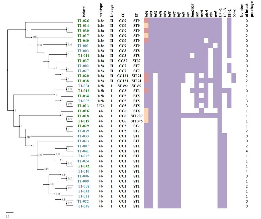
Figure 1. Phylogenetic
Phylogenetic tree
tree of
of 38
38 L.
L. monocytogenes
monocytogenes isolates
isolates from
from Chile
Chile inferred
inferred from
from single
single nucleotide
nucleotide
polymorphisms (SNPs) of whole genome sequencing data and the distribution
polymorphisms (SNPs) of whole genome sequencing data and the distribution of their genetic elementsof their genetic
elements associated with virulence and stress survival. Isolates from clinical samples
associated with virulence and stress survival. Isolates from clinical samples are colored in blue and are colored in
blue and
isolates isolates
from from non-clinical
non-clinical samples are samples
coloredare coloredColumns
in green. in green.onColumns
the rightonof the rightindicate
the tree of the tree
the
indicate the
presence presence
(purple) (purple)
or absence or absence
(white) (white)
of genetic of genetic
elements. elements.
Isolates Isolates stop
with premature withcodon
premature stop
mutations
codon mutations
(PMSCs) (PMSCs)
in inlA are in pink in
andinlA are inwith
isolates pink3 and isolates with
aa deletions 3 aa deletions
are yellow. are yellow.
The number of intactThe number
prophages
of intact prophages predicted
predicted with PHAST is added. with PHAST is added.
The MLST analysis differentiated the 38 Chilean isolates into 13 sequence types (STs). A total of
The MLST analysis differentiated the 38 Chilean isolates into 13 sequence types (STs). A total of
12 STs had been previously reported and were present in the Institut Pasteur whole genome MLST
12 STs had been previously reported and were present in the Institut Pasteur whole genome MLST
database and one novel ST was assigned (ST1395) (Table 1). Additionally, the 13 STs were grouped
database and one novel ST was assigned (ST1395) (Table 1). Additionally, the 13 STs were grouped in
in 11 CCs and 1 ST represented a singleton (Figure 1 and Table 1). In Lineage I, isolates were grouped
11 CCs and 1 ST represented a singleton (Figure 1 and Table 1). In Lineage I, isolates were grouped in
in CC1 (n = 15), CC2 (n = 2), CC3 (n = 1), CC5 (n = 3), CC6 (n = 3), and one singleton (ST392). In Lineage
CC1 (n = 15), CC2 (n = 2), CC3 (n = 1), CC5 (n = 3), CC6 (n = 3), and one singleton (ST392). In Lineage II,
II, isolates were grouped in CC7 (n = 2), CC8 (n = 2), CC9 (n = 6), CC37 (n = 1), and CC121 (n = 2). The
isolates were grouped in CC7 (n = 2), CC8 (n = 2), CC9 (n = 6), CC37 (n = 1), and CC121 (n = 2). The most
most common CCs in Lineage I and Lineage II were CC1 (60%) and CC9 (46%), respectively. Most of
common CCs in Lineage I and Lineage II were CC1 (60%) and CC9 (46%), respectively. Most of the
the CCs identified among Chilean isolates represent the most common CCs worldwide with CC1 and
CCs identified among Chilean isolates represent the most common CCs worldwide with CC1 and
CC9 as the most commonly and widely reported CCs in Europe and South/Central America [5,6].
CC9 as the most commonly and widely reported CCs in Europe and South/Central America [5,6].
Importantly, CC1 has also been associated with hyper virulence [16]. In addition, CCs were not
Importantly, CC1 has also been associated with hyper virulence [16]. In addition, CCs were not equally
equally distributed among isolates from different origins. CC1 was more common on isolates from
distributed among isolates from different origins. CC1 was more common on isolates from clinical
clinical cases and CC9 in non-clinical isolates. This result is in agreement with a recent retrospective
cases and CC9 in non-clinical isolates. This result is in agreement with a recent retrospective study that
study that analyzed over 6000 strains of L. monocytogenes in France [16].
analyzed over 6000 strains of L. monocytogenes in France [16].
3.2. Isolates of the Pulsed Field Gel Electrophoresis Type Causing the 2008–2009 Outbreaks Showed Genetic
Relatedness to Other Worldwide Clinical Isolates
The phylogenetic analysis of the core genome alignment identified a group within Lineage I of
clinical isolates (T1-006, T1-007, T1-008, T1-009, T1-010, T1-022, T1-025, T1-031, T1-033, T1-041, and T1-
043) and one non-clinical isolate (T1-042) that were clustered together (Figures S2 and S3, Table S3). AllGenes 2018, 9, 396 7 of 12
3.2. Isolates of the Pulsed Field Gel Electrophoresis Type Causing the 2008–2009 Outbreaks Showed Genetic
Relatedness to Other Worldwide Clinical Isolates
The phylogenetic analysis of the core genome alignment identified a group within Lineage I
of clinical isolates (T1-006, T1-007, T1-008, T1-009, T1-010, T1-022, T1-025, T1-031, T1-033, T1-041,
and T1-043) and one non-clinical isolate (T1-042) that were clustered together (Figures S2 and S3,
Table S3). All these isolates belong to the same CC1 and were obtained in different years (2008–2011)
and displayed different PFGE types. To gain insights into relatedness between these isolates,
an analysis of the whole genome to determine SNP differences among them was conducted using the
L. monocytogenes strain F2365 as the most closely related reference. SNP differences of these isolates
ranged from 17 SNPs to 198 SNPs (Table S3). Within this subgroup, T1-023, T1-024, and T1-028
presented the same PFGE type as the isolates that caused the 2008 outbreak (type 9). However,
these three isolates were found to differ between 66 SNPs to 122 SNPs (Table S3) from each other.
A previous study using WGS showed a lower diversity among epidemiologically linked isolates
(same PFGE type) with SNP differences less than 10 SNPs [49]. A cluster of three non-clinical isolates
(T1-016, T1-018, and T1-019), which clustered in CC6 and showed SNP differences that ranged from
three SNPs to 12 SNPs was found, which suggests these isolates are highly related even though these
isolates were not epidemiologically linked in this study. Prophage analysis on these isolates showed
one intact prophage identified in T1-016, T1-018, and T1-019. These two isolates (T1-018 and T1-019)
were different by only three SNPs.
3.3. Clinical and Non-Clinical L. monocytogenes Isolates Showed Distinct Virulence and Stress Survival
Genetic Elements
In this study, the distribution of selected virulence genes and genetic elements related with
stress survival were surveyed. Genes encoded in LIPI-1 (prfA, plcA, plcB, hly, and mpl) and the inlAB
operon, which encodes internalin A and B, were present in all 38 isolates (Figure 1). A previous study
conducted in Chile of L. monocytogenes isolated from foods (e.g., raw meat, cheese, and frozen seafood)
reported a different distribution of these genomic elements. In the previous study, the LIPI-1 cluster
and the inlAB operon were found to be associated with a given serotype and food group [24]. However,
methodologies and isolates between our study and this previous are different, which may explain
the difference in the results. Reports in France and China found these genes in all isolates and both
studies used WGS [16,50]. Other internalin family members (inlC, inlJ, inlH, inlD, inlE, and inlJ) were
detected in all isolates (Figure 1). However, inlG, inlF, and inlK were found not evenly distributed
among isolates. The genes inlG and inlK were found in 14 isolates and inlF in 12 isolates. Most of
these isolates were obtained from non-clinical sources, which are commonly represented by Lineage
II. The presence of inlG seems to be associated with Lineage II and our result agree with previous
studies using PCR that associated the presence of these internalins exclusively with Lineage II [51,52].
Additionally, an analysis looking at 81 distinct genes in the database of VirulenceFinder for Listeria
identified seven genes distinct to internalins (i.e., lmo2026, aut, actA, gtcA, vip, and ami) that showed
diversity in their content (Figure 1). Other virulence markers possibly associated with lineage is LIPI-3,
which was exclusively found in 15 isolates of the Lineage I of serotype 4b and one from serotype 1/2b
(Figure 1). LIPI-3 has been previously associated with Lineage I, serotype 4b [53,54], and with Lineage
III and Listeria innocua [55,56]. Conversely, the stress survival associated gene cluster known as SSI-1
was found in both lineages. SSI-1 was found in 37% of isolates and most of these isolates belonged to
CC9, CC8, and CC7 of Lineage II and to CC5 and CC3 (both Lineage I). However, SSI-2 was found only
in two isolates from CC121 (Lineage II). This is consistent with previous reports that indicate SSI-2
to be only associated with CC121 isolates [18]. The analysis of the plasmids identified that none of
the isolates contained a known plasmid. In addition, the only antimicrobial resistance gene identified
was the gene fosX, which confers resistance to fosfomycin identified in all 38 isolates. The number of
prophages detected with PHAST was very diverse and ranged from 0 to 4 intact prophages detected
(Figure 1).Genes 2018, 9, 396 8 of 12
3.4. Presence of One Novel PMSC Mutation in the inlA Gene Along with Additional PMSC Reported in Other
Countries in Isolates from Non-Clinical Samples
The WGS analysis showed that most of the isolates of this study (68%) contained a complete
InlA. All clinical isolates have a full-length inlA gene while 11 isolates from non-clinical samples
carried a PMSC mutation (Figure 1). Five isolates harbored a previously reported 9 nucleotide deletion,
which was predicted to encode a shorter (797 amino acids) version of InlA. This variant of inlA is
predicted to be fully functional. In vitro invasion assays have shown that these shorter variants have an
invasion ability comparable with that of full-length inlA isolates [57,58] and also have been reported in
isolates from clinical cases [14]. This type of deletion was found in isolates of serotypes 1/2c and 4b and
have been found in isolates of serotypes 1/2b in USA [59] as well as serotypes 4c and 1/2a in Canada
and Switzerland [60]. Additionally, six PMSCs were detected exclusively in isolates from non-clinical
origin, which belongs to serotypes 1/2c (3), 1/2a (2), and 1/2b (1) (Table 2). These mutations were
classified into four PMSC types that were previously described, which include one type 19 (resulting
in 325 aa protein product), one type 13 (resulting in a 527 aa protein product), two type 6 (resulting in
491 aa protein product), and one type 11 (resulting in a 684 aa protein product) [61–64]. In addition,
the presence of a novel PMSC type was found in one isolate (T1-012), which carried a non-sense
mutation at position 821 where one adenine base was deleted. This resulted in a frameshift mutation,
which codes for a 277-protein product (Table 2). This truncated protein might result in low virulence in
in vitro invasion assays due to the lack of the LPXTG motif, which is involved in anchoring the protein
to peptidoglycan in the cell wall [65]. Further studies are essential to confirm this.
Table 2. Length of InlA among Chilean L. monocytogenes.
Number InlA Mutation Type Nucleotide
Functional Reference
of Isolates Length (aa) (PMSC) Mutation Position
27 800 - - yes Glaser et al., 2001
5 797 NA NA yes Chen et al., 2011
1 684 11 2054 (G to A) no Rousseaux et al., 2002
1 527 13 1579 (Ato T) no Handa-Miya et al., 2007
2 491 6 1474 (Cto T) no Olier et al., 2002
1 325 19 976 (G to T) no Gelbicova et al., 2015
1 277 Novel 821 (deletion A) no In this study
4. Conclusions
Whole genome sequencing has proven to be a powerful subtyping tool for L. monocytogenes
especially in reference centers in North America and Europe. This study provides the first
characterization at a genomic level using WGS of clinical and non-clinical isolates of L. monocytogenes
isolated from Chile. Our results show the presence of isolates from Chile that represent clonal groups
associated with listeriosis worldwide, which supports the global distribution of key human diseases
associated with L. monocytogenes clonal groups. This study is the first of this type in South America,
so further efforts are necessary in order to implement WGS in routine surveillance in South America.
Supplementary Materials: The following are available online at http://www.mdpi.com/2073-4425/9/8/396/s1.
Figure S1. Location and distribution of isolates used in this study. (a) Map of Chile divided by regions and
red dots represent the locations where the isolates were obtained. (b) Circular representation of the percent of
isolates sequenced that were obtained from the different regions within Chile. Figure S2. Maximum likelihood
phylogenetic tree based on SNPs of core genome of Chilean L. monocytogenes using 55 L. monocytogenes sequences
as references. Phylogenetic tree representing the two lineages identified in this study, ID of the isolates from
clinical samples are colored in blue and ID of the isolates from non-clinical samples are colored in green. Figure S3.
L. monocytogenes phylogeny based on single nucleotide polymorphism (SNP). Phylogenetic tree of the 15 isolates
that clustered together that were obtained from clinical samples, mostly of the CC1. Isolates with their ID in blue
were obtained from human clinical samples and the one in green from non-clinical samples. The bootstrap was
added to the clades. Table S1. Sequencing statistics of 38 isolates of L. monocytogenes obtained from clinical and
non-clinical samples sequenced in this study. Table S2. L. monocytogenes used as references in the core genome
alignment for lineage determination. Table S3. Pairwise distance matrix of SNP number differences for 15 Chilean
isolates of L. monocytogenes clustered in CC1.Genes 2018, 9, 396 9 of 12
Author Contributions: V.T. and A.I.M.-S. conceived this project and designed the analysis. V.T. and H.C.d.B.
performed the bioinformatics analysis. J.C.H., G.G.-R., H.B.-T. contributed with material. V.T., G.G.-R., H.B.-T.,
M.T., and A.I.M.-S. contributed to the writing of this manuscript.
Funding: The study was funded by the Universidad Andres Bello Grant no. DI-638-15/I to V.T.
Acknowledgments: We thank Marc Allard from FDA for kindly sequencing the isolates analyzed in this study.
Conflicts of Interest: The authors declared no conflict of interest.
References
1. Swaminathan, B.; Gerner-Smidt, P. The epidemiology of human listeriosis. Microbes Infect. 2007, 9, 1236–1243.
[CrossRef] [PubMed]
2. Mead, P.S.; Slutsker, L.; Dietz, V.; McCaig, L.F.; Bresee, J.S.; Shapiro, C.; Griffin, P.M.; Tauxe, R.V. Food-related
illness and death in the United States. Emerg. Infect. Dis. 1999, 5, 607–625. [CrossRef] [PubMed]
3. Farber, J.M.; Peterkin, P.I. Listeria monocytogenes, a food-borne pathogen. Microbiol. Rev. 1991, 55, 476–511.
[CrossRef] [PubMed]
4. McLauchlin, J.; Mitchell, R.T.; Smerdon, W.J.; Jewell, K. Listeria monocytogenes and listeriosis: A review of
hazard characterisation for use in microbiological risk assessment of foods. Int. J. Food Microbiol. 2004, 92,
15–33. [CrossRef]
5. Ragon, M.; Wirth, T.; Hollandt, F.; Lavenir, R.; Lecuit, M.; Le Monnier, A.; Brisse, S. A new perspective on
Listeria monocytogenes evolution. PLoS Pathog. 2008, 4. [CrossRef] [PubMed]
6. Chenal-Francisque, V.; Lopez, J.; Cantinelli, T.; Caro, V.; Tran, C.; Leclercq, A.; Lecuit, M.; Brisse, S. Worldwide
distribution of major clones of Listeria monocytogenes. Emerg. Infect. Dis. 2011, 17, 1110–1112. [CrossRef]
[PubMed]
7. Cantinelli, T.; Chenal-Francisque, V.; Diancourt, L.; Frezal, L.; Leclercq, A.; Wirth, T.; Lecuit, M.; Brisse, S.
Epidemic clones of Listeria monocytogenes are widespread and ancient clonal groups. J. Clin. Microbiol. 2013,
51, 3770–3779. [CrossRef] [PubMed]
8. Vázquez-Boland, J.A.; Kuhn, M.; Berche, P.; Chakraborty, T.; Domi, G.; González-Zorn, B.; Wehland, J.
Listeria pathogenesis and molecular virulence determinants Listeria pathogenesis and molecular virulence
determinants. Clin. Microbiol. Rev. 2001, 14, 584–640. [CrossRef] [PubMed]
9. Vazquez-Boland, J.A.; Dominguez-Bernal, G.; Gonzalez-Zorn, B.; Kreft, J.; Goebel, W. Pathogenicity islands
and virulence evolution in Listeria. Microbes Infect. 2001, 3, 571–584. [CrossRef]
10. Gaillard, J.L.; Jaubert, F.; Berche, P. The inlAB locus mediates the entry of Listeria monocytogenes into
hepatocytes in vivo. J. Exp. Med. 1996, 183, 359–369. [CrossRef] [PubMed]
11. Raffelsbauer, D.; Bubert, A.; Engelbrecht, F.; Scheinpflug, J.; Simm, A.; Hess, J.; Kaufmann, S.H.E.; Goebel, W.
The gene cluster inlC2DE of Listeria monocytogenes contains additional new internalin genes and is important
for virulence in mice. Mol. Genet. Genom. 1998, 260, 144–158. [CrossRef]
12. Sabet, C.; Lecuit, M.; Cabanes, D.; Cossart, P. LPXTG protein InlJ, a newly identified internalin involved in
Listeria monocytogenes virulence. Infect. Immun. 2005, 73, 6912–6922. [CrossRef] [PubMed]
13. Neves, D.; Job, V.; Dortet, L.; Cossart, P.; Dessen, A. Structure of internalin InlK from the human pathogen
Listeria monocytogenes. J. Mol. Biol. 2013, 425, 4520–4529. [CrossRef] [PubMed]
14. Chen, Y.; Strain, E.A.; Allard, M.; Brown, E.W. Genome sequence of L. monocytogenes Strains J1816 and J1-220,
associated with human outbreaks. J. Bacteriol. 2011, 193, 3424–3425. [CrossRef] [PubMed]
15. Roche, S.M.; Grépinet, O.; Kerouanton, A.; Ragon, M.; Leclercq, A.; Témoin, S.; Schaeffer, B.; Skorski, G.;
Mereghetti, L.; Le Monnier, A.; et al. Polyphasic characterization and genetic relatedness of low-virulence
and virulent Listeria monocytogenes isolates. BMC Microbiol. 2012, 12. [CrossRef] [PubMed]
16. Maury, M.; Tsai, Y.H.; Charlier, C.; Touchon, M.; Chenal-Francisque, V.; Leclercq, A.; Criscuolo, A.;
Gaultier, C.; Roussel, S.; Brisabois, A.; et al. Uncovering Listeria monocytogenes hypervirulence by harnessing
its biodiversity. Nat. Genet. 2016, 48, 308–313. [CrossRef] [PubMed]
17. Nightingale, K.K.; Windham, K.; Martin, K.E.; Yeung, M.; Wiedmann, M. Select Listeria monocytogenes
subtypes commonly found in foods carry disctinct nonsense mutations in inlA. Appl. Environ. Microbiol.
2005, 71, 8764–8772. [CrossRef] [PubMed]Genes 2018, 9, 396 10 of 12
18. Harter, E.; Wagner, E.M.; Zaiser, A.; Halecker, S.; Wagner, M.; Rychli, K. The novel stress survival islet 2
(SSI-2), predominantly present in Listeria monocytogenes strains of ST121, is involved in alkaline and oxidative
stress response. Appl. Environ. Microbiol. 2017. [CrossRef] [PubMed]
19. Ryan, S.; Begley, M.; Hill, C.; Gahan, C.G.M. A five-gene stress survival islet (SSI-1) that contributes to the
growth of Listeria monocytogenes in suboptimal conditions. J. Appl. Microbiol. 2010, 109, 984–995. [CrossRef]
[PubMed]
20. MINSAL, Departamento de Epidemiología. Informe Listeriosis Actualizado el 15 de Septiembre 2011.
Available online: http://www.ispch.cl/sites/default/files/documento/2011/09/listeria2011.pdf (accessed
on 31 July 2018).
21. MINSAL, Departamento de Epidemiología; Informe año 2017. Situación Epidemiológica de Listeriosis en
Chile. 2017, pp. 1–11. Available online: http://epi.minsal.cl/wp-content/uploads/2018/04/INFORME-
ANUAL-LISTERIOSIS-2017_2018-03-09-RevIRO-SAF.pdf (accessed on 31 July 2018).
22. Saludes, M.; Troncoso, M.; Figueroa, G. Presence of Listeria monocytogenes in Chilean food matrices.
Food Control 2015, 50, 331–335. [CrossRef]
23. Cordano, A.M.; Jacquet, C. Listeria monocytogenes isolated from vegetable salads sold at supermarkets in
Santiago, Chile: Prevalence and strain characterization. Int. J. Food Microbiol. 2009, 132, 176–179. [CrossRef]
[PubMed]
24. Montero, D.; Bodero, M.; Riveros, G.; Lapierre, L.; Gaggero, A.; Vidal, R.M.; Vidal, M. Molecular epidemiology
and genetic diversity of Listeria monocytogenes isolates from a wide variety of ready-to-eat foods and their
relationship to clinical strains from listeriosis outbreaks in Chile. Front. Microbiol. 2015, 6, 1–8. [CrossRef]
[PubMed]
25. Bolger, A.M.; Lohse, M.; Usadel, B. Trimmomatic: A flexible trimmer for Illumina sequence data.
Bioinformatics 2014, 30, 2114–2120. [CrossRef] [PubMed]
26. Andrews, S. FastQC: A Quality Control Tool for High Throughput Sequence Data. Available online:
http://www.bioinformatics.babraham.ac.uk/projects/fastqc/ (accessed on 31 July 2018).
27. Bankevich, A.; Nurk, S.; Antipov, D.; Gurevich, A.A.; Dvorkin, M.; Kulikov, A.S.; Lesin, V.M.; Nikolenko, S.I.;
Pham, S.; Prjibelski, A.D.; et al. SPAdes: A new genome assembly algorithm and its applications to single-cell
sequencing. J. Comput. Biol. 2012, 19, 455–477. [CrossRef] [PubMed]
28. Aziz, R.K.; Bartels, D.; Best, A.A.; DeJongh, M.; Disz, T.; Edwards, R.A.; Formsma, K.; Gerdes, S.; Glass, E.M.;
Kubal, M.; et al. The RAST server: Rapid annotations using subsystems technology. BMC Genom. 2008, 9, 75.
[CrossRef] [PubMed]
29. Tatusova, T.; Dicuccio, M.; Badretdin, A.; Chetvernin, V.; Nawrocki, E.P.; Zaslavsky, L.; Lomsadze, A.;
Pruitt, K.D.; Borodovsky, M.; Ostell, J. NCBI prokaryotic genome annotation pipeline. Nucleic Acids Res.
2016, 44, 6614–6624. [CrossRef] [PubMed]
30. Treangen, T.J.; Ondov, B.D.; Koren, S.; Phillippy, A.M. The Harvest suite for rapid core-genome alignment
and visualization of thousands of intraspecific microbial genomes. Genome Biol. 2014, 15. [CrossRef]
31. Kaas, R.S.; Leekitcharoenphon, P.; Aarestrup, F.M.; Lund, O. Solving the problem of comparing whole
bacterial genomes across different sequencing platforms. PLoS ONE 2014, 9, 1–8. [CrossRef] [PubMed]
32. Doumith, M.; Buchrieser, C.; Glaser, P.; Jacquet, C.; Martin, P. Differentiation of the major Listeria
monocytogenes serovars by multiplex PCR. J. Clin. Microbiol. 2004, 42, 3819–3822. [CrossRef] [PubMed]
33. Larsen, M.V.; Cosentino, S.; Rasmussen, S.; Friis, C.; Hasman, H.; Marvig, R.L.; Jelsbak, L.; Sicheritz-Pontén, T.;
Ussery, D.W.; Aarestrup, F.M.; et al. Multilocus sequence typing of total-genome-sequenced bacteria.
J. Clin. Microbiol. 2012, 50, 1355–1361. [CrossRef] [PubMed]
34. Arndt, D.; Grant, J.R.; Marcu, A.; Sajed, T.; Pon, A.; Liang, Y.; Wishart, D.S. PHASTER: A better, faster version
of the PHAST phage search tool. Nucleic Acids Res. 2016, 44, W16–W21. [CrossRef] [PubMed]
35. Carattoli, A.; Zankari, E.; García-Fernández, A.; Larsen, M.V.; Lund, O.; Villa, L.; Aarestrup, F.M.; Hasman, H.
In silico detection and typing of plasmids using plasmidfinder and plasmid multilocus sequence typing.
Antimicrob. Agents Chemother. 2014, 58, 3895–3903. [CrossRef] [PubMed]
36. Zankari, E.; Hasman, H.; Cosentino, S.; Vestergaard, M.; Rasmussen, S.; Lund, O.; Aarestrup, F.M.;
Larsen, M.V. Identification of acquired antimicrobial resistance genes. J. Antimicrob. Chemother. 2012,
67, 2640–2644. [CrossRef] [PubMed]
37. Madden, T. The BLAST Sequence Analysis Tool. In The NCBI Handbook National Library of Medicine (US);
McEntyre, J., Ed.; National Center for Biotechnology Information: Bethesda, MD, USA, 2013.Genes 2018, 9, 396 11 of 12
38. Sievers, F.; Wilm, A.; Dineen, D.; Gibson, T.J.; Karplus, K.; Li, W.; Lopez, R.; McWilliam, H.; Remmert, M.;
Söding, J.; et al. Fast, scalable generation of high-quality protein multiple sequence alignments using Clustal
Omega. Mol. Syst. Biol. 2011, 7. [CrossRef] [PubMed]
39. Joensen, K.G.; Scheutz, F.; Lund, O.; Hasman, H.; Kaas, R.S.; Nielsen, E.M.; Aarestrup, F.M. Real-time
whole-genome sequencing for routine typing, surveillance, and outbreak detection of verotoxigenic
Escherichia coli. J. Clin. Microbiol. 2014, 52, 1501–1510. [CrossRef] [PubMed]
40. Cartwright, E.J.; Jackson, K.A.; Johnson, S.D.; Graves, L.M.; Silk, B.J.; Mahon, B.E. Listeriosis outbreaks and
associated food vehicles, United States, 1998–2008. Emerg. Infect. Dis. 2013, 19, 1–9. [CrossRef] [PubMed]
41. Orsi, R.H.; den Bakker, H.C.; Wiedmann, M. Listeria monocytogenes lineages: Genomics, evolution, ecology,
and phenotypic characteristics. Int. J. Med. Microbiol. 2011, 301, 79–96. [CrossRef] [PubMed]
42. Graves, L.M.; Swaminathan, B. PulseNet standardized protocol for subtyping Listeria monocytogenes by
macrorestriction and pulsed-field gel electrophoresis. Int. J. Food Microbiol. 2001, 65, 55–62. [CrossRef]
43. Chen, J.; Chen, Q.; Jiang, J.; Hu, H.; Ye, J.; Fang, W. Serovar 4b complex predominates among Listeria
monocytogenes isolates from imported aquatic products in China. Foodborne Pathog. Dis. 2010, 7, 31–41.
[CrossRef] [PubMed]
44. Braga, V.; Vázquez, S.; Vico, V.; Pastorino, V.; Mota, M.I.; Legnani, M.; Schelotto, F.; Lancibidad, G.; Varela, G.
Prevalence and serotype distribution of Listeria monocytogenes isolated from foods in Montevideo-Uruguay.
Braz. J. Microbiol. 2017, 48, 689–694. [CrossRef] [PubMed]
45. Vallim, D.C.; Barroso Hofer, C.; Lisbôa, R.D.C.; Victor Barbosa, A.; Alves Rusak, L.; Dos Reis, C.M.F.; Hofer, E.
Twenty years of Listeria in Brazil: Occurrence of Listeria species and Listeria monocytogenes serovars in food
samples in Brazil between 1990 and 2012. Biomed. Res. Int. 2015. [CrossRef] [PubMed]
46. Leclercq, A.; Chenal-Francisque, V.; Dieye, H.; Cantinelli, T.; Drali, R.; Brisse, S.; Lecuit, M. Characterization
of the novel Listeria monocytogenes PCR serogrouping profile IVb-v1. Int. J. Food Microbiol. 2011, 147, 74–77.
[CrossRef] [PubMed]
47. Burall, L.S.; Grim, C.J.; Datta, A.R. A clade of Listeria monocytogenes serotype 4b variant strains linked to
recent listeriosis outbreaks associated with produce from a defined geographic region in the US. PLoS ONE
2017, 12. [CrossRef] [PubMed]
48. Burall, L.S.; Grim, C.J.; Mammel, M.K.; Datta, A.R. A Comprehensive evaluation of the genetic relatedness
of Listeria monocytogenes serotype 4b variant strains. Front. Public Health 2017, 5, 1–10. [CrossRef] [PubMed]
49. Kwong, J.C.; Mercoulia, K.; Tomita, T.; Easton, M.; Li, H.Y.; Bulach, D.M.; Stinear, T.P.; Seemann, T.;
Howden, B.P. Prospective whole-genome sequencing enhances national surveillance of Listeria monocytogenes.
J. Clin. Microbiol. 2016, 54, 333–342. [CrossRef] [PubMed]
50. Zhang, J.; Cao, G.; Xu, X.; Allard, M.; Li, P.; Brown, E.; Yang, X.; Pan, H.; Meng, J. Evolution and diversity of
Listeria monocytogenes from clinical and food samples in Shanghai, China. Front. Microbiol. 2016, 7. [CrossRef]
[PubMed]
51. Jia, Y.; Nightingale, K.K.; Boor, K.J.; Ho, A.; Wiedmann, M.; McGann, P. Distribution of internalin gene profiles
of Listeria monocytogenes isolates from different sources associated with phylogenetic lineages. Foodborne
Pathog. Dis. 2007, 4, 222–232. [CrossRef] [PubMed]
52. Su, X.; Zhang, J.; Shi, W.; Yang, X.; Li, Y.; Pan, H.; Kuang, D.; Xu, X.; Shi, X.; Meng, J. Molecular
characterization and antimicrobial susceptibility of Listeria monocytogenes isolated from foods and humans.
Food Control 2016, 70, 96–102. [CrossRef]
53. Clayton, E.M.; Hill, C.; Cotter, P.D.; Ross, R.P. Real-time PCR assay to differentiate listeriolysin S-positive and
-negative strains of Listeria monocytogenes. Appl. Environ. Microbiol. 2011, 77, 163–171. [CrossRef] [PubMed]
54. Cotter, P.D.; Draper, L.A.; Lawton, E.M.; Daly, K.M.; Groeger, D.S.; Casey, P.G.; Ross, R.P.; Hill, C. Listeriolysin
S, a novel peptide haemolysin associated with a subset of lineage I Listeria monocytogenes. PLoS Pathog. 2008,
4. [CrossRef] [PubMed]
55. Shen, J.; Rump, L.; Zhang, Y.; Chen, Y.; Wang, X.; Meng, J. Molecular subtyping and virulence gene analysis
of Listeria monocytogenes isolates from food. Food Microbiol. 2013, 35, 58–64. [CrossRef] [PubMed]
56. Clayton, E.M.; Daly, K.M.; Guinane, C.M.; Hill, C.; Cotter, P.D.; Ross, P.R. Atypical Listeria innocua strains
possess an intact LIPI-3. BMC Microbiol. 2014, 14. [CrossRef] [PubMed]
57. Kanki, M.; Naruse, H.; Taguchi, M.; Kumeda, Y. Characterization of specific alleles in inlA and prfA of
Listeria monocytogenes isolated from foods in Osaka, Japan and their ability to invade Caco-2 cells. Int. J.
Food Microbiol. 2015, 211, 18–22. [CrossRef] [PubMed]Genes 2018, 9, 396 12 of 12
58. Kovacevic, J.; Arguedas-Villa, C.; Wozniak, A.; Tasara, T.; Allen, K.J. Examination of food chain-derived
Listeria monocytogenes strains of different serotypes reveals considerable diversity in inlA genotypes,
mutability, and adaptation to cold temperatures. Appl. Environ. Microbiol. 2013, 79, 1915–1922. [CrossRef]
[PubMed]
59. Gorski, L.; Parker, C.T.; Liang, A.S.; Walker, S.; Romanolo, K.F. The majority of genotypes of the virulence
gene inlA are intact among natural watershed isolates of Listeria monocytogenes from the central California
Coast. PLoS ONE 2016, 11, e0167566. [CrossRef] [PubMed]
60. Hingston, P.; Chen, J.; Dhillon, B.K.; Laing, C.; Bertelli, C.; Gannon, V.; Tasara, T.; Allen, K.; Brinkman, F.S.L.;
Hansen, L.T.; et al. Genotypes associated with Listeria monocytogenes isolates displaying impaired or enhanced
tolerances to cold, salt, acid, or desiccation stress. Front. Microbiol. 2017, 8, 369. [CrossRef] [PubMed]
61. Olier, M.; Pierre, F.; Lemaître, J.P.; Divies, C.; Rousset, A.; Guzzo, J. Assessment of the pathogenic potential
of two Listeria monocytogenes human faecal carriage isolates. Microbiology 2002, 148, 1855–1862. [CrossRef]
[PubMed]
62. Rousseaux, S.; Olier, M.; Piveteau, P.; Guzzo, J. Use of PCR-restriction fragment length polymorphism
of inlA for rapid screening of Listeria monocytogenes strains deficient in the ability to invade Caco-2 cells.
Appl. Environ. Microbiol. 2004, 70, 2180–2185. [CrossRef] [PubMed]
63. Handa-Miya, S.; Kimura, B.; Takahashi, H.; Sato, M.; Ishikawa, T.; Igarashi, K.; Fujii, T. Nonsense-mutated
inlA and prfA not widely distributed in Listeria monocytogenes isolates from ready-to-eat seafood products in
Japan. Int. J. Food Microbiol. 2007, 117, 312–318. [CrossRef] [PubMed]
64. Gelbíčová, T.; Koláčková, I.; Pantůček, R.; Karpíšková, R. A novel mutation leading to a premature stop
codon in inlA of Listeria monocytogenes isolated from neonatal listeriosis. New Microbiol. 2015, 38, 293–296.
[PubMed]
65. Dramsi, S.; Trieu-Cuot, P.; Bierne, H. Sorting sortases: A nomenclature proposal for the various sortases of
Gram-positive bacteria. Res. Microbiol. 2005, 156, 289–297. [CrossRef] [PubMed]
© 2018 by the authors. Licensee MDPI, Basel, Switzerland. This article is an open access
article distributed under the terms and conditions of the Creative Commons Attribution
(CC BY) license (http://creativecommons.org/licenses/by/4.0/).You can also read