A Short Review on the Role of the Metal-Graphene Hybrid Nanostructure in Promoting the Localized Surface Plasmon Resonance Sensor Performance - MDPI
←
→
Page content transcription
If your browser does not render page correctly, please read the page content below
sensors
Review
A Short Review on the Role of the Metal-Graphene
Hybrid Nanostructure in Promoting the Localized
Surface Plasmon Resonance Sensor Performance
Raed Alharbi 1,2, *, Mehrdad Irannejad 2,3 and Mustafa Yavuz 2
 1   Mechanical Engineering Department, Taibah University, Madina P.O. Box. 344, Saudi Arabia
 2   Mechanical and Mechatronics Engineering Department, University of Waterloo,
     Waterloo, ON N2L 3G1, Canada; pm07mi@gmail.com (M.I.); myavuz@uwaterloo.ca (M.Y.)
 3   OZ optics, Ltd., Ottawa, ON K0A 1L0, Canada
 *   Correspondence: r2alharb@uwaterloo.ca

 Received: 18 January 2019; Accepted: 15 February 2019; Published: 19 February 2019

 Abstract: Localized Surface Plasmon Resonance (LSPR) sensors have potential applications in
 essential and important areas such as bio-sensor technology, especially in medical applications
 and gas sensors in environmental monitoring applications. Figure of Merit (FOM) and Sensitivity (S)
 measurements are two ways to assess the performance of an LSPR sensor. However, LSPR sensors
 suffer low FOM compared to the conventional Surface Plasmon Resonance (SPR) sensor due to high
 losses resulting from radiative damping of LSPs waves. Different methodologies have been utilized
 to enhance the performance of LSPR sensors, including various geometrical and material parameters,
 plasmonic wave coupling from different structures, and integration of noble metals with graphene,
 which is the focus of this report. Recent studies of metal-graphene hybrid plasmonic systems have
 shown its capability of promoting the performance of the LSPR sensor to a level that enhances its
 chance for commercialization. In this review, fundamental physics, the operation principle, and
 performance assessment of the LSPR sensor are presented followed by a discussion of plasmonic
 materials and a summary of methods used to optimize the sensor’s performance. A focused review
 on metal-graphene hybrid nanostructure and a discussion of its role in promoting the performance of
 the LSPR sensor follow.

 Keywords: localized plasmons; sensor; graphene; metal; hybrid; sensitivity; figure of merit

1. Introduction
     The Surface Plasmonic Resonance (SPR) sensor is a common and commercialized sensor used in
different areas such healthcare and gas sensing [1,2]. When SP evanescent waves and incident light
couple together, Surface Plasmon Polaritons (SPP) result, which is the basic element in SPR sensor
principles. Any variation in permittivity of a sensing area shifts the resonance angle of the SPP and
the resonant intensity [3,4]. When the size of the plasmonic material decreases to the nano range, the
way in which free electrons couple with the incident wave differs. When metallic nanoparticles are
irradiated by an electromagnetic wave with a wavelength greater than the NP size, the metal’s free
electrons generate oscillations called Localized Surface Plasmons (LSPs). These collective oscillations
are restricted to the geometry of the nanostructure [5]. LSPs can be induced by direct illumination of
photons where there is no need for momentum matching between a photon and free electron [5]. The
only required condition to induce LSPs is conservation of the electron energy of the metal nanoparticle
(NP) with incident photon energy.
     In addition to the low cost of fabrication, the LSPR sensor offers a simple sensor setup and
detection of local change in the refractive index, which is not the case with SPR sensors [6,7]. However,

Sensors 2019, 19, 862; doi:10.3390/s19040862                                   www.mdpi.com/journal/sensors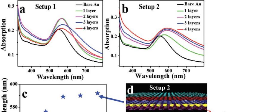
Sensors 2019, 19, 862                                                                                   2 of 15

damping in the radiation process in LSPR modes increases the resonance peak width, and accordingly
reduces sensor performance [8–10]. Different approaches have been reported to enhance LSPR sensor
performance, including variation of the geometry and material type of the nanostructure, surface
plasmon hybridization, and noble metal-2D material integrated structures such as metal-graphene
hybrid nanostructures. However, recently, metal-2D material integrated structures showed good
improvement in LSPR sensor performance, the focus point of this review paper. This review starts
by summarizing the fundamentals of the localized surface plasmon resonance sensor, followed by a
discussion of the material used in the plasmonic sensor. A summary of the different approaches used
to enhance the performance of the LSPR sensor follows, with additional focus on a metal-graphene
material integration approach.

2. Localized Surface Plasmon Resonance Sensor
    The Mie scattering theory, with the help of Maxwell’s equations, is a very useful way to explain
how metal NPs interact with incident light. Equation (1) shows the derived optical extinction for
metallic NP with a radius smaller than the wavelength of the incident wave (2r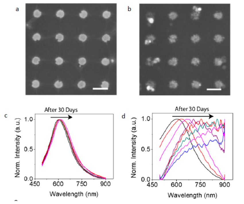
Sensors 2019, 19, 862 3 of 15
Sensors 2019, 16, x FOR PEER REVIEW 3 of 4
MetalNP
Figure1.1.Metal
Figure NPexternal
externalelectric
electricfield
fieldinteraction
interaction[17].
[17].
Mieresonance
Mie resonanceisisaagood goodexample
exampleofofthe thenonlinear
nonlinearopticalopticaleffecteffectwherewherethe theoptical
opticalresponse
responseof of
the medium is nonlinearly depending on the electric field applied
the medium is nonlinearly depending on the electric field applied [16]. In addition to the applied [16]. In addition to the applied
electric field,
electric field, there
there are area anumber
number of of
parameters
parameters thatthat
affect the plasmonic
affect the plasmonic resonance properties.
resonance When
properties.
the surrounding’s dielectric constant increases, screening of the surface
When the surrounding’s dielectric constant increases, screening of the surface electrons increases. electrons increases. This results
in the
This increase
results in theof the restoring
increase of theforce and, hence,
restoring force requires
and, hence, lower energylower
requires to excite
energyfreetocharges.
excite free The
resonance peak consequently shifts to the red region [14], as
charges. The resonance peak consequently shifts to the red region [14], as can be seen from can be seen from Equation (3). From
this equation,
Equation (3). it is also
From evident
this that the
equation, it increase
is also in the surrounding’s
evident that the increase permittivityin the or refractive
surrounding‘s index
reduces the resonance
permittivity or refractive frequency and hence
index reduces theresults in a longer
resonance frequency resonance and wavelength
hence results shift.
in Therefore,
a longer
resonance wavelength shift. Therefore, metallic NPs can be used as an LSPR sensor where bothforce
metallic NPs can be used as an LSPR sensor where both the polarization of the NP and restoring the
strength affect
polarization of thetheNP sensitivity of the force
and restoring sensor; accordingly,
strength sensor
affect the performance
sensitivity of the depends on the type
sensor; accordingly,
of materials
sensor and thedepends
performance geometrical properties
on the type of of the metal
materials andNPs. theAccording
geometrical to properties
the equation of mentioned
the metal
above, as the NP size increases, the resonance wavelength
NPs. According to the equation mentioned above, as the NP size increases, the resonance shifts to a longer wavelength, which is a
kind of tuning
wavelength of the
shifts toplasmonic
a longerresonancewavelength, [18]. In addition,
which is aincreasing
kind of the size ofofparticles
tuning increases
the plasmonic
resonance [18]. In addition, increasing the size of particles increases the radiative losses,intensity
the radiative losses, which increases the width of the resonance band, hence resulting in which
reductionthe
increases [19]. Furthermore,
width NP geometry
of the resonance band, has hencea crucial
resulting roleininintensity
tuning the resonance
reduction wavelength.
[19]. Furthermore, The
NP geometry
NP shape applies has an important
a crucial roleimpact
in tuningeffectthe on resonance
the plasmon resonance peak
wavelength. The NP position.
shapeInapplies
the casean of
important impact effect on the plasmon resonance peak position. In the case of a high symmetryis
a high symmetry spherical NP, only one dipolar resonance is induced. However, when the shape
altered, the
spherical NP,particle
only one becomes
dipolar increasingly
resonance asymmetric,
is induced. and a higher
However, whenorder theof shape
electricisdipole
altered, modesthe
is induced, which increases the complexity of optical response
particle becomes increasingly asymmetric, and a higher order of electric dipole modes is induced, of the NP [20]. For example, upon
exciting
which a nanorod,
increases the two distinct resonance
complexity of optical peaks response are observed,
of the NPone [20].from
Forthe transverse
example, upon and one from
exciting a
the longitudinal mode at different spectral positions [14]. Furthermore,
nanorod, two distinct resonance peaks are observed, one from the transverse and one from the resonance with higher order
modes, for example,
longitudinal mode at quadrupoles
different spectral and octopoles,
positions may [14]. be induced in the
Furthermore, case of an
resonance with inhomogeneous
higher order
distribution of the surface charges of the NPs [21–23]. Therefore,
modes, for example, quadrupoles and octopoles, may be induced in the case of an inhomogeneous engineering the shape of the NP is an
effective way
distribution oftothegenerate
surfaceLSP resonance
charges of themodes within Therefore,
NPs [21-23]. a visible and near-infrared
engineering the (NIR)
shaperegion
of the [24,25].
NP is
Nanostructure supported LSPR extinction can
an effective way to generate LSP resonance modes within a visible and near-infrared be used in refractometric sensing, like a
conventional
(NIR) SPR sensor. The detection principle in LSPR sensors is based on an intensity or
region [24,25].
wavelength shift
Nanostructure upon variationLSPR
supported in theextinction
permittivity can of abesensing
used region (proximity region
in refractometric sensing, of the
likeNP).a
In the transmission or reflection spectrum, the resonance peak
conventional SPR sensor. The detection principle in LSPR sensors is based on an intensity or position varies based on the refractive
index of theshift
wavelength sensing
upon area, as is confirmed
variation by Equation
in the permittivity of (3). Any change
a sensing regionof permittivity
(proximity in the
region of proximity
the NP).
of the NP will shift the resonance LSP peak, which can be employed
In the transmission or reflection spectrum, the resonance peak position varies based on the refractive to detect molecular interaction
between
index the sensing
of the NP and area, adsorbedas ismaterial.
confirmed Thebyamount
Equation of adsorbed
(3). Any materialchange ofcan be found through
permittivity in the
monitoring the plasmonic resonance shift. The performance of
proximity of the NP will shift the resonance LSP peak, which can be employed to detect molecular the sensor can be assessed through
Sensitivity between
interaction (S) and Figure
the NP of and
Meritadsorbed
(FOM) measurements.
material. The amount Sensitivity of (S) is defined
adsorbed as the can
material extent be of peak
found
shift in position
through monitoring or intensity to the change
the plasmonic resonance of the refractive
shift. index of a dielectric
The performance of the sensor medium.can be Therefore,
assesseda
through Sensitivity (S) and Figure of Merit (FOM) measurements. Sensitivity (S) is defined[26].
larger resonance shift at a small refractive index change results in increased bulk sensitivity as theIt is
known that increased sensitivity due to larger surface plasmon
extent of peak shift in position or intensity to the change of the refractive index of a dielectricresonance wavelength shifts results
in a wider
medium. resonancea peak,
Therefore, largerdue to dephasing
resonance shift atand radiative
a small damping,
refractive indexwhich change affect the in
results resolution
increased of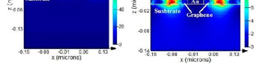
Sensors 2019, 19, 862                                                                                 4 of 15

detection [8]. Therefore, sensitivity measurement is not adequate to assess the performance of the
sensor. Thus, another parameter required to assess the LSPR sensors is called the Figure of Merit
(FOM). FOM is the sensitivity divided by bandwidth (Full Width at Half Maximum (FWHM)) in the
case of a wavelength shift, or by reference intensity in the case of an intensity shift. Equations (4)–(7)
are the sensitivity and FOM equations for both the wavelength shift and intensity shift [8].

                                                   ∆λ
                                          Sλ =        (nm/RIU )                                          (4)
                                                   ∆n
                                               ∆I
                                            SI =   ( RIU −1 )                                            (5)
                                               ∆n
                                                       Sλ
                                            FOMλ =                                                       (6)
                                                    FWHM
                                                       S
                                             FOM I = I                                                   (7)
                                                      Ire f .
where ∆λ is the change that occurs in the resonance wavelength (nm), ∆I is the change in intensity, ∆n
is the change in the sensing medium’s refractive index, and Sλ , FOMλ , S I , FOM I are the sensitivity
and figure of merit based on the wavelength and intensity shift, respectively. Therefore, a higher
resonance shift is preferred at a small refractive index change to achieve higher bulk sensitivity and
FOM. Thus, both a large ∆λ and narrower resonance peak is desired to promote the quality of an
LSPR-based sensor.

3. Plasmonic Material
      Metal is a candidate material for a plasmonic device. It has an enormous number of free electrons,
and upon excitation with electromagnetic wave, those free electrons produce collective oscillations,
which is the plasmon phenomena used in optical devices. Any plasmonic material should have the
ability to provide a negative real permittivity to be used in a plasmonic device. Free electrons in metal
provide this property, and metal therefore is mostly used as a plasmonic material [11]. However, due
to interband electronic transition in metals, they suffer large losses in the Visible (Vis) and Ultra-Violet
(UV) frequencies [27]. These losses affect the performance of plasmonic devices and limit their
applications. At optical frequencies, silver has minimum loss compared to other metals, and thus, it
is the best choice for a plasmonic device. At around a 500-nm wavelength, gold has larger loss than
silver; however, it is mostly used as a plasmonic material at a lower NIR region due to its chemical
stability [11]. Another example of a metal that is used as a plasmonic material is copper, which has
higher losses than silver and gold for most optical wavelengths. One of the applications of plasmonic
devices is LSPR sensors, in which gold is mostly used as the plasmonic material due to its chemical
stability [28]. It has been reported that some metals with higher optical losses than gold and silver have
been used as plasmonic materials when functionality affects the overall performance of the device,
such as platinum and palladium, where these were used as catalytic materials in addition to their use
as plasmonic materials [29,30]. Different review papers give additional details about the use of metal
as a plasmonic material [7,31,32].
      Graphene, a single layer of carbon, has been reported as a new emerging competitive plasmonic
material to metal. In the NIR-IR region, graphene has low losses and high confinement of surface
plasmons and, thus, has a good advantage over metals [33]. Furthermore, graphene can support SPs
in a flexible and curved structure. When combining graphene with metal, the tunability of a plasmonic
device is enhanced. As stated, metal has losses in the visible spectra region, and losses increase at
lower frequency, which is the opposite case to graphene [33]. Therefore, a graphene-metal hybrid
nanostructure increases the tunability of plasmonic devices in a wide range of frequency, starting
from visible down to the infrared region. Different reviews about plasmonics in graphene and its
application have been reported in the literature [33,34], which give good details about plasmonic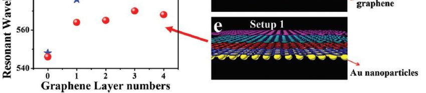
Sensors 2019, 19, 862                                                                                 5 of 15

physics and applications focusing on graphene itself. However, in this review, the focus is on a
metal-graphene hybrid nanostructure and the effect of the integration of metal with graphene on
plasmonic sensor performance.

4. Different Approaches Used to Enhance LSPR Sensor Performance
       The performance of plasmonic sensors is directly related to the amount of energy loss in the
metallic structure of a sensor. For example, using a metallic structure with a high loss results in
broadening of the resonance plasmonic waveband, and hence, the FOM of a sensor is decreased. It
is known that silver has a lower damping rate and thus has a lower absorption loss than gold and
copper [27]. Gold and copper are still considered to be low-loss materials, close to silver, compared
with aluminum, which has the highest losses in the visible spectrum [27,35,36]. A plasmonic sensor,
e.g., an LSPR sensor, requires a material that has lower losses to achieve better refractive index
sensitivity. When comparing gold and silver, silver offers better sensitivity than gold [37,38]. Upon
using a spherical NP with a diameter of 60 nm, silver exhibits better sensitivity (160 nm/RIU) (i.e. RIU:
refractive index unit) compared to that of gold (60 nm/RIU). The main reason for observing a higher
refractive index sensitivity in silver is attributed to a lower loss in silver and, hence, a lower damping
rate, which results in increasing scattering efficiency and therefore refractive index sensitivity [38].
However, losses of material are not the only physical property that needs to be considered in the design
of an LSPR sensor. The compatibility of the sensor with the sensing medium is also essential. If the
material used in an LSPR sensor is corrosive in an aqueous medium or oxidized in air, refractive index
sensitivity diminishes significantly [39]. Therefore, since gold has higher absorption loss than silver, it
is preferred in most LSPR applications. Silver [40] and other metals such as copper [41], aluminum [42],
or metal oxides such as zinc oxide [43] are corrosive in aqueous medium and oxidize in air, which
limits their application in LSPR sensor engineering. In addition to the material type of the NP used in
the LSPR sensors, NP size also affects the refractive index sensitivity of sensors. Particle size affects the
position of the surface plasmon peak, and it shifts towards larger wavelengths upon increasing the
size of the NPs, thus enhancing the sensitivity where it (i.e., sensitivity) is higher at larger wavelength
resonances. It has been verified that the SP peak of Au NPs can be redshifted up to 60 nm upon
increasing the particle radius from 10–100 nm [44]. This gives a good ability to engineer plasmonic
resonance by tuning the wavelength of the SP resonance of NPs for different applications. Therefore,
the proper choice of NP size must be considered to achieve better sensitivity of a plasmonic sensor.
       The shape of the NP is also used as a tunable parameter to improve LSPR sensor performance.
Changing the shape of NP causes resonance peak shift [17]. Tuning particle shape is a method used
to tune the wavelength of resonance at longer wavelengths and, hence, optimize the sensitivity of
an LSPR sensor. Mock et al. studied the effect of different shapes (sphere, cube, and triangle) of
silver NPs on the sensitivity of a single NP to the change in the refractive index of the surrounding
medium [38]. They reported that the triangle NP exhibited larger sensitivity (i.e., 350 nm/RIU) than a
spherical NP (i.e., 160 nm/RIU). Sun et.al [45] showed that a gold nanoshell shows higher sensitivity
(i.e., 406 nm/RIU) than a gold spherical NP with a sensitivity of 60 nm/RIU. The main reason for
observing higher sensitivity in the triangle [46], nanoshell [45], and nanocube [8] structures relative
to the spherical NPs [4] is due to the sharper edges of these NPs, which increase the intensity of the
localized electric field around the NPs. Changing the aspect ratio also helps to produce multi-modal
resonances at longer wavelengths, thus increasing sensor sensitivity. Hanarp et al. studied nanodisc
structures with aspect ratios starting from 1:1–5:1 at refractive indices change from 1.30–1.50 [47]. More
resonance shift was observed in the case of the 5:1 than in the case of the 1:1 aspect ratio, which offers
better sensor performance. Enhancement in sensitivity is acquired due to strongest excitation resulting
from the elongated nanodisc resonance mode [47]. Thus, the aspect ratio is another powerful key used
to tune the plasmonic properties of a nanostructure.
       By using two or more NPs separated by a specific distance in one-, two-, or three-dimensions, the
interaction between these incident waves and the excited surface plasmon wave from NPs resulted in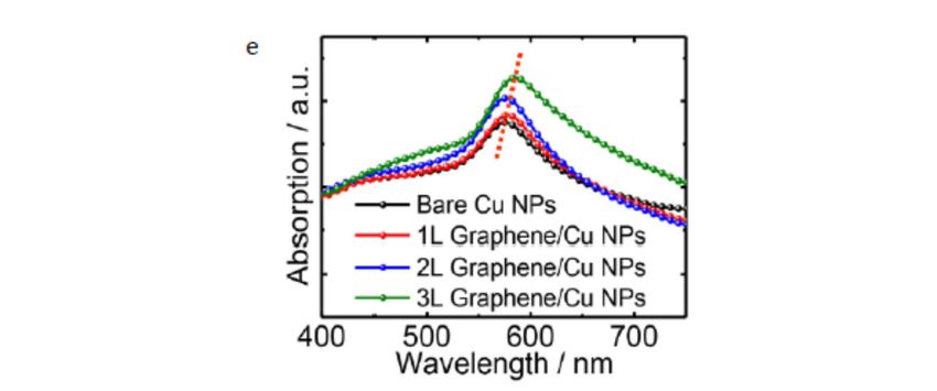
Sensors 2019, 19, 862                                                                                 6 of 15

an additional shift in the resonance wavelength to the red region as a result of the coupling interaction,
which enhances sensor sensitivity [48]. Erik et al. [49] studied the effect of inter-coupling between
two gold NPs (10 nm in diameter) on electric field enhancement. NPs’ spacing affects the position of
resonance peak and decreases the separation distance; the resonance wavelength shifts toward a longer
wavelength. However, upon using a structure with a larger separation distance between the NPs
than the NPs’ dimensions, the resonance wavelength shift is negligible, as discussed elsewhere [50].
Furthermore, upon reducing the distance between two consecutive NPs, the electric field in a near-field
regime is enhanced in the gap between NPs’ “hot spot”. Erik et al. also showed that the induced
electric field distribution between two consecutive NPs with a separation distance of 3 nm is stronger
than in the case of 10 nm. As mentioned earlier in this report, due to the nonlinear effect, the number
of parameters affects the plasmonic properties, such as field strength. In Erik et al.’s report, it was
shown that keeping NP’s geometry and material fixed, a lower inter-particle distance is preferred to
enhance the local field. Increasing the amount of scattering results in enhancement of the sensitivity of
the LSPR sensor [14,15].
      Plasmon mode hybridization is another promising method that has been applied to promote the
performance of LSPR sensors. Nanorings and nanoshells are the best nanostructures that represents the
plasmon hybridization effects [51]. In general, two different plasmonic modes are observed from the
hybridization phenomena: bonding and antibonding modes [52]. The bonding mode and anti-bonding
mode, which are observed at a lower and a higher frequency, respectively, result from the symmetrical
and asymmetrical coupling of induced plasmonic waves and incident light in a nanostructure [52]. The
geometry of the nanostructure plays a crucial rule in tuning hybridized plasmonic modes. Tao et al. [53]
showed that by using nanoring structures, larger sensitivity could be achieved than with nanodisc
structures. Sensitivity improvement is attributed to strong localization of the field in the ring’s center.
They also reported that the effect of ring width variation is more significant for the spectral properties
than other geometrical parameters, such as the ring size and the rings’ separation distance. As ring
width is increased, the plasmonic resonance peak narrows, which helps to enhance sensor performance
and, specifically, the FOM of the sensor due to the reduction of the FWHM of the resonance peak. There
are several other studies on nanostructures and the plasmonic hybridization effect, such as hollow
nanoshells [45], nanorice shells [54], and nanotube arrays [55], and using nanoparticle arrays; coupling
plasmonic waves and incident waves shows an improvement in plasmonic resonance properties, and
this could improve LSPR sensor performance [45,56].

5. Metal-Graphene Hybrid LSPR Sensor
     Longer wavelength resonance, high resonance intensity or amplitude, and sharper resonance
band (min FWHM) are indications of enhanced sensitivity and FOM of the LSPR sensor. Therefore,
engineering a plasmonic material by choosing a proper type, tuning the geometry of the NP, or
hybridizing it with another material in a way that improves one of the mentioned three plasmonic
properties could lead to promoting plasmonic sensor performance. Starting with the type of plasmonic
material, silver is the best choice among other plasmonic materials in terms of sensor performance
due to low optical loss compared to other metals in the visible range. This is due to the lack of the
interband transition as in gold and copper [11]. However, in addition to the oxidation problem, Ag2 S
forms on the Ag surface due to the reaction of carbonyl sulfide (OCS) and hydrogen sulfide (H2 S) with
the Ag surface; this affects the plasmonic properties of the Ag nanostructure and results in an increase
in material loss [57] Furthermore, another source of loss arises from scattering due to the increase in the
surface roughness of the Ag surface [58]. Gold for this reason is used more in applications even though
gold suffers from optical loss in the visible range. Passivating and encapsulating of the Ag surface to
prevent the formation of Ag2 S and maintaining the excellent plasmonic properties of Ag nanostructure
have been reported [59]. However, the thickness of the passivation layer is high, up to a level that
affects near-field interaction, which is very strong in the vicinity of the nanoparticle [59,60]. Therefore,
a passivation material that prevents the formation of Ag2 S and that is sufficiently thin is desired to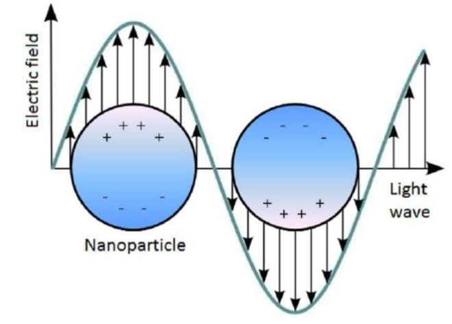
Sensors 2019, 19, 862                                                                              7 of 15

help solve the limitation of Ag in LSPR sensor applications. Leenaerts et al. studied the permeability
of a graphene sheet with different levels of defects for helium gas molecules [60]. They found that the
penetration of the defective graphene sheet for small atoms and molecules decreases exponentially
with the decrease in the size of sheet defects, and thus, a large defect size is preferred to increase
graphene permeability for gas molecules. This result led to the use of graphene as a passivation layer
for Ag NPs, in addition to the good plasmonic properties of graphene itself. Jason et al. studied the
effect of graphene passivation on the plasmonic properties of Ag NPs. They fabricated two sets of a
Ag nanoantenna array, and one of these was passivated with graphene by transferring CVD-grown
graphene on the Ag nanoantenna array, as shown in Figure 2a,b. From the SEM image of the passivated
and unpassivated array, it is clear how Ag2 S formed on the Ag surface affects the morphology of the
Ag nanoantenna, which affects plasmonic resonance over 30 days of measuring optical reflection. They
observed that the resonance peak shifts 216 nm after 30 days of fabrication for the unpassivated arrays,
while a very large reduction in resonance shift of 15 nm occurs in the case of graphene-passivated
Ag NPs; see Figure 2c,d. This provides high stability of the plasmonic properties of the Ag NPs over
the time it is passivated with graphene. To assess the effect of passivating Ag NPs with graphene
on LSPR sensor performance, they measured the sensitivity of passivated a Ag NP array and a Au
NP array by monitoring the resonance shift over change in the refractive index of the surrounding
medium. The sensor sensitivity of the Au NP array was found to be 102 nm/RIU and jumped to
162 nm/RIU for the graphene-passivated Ag NP array, which represent ~60% enhancement in the
sensor performance. This enhancement in performance of an Ag-based sensor after using graphene as
a passivation material means that graphene is a promising material to solve the problem of utilizing
an Ag-based LSPR sensor in different applications. Another example showing the effectiveness of
using graphene as a passivation material for an LSPR device is with passivating copper NPs, as
reported by Li et al. [61]. Copper is a good plasmonic material after silver and gold due to it having
higher loss, but its low cost compared to gold and silver gives it a good advantage. However, under
an ambient environment, copper oxidized rapidly with degradation in the LSPR intensity resulting
and, thus, a reduction in the sensing performance [62,63]. In order to overcome this issue, protecting
the Cu surface from oxidization is required. As mentioned, protecting the NPs with a thin layer of
material helps to detect the near-field effect of plasmonic NPs after being excited with light. Li et al.
transferred CVD graphene (1–3) layers on top of a Cu NP array fabricated on a quartz substrate, and
the absorption spectra were measured; see Figure 2e. They observed drastic field enhancement after
adding graphene, and adding more graphene layers enhanced the intensity of plasmonic resonance.
This enhancement is due to strong field localization at the graphene/copper interface that resulted
from the transfer of electrons from graphene to the surface of Cu due to the higher work function of Cu
compared to graphene, as reported in [64]. Furthermore, the resonance peak redshifted with increasing
graphene layers resulting from an increase in the refractive index of the Cu surrounding medium.
Both enhancements of plasmonic resonance intensity and redshift in the position are indicators of the
enhancement of the performance of this device when used as an LSPR sensor. Therefore, in addition
to protecting the Cu surface from oxidation, graphene by its plasmonic properties also enhances the
plasmonic properties of a sensor-based device.
shifting of resonance is a good behavior to enhance the sensitivity of the LSPR sensor, as mentioned;
    however, decreasing the amplitude of the resonance results in an increase in the FWHM of the
    resonance band, thus a reduction in the FOM of the sensor as explained in Equation 6. Therefore,
    during the design of a metal-graphene hybrid LSPR sensor, the size of metal NP and graphene has to
Sensors 2019, 19, 862                                                                            8 of 15
    be done in manner that gives longer wavelength resonance with a high amplitude                       or
    minimum FWHM.

     Figure 2. Scanning electron microscopy (SEM) images of a bare Ag nanoantenna (a) and a
         Figure 2. Scanning electron microscopy (SEM) images of a bare Ag nanoantenna (a) and a
     graphene-passivated Ag nanoantenna (b) after 30 days (scale bars are 200 nm), showing how graphene
         graphene-passivated Ag nanoantenna (b) after 30 days (scale bars are 200 nm), showing how
     protects Ag NPs from degradation [60]. Bare Ag nanoantenna’s (c) and graphene-passivated Ag NPs’
         graphene protects Ag NPs from degradation [60]. Bare Ag nanoantenna’s (c) and
     (d) normalized (Norm.) reflection spectra over 30 days, which shows how the passivation of Ag with
         graphene-passivated Ag NPs’ (d) normalized (Norm.) reflection spectra over 30 days, which shows
     graphene enhances the stability of Ag NPs [60]. Optical absorption spectra for the Cu NPs coated with
         how the passivation of Ag with graphene enhances the stability of Ag NPs [60]. Optical absorption
     a few graphene layers [61].
         spectra for the Cu NPs coated with a few graphene layers [61].
      Decorating graphene film with metal NPs is another approach used to tune the plasmonic
         Another
properties        metal-graphene
            of an LSPR  device. Xuhybrid     system that
                                     et al. fabricated Agstudied
                                                           NPs onthe
                                                                   topeffect of thegraphene
                                                                        of a CVD     hybrid nanostructure
                                                                                               film using a on
    the plasmonic  resonance   of the nanostructure   is that reported by  Nan   et al. [67].
thermally-assisted self-assembly method [65]. They observed that as the Ag NP size increased  They studied
                                                                                                      from the
50 nm–150 nm, plasmonic resonance redshifted from 446–495 nm. This redshift can be attributed to
two reasons. The first is the increase in the size of Ag NPs, for which is known that as the metal NP
size increases, longer wavelength resonance is produced [44]. Furthermore, Xu et al. reported that
as the graphene film thickness increases, plasmonic resonance redshift and amplitude are reduced.
Graphene is a good conductor, and thus, it facilitates energy concentrated in the Ag NP to transfer to
the graphene film, consequently decreasing the amplitude of plasmonic resonance [65,66]. Red shifting
of resonance is a good behavior to enhance the sensitivity of the LSPR sensor, as mentioned; however,
Sensors 2019, 19, 862 9 of 15
decreasing the amplitude of the resonance results in an increase in the FWHM of the resonance band,
thus a reduction in the FOM of the sensor as explained in Equation 6. Therefore, during the design of a
metal-graphene hybrid LSPR sensor, the size of metal NP and graphene has to be done in manner that
gives longer wavelength resonance with a high amplitude or minimum FWHM.
Another metal-graphene hybrid system that studied the effect of the hybrid nanostructure on the
Sensors 2019, 16, x FOR PEER REVIEW 9 of 4
plasmonic resonance of the nanostructure is that reported by Nan et al. [67]. They studied the effect of
two different gold-graphene hybrid systems on the plasmonic resonance of a nanostructure. The first
effect of two different gold-graphene hybrid systems on the plasmonic resonance of a nanostructure.
hybrid structure covered Au NPs with graphene (Figure 3e), and the second one was “sandwiched”
The first hybrid structure covered Au NPs with graphene (Figure 3e), and the second one was
Au NPs with graphene layers (Figure 3d). For both hybrid schemes, there is an obvious redshift
“sandwiched” Au NPs with graphene layers (Figure 3d). For both hybrid schemes, there is an
for the resonance peak (Figure 3a,b), but there is no clear effect on red shifting as graphene layers
obvious redshift for the resonance peak (Figure 3a,b), but there is no clear effect on red shifting as
increase (Figure 3c). The redshift in plasmonic resonance is attributed to electron transfer from
graphene layers increase (Figure 3c). The redshift in plasmonic resonance is attributed to electron
Au NPs to graphene. Consequently, modulating the electron transfer process could provide the
transfer from Au NPs to graphene. Consequently, modulating the electron transfer process could
ability to tune plasmonic resonance and, thus, improve sensor performance by producing longer
provide the ability to tune plasmonic resonance and, thus, improve sensor performance by
wavelength resonance.
producing longer wavelength resonance.
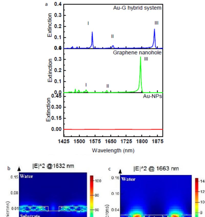
Figure3.3.Absorption
Figure Absorption spectra
spectra of gold
of gold NPs NPs coated
coated (a) and(a)encapsulated
and encapsulated (b) by graphene
(b) by graphene layers. (c)layers.
Gold
(c) Gold NP LSPR wavelengths for Setups 1 and 2. (d,e) The structures of the setups used
NP LSPR wavelengths for Setups 1 and 2. (d,e) The structures of the setups used in (b,a) [67]. in (b,a) [67].
AAplasmonic
plasmonicmaterial
materialthat
thathashashigh
highabsorption
absorptionin inthe
thevisible
visibleregion,
region,such
suchasasmetal,
metal,isispreferred
preferred
in
inplasmonic
plasmonicapplications
applicationssuch
suchas asan
anLSPR
LSPRsensor.
sensor.Graphene
Graphenehas hasvery
verylow
lowabsorption
absorptionin inthe
thevisible
visible
spectrum,
spectrum,and andthus, enhancing
thus, enhancing its absorption
its absorptionwill increase its chance
will increase to be used
its chance as aused
to be plasmonic material
as a plasmonic
for an LSPR
material forsensor
an LSPRin the visible
sensor region
in the [68].region
visible The hybrid of graphene
[68]. The hybrid ofwith metalwith
graphene enhances
metalgraphene
enhances
absorption, as reported as
graphene absorption, byreported
Wu et al. by[69].
Wu They fabricated
et al. [69]. Theygraphene nano
fabricated mesh samples
graphene and filled
nano mesh the
samples
empty partthe
and filled of the
emptymesh with
part Aumesh
of the nanodiscs.
with Au They observed
nanodiscs. a huge
They improvement
observed in the absorption
a huge improvement in the
of the hybrid
absorption ofnanostructure compared to
the hybrid nanostructure bare graphene.
compared This improvement
to bare graphene. can help to
This improvement enhance
can help to
the LSPR-based
enhance sensor fromsensor
the LSPR-based two sides.
fromFirst,
two enhancing
sides. First, absorption
enhancing will increase the
absorption localized
will increasefield
the
around thefield
localized nanostructure,
around the and thus, it will beand
nanostructure, morethus,
sensitive to be
it will a change
more in the surrounding
sensitive to a changerefractive
in the
index. Furthermore,
surrounding the increase
refractive in the absorption
index. Furthermore, the band could
increase in lead
the to a decreaseband
absorption in thecould
FWHM leadof to
thea
decrease in the FWHM of the resonance band, thus enhancing the resulting FOM of the sensor.
Therefore, integration of graphene with metal increases the opportunity for a graphene-based device
to be used in the visible region for different LSPR sensor applications.
Increasing the metal NP size results in red shifting in plasmonic resonance; for this reason,
having a fabrication method that can control the size of the metal NPs on top of graphene film isSensors 2019, 19, 862 10 of 15
resonance band, thus enhancing the resulting FOM of the sensor. Therefore, integration of graphene
with metal increases the opportunity for a graphene-based device to be used in the visible region for
different LSPR sensor applications.
Increasing the metal NP size results in red shifting in plasmonic resonance; for this reason, having
a fabrication method that can control the size of the metal NPs on top of graphene film is important.
Lee et al. introduced a tunable size and LSP resonance of Au NPs on top of a mono-layer of graphene
film based on the reduction potential between the Au3+ precursor and graphene [70]. They modulated
the Au NP size by the concentration of Au3+ precursor, the surface energy of graphene on the substrate,
the spin-coating speed, and the coating cycles. However, they found that Au NP size is strongly
affected by the concentration of Au3+ precursor and the coating cycle’s number. Upon increasing the
concentration of Au3+ and repeating the cycles of the reduction process, the plasmonic resonance of the
Au-graphene hybrid nanostructure was redshifted, and broadening in the band FWHM was observed.
After repeating the reduction process, the Au NP size was distributed between 20 and 120 nm, and the
resonance redshifted from 560–620 nm. This redshift in resonance wavelength is a tuning parameter to
enhance LSPR sensor performance; however, as mentioned in the previous section, the FWHM of the
resonance band has to be minimized to obtain a superior FOM of the sensor.
Recently, some published works showed the direct effect of hybrid graphene with metal on LSPR
sensor performance. Maurer et al. studied the effect of the graphene layer as a spacer between Au
film and Au NPs. They deposited graphene film on top of a gold film and then decorated the layer
with an Au nanodisc array [28]. They compared the optical extinction between AuNPs/Au film and
AuNPs/graphene film/Au film nanostructures. Enhancement in the localization field after adding the
graphene spacer led to an ~33% enhancement in the sensitivity of the sensor. Furthermore, a sharper
resonance width resulting from the Au-graphene hybrid structure was achieved and increased the
FOM from 2.1 for Au NPs/Au film to 2.8 for Au NPs/graphene/Au film nanostructures. This work
done by Maurer gives evidence of the effectiveness of the metal-graphene hybrid nanostructure on
the performance of the LSPR sensor. In one of our published works [25], the plasmonic properties of
the Au-graphene core-shell NP array on top of a quartz substrate have been theoretically investigated
using a Finite Difference Time Domain (FDTD) method. By comparing the extinction of the Au NP
array with the Au-graphene core-shell NP array, very strong enhancement was present in the resonance
band in the NIR region, in which sharpness reached an FWHM of 7.8 nm. This sharp resonance resulted
from a strong localization field enhancement after coating the Au NPs with a graphene shell due to
electron transfer between the Au NP and graphene shell, as shown clearly in electric field profiles.
Furthermore, the periodic arrangement of the hybrid NPs enhanced the interaction field between
them and enhanced the resonance, which is clear by the increase in the resonance amplitude when
the periodicity (i.e., distance between NPs) decreased. The FOM of this Au-graphene core-shell NPs
array nanostructure has been tested by measuring the optical transmission at a different refractive
index value (∆n = 0.01) of the surrounding medium. The results showed a huge improvement in the
FOM compared to the related published work in the field of metal-graphene hybrid LSPR sensors [28],
by achieving an FOM of 102.6. Furthermore, this improvement in the NIR region could be attributed
to the low loss of graphene compared to metal. Another hybrid system studied in our group is a
planner Au-graphene hybrid LSPR sensor [24]. In this hybrid nanostructure, the plasmonic properties
of the Au NP array/graphene film hybrid scheme have also been theoretically studied using an FDTD
method. The graphene in this nanostructure fills the gap between the Au NPs array, which shows a
huge improvement in the extinction in the NIR region (Figure 4), as reported in the previous published
work [25]. This indicates that the NIR region is good for the hybrid of graphene with metal. It was
observed that the intensity of the plasmonic resonance of this hybrid system is extremely sensitive to
very small changes in refractive index value (∆n = 0.001) and results in an FOM of 390 and a sensitivity
of 4380 nm/RIU. This improvement in the FOM of the LSPR sensor opens the door for the competition
of a commercialized SPR sensor and promotes the commercialization opportunity of an LSPR sensor.Sensors 2019, 19, 862 11 of 15
Sensors 2019, 16, x FOR PEER REVIEW 11 of 4
Figure 4. (a) Extinction spectrum for three different nanostructures; Au NP square array, nanohole
array perforated in 20 nm-thick graphene film, and Au NPs/G hybrid structure showing three main
resonance
Figure modes;
4. (a) Modespectrum
Extinction I (λ = 1532for
nm), Mode
three II (λ =nanostructures;
different 1632 nm), and Mode
Au NPIIIsquare
(λ = 1793 nm).
array, (b,c) are
nanohole
the electric
array field in
perforated profiles at the resonance
20 nm-thick graphene wavelength
film, and AuofNPs/G
Mode hybrid
II for the graphene
structure nanohole
showing structure
three main
and the Au-graphene
resonance modes; Mode hybrid
I (λ = structure,
1532 nm),respectively
Mode II (λ =[24].
1632 nm), and Mode III (λ = 1793 nm). (b,c) are
the electric field profiles at the resonance wavelength of Mode II for the graphene nanohole structure
and the Au-graphene hybrid structure, respectively [24].Sensors 2019, 19, 862 12 of 15
6. Conclusions
In this short review, the fundamental physics of the LSPR sensor was summarized followed by a
discussion of different methods used to enhance the performance of LSPR sensors. It was shown that
the size and shape of the nanoparticle give a broad tunability in optimizing the performance (sensitivity
and FOM) of the LSPR sensor. A smaller NP size (Sensors 2019, 19, 862 13 of 15
11. West, P.R.; Ishii, S.; Naik, G.V.; Emani, N.K.; Shalaev, V.M.; Boltasseva, A. Searching for Better Plasmonic
Materials. Laser Photonics Rev. 2010, 4, 795–808. [CrossRef]
12. Wu, B.; Mathews, N.; Sum, T. Plasmonic Organic Solar Cells: Charge Generation and Recombination; Springer:
Singapore, 2016.
13. Sönnichsen, C.; Franzl, T.; Wilk, T.; von Plessen, G.; Feldmann, J.; Wilson, O.; Mulvaney, P. Drastic Reduction
of Plasmon Damping in Gold Nanorods. Phys. Rev. Lett. 2002, 88, 077402. [CrossRef] [PubMed]
14. Dmitriev, A. Nanoplasmonic Sensors; Springer Science & Business Media: Berlin, Germany, 2012.
15. Enoch, S.; Bonod, N. Plasmonics: From Basics to Advanced Topics; Springer: Berlin, Germany, 2012.
16. Dieringer, J.A.; McFarland, A.D.; Shah, N.C.; Stuart, D.A.; Whitney, A.V.; Yonzon, C.R.; Young, M.A.;
Zhang, X.; Van Duyne, R.P. Introductory Lecture Surface Enhanced Raman Spectroscopy: New Materials,
Concepts, Characterization Tools, and Applications. Faraday Discuss. 2006, 132, 9–26. [CrossRef] [PubMed]
17. Hammond, J.L.; Bhalla, N.; Rafiee, S.D.; Estrela, P. Localized Surface Plasmon Resonance as a Biosensing
Platform for Developing Countries. Biosensors 2014, 4, 172–188. [CrossRef] [PubMed]
18. Link, S.; El-Sayed, M.A. Shape and Size Dependence of Radiative, Non-Radiative and Photothermal
Properties of Gold Nanocrystals. Int. Rev. Phys. Chem. 2000, 19, 409–453. [CrossRef]
19. Meier, M.; Wokaun, A. Enhanced Fields on Large Metal Particles: Dynamic Depolarization. Opt. Lett. 1983,
8, 581–583. [CrossRef]
20. Zhang, J.Z.; Noguez, C. Plasmonic Optical Properties and Applications of Metal Nanostructures. Plasmonics
2008, 3, 127–150. [CrossRef]
21. Krenn, J.; Schider, G.; Rechberger, W.; Lamprecht, B.; Leitner, A.; Aussenegg, F.; Weeber, J. Design of
Multipolar Plasmon Excitations in Silver Nanoparticles. Appl. Phys. Lett. 2000, 77, 3379–3381. [CrossRef]
22. Shuford, K.L.; Ratner, M.A.; Schatz, G.C. Multipolar Excitation in Triangular Nanoprisms. J. Chem. Phys.
2005, 123, 114713. [CrossRef]
23. Wei, H.; Reyes–Coronado, A.; Nordlander, P.; Aizpurua, J.; Xu, H. Multipolar Plasmon Resonances in
Individual Ag Nanorice. ACS Nano 2010, 4, 2649–2654. [CrossRef]
24. Alharbi, R.; Irannejad, M.; Yavuz, M. Au–Graphene Hybrid Plasmonic Nanostructure Sensor Based on
Intensity Shift. Sensors 2017, 17, 191. [CrossRef] [PubMed]
25. Alharbi, R.; Irannejad, M.; Yavuz, M. Gold-Graphene Core-Shell Nanostructure Surface Plasmon Sensors.
Plasmonics 2017, 12, 783–794. [CrossRef]
26. Miller, M.M.; Lazarides, A.A. Sensitivity of Metal Nanoparticle Surface Plasmon Resonance to the Dielectric
Environment. J. Phys. Chem. B 2005, 109, 21556–21565. [CrossRef] [PubMed]
27. Johnson, P.B.; Christy, R. Optical Constants of the Noble Metals. Phys. Rev. B 1972, 6, 4370. [CrossRef]
28. Maurer, T.; Nicolas, R.; Lévêque, G.; Subramanian, P.; Proust, J.; Béal, J.; Schuermans, S.; Vilcot, J.; Herro, Z.;
Kazan, M. Enhancing LSPR Sensitivity of Au Gratings through Graphene Coupling to Au Film. Plasmonics
2014, 9, 507–512. [CrossRef]
29. Tobiška, P.; Hugon, O.; Trouillet, A.; Gagnaire, H. An Integrated Optic Hydrogen Sensor Based on SPR on
Palladium. Sens. Actuators B Chem. 2001, 74, 168–172. [CrossRef]
30. Baldelli, S.; Eppler, A.S.; Anderson, E.; Shen, Y.; Somorjai, G.A. Surface Enhanced Sum Frequency Generation
of Carbon Monoxide Adsorbed on Platinum Nanoparticle Arrays. J. Chem. Phys. 2000, 113, 5432–5438.
[CrossRef]
31. Estevez, M.; Otte, M.A.; Sepulveda, B.; Lechuga, L.M. Trends and Challenges of Refractometric Nanoplasmonic
Biosensors: A Review. Anal. Chim. Acta 2014, 806, 55–73. [CrossRef]
32. Anker, J.N.; Hall, W.P.; Lyandres, O.; Shah, N.C.; Zhao, J.; Van Duyne, R.P. Biosensing with plasmonic
nanosensors. In Nanoscience and Technology: A Collection of Reviews from Nature Journals; World Scientific:
Singapore, 2010; pp. 308–319.
33. Luo, X.; Qiu, T.; Lu, W.; Ni, Z. Plasmons in Graphene: Recent Progress and Applications. Mater. Sci. Eng.
R Rep. 2013, 74, 351–376. [CrossRef]
34. Huang, S.; Song, C.; Zhang, G.; Yan, H. Graphene Plasmonics: Physics and Potential Applications.
Nanophotonics 2016, 6, 1191–1204. [CrossRef]
35. Cooper, B.; Ehrenreich, H.; Philipp, H. Optical Properties of Noble Metals. II. Phys. Rev. 1965, 138, A494.
[CrossRef]
36. Ehrenreich, H.; Philipp, H.; Segall, B. Optical Properties of Aluminum. Phys. Rev. 1963, 132, 1918. [CrossRef]Sensors 2019, 19, 862 14 of 15
37. Sun, Y.; Xia, Y. Increased Sensitivity of Surface Plasmon Resonance of Gold Nanoshells Compared to that of
Gold Solid Colloids in Response to Environmental Changes. Anal. Chem. 2002, 74, 5297–5305. [CrossRef]
[PubMed]
38. Mock, J.J.; Smith, D.R.; Schultz, S. Local Refractive Index Dependence of Plasmon Resonance Spectra from
Individual Nanoparticles. Nano Lett. 2003, 3, 485–491. [CrossRef]
39. Sepúlveda, B.; Angelomé, P.C.; Lechuga, L.M.; Liz–Marzán, L.M. LSPR–Based Nanobiosensors. Nano Today
2009, 4, 244–251. [CrossRef]
40. Guo, L.; Yin, Y.; Huang, R.; Qiu, B.; Lin, Z.; Yang, H.; Li, J.; Chen, G. Enantioselective Analysis of Melagatran
Via an LSPR Biosensor Integrated with a Microfluidic Chip. Lab Chip 2012, 12, 3901–3906. [CrossRef]
41. Sekhon, J.S.; Verma, S. Refractive Index Sensitivity Analysis of Ag, Au, and Cu Nanoparticles. Plasmonics
2011, 6, 311–317. [CrossRef]
42. Chan, G.H.; Zhao, J.; Schatz, G.C.; Van Duyne, R.P. Localized Surface Plasmon Resonance Spectroscopy of
Triangular Aluminum Nanoparticles. J. Phys. Chem. C 2008, 112, 13958–13963. [CrossRef]
43. Park, Y.R.; Liu, N.; Lee, C.J. Photoluminescence Enhancement from Hybrid Structures of Metallic
Single–Walled Carbon Nanotube/ZnO Films. Curr. Appl. Phys. 2013, 13, 2026–2032. [CrossRef]
44. Link, S.; El–Sayed, M.A. Size and Temperature Dependence of the Plasmon Absorption of Colloidal Gold
Nanoparticles. J. Phys. Chem. B 1999, 103, 4212–4217. [CrossRef]
45. Sun, Y.; Xia, Y. Synthesis of Gold Nanoshells and their use in Sensing Applications. MRS Online Proc.
Libr. Arch. 2003, 776. [CrossRef]
46. Mock, J.; Barbic, M.; Smith, D.; Schultz, D.; Schultz, S. Shape Effects in Plasmon Resonance of Individual
Colloidal Silver Nanoparticles. J. Chem. Phys. 2002, 116, 6755–6759. [CrossRef]
47. Hanarp, P.; Käll, M.; Sutherland, D.S. Optical Properties of Short Range Ordered Arrays of Nanometer Gold
Disks Prepared by Colloidal Lithography. J. Chem. Phys. B 2003, 107, 5768–5772. [CrossRef]
48. Ross, M.B.; Mirkin, C.A.; Schatz, G.C. Optical Properties of One–, Two–, and Three–Dimensional Arrays of
Plasmonic Nanostructures. T J. Chem. Phys. C 2016, 120, 816–830. [CrossRef]
49. Martinsson, E.; Sepulveda, B.; Chen, P.; Elfwing, A.; Liedberg, B.; Aili, D. Optimizing the Refractive Index
Sensitivity of Plasmonically Coupled Gold Nanoparticles. Plasmonics 2014, 9, 773–780. [CrossRef]
50. Jain, P.K.; Lee, K.S.; El–Sayed, I.H.; El-Sayed, M.A. Calculated Absorption and Scattering Properties of
Gold Nanoparticles of Different Size, Shape, and Composition: Applications in Biological Imaging and
Biomedicine. J. Chem. Phys. B 2006, 110, 7238–7248. [CrossRef] [PubMed]
51. Chung, T.; Lee, S.; Song, E.Y.; Chun, H.; Lee, B. Plasmonic Nanostructures for Nano–Scale Bio–Sensing.
Sensors 2011, 11, 10907–10929. [CrossRef] [PubMed]
52. Prodan, E.; Radloff, C.; Halas, N.J.; Nordlander, P. A Hybridization Model for the Plasmon Response of
Complex Nanostructures. Science 2003, 302, 419–422. [CrossRef]
53. Teo, S.L.; Lin, V.K.; Marty, R.; Large, N.; Llado, E.A.; Arbouet, A.; Girard, C.; Aizpurua, J.; Tripathy, S.;
Mlayah, A. Gold Nanoring Trimers: A Versatile Structure for Infrared Sensing. Opt. Express 2010, 18,
22271–22282. [CrossRef]
54. Tam, F.; Moran, C.; Halas, N. Geometrical Parameters Controlling Sensitivity of Nanoshell Plasmon
Resonances to Changes in Dielectric Environment. J. Chem. Phys. B 2004, 108, 17290–17294. [CrossRef]
55. McPhillips, J.; Murphy, A.; Jonsson, M.P.; Hendren, W.R.; Atkinson, R.; Höök, F.; Zayats, A.V.; Pollard, R.J.
High–Performance Biosensing using Arrays of Plasmonic Nanotubes. ACS Nano 2010, 4, 2210–2216.
[CrossRef] [PubMed]
56. Larsson, E.M.; Alegret, J.; Käll, M.; Sutherland, D.S. Sensing Characteristics of NIR Localized Surface Plasmon
Resonances in Gold Nanorings for Application as Ultrasensitive Biosensors. Nano Lett. 2007, 7, 1256–1263.
[CrossRef] [PubMed]
57. McMahon, M.; Lopez, R.; Meyer, H.; Feldman, L.; Haglund, R. Rapid Tarnishing of Silver Nanoparticles in
Ambient Laboratory Air. Appl. Phys. B 2005, 80, 915–921. [CrossRef]
58. Nagpal, P.; Lindquist, N.C.; Oh, S.H.; Norris, D.J. Ultrasmooth Patterned Metals for Plasmonics and
Metamaterials. Science 2009, 325, 594–597. [CrossRef] [PubMed]
59. Park, Y.; Cha, S.; Saito, Y.; Prinz, F.B. Gas–Tight Alumina Films on Nanoporous Substrates through Oxidation
of Sputtered Metal Films. Thin Solid Films 2005, 476, 168–173. [CrossRef]
60. Reed, J.C.; Zhu, H.; Zhu, A.Y.; Li, C.; Cubukcu, E. Graphene–Enabled Silver Nanoantenna Sensors. Nano Lett.
2012, 12, 4090–4094. [CrossRef] [PubMed]Sensors 2019, 19, 862 15 of 15
61. Li, Y.; Dong, F.; Chen, Y.; Zhang, X.; Wang, L.; Bi, Y.; Tian, Z.; Liu, Y.; Feng, J.; Sun, H. As-Grown
Graphene/Copper Nanoparticles Hybrid Nanostructures for Enhanced Intensity and Stability of Surface
Plasmon Resonance. Sci. Rep. 2016, 6, 37190. [CrossRef] [PubMed]
62. Chuang, C.; Aoh, J.; Din, R. Oxidation of Copper Pads and its Influence on the Quality of Au/Cu Bonds
during Thermosonic Wire Bonding Process. Microelectron. Reliab. 2006, 46, 449–458. [CrossRef]
63. Kim, D.; Yoo, S.M.; Park, T.J.; Yoshikawa, H.; Tamiya, E.; Park, J.Y.; Lee, S.Y. Plasmonic Properties of the
Multispot Copper–Capped Nanoparticle Array Chip and its Application to Optical Biosensors for Pathogen
Detection of Multiplex DNAs. Anal. Chem. 2011, 83, 6215–6222. [CrossRef]
64. Giovannetti, G.; Khomyakov, P.; Brocks, G.; Karpan, V.v.; Van den Brink, J.; Kelly, P.J. Doping Graphene with
Metal Contacts. Phys. Rev. Lett. 2008, 101, 026803. [CrossRef]
65. Xu, G.; Liu, J.; Wang, Q.; Hui, R.; Chen, Z.; Maroni, V.A.; Wu, J. Plasmonic Graphene Transparent Conductors.
Adv. Mater. 2012, 24, OP71–OP76. [CrossRef] [PubMed]
66. Liu, J.; Xu, G.; Rochford, C.; Lu, R.; Wu, J.; Edwards, C.M.; Berrie, C.L.; Chen, Z.; Maroni, V.A. Doped
Graphene Nanohole Arrays for Flexible Transparent Conductors. Appl. Phys. Lett. 2011, 99, 023111.
[CrossRef]
67. Nan, H.; Chen, Z.; Jiang, J.; Li, J.; Zhao, W.; Ni, Z.; Gu, X.; Xiao, S. The Effect of Graphene on Surface Plasmon
Resonance of Metal Nanoparticles. Phys. Chem. Chem. Phys. 2018, 20, 25078–25084. [CrossRef] [PubMed]
68. Chen, Z.; Li, X.; Wang, J.; Tao, L.; Long, M.; Liang, S.; Ang, L.K.; Shu, C.; Tsang, H.K.; Xu, J. Synergistic Effects
of Plasmonics and Electron Trapping in Graphene Short–Wave Infrared Photodetectors with Ultrahigh
Responsivity. ACS Nano 2017, 11, 430–437. [CrossRef] [PubMed]
69. Wu, Y.; Niu, J.; Danesh, M.; Liu, J.; Chen, Y.; Ke, L.; Qiu, C.; Yang, H. Localized Surface Plasmon Resonance
in Graphene Nanomesh with Au Nanostructures. Appl. Phys. Lett. 2016, 109, 041106. [CrossRef]
70. Lee, S.; hyung Lee, M.; Shin, H.; Choi, D. Control of Density and LSPR of Au Nanoparticles on Graphene.
Nanotechnology 2013, 24, 275702. [CrossRef] [PubMed]
71. Shen, Y.; Zhou, J.; Liu, T.; Tao, Y.; Jiang, R.; Liu, M.; Xiao, G.; Zhu, J.; Zhou, Z.; Wang, X. Plasmonic Gold
Mushroom Arrays with Refractive Index Sensing Figures of Merit Approaching the Theoretical Limit.
Nat. Commun. 2013, 4, 2381. [CrossRef] [PubMed]
© 2019 by the authors. Licensee MDPI, Basel, Switzerland. This article is an open access
article distributed under the terms and conditions of the Creative Commons Attribution
(CC BY) license (http://creativecommons.org/licenses/by/4.0/).You can also read