Cancer Association of South Africa (CANSA) - Cancer Association of ...
←
→
Page content transcription
If your browser does not render page correctly, please read the page content below
Cancer Association of South Africa (CANSA)
Fact Sheet
on
Cancer of the Brain and
Central Nervous System
Introduction
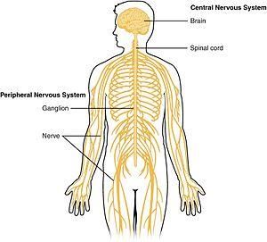
The human body cannot function without the nervous system.
The nervous system is a complex network that coordinates one’s
actions, reflexes, and sensations.
Broadly speaking, the nervous system is organised into two main
parts, the Central Nervous System (CNS) and the Peripheral
Nervous System (PNS).
[Picture Credit: Central Nervous System 1]
The Central Nervous System is the processing centre of the body
and consists of the brain and the spinal cord. Both of these are
protected by three layers of membranes known as meninges. For
further protection, the brain is encased within the hard bones of the skull, while the spinal cord is protected
with the bony vertebrae (backbones). A third form of protection is cerebrospinal fluid, which provides a buffer
that limits impact between the brain and skull or between spinal cord and vertebrae.
The brain plays a central role in controlling most of the bodily functions, including awareness, movements,
sensations, thoughts, speech, and memory. Some reflex movements occur via spinal cord pathways without
the participation of brain structures. The spinal cord is connected to a section of the brain called the brainstem
and runs through the spinal canal. Cranial nerves exit the brainstem. Nerve roots exit the spinal cord to both
sides of the body. The spinal cord carries signals (messages)
back and forth between the brain and the peripheral nerves.
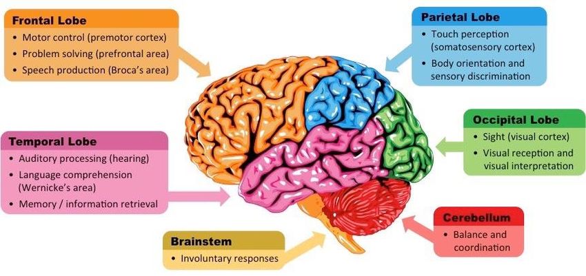
The brain can be divided into three basic units: the forebrain,
the midbrain and the hindbrain. These areas are: Occipital
lobe, Temporal lobe, Parietal lobe, Frontal lobe. Cerebral
cortex, Cerebellum, Hypothalamus,Thalamus,Pituitary gland,
Pineal gland, Amygdala, Hippocampas and the Mid- brain.
[Picture Credit: Central Nervous System 2]
Researched and Authored by Prof Michael C Herbst
[D Litt et Phil (Health Studies); D N Ed; M Art et Scien; B A Cur; Dip Occupational Health; Dip Genetic Counselling; Dip Audiometry and
Noise Measurement; Diagnostic Radiographer; Medical Ethicist]
Approved by Ms Elize Joubert, Chief Executive Officer [BA Social Work (cum laude); MA Social Work]
January 2021 Page 1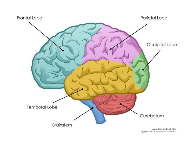
Various parts of the human brain have
                                                                                   been identified to be responsible for
                                                                                   specific functions/actions. This is a work
                                                                                   in progress and additional information is
                                                                                   being discovered.

                                                                                   [Picture Credit: Central Nervous System 3]

Cancer of the Brain and Central Nervous System
Cancer of the brain and spinal cord occurs when malignant cell within the brain and/or spinal cord multiply
and grow in an uncontrolled manner to form a mass of cancer tissue (tumour) that interferes
with brain functions such as muscle control, sensation, memory, and other normal body functions.

Tang, W., Fan, W., Lau, J., Deng, L., Shen, Z. & Chen, X. 2019. Emerging blood-brain-barrier-crossing
nanotechnology for brain cancer theranostics. Chem Soc Rev. 48 (11), 2967-3014, 2019 Jun 4.
 “Despite surgical and medical advances, the prognosis for most brain cancer patients remains dismal and the
median survival rarely exceeds 16 months. Drug delivery to the brain is significantly hindered by the existence
of the blood-brain barrier (BBB), which serves as a protective semi-permeable membrane for the central
nervous system. Recent breakthroughs in nanotechnology have yielded multifunctional theranostic
nanoplatforms with the ability to cross or bypass the BBB, enabling accurate diagnosis and effective treatment
of brain tumours. Herein, we make our efforts to present a comprehensive review on the latest remarkable
advances in BBB-crossing nanotechnology, with an emphasis on the judicious design of multifunctional
nanoplatforms for effective BBB penetration, efficient tumour accumulation, precise tumour imaging, and
significant tumour inhibition of brain cancer. The detailed elucidation of BBB-crossing nanotechnology in this
review is anticipated to attract broad interest from researchers in diverse fields to participate in the
establishment of powerful BBB-crossing nanoplatforms for highly efficient brain cancer theranostics.”

Incidence of Cancer of the Brain and Central Nervous System
According to the outdated National Cancer Registry (2017), known for under reporting, the following number
of Brain and Central Nervous System cancer cases was histologically diagnosed in South Africa during 2017:

 Group - Males                   Actual              Estimated         Percentage of
 2017                          No of Cases          Lifetime Risk       All Cancers
 All males                         262                  1:825              0,66%
 Asian males                        12                  1:613              1,24%
 Black males                        92                 1:2 294             0,69%
 Coloured males                     40                  1:530              0,83%
 White males                       118                  1:256              0,56%

Researched and Authored by Prof Michael C Herbst
[D Litt et Phil (Health Studies); D N Ed; M Art et Scien; B A Cur; Dip Occupational Health; Dip Genetic Counselling; Dip Audiometry and
Noise Measurement; Diagnostic Radiographer; Medical Ethicist]
Approved by Ms Elize Joubert, Chief Executive Officer [BA Social Work (cum laude); MA Social Work]
January 2021                                                                                                                     Page 2
Group - Females                       Actual                 Estimated            Percentage of
 2017                                No of Cases             Lifetime Risk          All Cancers
 All females                             215                    1:1 236                0,52%
 Asian females                            8                      1:978                 0,54%
 Black females                            79                    1:3 413                0,41%
 Coloured females                         26                     1:822                  0,57
 White females                           102                     1:315                 0,60%

The frequency of histologically diagnosed cases of Brain and Central Nervous System cancer in South Africa
for 2017 was as follows (National Cancer Registry, 2017):

 Group - Males                      0 – 19         20 – 29         30 – 39        40 – 49       50 – 59       60 – 69      70 – 79         80+
 2017                               Years           Years           Years          Years         Years         Years        Years         Years
 All males                            41             23              30             36            78            46           24             4
 Asian males                           1              1               2              1             3             4            0             0
 Black males                          32             12              11             12            15             8            2             0
 Coloured males                        1              5               3              1            10             9            2             0
 White males                           7              5              14             13            30            25           20             4

 Group - Females                    0 – 19         20 – 29         30 – 39        40 – 49       50 – 59       60 – 69      70 – 79         80+
 2017                               Years           Years           Years          Years         Years         Years        Years         Years
 All females                          43             12              23             29            39            37           30             4
 Asian females                         2              1               0              1             1             3            0             0
 Black females                        27              7              10             11            12             3            8             1
 Coloured females                      3              0               3              3             7             4            6             0
 White females                         9              4              10             14            19            27           16             3
N.B. In the event that the totals in any of the above tables do not tally, this may be the result of uncertainties as to the age, race or sex of the
individual. The totals for ‘all males’ and ‘all females’, however, always reflect the correct totals.

According to Bruni, et al., (2019), the burden of Brain and Central Nervous System cancer for South Africa for
2018 is estimated as (based on Globocan estimates):
   • Annual number of Brain and Central Nervous System cancer cases                       900
   • Annual number of Brain and Central Nervous System cancer deaths                      719

Risk Factors for Cancer of the Brain and Central Nervous System
Most of the time, the cause of a brain tumour is unknown, but the following factors may raise a person’s risk
of developing a brain and/or central nervous system tumour:

Age – these type of tumours are more common in children and older adults, although people of any age can
develop a brain or CNS tumour
Sex - in general, men are more likely than women to develop a brain and CNS tumours - some specific types
of brain tumours, such as meningioma, are more common in women
Home and work exposures - exposure to solvents, pesticides, oil products, rubber, or vinyl chloride may
increase the risk of developing a brain and CNS tumour – scientific evidence to fully support this possible link
is not yet available
Family history - approximately 5% of brain and CNS tumours may be linked to hereditary genetic factors or
conditions, including:
• Li-Fraumeni syndrome
• Neurofibromatosis
Researched and Authored by Prof Michael C Herbst
[D Litt et Phil (Health Studies); D N Ed; M Art et Scien; B A Cur; Dip Occupational Health; Dip Genetic Counselling; Dip Audiometry and
Noise Measurement; Diagnostic Radiographer; Medical Ethicist]
Approved by Ms Elize Joubert, Chief Executive Officer [BA Social Work (cum laude); MA Social Work]
January 2021                                                                                                                     Page 3
•   Nevoid basal cell carcinoma Syndrome
•   Tubeculous sclerosis
•   Turcot Syndrome
•   Von Hippel-Lindau Disease

Scientists have also found “clusters” of brain and CNS tumours within some families without a link to these
known hereditary conditions

Exposure to infections, viruses, and allergens - infection with the Epstein-Barr virus (EBV) increases the risk of
CNS lymphoma. In other research, high levels of a common virus called cytomegalovirus (CMV) have been
found in brain tumour tissue

Electromagnetic fields - most studies evaluating the role of electromagnetic fields, such as energy from power
lines or from cell phone use, show no link to an increased risk of developing a brain tumour in adults. Because
of conflicting information regarding risk in children, the World Health Organization (WHO) and Cancer
Association of South Africa (CANSA) recommends limiting cell phone use and promotes the use of a hands-
free headset for both adults and children

Race and ethnicity – it would appear that white people are more likely to develop gliomas but less likely to
develop meningioma than black people. Also, people from northern Europe are more than twice as likely to
develop a brain tumour as people in Japan

Ionizing radiation - previous treatment to the brain or head with ionizing radiation, including X-rays, has been
shown to be a risk factor for a brain tumour

Head injury and seizures - serious head trauma has long been studied for its relationship to brain tumours

N-nitroso compounds - some studies of diet and vitamin supplementation seem to indicate that dietary N-
nitroso compounds may raise the risk of both childhood and adult brain tumours. Dietary N-nitroso
compounds are formed in the body from nitrites or nitrates found in some cured meats, cigarette smoke, and
cosmetics

Veillon, L., Fakih, C., Abou-El-Hassan, H., Kobeissy, F. & Mechref, Y. 2018.
“Protein glycosylation is a posttranslational modification that affects more than half of all known proteins.
Glycans covalently bound to biomolecules modulate their functions by both direct interactions, such as the
recognition of glycan structures by binding partners, and indirect mechanisms that contribute to the control
of protein conformation, stability, and turnover. The focus of this Review is the discussion of aberrant
glycosylation related to brain cancer. Altered sialylation and fucosylation of N- and O-glycans play a role in the
development and progression of brain cancer. Additionally, aberrant O-glycan expression has been implicated
in brain cancer. This Review also addresses the clinical potential and applications of aberrant glycosylation for
the detection and treatment of brain cancer. The viable roles glycans may play in the development of brain
cancer therapeutics are addressed as well as cancer-glycoproteomics and personalized medicine. Glycoprotein
alterations are considered as a hallmark of cancer while high expression in body fluids represents an
opportunity for cancer assessment.”

Bytnar, J.A., Lin, J., Shriver, C.D. & Zhu, K. 2019.
Purpose: Racial disparity with shorter survival for Blacks than Whites is well known for many cancers.
However, for brain cancer, some national cancer registry studies have shown better survival among Blacks

Researched and Authored by Prof Michael C Herbst
[D Litt et Phil (Health Studies); D N Ed; M Art et Scien; B A Cur; Dip Occupational Health; Dip Genetic Counselling; Dip Audiometry and
Noise Measurement; Diagnostic Radiographer; Medical Ethicist]
Approved by Ms Elize Joubert, Chief Executive Officer [BA Social Work (cum laude); MA Social Work]
January 2021                                                                                                                     Page 4
compared to Whites. This study aimed to systematically investigate whether Blacks and Whites differ in
survival and also in tumor characteristics and treatment for neuroepithelial brain tumors.
Methods: The National Cancer Institute's Surveillance Epidemiology and End Results (SEER) database was used
to identify non-Hispanic White and Black patients diagnosed with malignant, histologically confirmed
neuroepithelial brain cancer from 2004 through 2015. Racial differences in brain cancer survival were
compared using Kaplan-Meier curve and Cox proportional hazard models. The associations of race with tumor
and treatment characteristics (location, size, grade, surgical type) were examined using multinomial logistic
regression.
Results: After adjusting for demographic, tumor, and treatment factors, there were no significant differences
in survival for non-Hispanic Blacks compared to non-Hispanic Whites [hazard ratio (HR) 1.05, 95% confidence
interval (CI) 0.99-1.10]. Non-Hispanic Blacks had higher odds of being diagnosed with tumors of unknown
grade [odds ratio (OR) 1.16, 95% CI 1.05-1.29], unknown size (OR 1.14, 95% CI 1.01-1.29), infratentorial (OR
1.12, 95% CI 1.01-1.24) or overlapping area (OR 1.39, 95% CI 1.14-1.70), and lower odds of having a total
surgical resection (OR 0.83, 95% CI 0.74-0.93).
Conclusion: Non-Hispanic Blacks do not exhibit longer brain cancer-specific survival than non-Hispanic Whites.
They were more likely to have tumors of unknown size or grade and less likely to receive total surgical
resection, which may result from racial differences in access to and use of healthcare.

Signs and Symptoms of Cancer of the Brain and Central Nervous System
Brain tumour symptoms depend on the size, location, and type of tumour. General brain tumour symptoms
may include:
•       headache
•       nausea or vomiting
•       loss of motor coordination, such as trouble walking
•       feeling sleepy
•       feelings of weakness
•       appetite changes
•       convulsions or seizures
•       issues with your vision, hearing, or speech
•       difficulty concentrating
•       mood swings or behaviour changes

Diagnosis of Cancer of the Brain and Central Nervous System
A treating physician or oncologist may recommend a number of tests and procedures, including:

•   A neurological examination
•   Imaging tests – Magnetic Resonance Imaging (MRI); Computerized Tomography (CT) scan; Positron
    Emission Tomography (PET) scan
•   Tests to find cancer in other parts of your body - if it is suspected that a brain tumour may be a result of
    cancer that has spread from another area of your body, the doctor may recommend tests and procedures
    to determine where the cancer originated
•   Collecting and testing a sample of abnormal tissue (biopsy) - a stereotactic needle biopsy may be done for
    brain tumours in hard to reach areas or very sensitive areas within your brain that might be damaged by
    a more extensive operation.

Researched and Authored by Prof Michael C Herbst
[D Litt et Phil (Health Studies); D N Ed; M Art et Scien; B A Cur; Dip Occupational Health; Dip Genetic Counselling; Dip Audiometry and
Noise Measurement; Diagnostic Radiographer; Medical Ethicist]
Approved by Ms Elize Joubert, Chief Executive Officer [BA Social Work (cum laude); MA Social Work]
January 2021                                                                                                                     Page 5
Müller Bark, J., Kulasinghe, A., Chua, B., Day, B.W. & Punyadeera, C. 2020. “Gliomas are the most common tumours of the central nervous system and the most aggressive form is glioblastoma (GBM). Despite advances in treatment, patient survival remains low. GBM diagnosis typically relies on imaging techniques and postoperative pathological diagnosis; however, both procedures have their inherent limitations. Imaging modalities cannot differentiate tumour progression from treatment-related changes that mimic progression, known as pseudoprogression, which might lead to misinterpretation of therapy response and delay clinical interventions. In addition to imaging limitations, tissue biopsies are invasive and most of the time cannot be performed over the course of treatment to evaluate 'real-time' tumour dynamics. In an attempt to address these limitations, liquid biopsies have been proposed in the field. Blood sampling is a minimally invasive procedure for a patient to endure and could provide tumoural information to guide therapy. Tumours shed tumoural content, such as circulating tumour cells, cell-free nucleic acids, proteins and extracellular vesicles, into the circulation, and these biomarkers are reported to cross the blood-brain barrier. The use of liquid biopsies is emerging in the field of GBM. In this review, we aim to summarise the current literature on circulating biomarkers, namely circulating tumour cells, circulating tumour DNA and extracellular vesicles as potential non-invasively sampled biomarkers to manage the treatment of patients with GBM.” Treatment of Cancer of the Brain and Central Nervous System Tumours that start in the brain (primary brain tumours) are not the same as tumours that start in other organs, such as the lung or breast, and then spread to the brain (metastatic or secondary brain tumours). In adults, metastatic tumours to the brain are actually more common than primary brain tumours. These tumours are not treated the same way. For example, breast or lung cancers that spread to the brain are treated differently from tumours that start in the brain. The mainstay for treatment of Brain and CNS Cancers include: • Surgery • Chemotherapy • Radiation therapy For specific treatment of the various Brain and CNS cancers, kindly consult the specific Fact Sheet for the various Brain and CNS cancers. Baliga, S. & Yock, T.I. 2020. “In pediatric brain tumors, the intensification of chemotherapy has allowed for a reduction in radiotherapy (RT) volume to an involved field approach, particularly in patients with medulloblastoma. For patients with low-grade gliomas, the trend has remained to delay RT with chemotherapy; however, when RT is used, typically smaller clinical target volume margins are used. For patients with extracranial tumors, intensive chemotherapy to address systemic disease with local control is considered standard. Proton beam therapy shows significant promise in addressing both short-term and long-term toxicities in both central nervous system (CNS) and non-CNS pediatric tumors.” Houillier, C., Soussain, C., Ghesquières, H., Soubeyran, P., Chinot, O., Taillandier, L., Lamy, T., Choquet, S., Ahle, G., Damaj, G., Agapé, P., Moluçon-Chabrot, C., Amiel, A., Delwail, V., Fabbro, M., Jardin, F., Chauchet, A., Moles-Moreau, M.P., Morschhauser, F., Casasnovas, O., Gressin, R., Fornecker, L.M., Abraham, J., Marolleau, J.P., Tempescul, A., Campello, C., Colin, P., Tamburini, J., Laribi, K., Serrier, C., Haioun, C., Chebrek, S., Schmitt, A., Blonski, M., Houot, R., Boyle, E., Bay, J.O., Oberic, L., Tabouret, E., Waultier, A., Martin-Duverneuil, N., Touitou, V., Cassoux, N., Kas, A., Mokhtari, K., Charlotte, F., Alentorn, A., Feuvret, L., Researched and Authored by Prof Michael C Herbst [D Litt et Phil (Health Studies); D N Ed; M Art et Scien; B A Cur; Dip Occupational Health; Dip Genetic Counselling; Dip Audiometry and Noise Measurement; Diagnostic Radiographer; Medical Ethicist] Approved by Ms Elize Joubert, Chief Executive Officer [BA Social Work (cum laude); MA Social Work] January 2021 Page 6
Le Garff-Tavernier, M., Costopoulos, M., Mathon, B., Peyre, M., Delgadillo, D., Douzane, H., Genet, D.,
Aidaoui, B., Hoang-Xuan, K. & Gyan, E. 2020.
Objective: Real-life studies on patients with primary CNS lymphoma (PCNSL) are scarce. Our objective was to
analyze, in a nationwide population-based study, the current medical practice in the management of PCNSL.
Methods: The French oculo-cerebral lymphoma network (LOC) database prospectively records all newly
diagnosed PCNSL cases from 32 French centers. Data of patients diagnosed between 2011 and 2016 were
retrospectively analyzed.
Results: We identified 1,002 immunocompetent patients (43% aged >70 years, median Karnofsky
Performance Status [KPS] 60). First-line treatment was high-dose methotrexate-based chemotherapy in 92%
of cases, with an increasing use of rituximab over time (66%). Patients 60 years of age, WBRT and HCT-
ASCT consolidation were administered in only 9% and 2%, respectively. The complete response rate to initial
chemotherapy was 50%. Median progression-free survival was 10.5 months. For relapse, second-line
chemotherapy, HCT-ASCT, WBRT, and palliative care were offered to 55%, 17%, 10%, and 18% of patients,
respectively. The median, 2-year, and 5-year overall survival was 25.3 months, 51%, and 38%, respectively
(60 years: 15.4 months, 44%, and 28%). Age, KPS, sex, and
response to induction CT were independent prognostic factors in multivariate analysis.
Conclusions: Our study confirms the increasing proportion of elderly within the PCNSL population and shows
comparable outcome in this population-based study with those reported by clinical trials, reflecting a notable
application of recent PCNSL advances in treatment.

Kaina, B. & Christman M. 2019.
“Alkylating agents have been used since the 60ties in brain cancer chemotherapy. Their target is the DNA and,
although the DNA of normal and cancer cells is damaged unselectively, they exert tumor-specific killing effects
because of downregulation of some DNA repair activities in cancer cells. Agents exhibiting methylating
properties (temozolomide, procarbazine, dacarbazine, streptozotocine) induce at least 12 different DNA
lesions. These are repaired by damage reversal mechanisms involving the alkyltransferase MGMT and the alkB
homologous protein ALKBH2, and through base excision repair (BER). There is a strong correlation between
the MGMT expression level and therapeutic response in high-grade malignant glioma, supporting the notion
that O6-methylguanine and, for nitrosoureas, O6-chloroethylguanine are the most relevant toxic damages at
therapeutically relevant doses. Since MGMT has a significant impact on the outcome of anti-cancer therapy,
it is a predictive marker of the effectiveness of methylating anticancer drugs, and clinical trials are underway
aimed at assessing the influence of MGMT inhibition on the therapeutic success. Other DNA repair factors
involved in methylating drug resistance are mismatch repair, DNA double-strand break (DSB) repair by
homologous recombination (HR) and DSB signaling. Base excision repair and ALKBH2 might also contribute to
alkylating drug resistance and their downregulation may have an impact on drug sensitivity notably in cells
expressing a high amount of MGMT and at high doses of temozolomide, but the importance in a therapeutic
setting remains to be shown. MGMT is frequently downregulated in cancer cells (up to 40% in glioblastomas),
which is due to CpG promoter methylation. Astrocytoma (grade III) are frequently mutated in isocitrate
dehydrogenase (IDH1). These tumors show a surprisingly good therapeutic response. IDH1 mutation has an
impact on ALKBH2 activity thus influencing DNA repair. A master switch between survival and death is p53,
which often retains transactivation activity (wildtype) in malignant glioma. The role of p53 in regulating
survival via DNA repair and the routes of death are discussed and conclusions as to cancer therapeutic options
were drawn.”

Rick, J.W., Shahin, M., Chandra, A., Dalle, Ore, C.D., Yue, J.K., Nguyen, A., Yagnik, G., Sagar, S., Arfaue, S. &
Aghi, M. 2019.

Researched and Authored by Prof Michael C Herbst
[D Litt et Phil (Health Studies); D N Ed; M Art et Scien; B A Cur; Dip Occupational Health; Dip Genetic Counselling; Dip Audiometry and
Noise Measurement; Diagnostic Radiographer; Medical Ethicist]
Approved by Ms Elize Joubert, Chief Executive Officer [BA Social Work (cum laude); MA Social Work]
January 2021                                                                                                                     Page 7
“Metastases from cells outside of the central nervous system are the most common cancer found in the brain
and are commonly associated with poor prognosis. Although cancer treatment is improving overall, central
nervous system metastases are becoming more prevalent and require finesse to properly treat. Physicians
must consider the biology of the primary tumor and the complex neurological environment that the metastasis
resides in. This can be further complicated by the fact that the practice of cancer management is constantly
evolving and therapy that works outside of the blood-brain barrier may not be effective inside of it. Therefore,
this review seeks to update the reader on recent advancements made on the three most common sources of
brain metastases: lung cancer, breast cancer, and melanoma. Each of these malignancies has been the subject
of intriguing and novel avenues of therapy which are reviewed here.”

Rassy, E., Zanaty, M., Azoury, F. & Paylidis, N. 2019.
“Cancer of unknown primary accounts for 3-5% of all cancers for which an adequate investigation does not
identify the primary tumor. The particular subset of brain metastasis in cancer of unknown primary (BMCUP)
is a clinical challenge that lacks standardized diagnostic and therapeutic options. It is diagnosed predominantly
in male patients in the sixth decade of age with complaints of headache, neurological dysfunction, cognitive
and behavioral disturbances and seizures. The therapeutic approach to patients with BMCUP relies on local
control and systemic treatment. Surgery or stereotactic radiosurgery and/or whole brain radiation therapy
seems to be the cornerstone of the treatment approach to BMCUP. Systemic therapy remains essential as
cancers of unknown primary are conceptually metastatic tumors. The benefits of chemotherapy were
disappointing whereas those of targeted therapies and immune checkpoint inhibitors remain to be evaluated.
In this Review, we address the advances in the diagnosis and treatment of BMCUP.”

About Clinical Trials
Clinical trials are research studies that involve people. They are conducted under controlled conditions. Only
about 10% of all drugs started in human clinical trials become an approved drug.

Clinical trials include:
     • Trials to test effectiveness of new treatments
     • Trials to test new ways of using current treatments
     • Tests new interventions that may lower the risk of developing certain types of cancers
     • Tests to find new ways of screening for cancer

The South African National Clinical Trials Register provides the public with updated information on clinical
trials on human participants being conducted in South Africa. The Register provides information on the
purpose of the clinical trial; who can participate, where the trial is located, and contact details.

Medical Disclaimer
This Fact Sheet is intended to provide general information only and, as such, should not be considered as a
substitute for advice, medically or otherwise, covering any specific situation. Users should seek appropriate
advice before taking or refraining from taking any action in reliance on any information contained in this Fact
Sheet. So far as permissible by law, the Cancer Association of South Africa (CANSA) does not accept any liability
to any person (or his/her dependants/estate/heirs) relating to the use of any information contained in this
Fact Sheet.

Researched and Authored by Prof Michael C Herbst
[D Litt et Phil (Health Studies); D N Ed; M Art et Scien; B A Cur; Dip Occupational Health; Dip Genetic Counselling; Dip Audiometry and
Noise Measurement; Diagnostic Radiographer; Medical Ethicist]
Approved by Ms Elize Joubert, Chief Executive Officer [BA Social Work (cum laude); MA Social Work]
January 2021                                                                                                                     Page 8
Whilst the Cancer Association of South Africa (CANSA) has taken every precaution in compiling this Fact Sheet, neither it, nor any contributor(s) to this Fact Sheet can be held responsible for any action (or the lack thereof) taken by any person or organisation wherever they shall be based, as a result, direct or otherwise, of information contained in, or accessed through, this Fact Sheet. Sources and References Consulted and/or Utilised Baliga, S. & Yock, T.I. 2020. Pediatric cancer. Hematol Oncol Clin North Am. 2020 Feb;34(1):143-159. Bruni, L., Albero, G., Serrano, B., Mena, M., Gómez, D., Muñoz, J., Bosch, F.X.& de Sanjosé, S. 2019. ICO/IARC Information Centre on HPV and Cancer (HPV Information Centre). Human Papillomavirus and Related Diseases in South Africa. Summary Report 17 June 2019. [Date Accessed] Brain and Central Nervous System https://www.emedicinehealth.com/anatomy_of_the_central_nervous_system/article_em.htm https://www.healthline.com/human-body-maps/brain#symptoms https://www.sciencemag.org/news/2016/07/updated-human-brain-map-reveals-nearly-100-new-regions https://www.nlp-secrets.com/regions-of-the-brain.php https://www.smithsonianmag.com/smart-news/new-brain-map-doubles-known-number-regions-180959869/ https://www.cancer.net/cancer-types/central-nervous-system-tumors-childhood/diagnosis https://www.mayoclinic.org/diseases-conditions/brain-tumor/diagnosis-treatment/drc-20350088 https://www.cancer.org/content/dam/CRC/PDF/Public/8569.00.pdf https://www.cancer.org/cancer/brain-spinal-cord-tumors-adults/detection-diagnosis-staging/how-diagnosed.html https://www.uptodate.com/contents/overview-of-the-clinical-features-and-diagnosis-of-brain-tumors-in-adults https://www.cancer.org/cancer/brain-spinal-cord-tumors-adults/about/types-of-brain-tumors.html https://www.umc.edu/Healthcare/Cancer/Cancer_Types/Brain%20and%20Central%20Nervous%20System%20Cancers.html https://www.ohsu.edu/brain-institute/medical-treatment-brain-and-central-nervous-system-tumors https://www.ucsfhealth.org/conditions/brain-tumor/treatment http://www.columbianeurology.org/neurology/staywell/document.php?id=36972 Bytnar, J.A., Lin, J., Shriver, C.D. & Zhu, K. 2019. Racial differences in brain cancer characterisitcs and survi8val: an analysis of SEER Data. Cancer Causes Control, 30 (12), 1283-1291, Dec 2019. Central Nervous System 1 https://en.wikipedia.org/wiki/Central_nervous_system Central Nervous System 2 https://www.thinglink.com/scene/741488757735161858 Central Nervous System 3 https://ib.bioninja.com.au/options/option-a-neurobiology-and/a2-the-human-brain/brain-sections.html Houillier, C., Soussain, C., Ghesquières, H., Soubeyran, P., Chinot, O., Taillandier, L., Lamy, T., Choquet, S., Ahle, G., Damaj, G., Agapé, P., Moluçon-Chabrot, C., Amiel, A., Delwail, V., Fabbro, M., Jardin, F., Chauchet, A., Moles-Moreau, M.P., Morschhauser, F., Casasnovas, O., Gressin, R., Fornecker, L.M., Abraham, J., Marolleau, J.P., Tempescul, A., Campello, C., Colin, P., Tamburini, J., Laribi, K., Serrier, C., Haioun, C., Chebrek, S., Schmitt, A., Blonski, M., Houot, R., Boyle, E., Bay, J.O., Oberic, L., Tabouret, E., Waultier, A., Martin-Duverneuil, N., Touitou, V., Cassoux, N., Kas, A., Mokhtari, K., Charlotte, F., Alentorn, A., Feuvret, L., Le Garff-Tavernier, M., Costopoulos, M., Mathon, B., Peyre, M., Delgadillo, D., Douzane, H., Genet, D., Aidaoui, B., Hoang-Xuan, K. & Gyan, E. 2020. Management and outcome of primary CNS lymphoma in the modern era: an LOC network study. Neurology. 2020 Mar 10;94(10):e1027-e1039. Kaina, B. & Christman M. 2019. DNA Repair in Personalized Brain Cancer Therapy With Temozolomide and Nitrosoureas. DNA Repair (Amst). 78, 128-141, Jun 2019. Researched and Authored by Prof Michael C Herbst [D Litt et Phil (Health Studies); D N Ed; M Art et Scien; B A Cur; Dip Occupational Health; Dip Genetic Counselling; Dip Audiometry and Noise Measurement; Diagnostic Radiographer; Medical Ethicist] Approved by Ms Elize Joubert, Chief Executive Officer [BA Social Work (cum laude); MA Social Work] January 2021 Page 9
Müller Bark, J., Kulasinghe, A., Chua, B., Day, B.W. & Punyadeera, C. 2020. Circulating biomarkers in patients with glioblastoma. Br J
Cancer. 2020 Feb;122(3):295-305.

Rassy, E., Zanaty, M., Azoury, F. & Paylidis, N. 2019. Advances in the management of brain metastases from cancer of unknown
primary. Future Oncol. 15 (23), 2759-2768, Aug 2019.

Rick, J.W., Shahin, M., Chandra, A., Dalle, Ore, C.D., Yue, J.K., Nguyen, A., Yagnik, G., Sagar, S., Arfaue, S. & Aghi, M. 2019. Systemic
therapy for brain metastases. Crit Rev Oncol Hematol, 142, 44-50, Oct 2019.

Tang, W., Fan, W., Lau, J., Deng, L., Shen, Z. & Chen, X. 2019. Emerging blood-brain-barrier-crossing nanotechnology for brain cancer
theranostics. Chem Soc Rev. 48 (11), 2967-3014, 2019 Jun 4.

Veillon, L., Fakih, C., Abou-El-Hassan, H., Kobeissy, F. & Mechref, Y. 2018. Glycosylation changes in brain cancer. ACS Chem Nerusci. 9
(1), 51-72, 2018 Jan 17.

Researched and Authored by Prof Michael C Herbst
[D Litt et Phil (Health Studies); D N Ed; M Art et Scien; B A Cur; Dip Occupational Health; Dip Genetic Counselling; Dip Audiometry and
Noise Measurement; Diagnostic Radiographer; Medical Ethicist]
Approved by Ms Elize Joubert, Chief Executive Officer [BA Social Work (cum laude); MA Social Work]
January 2021                                                                                                                   Page 10
You can also read