Craniosynostosis in the Middle Pleistocene human Cranium 14 from the Sima de los Huesos, Atapuerca, Spain - Cenieh
←
→
Page content transcription
If your browser does not render page correctly, please read the page content below
SEE COMMENTARY
Craniosynostosis in the Middle Pleistocene human
Cranium 14 from the Sima de los Huesos,
Atapuerca, Spain
Ana Graciaa,1, Juan Luis Arsuagaa,b,1, Ignacio Martı́neza,c, Carlos Lorenzod, José Miguel Carreteroe,
José Marı́a Bermúdez de Castrof, and Eudald Carbonelld
aCentro Mixto UCM-ISCIII de Evolución y Comportamiento Humanos, c/ Sinesio Delgado 4, Pabellón 14, 28029 Madrid, Spain; bDepartamento de
Paleontologı́a, Facultad de Ciencias Geológicas, Universidad Complutense de Madrid, 28040 Madrid, Spain; cÁrea de Paleontologı́a, Departamento de
Geologı́a, Universidad de Alcalá de Henares, 28871 Alcalá de Henares, Spain; dInstitut de Paleoecologia Humana i Evolució Social-Area de Prehistoria,
Facultat de Lletres, Universitat Rovira i Virgili, Plaça Imperial Tarraco 1, 43005 Tarragona, Spain; eDepartamento de Ciencias Históricas y Geografı́a,
Facultad de Humanidades y Educación, Universidad de Burgos, 09001 Burgos, Spain; and fCentro Nacional de Investigación sobre la Evolución Humana,
Avenida de la Paz 28, 09004 Burgos, Spain
Contributed by Juan Luis Arsuaga, February 12, 2009 (sent for review October 7, 2008)
We report here a previously undescribed human Middle Pleisto-
cene immature specimen, Cranium 14, recovered at the Sima de los
Huesos (SH) site (Atapuerca, Spain), that constitutes the oldest
evidence in human evolution of a very rare pathology in our own
species, lambdoid single suture craniosynostosis (SSC). Both the
ecto- and endo-cranial deformities observed in this specimen are
severe. All of the evidence points out that this severity implies that
the SSC occurred before birth, and that facial asymmetries, as well
as motor/cognitive disorders, were likely to be associated with this
condition. The analysis of the present etiological data of this
specimen lead us to consider that Cranium 14 is a case of isolated
SSC, probably of traumatic origin. The existence of this patholog-
ical individual among the SH sample represents also a fact to take
ANTHROPOLOGY
into account when referring to sociobiological behavior in Middle
Pleistocene humans.
human evolution 兩 paleopathology 兩 sociobiology 兩
congenital skull deformation
P aleopathology is the study of past diseases. In paleoanthro-
pology, this is generally restricted to lesions of the bones and
teeth. However, paleopathology can also inform about past
human behavior in healthy individuals (ref. 1 and references
therein), being cautious with the interpretation of health status
of individuals and population based on any paleontological data
(2, 3). Some pathological lesions produced by or related to
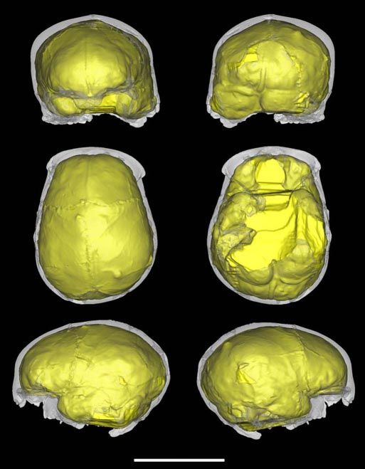
Fig. 1. Cranium 14 from SH site. (A) Frontal view, showing the left glenoid
human activities, including dietary aspects, can be treated, and
and mastoid regions well below those of the right side. (B) Left lateral view.
social support can be provided by relatives or other members of Note the rounded profile, and the vertical forehead. (C) Superior view. The
the social group. Also, it is possible to hypothesize whether an projection of the torus supraorbitalis can be clearly seen. (D) Inferior view,
affected individual would have been able to keep up with the revealing the characteristic deformities of this craniosynostosis: The posterior
group and provide for themselves within a hunter gatherer part of the cranium is twisted to the left with respect to the sagittal plane; the
context, or whether long term survival due to a serious illness or left glenoid cavity is more anteriorly placed than the right one. (Scale bar, to
injury was impossible without assistance from other members of 5 cm.)
the social group. Neanderthals as well as other Pleistocene
hominins have been claimed by some authors to likely have
shown social caring for ill/nonautonomous individuals, in the The new cranium was recovered in many pieces during the
same way as only modern humans have (1, 4). Here, we discuss 2001 and 2002 field seasons [Fig. S1 A and B and Movie S1; for
a Middle Pleistocene case of a serious congenital skull defor- fragments and labels, see Materials and Methods], and has been
mation that may have required extra conspecific care for the reconstructed during the subsequent years. As it happens with
individual to survive for a number of years before he/she died at
the end of childhood.
Author contributions: A.G. designed research; A.G., I.M., C.L., and J.L.A. performed re-
search; A.G., J.L.A., I.M., C.L., J.M.C., J.M.B.d.C., and E.C. contributed new reagents/analytic
Description of Cranium 14 tools; A.G. and J.L.A. analyzed data; and A.G., J.L.A., and I.M. wrote the paper.
The new Cranium 14 (Figs. 1 and 2) is part of a hominin sample The authors declare no conflict of interest.
of at least 28 individuals of a European Middle Pleistocene
Downloaded at Consorcio CENIEH on August 3, 2020
See Commentary on page 6429.
Homo population that are being unearthed at the Sima de los 1To whom correspondence may be addressed. E-mail: agracia@isciii.es or jlarsuaga@
Huesos (SH) site (Atapuerca, Spain) since 1976 (5, 6). The most isciii.es.
recent attempts to date the site have provided a firm minimum This article contains supporting information online at www.pnas.org/cgi/content/full/
age of 530 kya for the fossil human assemblage (7). 0900965106/DCSupplemental.
www.pnas.org兾cgi兾doi兾10.1073兾pnas.0900965106 PNAS 兩 April 21, 2009 兩 vol. 106 兩 no. 16 兩 6573– 6578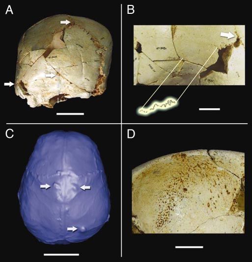
Fig. 2. Relevant features in Cranium 14. (A) Posterior view, showing the
                                                   parallelogram profile and the ipsilateral occipito-mastoid bulge, both diag-
                                                   nostic features of the left lambdoid suture premature fusion. Note the devi-
                                                   ation of the sagittal plane with respect to the sagittal suture plane, showing
                                                   that the Inion, the occipital crest and other medial structures of the nuchal
                                                   plane are positioned 10 mm or more to the left. From top to bottom, the white
                                                   arrows mark the craniometric points Lambda, Inion, and left Asterion, respec-
                                                   tively. Discontinuous lines show the displacement of the sagittal suture and          Fig. 3. Virtual endocast of Cranium 14. From top to bottom, left to right:
                                                   the occipital midline. (Scale bar, 5 cm.) (B) Close-up of part of the left lambdoid   frontal, posterior, superior, inferior, left lateral, and right lateral views. Note
                                                   synostosis. The section where the suture is almost completely obliterated is          the general bilateral asymmetry, the occipitomastoid bulging of the left side,
                                                   enhanced above. (Scale bar, 2 cm.) (C) Virtual endocast of Cranium 14, superior       the anteriorly placed left temporal lobe compared with the right, and the
                                                   view. Upper arrows point to the bregmatic arachnoid granulations. Lower               protruding left occipital pole. (Scale bar, 10 cm.)
                                                   arrow points to the obelionic arachnoid granulation. (Scale bar, 5 cm.) (D)
                                                   Cribra orbitalia on Cranium 14 right orbital roof. (Scale bar, 1 cm.)
                                                                                                                                         the spheno-occipital syncondrosis, and appears always fused in
                                                                                                                                         all of the SH adult specimens (12). Thus, based on these two
                                                   the other fossils from this site, the pieces show different degrees                   syncondroses, Cranium 14 had not reached adulthood in any
                                                   of breakage, but are in very good state of preservation, allowing                     case, and its age at death was well ⬍18-years-old.
                                                   accurate reconstructions, for example, see Cranium 5 (6). It is                          However, the 3D CT scan reconstructions of the specimen
                                                   especially important to point out that no postmortem deforma-                         (Fig. 2C and Fig. 3; for virtual reconstruction, see Materials and
                                                   tion has been found in either this particular specimen, nor in any                    Methods), have allowed us to estimate its endocranial volume at
                                                   other fossils of this collection (8).                                                 ⬇1,200 cm3. This value is within the SH adult range of variation
                                                      Cranium 14 consists of an almost complete neurocranium                             (Cranium 5 is ⬇1,100 cm3 and Cranium 4 is 1,390 cm3), and it
                                                   lacking the face, the petrous and mastoid processes of the left                       is nearly identical to that estimated for the 13.5-years-old
                                                   temporal bone, the right occipital condyle, the ethmoid bone,                         Cranium 6 (6, 13). If brain development of SH hominids is
                                                   and the central part of the sphenoid bone. After reconstruction,                      similar to that of modern humans, as it has been suggested in
                                                   Cranium 14 revealed a premature suture fusion between the left
                                                                                                                                         some studies (14–16), the individual represented by Cranium 14
                                                   parietal and occipital bones (Fig. 2 A and B; Fig. S2), which is
                                                                                                                                         was at least 5- to 8-years-old, because it had reached an adult
                                                   the core of the present study.
                                                                                                                                         brain size by the time he/she died.
                                                   Age at Death                                                                             Last, we have observed that the torus supraorbitalis is much
                                                   Cranium 14 belonged to an immature individual. There are two                          more gracile in immature specimens of the SH sample than it is
                                                   completely open syncondrosis preserved: the spheno-occipital (Fig.                    in adult individuals (6). The most important age-related thick-
                                                   S3) and the jugular (both sides). The occipital surface of the                        ness differences in the supraorbital torus are found at the medial
                                                   spheno-occipital one shows the characteristic immature irregular                      point (TMOP) and the lateral point (TLP). To try a more
                                                   and rugous pattern of unfused syncondrosis, without any bridging                      accurate estimation of the age at death of Cranium 14, we have
                                                   even in the endocranial border (9). The time of closure of the                        compared its torus supraorbitalis thickness to other two imma-
                                                   spheno-occipital syncondrosis among modern human populations                          ture specimens that have associated dentitions, both with an
                                                   is variable, ranging from the extreme cases that started at ⬇11- to                   estimated age at death ranging from 12.5- to 14.5-years-old (6,
Downloaded at Consorcio CENIEH on August 3, 2020

                                                   13- up to 18-years-old, depending on the reference sample/author                      17, 18): Cranium 6, which corresponds to SH dental individual
                                                   (9, 10).                                                                              XX, and Cranium 9, whose left frontal fragment AT-626 was
                                                      Although the closing time of the jugular syncondrosis is even                      published earlier (6), and now has been associated to SH dental
                                                   more variable (9, 11), its lower limit is well inside the range of                    individual XVI.

                                                   6574 兩 www.pnas.org兾cgi兾doi兾10.1073兾pnas.0900965106                                                                                                         Gracia et al.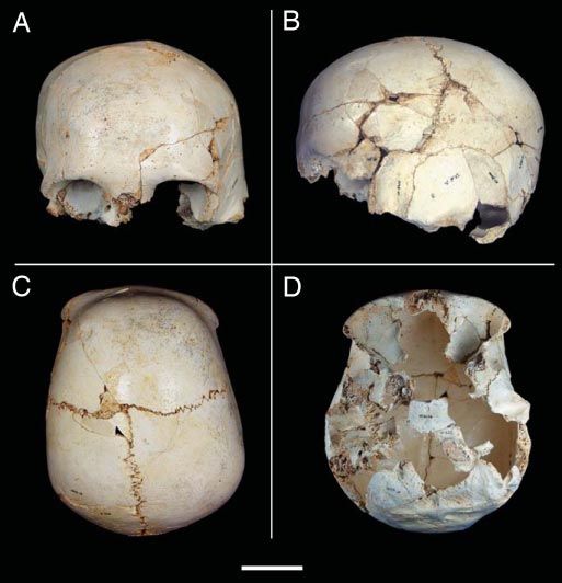
SEE COMMENTARY
Table 1. Supraorbital thickness of some SH specimens
Supraorbital
thickness
SH individuals no. Age at death, y TLP TMOP Source
Immature individuals
Cranium 6 XX 12.5–14.5 8 7.4 Authors, ref. 6
Cranium 9 XVI 12.5–14.5 6.5 6.8 Authors, ref. 6
Cranium 14 — 5–12.5 5 4 Authors, this study
Adult individuals
Cranium 4 — Adult 12.0 11.5 Authors, ref. 6
Cranium 5 XXI ⬎35 14.6 14.1 Authors, ref. 6
SH individual no. is from refs. 17 and 18. Although Cranium 4 does not have associated dentition, it is the oldest specimen of the SH
sample using age-cranial suture closure assesment. TLP, thickness at the lateral point; TMOP, thickness at the midorbital point; y, years
old.
Cranium 14 supraorbital thickness values at TMOP and TLP reconstructed endocast (Fig. 3). The foramen magnum, the
are well below those of Cranium 6 and Cranium 9 (Table 1); thus, external occipital crest, the Inion, and the suprainiac area are
its age at death should have been lower than that of those off-set and twisted 8 ° to the left with respect to the plane of the
individuals. sagittal suture (Figs. 1D, 2 A, and 3). The relative positions of the
After all these criteria, we conclude that the most probable age glenoid cavities, which is an indirect measure of ear symmetry,
at death for Cranium 14 is somewhat below the lower limit is displaced with respect to both the foramen magnum, the
estimated for Cranium 6 and Cranium 9 (i.e., ⬍12.5-years-old), sagittal suture plane and also a coronal plane (Figs. 1 A and D,
and, compared with the development of living populations (14, 2 A, and 3; compare coronal slices 80/100 in Fig. S5). The left
15), older than 5- to 8-years-old. glenoid cavity is located 4.4 mm below and 10.1 mm anterior to
that on the right side. The frontal bone morphology of all archaic
Description of the Pathological Condition Homo fossils (i.e., non-Homo sapiens) displays a receding frontal
Cranium 14 has almost complete fusion of the left lambdoid squama, and its maximum curvature is always located along the
suture, except for the first ⬇31 mm below the estimated Lambda sagittal plane, whereas the modern human frontal squama is
ANTHROPOLOGY
(Fig. 2 A and B; Fig. S2). The total arc length of the right usually vertical, and the two frontal bosses are well defined. As
(nonfused) lambdoid suture (⬇85 mm) is much shorter than the in some cases of lambdoid single suture craniosynostosis (SSC)
left side lambdoid arc length (⬇114 mm). On the right lamb- (20), Cranium 14 frontal squama presents a little right (con-
doidal suture, there is, at least, one small wormian bone (Fig. 2 A; tralateral) frontal projection. Also, Cranium 14 displays verti-
Fig. S4A). Starting at the lowest nonfused left lambdoidal suture calization of the forehead and individualization of the two
end, a well-marked horizontal depression crosses from one frontal bosses, which superficially makes this specimen resemble
lambdoid suture to the other at the occipital apex (Fig. 2 A; Fig. the condition seen in modern humans (Fig. 1 A and B).
S4A). In our opinion, it is a remainder of a complete fusion of The endocranial surface of Cranium 14 presents strong asym-
the inferior suture of a triangular-shaped wormian bone. These
metries (Figs. 2C and 3; Fig. S5), particularly in the region of the
triangular-shaped lambdoid wormian bones are frequent within
temporal lobes, with the left side shorter and more anteriorly
the SH population (6, 19), but they never appear fused, not even
placed than the right side (Fig. 3; see slice 120 in Fig. S5). The
in the older individuals (Fig. S4B).
endocranial convolutions are abnormally projected and much
In addition to the left unilateral lambdoid synostosis, Cranium
more deeply marked at the distal end of the occipital poles, at
14 shows contralateral parietal bossing, a very conspicuous
ipsilateral occipitomastoid bulge, and an ipsilateral inferior tilt the endometopic region, at the left parietal tuber, and at the
of the skull base (Figs. 2 A and 4; Fig. S5); also seen in the cerebellar poles. On the internal table of the frontal bone,
perpendicular to the sulcus sagitalis, an anomalous and smooth
capillary network is present. Three very broad and developed
subarachnoid fossae are visible on the parietal bones (Fig. 2C).
Two of them are more or less symmetrically placed at the end of
the bregmatic branches of the middle meningeal system, and the
third one, which is the largest and deepest of the three, is located
on the obelionic region of the right parietal bone.
Differential Diagnosis and Possible Etiological Factors
Cranium 14 displays a premature lambdoid suture fusion, an
uncommon pathology included among birth defects known as
craniosynostosis (20, 21), specifically a type described as SSC
(21–25). Most modern understanding of craniosynostosis is
referenced from the 1851 writings of Virchow (26). Unilateral
lambdoid synostosis is a rare pathology, occurring ⬍6 in every
200,000 individuals in living humans (27, 28).
There is abundant literature about plagiocephalia in modern
Downloaded at Consorcio CENIEH on August 3, 2020
populations due to its interest regarding the sudden infant death
Fig. 4. Diagnostic profiles of unilambdoid synostosis of Cranium 14. (A) syndrome (24, 21 and all references therein); thus, a lot of
Trapezoid-shape in vertex view. (B) Parallelogram profile in posterior view. attention has been paid to make the differential diagnosis of
(Scale bar, 5 cm.) plagiocephalia (deformational or positional plagiocephalia
Gracia et al. PNAS 兩 April 21, 2009 兩 vol. 106 兩 no. 16 兩 6575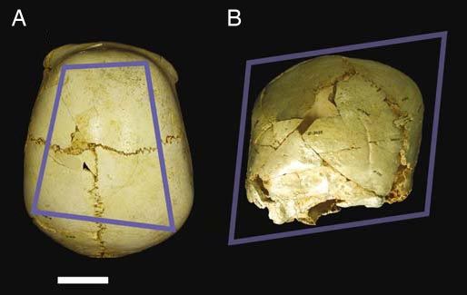
versus a real unilateral lambdoidal synostosis (20–23, 29, 30)) in       and 34 fetal weeks (9). These data suggest that the synostosis of
                                                   living children, because the treatment differs depending on it.          this suture in Cranium 14 was initiated during the third term of
                                                      Cranium 14 displays a trapezoidal-shaped and parallelogram            fetal development.
                                                   profiles in vertex and posterior views, respectively (Fig. 4 A and          Thus, considering that there is some evidence about the timing
                                                   B), which are definitive diagnostic signs of unilateral lambdoid         of the initiation of the fusion, the possible etiology of the SSC
                                                   synostosis in modern children (20–32), as well as the 8 ° of the         of Cranium 14 can be summarized as follows. The first case
                                                   off-set ipsilateral deviation of the intersection of posterior fossa     would be premature SC of traumatic origin due either to (i)
                                                   axis and the anterior fossa axis (29). This deformation is the           intrauterine stress/constrait (37), (ii) torticollis congenita (21,
                                                   result of the premature fusion reaction that produces: ‘‘First, in       38), or (iii) intrauterine trauma (21) and would exclude other
                                                   true lambdoid synostosis, the contralateral posterior bossing            frequently quoted, labor complications (39, 40). The second case
                                                   more laterally and superiorly, in the parietal region. Second,           would be premature SC due to a metabolic disease, either (i)
                                                   frontal bossing is not a striking feature, but when it occurred, it      rickets and/or (ii) anemia. Cranium 14 also shows the most
                                                   was contralateral rather than ipsilateral. (…) Third, ipsilateral        pronunced example of cribra orbitalia (Fig. 2D) within the entire
                                                   occipitomastoid bossing was consistently present in lambdoid             SH sample (compare with ref. 41). The right orbit is severely
                                                   synostosis, whereas it was conspicuously absent in deformational         affected, showing extensive pitting on the roof. Both anemia and
                                                   posterior plagiocephaly’’ (20). As it has been established for           rickets are claimed to be metabolic causes associated with the
                                                   other craniosynostotic cases in modern populations, the mis-             presence of cribra orbitalia (42, 43). Since rickets and anemia
                                                   shapen cranial abnormalities present in Cranium 14 constitute a          have been related to some cases of craniosynostosis (34, 44, 45),
                                                   secondary response to the lambdoid synostosis, rather than a             we cannot exclude these metabolic diseases as the possible
                                                   causal factor (24). The same must be true for the presence of the        trigger factor of the premature suture fusion. Nevertheless, we
                                                   small right lambdoid ossicle (Fig. S4A). As has been documented          consider these options less probable, because except for some
                                                   for intentionally deformed modern human skulls (33), we con-             genetic vitamin D deficiencies, these pathological conditions
                                                   sider the presence of small lambdoid ossicles to be further              develop after birth, and in Cranium 14, the synostosis seems to
                                                   evidence of an anomalous compensatory rapid growth that also             have started during the fetal stage. Also, in Cranium 14 there are
                                                   produced the contralateral displacement of the right parietal            no other cranial signs usually associated to rickets or anemia,
                                                   tuber. Unlike many other synostotic cases (20), the fused suture         such as porotic hyperostosis.
                                                   is not ridged in this specimen. The same holds true for other
                                                   lambdoid synostosis studied in modern humans, where no sig-              Discussion
                                                   nificant ridging was found (34). The fusion seems to have been           There are some references about pathological specimens from the
                                                   edge to edge, and the exocranial (Fig. 2 A; Fig. S2) and endocra-        Pleistocene hominid fossil record with serious developmental/
                                                   nial surfaces of the synostosed suture appear continuous and             degenerative abnormalities. Among them, the oldest is dated in 1.77
                                                   smooth.                                                                  kya and corresponds to that of the edentulous D3444/D3900
                                                      The parietal, temporal, and occipital deformations seen in this       individual from the Dmanisi site, who ‘‘apparently survived for a
                                                   fossil are very similar to other ‘‘true’’ plagiocephalic cases (e.g.,    lengthy period without consuming foods that required heavy chew-
                                                   see figures 6 and 7 in ref. 22), and, with regard to the frontal bone    ing’’ (46). In this study, the survival of this specimen was interpreted
                                                   morphology, in this case, the frontal squama compensatory                as a possible evidence of conspecific care. Another case of some
                                                   deformation does not result in a conspicuous contralateral               degree of survival after an important disorder is that of the
                                                   bossing, as it has sometimes been found in modern human                  individual represented by the Hulu 1 cranium, that presents signs
                                                   populations (20–24). On the contrary, as described above, the            of a healed pathological lesion on the neurocranial vault surface (1).
                                                   compensatory distortion on the frontal bone of Cranium 14                With respect to malformations, there are two cases of special
                                                   resulted in a modern-like frontal bone morphology (Fig. 1 A and          interest: the cranium of Salé, whose occipital deformation was
                                                   B). The same holds true with respect to the glenoid cavity               interpreted as a result of torticollis congenita (47), and the temporal
                                                   asymmetry, which more frequently presents a posterior position           bone pathology from Singa 1, which lacks the structures of the bony
                                                   for the ipsilateral glenoid fossa, but, in other cases, it can be        labyrinth (48). This Middle Pleistocene specimen also presents
                                                   found anteriorly placed or even not displaced at all (21).               parietal diploic expansion, and was previously cited as a probable
                                                      With regard to the etiology of SSC, it is very difficult to           case of premature synostosis of the sagittal suture (49). In our
                                                   establish it, even for modern humans. Nevertheless, we have              opinion, premature sagittal synostosis was not present in the Singa
                                                   some arguments that can be applied to the case of Cranium 14             1 specimen, because, in almost all sagittal synostosis cases, the
                                                   SSC.                                                                     vertex cranial shape should be elongated antero-posteriorly
                                                      The premature unilambdoidal fusion found in Cranium 14 can            (scaphocephalia) and, in posterior view, with a disproportionally
                                                   be the result of primary or secondary craniosynostosis, being            narrow cranium (27), which is not the case of this specimen.
                                                   defined the former as the ‘‘polar term of the pair’’ sensu Cohen (21),      As it has been claimed by DeGusta (50), inferences made
                                                   where ‘‘sutural obliteration is secondary to a known disorder.’’         about behavior in fossil hominid populations from skeletal
                                                      In a suture fused prematurely, growth is mainly restricted            pathologies must be tested with a comparative approach, but it
                                                   orthogonal to the fused suture, from the ossification center to the      is clear that the fossil evidence of individual cases that survived
                                                   fused suture (34–36). Although growth does not completely                with different degrees of impairment has increased a lot (1 and
                                                   stop, almost no change is seen during development. In response           references therein), and have been pointed out in some cases as
                                                   to a prematurely closed suture, the remaining sutures undergo            evidence of conspecific care (4, 46).
                                                   compensatory growth in a very fixed pattern (35, 36). In                    The present pathology of Cranium 14 from the SH site must
                                                   Cranium 14, the primary ossification center (parietal protuber-          also be considered from this point of view, due to the handi-
                                                   ance) of the left parietal bone is very close in straight line           capping lesions that this individual could have had. It is obvious
                                                   (perpendicular) to the fused lambdoid suture (arc, ⬇30 mm;               that the SH hominin species did not act against the abnormal/ill
                                                   chord, ⬇28 mm) (Fig. 2 A; Fig. S2). According to this criteria,          individuals during the infancy, as has happened along our own
                                                   and assuming that the newborn parietal is square shaped (9) and          history many times and in many cultures, and can be illustrated,
Downloaded at Consorcio CENIEH on August 3, 2020

                                                   that fetal development and newborn-brain size were similar to            for example, with the case of the elevated frequency of cranio-
                                                   that of modern humans (15, 16), the distance in Cranium 14               synostosis found in the cemetery of the Medieval Hospital of St.
                                                   would correspond to a modern H. sapiens fetal parietal bone with         James and St. Mary Magdalene (Chichester, England), that
                                                   a maximum width of ⬇60 mm, size that is reached between 28               worked as an almshouse since AD 1450, where children with

                                                   6576 兩 www.pnas.org兾cgi兾doi兾10.1073兾pnas.0900965106                                                                                   Gracia et al.
SEE COMMENTARY
deforming congenital conditions among others were aban- AT-3413, AT-3416, AT-3430, AT-3431, AT-3432, AT-3434, AT-3435, AT-3436,
doned (51). AT-3439, AT-3442, AT-3451, AT-3893, AT-3896, AT-3898, AT-4234, AT-4236,
The premature sutural synostosis, as well as the abnormal AT-6224, and AT-6225.
endocranial features found in Cranium 14, especially the pres-
Virtual Reconstruction of Cranium 14 and Its Endocranium. CT image data of
ence of dilatation of subarachnoid spaces, were probably asso-
Cranium 14 was scanned with a YXLON Compact (YXLON International X-Ray)
ciated with an elevated intracranial pressure, as it has been industrial multislice computed tomography (CT) scanner, located in Burgos
described for SSC cases in living humans (24, 28, 31, 32, 52, 53). University, Spain. The specimen was aligned along a cranial-caudal axis with
Most cases of craniosynostosis, both of genetic or epigenetic the browridge facing upwards, to obtain para-coronal slices. Scanning pa-
origin (syndromes, torticollis congenita, genetic or metabolic rameters were: scanner energy 160 kV and 4 mA. Slice thickness was collimated
diseases, intrauterine position stress, traumas, labor complica- to 0.5 mm, interslice spacing was 0.5 mm, and field of view 299.47 mm,
tions; see refs. 21, 24, 31–41), are normally present before birth, reconstruction interval of 0.5 mm; 390 slices were obtained as a 1,024 ⫻ 1,024
and are noticeable externally during the first year of life. Also, matrix of 32-bit Float format with a pixel size of 0.216 mm, and transferred for
genetic and severe nongenetic cases worsen during growth, processing. By using commercially available software package Mimics v.10.0,
(Materialise), the CT image data of Cranium 14 were visualized, studied,
including both the cognitive capabilities and the aesthetics of the
measured, and processed to make a virtual endocast. The internal braincase
individual (21, 22, 24, 25, 27, 30–32, 45). And finally and
was enclosed manually to close any contour gaps in the skullcap, and to
probably most importantly, there are known cases of premature ‘‘complete’’ any missing areas in Cranium 14 to the minimum surface. The
SSC associated with a variable degree of mental retardation virtual 3D endocast was created with a cavity fill operation within the Mimics
and/or elevated intracranial pressure (20–26, 31, 52–58). 3D Segmentation module. The estimated encephalic volume, which is a min-
Despite the severe pathological signs present in Cranium-14 imum value, is 1,200 cm3. This value is the same as that obtained for Cranium
child already described, it is not possible to estimate the degree 6, which was recalculated in a previous study, yielding an estimation of 1,200
of mental retardation. cm3 (59).
In conclusion, Cranium 14 is the earliest documented case of
craniosynostosis with resulting neurocranial, brain deformities, ACKNOWLEDGMENTS. We thank the Atapuerca excavation team, especially
that of the Sima de los Huesos; the Burgos University for providing the CT
and, very likely, asymmetries of the facial skeleton. Despite these scanner; L. Rodrı́guez, E. Santos, and R. Garcı́a for their technical assistance in
handicaps, this individual survived for ⬎5 years, suggesting that CT scanning; Y. Rak, W. Hylander, A. Bartsiokas, R. Quam, G. Cuenca-Bescós,
her/his pathological condition was not an impediment to receive M. Martı́n-Loeches, and the 2 reviewers, who provided helpful comments;
the same attention as any other Middle Pleistocene Homo child. A. Bonmatı́, F. Gracia, J. Lira, and J. Trueba (Madrid Scientific Films) for their
help with the figures, photographs, and video; and Kennis & Kennis for their
portrait of Cranium 14. This work was supported by Ministerio de Ciencia y
Materials and Methods Tecnologı́a Spanish Government Grant CGL2006-13532-C03-02. The field ex-
Cranium 14. Cranium 14 is composed of the following labeled fragments: cavation work was supported by Junta de Castilla y León and Fundación
AT-2044, AT-3239, AT-3400, AT-3403, AT-3405, AT-3406, AT-3411, AT-3412, Atapuerca.
ANTHROPOLOGY
1. Shang H, Trinkaus E (2008) An ectocranial lesion on the Middle Pleistocene human 18. Bermúdez de Castro JM, Martinón-Torres M, Lozano M, Sarmiento S, Muela A (2004)
cranium from Hulu cave, Nanjing, China. Am J Phys Anthrop 135:431– 437. Paleodemography of the Atapuerca-Sima de los Huesos hominin sample: A revision
2. Wood JW, Milner GR, Harpending HC, Weiss KM (1992) The Osteological Paradox: and new approaches to the paleodemography of the European Middle Pleistocene
Problems of Inferring Prehistoric Health from Skeletal Samples. Curr Anthropol population. J Anthrop Res 60:5–26.
33:343–370. 19. Manzi G, Gracia A, Arsuaga JL (2000) Cranial discrete traits in the Middle Pleistocene
3. Brothwell DR (1981) Digging up Bones: The Excavation, Treatment and Study of humans from Sima de los Huesos (Sierra de Atapuerca, Spain). Does hypostosis repre-
Human Skeletal Remains, British Museum of Natural History (Oxford Univ Press, sent any increase in ‘‘ontogenetic stress’’ along the Neanderthal lineage? J Hum Evol
Oxford), pp 1–208. 38:425– 446.
4. Lebel S, Trinkaus E (2002) Middle Pleistocene human remains from the Bau de 20. Huang MHS, et al. (1996) The differential diagnosis of posterior plagiocephaly: True
l’Aubesier. J Hum Evol 43:659 – 685. lambdoid synostosis versus positional molding. Plast Reconst Surg 98:765–774.
5. Arsuaga JL, et al. (1997) Sima de los Huesos (Sierra de Atapuerca, Spain). The site. J Hum 21. Cohen MM, Jr, MacLean RE (2000) Craniosynostosis. Diagnosis, Evaluation and Man-
Evol 33:109 –127. agement, eds Cohen MM, Jr, MacLean RE (Oxford Univ Press, New York), pp 119 –143.
6. Arsuaga JL, Martı́nez I, Gracia A, Lorenzo C (1997) The Sima de los Huesos crania (Sierra 22. Smartt JM, Reid R, Singh DJ, Bartlett SP (2007) True lambdoid craniosynostosis: Long-
de Atapuerca, Spain). A comparative study. J Hum Evol 33:219 –281. term results of surgical and conservative theraphy. Plast Reconst Surg 120:993–1003.
7. Bischoff JL, et al. (2007) High-resolution U-series dates from the Sima de los Huesos 23. Mulliken JB, et al. (1999) Analysis of posterior plagiocephaly: Deformational versus
hominids yields 600 kyrs: Implications for the evolution of the early Neanderthal synostotic. Plast Reconst Surg 103:371–380.
24. Panchal J, Uttchin V (2003) Management of craniosynostosis. Plast Reconst Surg
lineage. J Archaeol Sci 34:763–770.
111:2032–2048.
8. Arsuaga JL, Carretero JM, Gracia A, Martı́nez I (1990) Taphonomical analysis of the
25. Speltz ML, Kapp-Simon KA, Cunningham M, Marsh J, Dawson G (2004) Single-Suture
human remains from the Sima de los Huesos Middle Pleistocene site (Atapuerca/Ibeas,
Craniosynostosis: A Review of Neurobehavioral Research and Theory. J Pediatr Psychol
Spain). Hum Evol 5:505–513.
29:651– 668.
9. Scheuer L, Black S, eds (2000) Developmental Juvenile Osteology, eds Scheuer L, Black
26. Cohen MM, Jr (2000) in Craniosynostosis. Diagnosis, Evaluation and Management, eds
S, (Academic, London), pp 36 –170.
Cohen MMJ, MacLean RE (Oxford Univ Press, New York), pp 103–110.
10. Bonmatı́ A, Arsuaga JL, Lorenzo C (2008) Revisiting the Developmental Stage and
27. Cohen MM, Jr (2000) Craniosynostosis. Diagnosis, Evaluation and Management, eds
Age-at-Death of the ‘‘Mrs. Ples⬙ (Sts 5) and Sts 14 Specimens from Sterkfontein (South
Cohen MMJ, MacLean RE (Oxford Univ Press, New York), pp 112–118.
Africa): Do They Belong to the Same Individual? Anat Rec 291:1707–1722.
28. Rekate HL (1998) Occipital plagiocephaly: A critical review of the literature. J Neuro-
11. Hershkovitz I, et al. (1997) The elusive petroexoccipital articulation. Am J Phys An-
surg 89:24 –30.
thropol 103:365–373.
29. Sze RW, et al. (2005) MDCT Diagnosis of the child with posterior plagiocephaly. Am J
12. Martı́nez I, Arsuaga JL (1997) The temporal bones from Sima de los Huesos Middle Rad 185:1342–1346.
Pleistocene site (Sierra de Atapuerca, Spain). A phylogenetic approach. J Hum Evol 30. Ehert FW, Whelan MF, Ellenbogen RG, Cunningham ML, Gruss JS (2004) Differential
33:283–318. diagnosis of the trapezoid-shaped head. Cleft Palate Craniofac J 41:13–19.
13. Arsuaga JL, et al. (1997) Size variation in Middle Pleistocene Humans. Science 31. Kapp-Simon KA, Speltz ML, Cunningham ML, Patel PK, Tomita T (2007) Neurodevel-
277:1086 –1088. opment of children with single suture craniosynostosis: A review. Childs Nerv Syst
14. Hublin JJ, Coqueugniot H (2006) Absolute or proportional brain size: That is the 23:269 –281.
question. A reply to Leigh’s comments. J Hum Evol 50:109 –113. 32. Kabbani H, Raghuveer TS (2004) Craniosynostosis. Am Fam Physic 69:2863–2870.
15. DeSilva JM, Lesnik JJ (2008) Brain size at birth throughout human evolution: A 33. Dean V (2004) Effects of different kinds of cranial deformation on the incidence of
new method for estimating neonatal brain size in hominins. J Hum Evol 55:1064 – wormian bones. Am J Phys Anthrop 123:146 –155.
1074. 34. Cohen MM, Jr (2000) Craniosynostosis. Diagnosis, Evaluation and Management, eds
Downloaded at Consorcio CENIEH on August 3, 2020
16. Ponce de León MS, et al. (2008) Neanderthal brain size at birth provides insights into Cohen MMJ, MacLean RE (Oxford Univ Press, New York), pp 51– 68.
the evolution of human life history. Proc Natl Acad Sci USA 105:13764 –13768. 35. Mathijssen IM, et al. (1999) Tracing craniosynostosis to its developmental stage
17. Bermúdez de Castro JM, et al. (2003) Rates of anterior tooth wear in Middle Pleistocene through bone center displacement. J Craniofac Gen Dev Biol 19:57– 63.
hominins from Sima de los Huesos (Sierra de Atapuerca, Spain). Proc Natl Acad Sci USA 36. Delashaw JB, Persing JA, Broaddus WC, Jane JA (1989) Cranial vault growth in cranio-
100:11992–11996. synostosis. J Neurosurg 70:159 –165.
Gracia et al. PNAS 兩 April 21, 2009 兩 vol. 106 兩 no. 16 兩 657737. Higginbottom MC, Jones KL, James HE (1980) Intrauterine constraint and craniosyn- 49. Brothwell DR (1974) in The Upper Pleistocene Singa Skull: A Problem in Paleontolog-
ostosis. Neurosurgery 6:39 – 44. ical Interpretation in Bevoelkerungsbiologie, eds Bernard W, Kandler A (Carl Fisher,
38. Raco A, et al. (1999) Congenital torticollis in association with craniosynostosis. Childs Stutgart), pp 534 –545.
Nerv Syst 15:163–168. 50. DeGusta D (2002) Comparative skeletal pathology and the case for conespecific care in
39. Bermejo E, et al. (2005) Craniofacial dyssostosis: Description of the First four Spanish Middle Pleistocene hominids. J Archaeol Sci 29:1435–1438.
Cases and Review. Am J Med Genet 41– 48. 51. Storm RA (2007) High Prevalence of Premature Craniosynostosis in the Medieval
40. Shahinian HK, et al. (1998) Obstetrical factors governing the etiophatogenesis of Hospital of St. James and St. Mary Magdalene, Chichester, England, Seventy-Sixth
lambdoid synostosis. Am J Perinat 15:281–286. Annual Meeting of the American Association of Physical Anthropologists, March
41. Pérez PJ, Gracia A, Martı́nez I, Arsuaga JL (1997) Paleopathological evidence of the 28 –31, 2007, Philadelphia, PA, p 226.
cranial remains from the Sima de los Huesos Middle Pleistocene site (Sierra de Atapu- 52. Thompson DNP, Malcolm GP, Jones BM, Harkness WJ, Hayward RD (1995) Intracranial
erca, Spain). Description and preliminary inferences. J Hum Evol 33:409 – 421. pressure in single-suture craniosynostosis. Pediatr Neurosurg 22:235–240.
42. Stuart-Macadam P (1987) A radiographic study of porotic hyperostosis. Am J Phys 53. Martı́nez-Lage JF, Alamo L, Poza M (1999) Raised intracranial pressure in minimal forms
Anthrop 74:511–520. of craniosynostosis. Childs Nerv Syst 15:11–16.
43. Schultz M (2003) Light microscopic analysis in skeletal paleopathology. Identification 54. Cinalli G, et al. (1998) Hydrocephalus and Craniosynosotosis. J Neurosurg 88:209 –214.
of Pathological Conditions in Human Skeletal Remains, ed Ortner DJ (Academic, San 55. Chadduck WM, Chadduck JB, Boop FA (1992) The subarachnoid spaces in craniosyn-
Diego), pp 73–108. ostosis. Neurosurgery 30:867– 871.
44. Duggan C, Keener E, Gay B (1970) Secondary Craniosynostosis. Am J Roentgenol 56. Rasmussen SA, Yazdy MM, Frı́as JL, Honein MA (2008) Priorities for public health
19:277–293. research on craniosynostosis: Summary and recommendations from a centers for
45. Portillo S, Konsol O, Pico P (2004) Cranial Deformity. Importance in General Pediatrics disease control and prevention-sponsored meeting. Am J Med Genet 146A:149 –158.
(Translated from Spanish). Arch Argent Pediatr 102:190 –202. 57. Coussens AK, et al. (2007) Unravelling the molecular control of calvarial suture fusion
46. Lordkipanidze D, et al. (2005) The earliest toothless hominin skull. Nature 434:717– in children with craniosynostosis. BioMed Centr Genom 8:458 – 482.
718. 58. Becker DB, et al. (2005) Speech, cognitive and behavioral outcomes in nonsyndromic
47. Hublin JJ (1991) Origin of archaic Homo Sapiens: North-West Africa and Western craniosynostosis. Plast Reconst Surg 116:400 – 407.
Europe (Translated from French). PhD thesis (University of Bordeaux I), 2 vol. 59. Arsuaga JL, Martı́nez I, Gracia A (2001) Phylogenetic Analysis of the Hominids from the
48. Spoor F, Stringer C, Zonneveld F (1998) Rare temporal bone pathology of the Singa Sierra de Atapuerca (Sima de los Huesos and Gran Dolina TD-6): Cranial evidence
calvaria from Sudan. Am J Phys Anthrop 107:41–50. (Translated from French). L’Anthropologie 105:161–178.
Downloaded at Consorcio CENIEH on August 3, 2020
6578 兩 www.pnas.org兾cgi兾doi兾10.1073兾pnas.0900965106 Gracia et al.You can also read