Charcot identifies and illustrates amyotrophic lateral sclerosis
←
→
Page content transcription
If your browser does not render page correctly, please read the page content below
Free Neuropathology 2:12 (2021)                                                                                                                                        Charles Duyckaerts et al
doi: https://doi.org/10.17879/freeneuropathology-2021-3323                                                                                                                        page 1 of 10

                                                                                                                                                                                Flashback

Charcot identifies and illustrates amyotrophic lateral sclerosis
Charles Duyckaerts1,2, Thierry Maisonobe1,3, Jean-Jacques Hauw4, Danielle Seilhean1,5
1
    Raymond Escourolle Neuropathology Department, La Salpêtrière Hospital, Assistance Publique des Hôpitaux de
    Paris, Sorbonne Université, Paris, France
2
    Alzheimer-Prions team, Paris Brain Institute ICM (INSERM U1127, CNRS UMR7225), Sorbonne Université-
    UMRS1127), Paris, France
3
    Clinical Neurophysiology Department, La Salpêtrière Hospital, Assistance Publique des Hôpitaux de Paris, Sor-
    bonne Université, Paris, France
4
    Académie Nationale de Médecine, Paris, France
5
    ALS: Causes and mechanisms of motor neuron degeneration, Paris Brain Institute ICM (INSERM U1127, CNRS
    UMR7225, Sorbonne Université-UMRS1127), Paris, France

Corresponding author:
Charles Duyckaerts · Raymond Escourolle Neuropathology Department · La Salpêtrière Hospital · Assistance Publique des Hôpitaux de
Paris · Sorbonne Université · Paris · France
charles.duyckaerts@gmail.com

Submitted: 31 March 2021 · Accepted: 08 May 2021 · Copyedited by: Lauren Walker and Henry Robbert ·                                                                    Published: 18 May 2021

Abstract
Jean-Martin Charcot described what he called amyotrophic lateral sclerosis in his 12th and 13th lessons published
in 1873 by Bourneville. He distinguished the symptoms that were related to the lesion of the anterior horn of the
spinal cord and those that were due to the degeneration (that he named “sclerosis”) of its lateral column. He
thought that “inflammation” progressed from the lateral column to the anterior horn (but the term inflammation
is not to be taken in the current meaning): the lesion of the anterior horn was thus “deuteropathic”. An album
containing drawings made by Charcot is kept in La Salpêtrière Neuropathology Department. Four drawings are
pasted on one of its pages, showing the degeneration of the pyramidal tract. They constitute the original of the
engravings illustrating Charcot’s 12th lesson. The illustration of the fascicular atrophy of the adductor pollicis pre-
sented in the album does not appear in the lessons, even though this alteration is widely discussed and linked to
the lesion of the anterior horn, which was supposed to ensure the “nutrition” of the muscle. The technique used
by Charcot and his interpretation of the microscopic pictures, as exposed in his lessons, are discussed.

Keywords: Amyotrophic lateral sclerosis, Charcot, History of medicine, Pyramidal tract

Introduction                                                                                                conception are remarkably summarized in the sec-
                                                                                                            ond volume of his Lessons on the diseases of the
     Jean-Martin Charcot identified amyotrophic                                                             nervous system, edited by Bourneville (Charcot,
lateral sclerosis (ALS) on clinical-pathological                                                            1873) and in his Lessons on cerebral localization, ed-
grounds. The data, which he collected, and his                                                              ited by Bourneville et Brissaud (Charcot, 1876). The

Copyright: © 2021 The author(s). This is an open access article distributed under the terms of the Creative Commons Attribution 4.0 International License (https://creativecommons.org/licenses/by/4.0/),
which permits unrestricted use, distribution, and reproduction in any medium, provided the original author and source are credited, a link to the Creative Commons license is provided, and any changes are
indicated. The Creative Commons Public Domain Dedication waiver (https://creativecommons.org/publicdomain/zero/1.0/) applies to the data made available in this article, unless otherwise stated.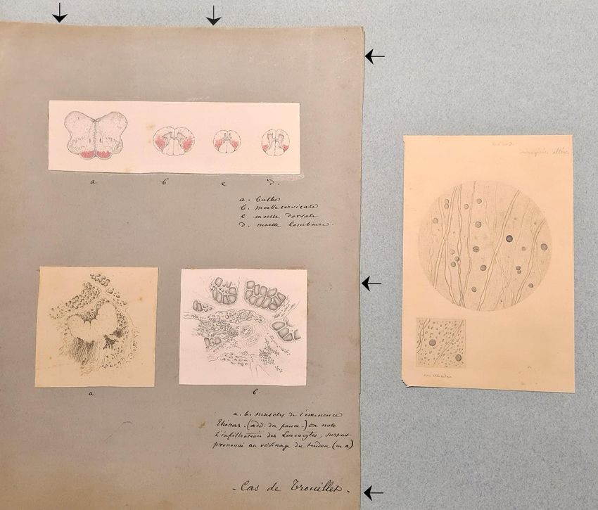
Free Neuropathology 2:12 (2021)                                                                     Charles Duyckaerts et al
doi: https://doi.org/10.17879/freeneuropathology-2021-3323                                                     page 2 of 10

Neuropathology Laboratory of La Salpêtrière Hospi-
tal keeps an album containing original drawings by
Charcot. One of its pages concerns an ALS case. This
paper compares the two documents: the drawings
and the description of the disease, both by Charcot.

The album
      The album bears the title “Charcot Museum,
Pathological anatomy”, handwritten in ink on the
black hardcover (Fig. 1). It contains 100 light-blue
pages of a format known as “Couronne” in France
                                                             Fig. 1. Hardcover of the album in which Charcot’s drawings were
(37 x 47 cm), commonly used by the artists of the            collected.
time. Pages 15 to 100 are blank. They were num-
bered with stencils. “Foxing” (i.e. brown spots and
browning of the edges) suggests they are original.           generally concerns a particular topic (Microaneu-
The first fourteen pages have been restored, as well         rysm, multiple sclerosis, myelitis, lateral funiculus in
as the binding, some twenty years ago, and were re-          a foetus, syringomyelia, Pott’s disease, vertebral
numbered in pencil. The drawings, usually on white           metastases, anatomy of the posterior funiculi, lead
papers, are pasted on those pages. There are also a          paralysis, tumor of the spinal cord, ALS, cysticerco-
few engravings, some taken from Charcot’s articles.          sis). The album has never been the subject of a spe-
All the drawings have the same style and, although           cific study, although some of its pictures have been
not signed, are reputed to be by Charcot himself.            published occasionally (the drawing of the central
Charcot had hesitated between medicine and the               nervous system of the ALS case under investigation
fine arts. He left numerous drawings of pathological         here has been published in Seilhean, 2020).
anatomy and sketches of patients (see for instance,
the case Bachère, Charcot, 1892, fig. 64, p 337,             Cases and samples
https://gallica.bnf.fr/ark:/12148/bpt6k55646784/f3
43.item.texteImage; or a case of Parkinson’s disease               Three original drawings of the ALS case, p 12 of
ibidem, fig. 63, p 336; the illustration of “a labioglos-    the album (Fig. 2), made on white papers, are pasted
sal spasm”, Charcot, 1892, fig. 19 and 20, p 211,            on a grey (faded green) cardboard and concern a
https://gallica.bnf.fr/ark:/12148/bpt6k55646784/f2           case, identified as “Trouillet’s case” at its bottom
17.image.r=fig; or the picture of the face in a case of      (Fig. 2 and Fig. 4). They include sections of the me-
Parkinson’s disease, Charcot, 1892, fig. 60, p 334,          dulla oblongata and spinal cord, shown in the supe-
https://gallica.bnf.fr/ark:/12148/bpt6k55646784/f3           rior part of the card (Fig. 3) and labeled a, b, c, d, and
40.image.r=fig).                                             two views of the adductor pollicis muscle, which oc-
                                                             cupy the inferior part and are labeled a and b (Fig.
      These drawings demonstrate his talent for illus-
                                                             4). A microscopic view of “altered slender columns”
trating the cases that he examined. The style of the
                                                             of the spinal cord (Fig. 5), possibly from another
drawings found in the album is identical to that of
                                                             case, has been directly pasted on the right side of
the drawings found in the notes that he prepared for
                                                             the page as shown in Fig. 2.
his “lessons” (still visible at Charcot library, now lo-
cated at Paris Brain Institute-ICM, https://instit                The views of “Trouillet’s case” are not dated
utducerveau-icm.org/en/actualite/charcot-library):           but, as we found a great similarity between the
there is no reason to question their authorship. The         drawings of the album and the engravings of the 12th
drawings, initially made on loose sheets, were sec-          lesson (Charcot, 1873, pp 221, fig. 16-19), we are in-
ondarily collected and pasted in the album that was          clined to believe that they are anterior to 1873. We
passed down from Charcot’s own laboratory to the             could not elucidate the identity of that Trouillet,
current department of neuropathology. Each page              probably the doctor who referred the patient to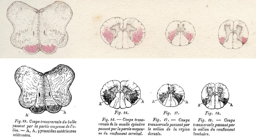
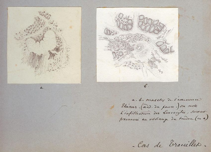
Free Neuropathology 2:12 (2021)                                                                                    Charles Duyckaerts et al
doi: https://doi.org/10.17879/freeneuropathology-2021-3323                                                                    page 3 of 10

Fig. 2. General view of page 12 of the album dealing with amyotrophic lateral sclerosis. Three drawings are pasted on a grey (faded green)
cardboard (arrows). They show sections of the central nervous system (at the top) and of the adductor pollicis (at the bottom). In the
lower right corner of the card, “Cas de Trouillet” (Trouillet’s case) can be read. An additional drawing on the right illustrates two micro-
scopic aspects of an “altered slender column [of the spinal cord]”.

Charcot. Charcot gave the names of the two ALS                            (Charcot, 1873, p 416) with Charcot’s commentary
cases that he published previously, as was usual at                       “with the collaboration of Gombault” as if he, Char-
the time. The first case, Catherine Aubel, was pub-                       cot, known for his authoritarianism, was the first co-
lished in a paper entitled “Two cases of progressive                      author. Trouillet is not mentioned in those two arti-
muscular atrophy with lesions of the grey matter                          cles. As Charcot indicated that his lesson was based
and of the anterolateral fascicles of the white mat-                      on the study of five clinical-pathological cases (Char-
ter” (Charcot and Joffroy, 1869, first case, p 354 re-                    cot, 1873, p 228, footnote 1) and as three cases are
produced in Charcot, 1873, p 402). The disease of                         identified (Catherine Aubel, Elisabeth P. and Trouil-
the second case of that paper (named A.C.) is not re-                     let’s case), two remain unidentified and, as far as we
ported in the lessons and is not identified as ALS by                     know, unpublished.
Charcot. The second case identified as ALS, Elisabeth
P., was published with only Gombault as its author
(Gombault, 1871). It is reproduced in the Lessons

Free Neuropathology 2:12 (2021)                                                                                     Charles Duyckaerts et al
doi: https://doi.org/10.17879/freeneuropathology-2021-3323                                                                     page 4 of 10

Fig. 3.
Upper part: Page 12 of the album. Trouillet’s case (see Fig. 2). Four sections of the central nervous system. The section are labeled a, b,
c, d by Charcot himself. The legend reads:
    a: bulbe
    b: moelle cervicale
    c: moelle dorsale
    d: moelle lombaire
which means: a: medulla oblongata; b: spinal cord at cervical level; c: at thoracic level, d: at lumbar level.
Lower part: below the original drawings of Charcot found in the album, the engravings of lesson 12 (Charcot, 1873, pp 221, fig. 16-19).
The legend in French reads:
    Fig. 19. Cross section of the medulla through the middle part of the olive. A, A, Sclerotic anterior pyramids
    Fig. 16. – Transverse section of the spinal cord through the middle part of the cervical enlargement.
    Fig. 17. – Transverse section through the middle of the dorsal region.
    Fig. 18. – Transverse section through the middle of the lumbar enlargement.

Preparation of microscopic sections by                                      chromic acid” (the fixation properties of formalde-
                                                                            hyde were described by F. Blum two decades later –
Charcot (et Bouchard)                                                       Blum, 1893). The microtome, as we know it today,
                                                                            was probably not in use in Paris at the time. Its in-
      The technique that has been used to obtain the
                                                                            vention by Purkinje in 1841 (Chvátal, 2017) or His in
illustrated sections is not reported. We can rely on
                                                                            1866 (Dupont, 2018) is controversial but it is clear
two articles in which Charcot indicated how he pro-
                                                                            that it was commonly used much later. The manual
ceeded (Charcot and Joffroy, 1869 reproduced in
                                                                            of histological techniques by Mathias Duval (1878),
Charcot, 1873, pp 402-416; Charcot, 1865).
                                                                            for instance, does not mention microtomes (in the
    The central nervous system was fixed for                                current meaning) and concludes: “A large number of
“nearly one month” with a “much diluted solution of                         instruments have been invented [to cut fixed
Free Neuropathology 2:12 (2021) Charles Duyckaerts et al
doi: https://doi.org/10.17879/freeneuropathology-2021-3323 page 5 of 10
pieces]. One comes always back to the simple razor manual of Mathias Duval (1878) indicates that one
which is more efficient than all complicated ‘disco- common practice was to cover the section with a co-
tomes’ [we put in quotes] if the habit required to use verslip (much thicker than they are today) and to in-
it skillfully has been acquired.” The fixed sample was troduce between the slide and the coverslip, on one
usually stuck in elderberry pith and cut, freehand, side, a drop of glycerol that would diffuse and push
with a razor blade. the water out on the other side. The coverslip could
then be sealed with wax or bitumen of Judea (Duval,
The sections were not dehydrated and 1878).
mounted in balsam or resin, as they are today. The
Fig. 4. Two drawings, labeled a and b, are visible in the lower part of the page.
a: one aspect of the adductor pollicis. A tendon appears as an unstained mass in the center of the section. Numerous atrophic fibers
are visible.
b: another aspect of the adductor pollicis. Fascicular atrophy of the muscle fibers are clearly seen around an artery in the center of
the section.
The legend, written by Charcot, reads: “a, b muscles de l’éminence thénar (add du pouce). On note l’infiltration des leucocytes, surtout
prononcée au voisinage du tendon en a.” which means: “a, b muscles of the thenar eminence (add[uctor] of the thumb). One notes the
infiltration by leucocytes, prominent around the tendon in a.” Modern pathologists would certainly be circumspect concerning the infil-
tration by leucocytes that would need a higher power view and more specific stains, to be ascertained. At the bottom of the drawing,
Charcot has written “Cas de Trouillet” (Case of Trouillet). Trouillet was probably the doctor who addressed the patient to Charcot.Free Neuropathology 2:12 (2021) Charles Duyckaerts et al
doi: https://doi.org/10.17879/freeneuropathology-2021-3323 page 6 of 10
The staining was obtained with a few drops of The method used to prepare the muscle sam-
“a concentrated solution of ammoniacal carmine”. ples that were drawn in Trouillet’s case is also not
“[…] the sick parts take a violet color, darker when indicated. Charcot and Joffroy examined the muscle
the alteration is more severe. The stain of the directly and immediately (“in the fresh state”), dis-
healthy part remains unchanged.” (Charcot, 1865, p sociating the fibers with needles in the case of Cath-
31). “This procedure, to make more sensitive to the erine Aubel (Charcot and Joffroy, 1869, p 362). Vul-
eye the alterations of sclerosis, which has been pian (1869), at the same time, mentions that he ex-
erased by the maceration in chromic acid, belongs amined the muscle fibers after fixation in a dilute so-
to M. Bouchard”, then an intern in Charcot’s depart- lution of chromic acid. In the case of Trouillet, this
ment and later professor of general pathology at fixation also must have been applied, as Charcot’s
Paris Medicine Faculty. What did ammoniacal car- careful drawings necessarily took a certain amount
mine stain in sections fixed by chromic acid? We of time that an unmounted and unfixed preparation
have no definite answer to this question. Astro- would not have allowed.
gliosis is a good candidate. Several methods were
developed later to stain “fibrous nevroglia” with
crystal violet or other phenyl methane dyes (such as
Weigert’s or Holzer’s methods), but the intimate
mechanism of the staining remained unknown (Pro-
escher, 1934).
The lower square view (at the highest magnifi-
cation) shows “altered slender columns [literally
cords]”, most probably the lateral column, after
treatment by acetic acid (as mentioned: “après
acide acétique”), a practice that was usual at the
time. Mathias Duval (1878) writes in his manual (p
211-212) “The most important [of the “insulating re-
agents”] and the most used is acetic acid: its special
action is to swell and make the connective or lami-
nous fibers disappear, and as there are few tissues
where these fibers are not in a certain abundance
and do not veil the other elements, there are few
preparations in which acetic acid is not used.”
Charcot mentions that “We have taken for
comparison some very nice preparations of healthy
spinal cord which we owe to the kindness of Mr.
Lockhart Clarke” (Charcot and Joffroy, 1869, p 366).
It is interesting to note that the technique used by
Lockhart Clarke was different: the fixation of the tis-
sue was obtained with spirit of wine; the section was
then treated with one part of acetic acid and three
parts of spirit of wine before being cleared in turpen-
tine oil and observed under a coverslip: in other
words, the fixation was alcoholic and there was no Fig. 5. Microscopic view of an “altered slender column” of the
staining. Lockhart Clarke’s pictures of the spinal cord spinal cord. At the top, one reads “350 to 400 D[iameters]” (the
were of an exceptional precision and beauty (Lock- magnification) and below the lower view “After acetic acid”
hart Clarke, 1851, 1858). A case of Lockhart Clarke is (“Après acide acétique” – see text for explanation).
cited by Charcot as possible cases of ALS (Turner et
al., 2010; Radcliffe and Lockhart Clarke, 1862).Free Neuropathology 2:12 (2021) Charles Duyckaerts et al
doi: https://doi.org/10.17879/freeneuropathology-2021-3323 page 7 of 10
The drawings the lesion of the anterior horn and was related to
atrophy of the muscle fibres, visible at microscopic
The drawing of Trouillet’s case shows in red the examination, contrarily to what is observed in mus-
pyramids of the medulla oblongata and the lateral cle atrophy due to inactivity. “Motor inertia caused
column of the spinal cord, over-colored by am- in the lower limbs by the suppression of cerebral ac-
mionacal carmine. Charcot insisted upon the sparing tion may be complete, absolute. Muscles, however,
of a fascicle of white matter, outside the pyramidal in such cases, do not lack nutrition or only in the long
tract and just below the surface of the spinal cord: run because of protracted inaction” (Charcot, 1873,
“On transverse sections, at the level of the cervical p 199). The anterior horn provides the “nutrition”
enlargement, the symmetrical alteration is greater for the muscle – “nutrition” is a term Charcot uses
in width than anywhere else. The area invaded by repeatedly in the study of the lesions of the anterior
the sclerosis reaches anteriorly, and even exceeds horn. The atrophy of denervated muscles had been
the external angle of the anterior horn. Posteriorly, previously described among others by Vulpian
it almost reaches the posterior grey matter. Later- (1869), a colleague and friend of Charcot.
ally, however, it is constantly separated from the
Charcot thought that the lesion of the anterior
cortical layer of the spinal cord by a spared bundle
horn was responsible for the fasciculations that he
of white matter” (Charcot, 1873, pp 220-221). The
named “fibrillar twitching”. (He added a remark that
microscopic view shown on the right of the page
experienced neurologists could still make today: “I
(Fig. 5) may have belonged to another case and illus-
could add that [such fibrillar twitching] is not only
trate “altered slender columns” [of the spinal cord]
seen in progressive muscular atrophy, but also in
at a magnification of “350 to 400 D[iameters]”. It
healthy subjects. It may then constitute one of the
shows oligodendroglial nuclei and scanty myelin
symptoms of a peculiar form of hypochondria, ra-
sheaths. The loss of fibers that it probably intended
ther frequent, incidentally, in medical students.”) He
to show is impossible to ascertain without compari-
considered that the involvement of the anterior
son with a normal case.
horn spared the “faradic contractility” (contrarily to
Two views of the adductor pollicis muscle are the primary involvement of the muscle).
shown (Fig. 4, a and b). The fascicular atrophy is ev-
In his view, the involvement of the anterior
ident on panel b. Charcot reported “infiltration by
horn was “secondary” to the lesion of the lateral col-
leukocytes, predominant near the tendon” (Fig. 4,
umn (hence, the term “deuteropathic” applied to
a). The tendon is the unstained mass in the center.
the “chronic spinal amyotrophy” observed in ALS).
It is difficult to ascertain the presence of leucocytes.
“The progression of the inflammatory lesion from
It could as well be atrophic muscle fibers.
the fascicles of the white matter to the grey matter
very likely takes place […] through the nervous tubes
Charcot’s interpretation that physiologically connect more or less directly the
two regions”. What was the meaning of the word
Charcot describes the effects of the lesions of “inflammation” at that time? Certainly not the
the anterior horn of the spinal cord in his 11th lesson, meaning of “neuroinflammation” as we understand
and of the lateral column of the spinal cord in the it today. Cohnheim (1873) was just publishing his ob-
12th and 13th lessons, the association of both lesions servation on the course of inflammation as observed
affecting anterior horn and lateral column, defining in thin, transparent tissue. With this experimental
ALS. model, he could observe diapedesis. Metchnikoff
The lesion of the anterior horn and muscle at- had not yet discovered phagocytosis (1893, Metch-
rophy: According to Charcot’s description, the “large nikoff, 1968). It is also worth mentioning here that
cells” of the anterior horn appear too few and the microglia have been described much later by del
atrophic, and the “neuroglia” is “sclerotic” (Charcot, Rio Hortega (1919). The use of the term inflamma-
1873, p 223). The anterior roots and the nerves are tion by Charcot does not correspond, from today’s
atrophic. Charcot had understood that the muscle perspective, to his neuropathological description of
atrophy, visible at clinical examination, was due to the lesions: he did not describe leucocyte infiltrationFree Neuropathology 2:12 (2021) Charles Duyckaerts et al
doi: https://doi.org/10.17879/freeneuropathology-2021-3323 page 8 of 10
of the white or grey matter of the spinal cord. How The spared bundle of white matter, outside of
could an “inflammatory lesion” progress “through the pyramidal tract, precisely illustrated in the al-
the nervous tubes”? In this context, was the term bum (Fig. 3) is in his 1875 lessons (Charcot, 1876)
“inflammation” used as a synonym for “lesion”? correctly identified as the dorsal spinocerebellar
tract (Flechsig’s bundle or direct spinocerebellar
The lesion of the “lateral fascicle”, named later tract): “As for the base of the triangle [made by the
“pyramidal tract”: Although the plantar reflex had pyramidal tract], directed outwards, it is separated
not yet been described by Babinski (1896), Charcot from the pia-mater by a band of nervous substance
believed that the motor deficit was initially due to forming a sort of mantle for it and consisting of the
the lesion of the lateral fascicles, rather than to the direct cerebellar fascicles. But this arrangement oc-
lesion of the anterior horns. Muscle stiffness was a cupies only the upper half of the medullary cord; be-
sign of the involvement of the same fascicle. The au- low the dorsal region, the cerebellar bundles end
tonomy of the “system of the lateral fascicles” or and, in the lumbar region, where there is no trace of
simply of the “lateral fascicle” was discussed at them left, the crossed pyramidal bundles touch the
length. He uses embryology to support his reasoning pia-mater” (Charcot, 1876, pp 191-192). Charcot’s
(Charcot, 1873, 12th lesson, pp 215-218), showing description of the motor symptoms, based on his
that both the lateral fascicle and “Türck’s bundle”, clinical-pathological method (Goetz, 2000) is now
localized in the anterior column and altered in some fully achieved and underlines the systematic aspect
cases of ALS, developed late in the spinal cord, in an of the atrophy, and its functional significance.
“autonomous” manner.
Charcot underlined the absence of sensory def-
The description of the anatomy is both more icit in ALS. He knew that the lesion of the posterior
precise and functional in his lessons of 1876 (Char- columns were linked with “locomotor ataxia” but
cot, 1876; translated in English in 1883 - Charcot, there is a stark contrast, in the lessons dealing with
1883), probably in relation with his recent acquaint- “localization”, between the knowledge that Charcot
ance with Flechsig’s work, abundantly referred to had of the pyramidal tract and the difficulties that
but without quotation. Flechsig, head of the histol- he still met in understanding the somatosensory
ogy department and later professor of psychiatry in pathways. He could state, for example, that for sen-
Leipzig, used the chronology of myelination during sitivity, there is “indifferent conduction through this
development to recognize the spinal cord fascicles. or that element of the spinal cord” (Charcot, 1876, p
They are correctly identified in Charcot’s Figure 46 282).
“after Flechsig” (Charcot, 1876, p 179): the term
“system of the lateral fasciculus” (Charcot, 1873, p In Charcot’s view the typical progression of ALS
220 with figures on p 221) is replaced by that of the started in the upper limbs on one side, affecting sec-
crossed “pyramidal tract” (Charcot, 1876, p 179), ondarily the lower limbs, sparing the nerves inner-
due to Türck, who, as a professor of neurology in Vi- vating the vesical and rectal muscles, and ending
enna, developed the method of secondary degener- with “labio-glosso-pharyngeal” paralysis, the nerves
ation to follow the spinal cord bundles (Türck, 1851). located above the VIIth cranial nerve being unaf-
Charcot shows pictures of the spinal cord stained by fected. The various clinical aspects of ALS remained,
osmic acid: “Thus, on a plate of Flechsig that I am at the time, to be described.
passing before your eyes and relating to the spinal
cord of a newborn, you can see the following partic- A note on the historical context
ularities: all the parts tinted in black are the devel-
oped parts: the cylinder-axis is surrounded by mye- Charcot used to quote rather extensively the
lin sheath…” The pyramidal tracts, still unmyelin- literature, at least according to the standards of the
ated, remain white (Charcot, 1876, fig. 48 and 49, p time. His library, still visible today, testifies of the ex-
188-189). Charcot had understood that the “auton- tent of his interests. He cited several cases of the lit-
omous development of the pyramidal tracts” was erature from France, Germany and the United King-
related to a late myelination. dom in support of his analysis. The second volumeFree Neuropathology 2:12 (2021) Charles Duyckaerts et al
doi: https://doi.org/10.17879/freeneuropathology-2021-3323 page 9 of 10
of the lessons, however, was published in 1873, Conclusion
three years only after the siege of Paris by the Prus-
sian troops. People caught in Paris during the siege It may finally be considered that the wish that
were hungry and ate dogs, horses, rats and even el- Charcot expressed in his lesson has been fulfilled: “I
ephants and antelopes from the zoo (as told by Vic- must first declare that the reports on which my de-
tor Hugo in his book “Choses Vues” – Hugo, 1900 scription is based, are still few, twenty at the most.
passim and pp 299, 308)! Charcot had sent his wife But one must notice that it was also the case, some
and his three children to Normandy, outside Paris time ago, concerning progressive locomotor ataxia.
where he, personally, continued to work at the hos- The clinical picture drawn by Duchenne (de Bou-
pital (Bonduelle et al. 1996, p. 296). If Charcot used logne) with the help of a small number of facts,
to remain on purely scientific grounds, it is not im- nearly 20 years ago, has not aged. [...] May my de-
possible that the comments on a paper by Nikolaus scription of amyotrophic lateral sclerosis experience
Friedreich (professor of pathological anatomy in the same fate!” (Charcot, 1873, p 228)
Würzburg, later professor of pathology and therapy
in Heidelberg) were tinged with a certain resent-
Acknowledgement
ment: “Such a neglect in matters of nosographic dis-
tinctions, especially in a question in itself rather ob- The help of Véronique Leroux-Hugon, previous
scure, is at least regrettable and can only maintain curator of Charcot Library, and of Florian Horrein,
the confusion” (Charcot, 1873, footnote 1, p 208). the current curator, is greatly acknowledged. Char-
Such a critical remark is counterbalanced by a good cot’s album belongs to the “Musée de l’Assistance
knowledge of the German literature. It is Charcot Publique-Hôpitaux de Paris”, which we thank, and
who names the direct corticospinal tract Türck’s kept in the Neuropathology Department of La Salpê-
bundle (referring to Türck, probably Türck, 1851) trière Hospital.
and underlines the importance of Flechsig (unfortu-
nately without quotation) in the functional under-
standing of the disease that he was identifying
(Charcot, 1876, p 191). Flechsig published an early
and impressive synthesis of his work in 1876 (Flech-
sig, 1876); he was only 29.
References
Babinski J. (1896) Sur le réflexe cutané plantaire dans certaines Charcot J.-M. (1876) Leçons sur les localisations dans les maladies du
affections du système nerveux central. C R Seances Soc Biol. 3: 207-208. cerveau et de la moëlle épinière, faites à la faculté de médecine de Paris,
recueillies et publiées par Bourneville et Brissaud (1875). A. Delahaye &
Blum F. (1893) Der Formaldehyd als Hartungsmittel. Z Wiss Mikrosk. 10: Lecrosnier Publisher, Paris. [The book may be accessed on the internet:
314-315. https://gallica.bnf.fr/ark:/12148/bpt6k773614/f177. item ]
Bonduelle M., Gelfand T., Goetz C.G. (1996) Charcot, un grand médecin Charcot J.-M. (1883) Lectures on the localization of cerebral and spinal
dans son siècle. Michalon Publisher, Paris. diseases delivered at the faculty of medicine of Paris translated and
Charcot J.-M. (1865) Sclérose des cordons latéraux de la moelle edited by Walter Baugh Madden. The new Sydenham Society. Volume
épinière, chez une femme hystérique, atteint de contracture CIL. [The book may be accessed on the internet: https://archive.org/
permanente des quatre membres. Bull Mém Soc Méd Hôp Paris. 2: 24- details/lecturesondiseas02char ]
35. [The document may be accessed on the internet: https://gallica. Charcot J.-M. (1892) Leçons du mardi à La Salpêtrière. Policlinique 1887-
bnf.fr/ark:/12148/bpt6k5440456t/f36.item ] 1888. Notes de cours de MM. Blin, Charcot et H. Colin, élèves du service.
Charcot J.-M. (1873) Leçons sur les maladies du système nerveux faites Vve Babé Publisher, Paris. [The book may be accessed on the internet:
à La Salpêtrière par J.M. Charcot, recueillies et éditées par Bourneville. https://gallica.bnf.fr/ark:/12148/bpt6k55646784/f4.item.r=fig ]
IIe série. Adrien Delahaye Publisher, Paris. [The book may be accessed
on the internet: https://gallica.bnf.fr/ark:/12148/bpt6k6149090r/
f12.item ]Free Neuropathology 2:12 (2021) Charles Duyckaerts et al
doi: https://doi.org/10.17879/freeneuropathology-2021-3323 page 10 of 10
Charcot J.-M., Joffroy A. (1869) Deux cas d’atrophie musculaire Lockhart Clarke J. (1858) Researches into the intimate structure of the
progressive avec lésions de la substance grise et des faisceaux brain, human and comparative. First series. On the structure of medulla
antérolatéraux de la moelle épinière. Arch Physiol Norm Pathol. 2 : 354- oblongata. Philos Trans R Soc Lond. 148: 231-259 [The paper may be
367, 629-649, 744-760 (Note that the paper was published in three accessed on the internet: https://wellcomelibrary.org/item/b2229
parts, in different issues of the journal.) [The paper may be accessed on 7169-]
the internet: https://gallica.bnf.fr/ark:/12148/bpt6k432734s/f354.
item ] Metchnikoff E. (1968) Lectures on the Comparative Pathology of
Inflammation, Delivered at the Pasteur Institute in 1891 Dover
Chvátal A. (2017) Jan Evangelista Purkyně (1787-1869) and his Publications Publisher, Mineola.
instruments for microscopic research in the field of neuroscience. J Hist
Neurosci. 26: 238-256. Proescher F. (1934) Contribution to the staining of neuroglia. Stain
Technology. 9: 33-38 [The paper may be accessed on the internet:
Cohnheim J. (1873) Neue Untersuchungen über die Entzündung. August https://doi.org/10.3109/10520293409116130 ]
Hirschwald, Publisher, Berlin.
Radcliffe C.B., Lockhart Clarke J. (1862) An important case of paralysis
Dupont J.C. (2018) Historical perspective on neuroembryology: Wilhelm and muscular atrophy with disease of the nervous centres. Brit &
His and his contemporaries. Genesis. 56: 6-7. Foreign Medico-Chirurgical Rev. 30: 215–225
Duval M. (1878) Précis de technique microscopique et histologique ou Río-Hortega P. (1919) El “Tercer elemento” de los centros nerviosos. I.
introduction à l’anatomie générale. J.-B. Baillière et Fils Publisher, Paris. La microglía en estado normal, Boletín de la Sociedad Española de
[The book may be accessed on the internet: https://gallica.bnf.fr/ Biología. 8: 67-82
ark:/12148/bpt6k9618339x.texteImage ]
Seilhean D. (2020) Neuropathology in Pitié-Salpêtrière hospital: past,
Flechsig P. (1876) Die Leitungsbahnen im Gehirn und Rückenmark des present and prospect. Neuropathology. 40: 3–13. doi:10.1111/
Menschen auf Grund entwickelungsgeschichtlichter Untersuchungen. neup.12616. [The paper may be accessed on the internet:
Wilhelm Engelmann Publisher, Leipzig. https://onlinelibrary.wiley.com/doi/full/10.1111/neup.12616 ]
Goetz C.G. (2000) Amyotrophic lateral sclerosis: early contributions of Türck L. (1852) Über secundäre Erkrankung einzelner
Jean-Martin Charcot. Muscle Nerve. 23: 336-43. Rückenmarkstränge und ihrer Fortsetzung zum Gehirne. Akad
Wissensch Wien Math Naturwiss Class Sitzungber. 8: 511-534.
Hugo V. (1900) Choses vues. Nouvelle série. Calmann-Lévy Publisher,
Paris. [The book may be accessed on the internet: Turner M.R., Swash M., Ebers G.C. (2010) Lockhart Clarke’s contribution
https://gallica.bnf.fr/ark:/12148/bpt6k141436z.r=Victor%20Hugo%20c to the description of amyotrophic lateral sclerosis. Brain. 133: 3470-
hoses%20vues?rk=21459 ] 3479. [The paper may be accessed on the internet:
https://academic.oup.com/brain/article-pdf/133/11/3470/882976/aw
Gombault M. (1871) Sclérose symétrique des cordons latéraux de la q097.pdf ]
moelle et des pyramides antérieures dans le bulbe. – Atrophie des
cellules des cornes antérieures de la moelle.—Atrophie musculaire Vulpian A. (1869) Sur les modifications que subissent les muscles sous
progressive. – Paralysie glosso-laryngée, Arch Physiol Norm Pathol. 4: l’influence de la section de leurs nerfs. Arch Physiol Norm Pathol. 2: 558-
509-518 [The paper may be accessed on the internet: https://gallica. 578. [The paper may be accessed on the internet: https://gallica.
bnf.fr/ark:/12148/bpt6k432736j/f517.item ] bnf.fr/ark:/12148/bpt6k432734s/f558.item ]
Lockhart Clarke J. (1851) Researches into the structure of the spinal
chord. 141: 607-621 [The paper may be accessed on the internet:
https://royalsocietypublishing.org/doi/pdf/10.1098/rstl.1851.0029 ]You can also read