Contributions of the Monkey Inferior Temporal Areas TE and TEO to Visual Categorization
←
→
Page content transcription
If your browser does not render page correctly, please read the page content below
Cerebral Cortex, 2021;00: 1–10

                                                                               doi: 10.1093/cercor/bhab129
                                                                               Original Article

                                                                                                                                                     Downloaded from https://academic.oup.com/cercor/advance-article/doi/10.1093/cercor/bhab129/6275470 by guest on 24 September 2021
ORIGINAL ARTICLE

Contributions of the Monkey Inferior Temporal Areas
TE and TEO to Visual Categorization
Tsuyoshi Setogawa1,2,† , Mark A. G. Eldridge1,† , Grace P. Fomani1 ,
Richard C. Saunders1 and Barry J. Richmond1
1 Laboratory of Neuropsychology, National Institute of Mental Health, National Institutes of Health, Bethesda,

MD 20892, USA and 2 System Emotional Science, Faculty of Medicine, University of Toyama, Toyama 930-0194,
Japan
Address correspondence to Barry J. Richmond, National Institutes of Health, National Institute of Mental Health, Bldg 49, Bethesda, MD 20892, USA.
Email: barryrichmond@mail.nih.gov
† Tsuyoshi   Setogawa and Mark A. G. Eldridge have contributed equally to this work

Abstract
The ability to categorize images is thought to depend on neural processing within the ventral visual stream. Recently, we
reported that after removal of architectonic area TE, the terminal region of the ventral stream, monkeys were still able to
categorize images as cats or dogs moderately well. Here, we investigate the contribution of TEO, the architectonically
defined region located one step earlier than area TE in the ventral stream. Bilateral removal of TEO caused only a mild
impairment in categorization. However, combined TE + TEO removal was followed by a severe, long-lasting impairment in
categorization. All of the monkeys tested, including those with combined TE + TEO removals, had normal low-level visual
functions, such as visual acuity. These results support the conclusion that categorization based on visual similarity is
processed in parallel in TE and TEO.

Key words: aspiration, inferior temporal cortex, rhesus monkey, visual categorization

Introduction                                                                   1997; Vogels et al. 1997; Buffalo et al. 1999, 2000; Gainotti 2000;
                                                                               Matsumoto et al. 2016).
Primates, including humans, can quickly group images based on
visual similarity. The ability to perform this type of categoriza-                 Behaviorally, categorization is accomplished quickly, accu-
tion is thought to arise from the activity of the neurons in the               rately, and seemingly without conscious effort, even for stimuli
ventral visual stream. The ventral visual stream is a sequentially             that have never before been seen. This categorization based
connected set of visual areas extending from primary visual                    on similarity makes it possible to infer the significance of
cortex (V1) through other visual areas including V2 and V4,                    objects, both those that are familiar as well as those never
and ending in the inferior temporal cortex, areas TEO and TE.                  seen before, for example, prey or predator, tasty (yellow banana)
Representations of images are built up from simple features in                 versus not so tasty foods (green banana). Monkeys can also
V1, through intermediate associations of features in V2 and V4,                categorize pictures of natural objects (e.g., dogs vs. cats), and
to information about whole, complex images in inferior tem-                    artificial objects, (e.g., cars vs. trucks) (Vogels 1999; Freedman
poral cortex (Iwai and Mishkin 1968; Ungerleider and Mishkin                   et al. 2001, 2002; Minamimoto et al. 2010;Matsumoto et al. 2016;
1982; Sigala and Logothetis 2002; Afraz et al. 2006; Kiani et al.              Eldridge et al. 2018).
2007; Sato et al. 2013). Neurophysiological and lesion studies                    Recently, we showed that removing TE caused only modest
implicate IT cortex in high-level visual processing, for example,              impairments in visual categorization using a visually cued two-
visual object recognition, object discrimination, and memory of                interval forced choice paradigm (13.0% increase in error rate in
complex objects (Fujita et al. 1992; Tanaka 1996; Buckley et al.               the categorization task compared to the control) (Matsumoto

Published by Oxford University Press 2021.
This work is written by US Government employees and is in the public domain in the US.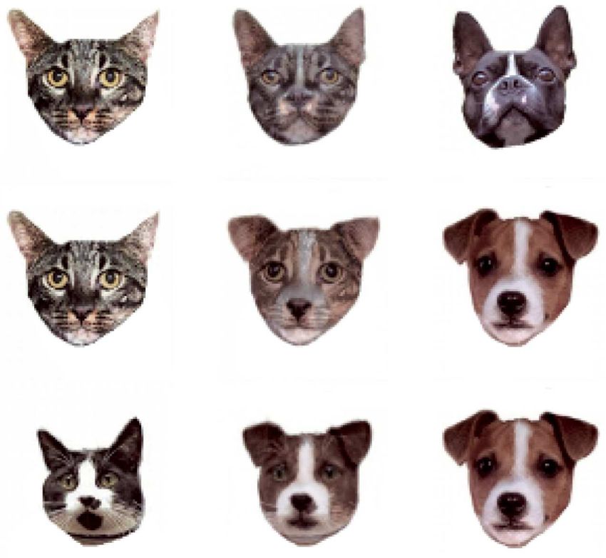
2     Cerebral Cortex, 2021, Vol. 00, No. 00

et al. 2016; Eldridge et al. 2018). The partial sparing of cat-       dark room. Experimental control and data acquisition were per-
egorization after the TE removals surprised us. One possible          formed using the real-time experimental system “REX” adapted
explanation is that TEO, the architectonically identified region      for the QNX operating system (Hays et al. 1982). Visual stimuli
just before TE in the ventral stream hierarchy, can substitute for    were presented by “Presentation” (Neurobehavioral Systems,
some of the missing functionality after TE removal. Although          Inc.) running on a Windows computer.
TEO is physically smaller than TE, neurons in TEO represent
high-level visual properties, and the region has a full represen-
                                                                      Task Procedures
tation of the visual field. Allman and Kaas described the re-
representation of the visual field as the standard for recogniz-      Monkeys were initially trained to grasp and release a touch

                                                                                                                                             Downloaded from https://academic.oup.com/cercor/advance-article/doi/10.1093/cercor/bhab129/6275470 by guest on 24 September 2021
ing a visual area; hence, TEO should be considered a discrete         sensitive bar to earn fluid rewards. After this initial shaping,
visual area (Allman and Kaas 1974). It contains neurons with          a red/green color discrimination task was introduced (Bowman
large receptive fields, although still smaller than those typically   et al. 1996). The trial began with a bar press, and 100 ms later, a
reported for area TE, and unlike TE, TEO seems to be retinotopi-      small red target square (0.5◦ × 0.5◦ ) was presented at the center
cally organized. It has been also suggested that TEO is important     of the display (overlaying a white noise background). Animals
for visual feature analysis and integration, whereas perhaps TE       were required to continue grasping the touch bar until the color
plays a more important role when memory for a whole object is         of the target square changed from red to green. Color changes
required (Iwai and Mishkin 1968). This led us to speculate that       occurred randomly 2000–3000 ms after bar touch. Rewards were
TEO may make a critical contribution to visual categorization.        delivered if the bar was released between 200 and 1000 ms after
To test this hypothesis, we compared performance on a visual          the color change; bar releases occurring either before or after
categorization task across four groups of monkeys: those with         this epoch were counted as errors. All correct responses were
TEO removals, those with TE removals (data reproduced from            followed by visual feedback (target square color changed to blue)
Eldridge et al), those with TE + TEO removals, and unoperated         after bar release and reward delivery 200–400 ms after visual
controls. The TEO-removal group showed mild impairments               feedback. There was a 2-s intertrial interval (ITI), regardless of
that disappeared after 1–3 days of practice (13.4% increase in        the outcome of the previous trial.
error rate in the categorization task compared to the control).           After an animal reached criterion in the red/green color dis-
The TEO plus TE group was severely impaired (31.7% increase           crimination task (two consecutive days with >85% correct per-
in error rate in the categorization task compared to the control).    formance), the monkeys progressed to category training (Fig. 1).
The degree of deficit was approximately equal to the sum of the       In the first phase of category training, 20 dog and 20 cat images
effects for each of TEO and TE.                                       were used. Each trial began when the animal grasped the touch
                                                                      sensitive bar. If the monkey released the bar during the green
                                                                      target when a dog was presented, the monkeys received one
Materials and Methods                                                 drop of liquid reward (Fig. 1a). If the monkey released the bar
                                                                      during the green target when a cat was presented, there was a
Subjects
                                                                      4000–6000 ms time-out with no reward. If the monkey released
Subjects were eight adult rhesus monkeys (Macaca mulatta).            the bar during the red target when either category of stimulus
Three monkeys (one male; weighing 11.6 kg, two females; weigh-        was present, no reward was delivered, and the monkey could
ing 5.4 and 9.3 kg) received bilateral aspiration removals of area    initiate a new trial after the standard ITI. Therefore, the optimal
TEO (Supplementary Fig. 1). Two monkeys (one male; weighing           behavior is to release during the red target for the trials on which
9.6 kg, one female; weighing 5.5 kg) received bilateral aspiration    cats are presented, essentially skipping on to the next randomly
removals of areas TE and TEO (Supplementary Fig. 2). After col-       selected trial, and release during the green target for the trials
lecting behavioral data from monkeys with TEO removals, one           on which dogs are presented to obtain a reward. This design
of the three monkeys (monkey M) received additional bilateral         is effectively a visually-cued two-interval forced choice (2-IFC)
aspiration removals of area TE (Supplementary Fig. 2). These          task, with asymmetrical reward. The 20 dog and 20 cat stimuli
five monkeys performed a visual categorization task before and        were repeated multiple times per session. In the second phase of
after surgery; the data collected before surgery were used as a       category training, the monkeys were presented with four larger
within-subject control. The five monkeys with TEO or TE + TEO         sets of trial-unique images (240 cats and 240 dogs), to confirm
removals received additional testing in tasks not used prior to       that the monkeys were able to classify stimuli based on visual
surgery (see Results); three unoperated monkeys (three males;         perceptual categorization (cat–dog trial-unique task).
weighing 7.8–9.5 kg) were used as controls for these additional           For the perceptually challenging tests of categorization,
experiments. All experimental procedures conformed to the             we used 20 sets of morphed stimuli, as in our previous study
Institute of Medicine Guide for the Care and Use of Laboratory        (Eldridge et al. 2018) (Fig. 1b). For the experiments with morphed
Animals and were performed under an Animal Study Proposal             stimuli, releasing the bar during the green target resulted in
approved by the Animal Care and Use Committee of the National         a 4000–6000 ms time-out with no reward if the stimulus was
Institute of Mental Health.                                           more cat-like (i.e., 50% dog). The outcome of trials on
                                                                      which a stimulus at the category boundary (i.e., =50% dog)
Experimental Conditions
                                                                      was presented was determined probabilistically; 50% of trials
Monkeys sat in a primate chair facing a 22-inch computer mon-         resulted in a reward delivery, 50% resulted in a 4000–6000 ms
itor (Samsung 2233RZ) placed 57 cm from their eyes. A touch           time-out. We collected behavioral data for 10 days using the
sensitive bar was attached to the front panel of the primate          same set of morphed images.
chair at the level of the monkey’s hand. A water reward was               For the simple discrimination task, two cues were used;
dispensed from a stainless steel tube that was positioned at the      these were black and white block (“Walsh”) patterns (13◦ × 13◦ )
monkey’s lips. Experiments were conducted in a sound-isolated         (Fig. 4a). These cues signaled whether a release during the green
Contributions of the Monkey Inferior Temporal Areas TE and TEO to Visual Categorization Setogawa et al. 3
Downloaded from https://academic.oup.com/cercor/advance-article/doi/10.1093/cercor/bhab129/6275470 by guest on 24 September 2021
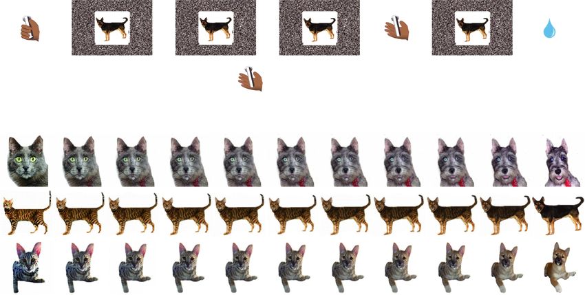
Figure 1. Task and visual stimuli. (a) A single trial of the visual categorization task (rewarded trial). By touching the bar, a morphed cat–dog image was presented. When
the dog-like image was presented (shown as Fig. 1a, 50–100% in Fig. 1b), the animal was required to continue grasping the touch bar until the color of the target square
changed from red to green to obtain water reward. When the cat-like image was presented (0–50% in Fig. 1b), the animal was required to release the touch bar during
the red target period to skip the current trial; otherwise, it received a time-out with no reward (see Materials and Methods). (b) Three examples of the cat–dog morphed
images.
target would result in the delivery of a drop of liquid reward, or distribution of the morph level, and vice versa (11 levels, 0, 10,
a 4000–6000 ms “time-out.” Monkeys could avoid the predicted 20, 30, 40, 50, 60, 70, 80, 90, and 100% dog) (Fig. 3g). We tested the
outcome by releasing the lever before the red target transitioned trial-unique morphed-stimuli categorization task for 1 day.
to green; a new trial could then be initiated after the standard ITI.
We tested each group of monkeys for one session on this task. Data Analysis
The “R” statistical programming language (R Foundation for
Statistical Computing, R Development Core Team, 2017) was
Visual Cues
used for all statistical analyses.
All visual cues were jpeg- or pcx-format photos (200 × 200 pix- A generalized linear mixed model (GLMM) analysis with a
els). The training sets of dogs/cats used in this study are the binomial link function was performed for analyzing the catego-
same as in our previous report (Minamimoto et al. 2010). The rization performance
images used in the main visual categorization task were gen-
erated from a subset of the training images, in which pairs of
P = γ0 + γ1 Level ∗ γ2 Condition + 1|Subject , (1)
cats and dogs were used to create cat–dog morph sequences
using FantaMorph software (Abrosoft). For the main catego-
rization task, 20 cat and 20 dog images were morphed with where P is trial-by-trial categorization performance (0 indicating
the distribution of stimuli concentrated around the category the trial was reported as cat and 1 indicating the trial was
boundary (11 levels, 0, 25, 35, 40, 45, 50, 55, 60, 65, 75, and 100% reported as dog), “Level” is the morph level, “Condition” is the
dog) (Fig. 1b). For the masked stimulus task, the same set of lesion group (TE + TEO removals, TEO removals, TE removals,
cat–dog morph series was used as in main categorization task, and control) or the masked/no masked condition, γ 0 is the
but on four-fifths of trials, the stimuli were overlaid with one intercept, γ 1 and γ 2 are the coefficients estimated by GLMM, and
of four coarse black-block masks (Fig. 3a). For the trial-unique (1|Subject) is the random effect for each monkey.
morphed-stimuli categorization task, a new set of 20 cats and 20 For analyzing the reaction time during the red target or the
dogs was used to create new cat–dog morph series. In this task, green target period, we used before and after surgery data of the
each cat image was morphed with two dog images with equal main visual categorization task (10 days each) and conducted4 Cerebral Cortex, 2021, Vol. 00, No. 00
a generalized linear model (GLM) analysis with a Gaussian link effect of condition: z = −10.48, P = 2.0 × 10⁻¹⁶ ) (Fig. 2a—red and
function as follows: b—top). By the third day, the performance of all three mon-
keys had returned to their pre-lesion levels (Fig. 2b—top). This
transient impairment in the TEO-removal group was smaller
RT = α0 + α1 Level ∗ α2 Condition, (2)
than seen for monkeys with TE removals reported previously
(Fig. 2a—orange; Eldridge et al. 2018).
where RT is logarithm of the reaction time, “Level” is the morph
level (we removed the choice reaction time data on the catego-
rization boundary, i.e., 50% dog, for this analysis; 0, 25, 35, 40, and Categorization after TE + TEO Removals
Downloaded from https://academic.oup.com/cercor/advance-article/doi/10.1093/cercor/bhab129/6275470 by guest on 24 September 2021
45% dog were used for the red target period, 55, 60, 65, 75, and We removed TE and TEO bilaterally in two monkeys and added
100% dog were used for the green target period), “Condition” is a TE lesion to one of the monkeys that had previously received
the lesion group (TE + TEO removals, TEO removals, and control), a TEO removal (monkey M). The performance of the monkey
α 0 is the intercept, and α 1 and α 2 are the coefficients estimated with the two-stage lesion was indistinguishable from that of
by GLM. the monkeys with one-stage TE + TEO removals in this and all
For analyzing the processing time, we used a GLM with a subsequent tasks; hence, the data from these three monkeys
Gaussian link function as follows: were pooled. For the TE + TEO-removal group, categorization
performance was severely impaired (GLMM, Eq. 1; TE + TEO
PT = β0 + β1 Level ∗ β2 Condition, (3) vs. control, effect of condition: z = −48.35, P = 2.0 × 10⁻¹⁶ )
(Fig. 2a—green). Performance recovered partially with experi-
ence, but remained significantly poorer than the TEO or TE
where PT is logarithm of the processing time, “Level” is the
groups by the 10th day of postoperative testing (GLMM, Eq. 1;
morph level (0, 25, 35, 40, and 45% dog), “Condition” is the lesion
TE + TEO vs. TE, effect of condition: z = 11.16, P = 2.0 × 10⁻¹⁶ ;
group (TE + TEO removals, TEO removals, and control), β 0 is
TE + TEO vs. TEO, effect of condition: z = 11.04, P = 2.0 × 10⁻¹⁶ )
the intercept, and β 1 and β 2 are the coefficients estimated by
(Fig. 2b—bottom). As shown in Figure 2a,b, the categorization
GLM.
performance was asymmetrical: higher percentages of cat
stimuli were categorized as dog than dog stimuli categorized
Order of Testing as cat. This asymmetry likely reflects the asymmetric reward
All monkeys received basic categorization training prior to structure of the task; monkeys with impaired ability to
surgery. Monkeys received 10 sessions of the main visual categorize—for example, as the result of a lesion—tend to
categorization task using morphed stimuli before and after present a bias toward releasing during the green interval
surgery. Three monkeys received bilateral TEO aspiration because only the dog stimuli are associated with reward.
removals, and two received bilateral TE + TEO removals. After
collecting behavioral data from monkeys with TEO removals, Reaction Times
one of the three monkeys (monkey M) received an additional
bilateral aspiration removal of area TE. Thus, for the main The pattern of reaction times indicates that monkeys in all
categorization task, pre- and post-op data were compared groups were sensitive to the degree of mixing in the morphs
within-subjects. The subsequent experiments were adminis- when responding to a cat (Fig. 2c for monkey M). When the cat-
tered only postoperatively. The performance of the monkeys like image was presented, the reaction time was defined as the
with aspiration removals on these latter tasks was compared time between onset of the stimulus and bar release. When the
to that of a group of control monkeys that had received parallel dog-like image was presented, the reaction time was defined as
training. After the main visual categorization task was tested, we the time between onset of the green cue and bar release. The
conducted the simple discrimination task (1 day) and a contrast reaction time following the green target was constant across
sensitivity task (5 days) for assessing low-level visual functions. morph level (55–100% dog), presumably because the monkey
Then, the cat–dog trial-unique task (1 day) and the masked had already made a decision that the presented stimulus was
stimulus task were tested (10 days). Finally, we tested the trial- dog-like before the red target changed to green (generalized
unique morphed-stimuli categorization task (1 day). All tests linear model [GLM], monkey M, Eq. 2; effect of morph level:
after lesion were conducted within 3 months. t = 0.75, P = 0.46). We interpret the response time during the
green target period as a basic visual–motor reaction time. This
visual–motor reaction time was indistinguishable across lesion
Results groups, suggesting that motor skill was not affected by TEO
We tested eight monkeys, three with TEO removals, three with or TE + TEO removals (GLM, monkey M, Eq. 2; effect of task
combined TE + TEO removals (including one from the previous condition: t = 0.91, P = 0.36).
group after a second surgery to remove TE bilaterally), and three To characterize the reaction times to more cat-like stimuli
normal unoperated controls, on a visual categorization task (i.e., lever releases during the red target period), we introduced
using 20 sets of cat/dog morphs (Fig. 1). Monkeys learned to a measure we term “processing time” that was calculated by
categorize morphed images as either “cat-like” or “dog-like” to subtracting the average visual–motor reaction times (55–100%
avoid a time-out or to obtain a liquid reward. dog) from the reaction times for each cat-like level (0–45% dog)
(Fig. 2d—monkey M and e—all monkeys). Because the visual–
motor reaction times were different among animals, the pro-
Categorization after TEO Removals
cessing time provides a normalized measure of the time it
We collected pre- and postoperative behavioral data for 10 days takes the monkeys to decide whether an image is more cat-
from the TEO-removal group. There was a mild impairment in like. The processing time for the TE + TEO group was signif-
categorization ability for the first 2 days of postoperative testing icantly longer than the control group (GLM, Eq. 3; effect of
(generalized linear mixed model [GLMM], Eq. 1; TEO vs. control, condition: t = 10.5, P = 2.0 × 10⁻¹⁶ ). The processing times for theContributions of the Monkey Inferior Temporal Areas TE and TEO to Visual Categorization Setogawa et al. 5
Downloaded from https://academic.oup.com/cercor/advance-article/doi/10.1093/cercor/bhab129/6275470 by guest on 24 September 2021
Figure 2. Categorization performance. (a) Percentage identified as dog in the TE + TEO, TE, TEO, and control groups on the first experimental day (TE data have been
previously reported [Eldridge et al. 2018]). (b) Day-by-day task performance for 10 days in the TE + TEO and TEO groups. A deep red indicates the first day and a deep
blue indicates the last day. A gray dashed line indicates the task performance before surgery (blue line in Fig. 2a). (c) An example of the reaction time (monkey M). The
vertical gray line indicates the category boundary, that is, 50% dog. (d) An example of the processing time (monkey M). (e) The average processing time of all animals.
Error bar: SEM.
TEO and control groups were indistinguishable (GLM, Eq. 3; effect overlaid with one of four coarse black-block masks on four-
of condition: t = 0.67, P = 0.51). These results indicate that the fifths of the trials (Fig. 3a). If the animals with TEO or TE + TEO
TE + TEO-removal group takes longer to process the stimuli even lesions rely on a limited set of (or even single) diagnostic features
when the animals have previously seen them. to categorize a presented image, their performance should be
impaired by masking. Consistent with our hypothesis, both
TEO and TE + TEO-removal groups showed severe impairments
Role of Experience in Categorization in categorizing masked stimuli relative to the interleaved
We tested the possibility that the monkeys with TEO or TE + TEO unmasked trials (GLMM, Eq. 1; Mask vs. No mask in TE + TEO,
removals had compensated for impaired visual categorization effect of condition: z = −8.56, P = 2.0 × 10⁻¹⁶ ; Mask vs. No
by memorizing one or more simple features of each morph mask in TEO, effect of condition: z = −14.23, P = 2.0 × 10⁻¹⁶ )
series (e.g., the “ear” of the stimuli in the first row of Fig. 1b). (Fig. 3b,c,e). Conversely, the performance of the control group
To examine this, we introduced two manipulations to the was only mildly affected by masking (GLMM, Eq. 1; Mask vs. No
categorization task; a masked stimulus set and a trial-unique mask in Control, effect of condition: z = −7.73, P = 1.1 × 10⁻¹⁴ )
stimulus set. In the masked stimulus task, the stimuli were (Fig. 3f ).6 Cerebral Cortex, 2021, Vol. 00, No. 00
Downloaded from https://academic.oup.com/cercor/advance-article/doi/10.1093/cercor/bhab129/6275470 by guest on 24 September 2021
Figure 3. Categorization performance in the mask categorization task and the trial-unique categorization task. (a) Examples of the visual stimuli in the mask
categorization task. Four checker-board masks were placed over each of the stimuli used in the main categorization task and presented interleaved with an unmasked
version of each stimulus. (b) Categorization performance of the TE + TEO, TE, TEO, and control groups (TE data have been previously reported [Eldridge et al. 2018]) in
the mask categorization task. (c–f ) Categorization performance on masked (mean of all masks) versus unmasked stimuli for (c) TE + TEO, (d) TE, (e) TEO, and (f ) control
groups. (g) Examples of the visual stimuli in the trial-unique categorization task. Each cat was morphed with two dogs, and vice versa. Examples at the 0%, 50%, and
100% dog level are shown. (h) Categorization performance of the TE + TEO, TE, TEO, and control groups (TE data have been previously reported [Eldridge et al. 2018])
in the trial-unique categorization task. Error bar: SEM. (i) Categorization performance of the TE + TEO, TE, TEO, and control groups in the cat–dog trial-unique task
(no-morphed 240 cats and 240 dogs).Contributions of the Monkey Inferior Temporal Areas TE and TEO to Visual Categorization Setogawa et al. 7
Downloaded from https://academic.oup.com/cercor/advance-article/doi/10.1093/cercor/bhab129/6275470 by guest on 24 September 2021
Figure 4. Low-level visual functions. (a) The visual stimuli of the cue discrimination task. (b) Discrimination performance of the TE + TEO, TEO, and control group.
∗ P < 2.2 × 10⁻¹⁶ . (c) The contrast sensitivity curves of the TE + TEO, TEO, and control group. Error bar: SEM.
For the trial-unique stimulus task, we prepared a large set df = 1, P = 2.0 × 10⁻¹⁶ ; TEO: χ 2 = 804.5, df = 1, P = 2.0 × 10⁻¹⁶ ; Con-
of novel morph images as trial-unique stimuli. A key difference trol: χ 2 = 1925.5, df = 1, P = 2.0 × 10⁻¹⁶ ) (Fig. 3i). This result con-
from the stimulus set used for main experiment was that each firms that the TE + TEO removal induces a severe impairment in
cat image was morphed with two dog images, and vice versa visual categorization, rather than impairment of the stimulus–
(Fig. 3g; Eldridge et al. 2018). This manipulation reduces the reward association learning.
utility of a strategy focused on a single memorized feature. We
tested this trial-unique categorization task for one day (a single
Low-Level Visional Function after TEO and TE + TEO
session). Consistent with the results of the masked stimulus
Removal
task, the categorization performance of the TE + TEO-removal
group was severely impaired compared to the other groups Two tasks were used to assess low-level visual functions: cue dis-
(e.g., TE + TEO vs. TE, GLMM, Eq. 1; effect of condition: z = 5.4, crimination and contrast sensitivity. In the cue discrimination
P = 5.5 × 10⁻⁸ ) (Fig. 3h). The degree of impairment in both the task, two different Walsh patterns were used; one cue associated
masked stimulus and the trial-unique stimulus tasks was con- with reward and the other associated with time-out (Fig. 4a).
sistent with the main experiment (Fig. 2a), the order of impair- All groups of monkeys distinguished between the rewarded and
ment from greatest to least was TE + TEO, TE, and TEO (Fig. 3b,h). unrewarded cues ([number of no-reward trials accepted/all no-
The processing time in the trial-unique task was also ana- reward trials]: TE + TEO; 6/1137 (0.4%), TEO; 6/1267 (0.5%), con-
lyzed. For the TE–TEO group, the processing time was signifi- trol; 3/686 (0.5%), [number of rewarded trials accepted/all reward
cantly longer than the control group (GLM, Eq. 3; effect of con- trials]: TE + TEO; 1310/1498 (87.4%), TEO; 1342/1433 (93.6%), con-
dition: t = 2.69, P = 7.4 × 10⁻³ ) (Supplementary Fig. 3). The pro- trol; 781/882 (88.5%)) (reward vs. no-reward, χ 2 -test, TE + TEO:
cessing time for the TEO group was also significantly longer df = 2, P < 2.2 × 10⁻¹⁶ , TEO: df = 2, P < 2.2 × 10⁻¹⁶ , control: df = 2,
than the control group (GLM, Eq. 3; effect of condition: t = 4.09, P < 2.2 × 10⁻¹⁶ ) (Fig. 4b). In the contrast sensitivity test (Mat-
P = 5.1 × 10⁻⁵ ) (Supplementary Fig. 3). These results indicate that sumoto et al. 2016), full-screen sine wave gratings (i.e., the local
both the group with TE + TEO removals and the group with TEO- intensity was modulated by a one-dimensional [vertical] sine
only removals take longer to process the stimuli, whether they wave across the screen) were presented that covered a range
are trial-unique or familiar. of frequencies (16, 8, 4, 2, 1, 0.5, 0.25, and 0.125 cycles/degree)
Because two adjacent morphed stimuli are visually simi- and contrasts (1, 0.64, 0.32, 0.16, 0.08, 0.04, 0.02, 0.01, 0.005). Con-
lar (e.g., 35% dog and 40% dog in Fig. 1b), it is possible that trast was calculated as: LP − LT/(LP + LT), where LP represents
the subjects learn stimulus–reward associations within a single peak luminance and LT trough luminance. The space-average
session instead of generalizing from previous experience with luminance was kept constant across stimuli. The task took
categorical exemplars. To examine this possibility, we included the form of a signal detection paradigm, whereby the monkey
a single session of the cat–dog trial-unique task (nonmorphed was required to release the lever immediately if a grating was
240 cats and 240 dogs) in which completely novel images were detected (during the presentation of the red target) to obtain
used; the subjects can only solve this task via visual percep- a reward, or otherwise to continue to hold the lever until the
tual generalization. We observed the same ranking of results target turned green, and then to release the lever to obtain a
as obtained at the 0% and 100% morph level in the morphed reward. This is a two-interval forced choice task, with symmetric
categorization tasks (χ 2 -test, % identified as cat vs. % identified reward. Gratings were presented for 500 ms on 50% of trials. If the
as dog, TE + TEO: χ 2 = 356.1, df = 1, P = 2.0 × 10⁻¹⁶ ; TE: χ 2 = 719.7, monkey released the lever during the presence of the red target8 Cerebral Cortex, 2021, Vol. 00, No. 00
when no grating had been presented or released on green when stations to TE without passing through area TEO. The most direct
a grating had been presented (i.e., both incorrect responses), path would be from connections to TE arising at earlier stages
a 4–6 s time-out was incurred. Grating contrast sensitivity, a such as V4 (Distler et al. 1993; Ungerleider et al. 2008), assuming
test designed to assess the visual acuity of human subjects that those connections bypassing TEO have enough bandwidth
(Blackmore and Campbell 1969), was indistinguishable across to carry sufficient information for TE to analyze images. Previous
all three groups (GLM, group: t = 0.07, df = 2, P = 0.95) (Fig. 4c). studies have suggested that four-legged animals are more likely
The contrast sensitivities of the three groups were similar to to be confused among one another than “simpler” contrasts,
those of humans (Blackmore and Campbell 1969) and monkeys such as fruits versus tables or cars versus chairs (Cadieu et al.
that received TE (Matsumoto et al. 2016) or rhinal cortex lesion 2014). We elected cats and dogs as the categories for comparison
Downloaded from https://academic.oup.com/cercor/advance-article/doi/10.1093/cercor/bhab129/6275470 by guest on 24 September 2021
(Eldridge et al. 2018). in the present study on the basis that they were likely to yield
high levels of confusion. We have previously demonstrated that
a linear classifier performs more accurately on human face ver-
Discussion sus monkey face and car versus truck comparisons than it does a
Above we have shown that selective bilateral removal of the cat versus dog comparison (Matsumoto et al. 2016). To maximize
inferior temporal cortex (TE + TEO combined) interferes with the perceptual difficulty in the present study, we used morphed
categorical discrimination when tested with sets of trial-unique pairs of cats and dogs to create even more category-ambiguous
cats and dogs, visually degraded morphed images, and trial- intermediate images (Eldridge et al. 2018). Our expectation is
unique morphed images. The severity of the deficit is differ- that “simpler” comparisons could be performed at earlier stages
ent if either of the two subregions of inferior temporal cor- of the visual system (e.g., bilateral removals of area TE produce
tex, areas TEO and TE, is removed independently. There was no impairment in the ability to categorize human vs. monkey
a significant deficit in the categorization of all trial-unique faces [Matsumoto et al. 2016]).
images after TEO removals, and a slightly more severe deficit The greatest reduction in categorization accuracy occurred
after TE removals. After removal of either area, the monkeys’ when we used previously unseen stimuli. Because the stimuli
performance improved quickly with repetitions of an image presented in this phase of the experiment were new, the
set; however, only the TEO-removal group recovered to control only means by which the monkeys could have accurately
levels of performance. When TEO and TE are both removed, classified them was to generalize from previously experienced
the monkeys are severely impaired, and while they show some exemplars. The data from using new images show that
improvement with additional practice on a single image set, they monkeys with combined TE + TEO removals exhibit a deficit
remain severely impaired. It appears that TEO and TE lesions in categorization accuracy that approximates a sum of the
have an additive effect on the severity of the deficit, consistent deficits observed following removal of either subregion of IT
with models derived from single unit recordings taken from alone. Thus, it appears that TE and TEO work in parallel, and
subregions TEO and TE of IT cortex (Majaj 2015). The improve- with minimum redundancy, to encode category membership of
ment with repeated image set presentation raises a difficulty a novel stimulus. Even the monkeys with complete TE + TEO
for the experimentalist trying to study perception or perceptual removals are able to categorize at above-chance levels with
categorization—the only presentation that can be assured to practice; on the first test session after the removals, they
rely solely on perception/categorical memory is the first one. performed at chance. The rapid increase in performance with
Every subsequent encounter is confounded by the possibility of practice, plus the increased processing time observed for both
recollective processes. lesion groups (see Fig. 2e and Supplementary Fig. 3), suggests
The canonical description of visual image processing by the that compensatory mechanisms may be invoked that preserve
brain posits that simple features, such as oriented edges or some degree of categorization accuracy at the cost of increased
lines, are represented in caudal regions, beginning with area decision time.
V1 (Hubel and Wiesel 1959), and that representations become Our data also demonstrate that the deficits observed in all
increasingly more complex as information converges along a treatment groups were ameliorated with increased familiarity to
ventral pathway in a sequential, feed-forward manner, culmi- a stimulus set; classification accuracy improved with repeated
nating in the representation of whole objects in inferior tem- postlesion exposure to the morphed stimulus set (Fig. 2b,c, and
poral cortex (Gross et al. 1972; Ungerleider and Mishkin 1982). Eldridge et al. 2018). The results here show that the ability of
Two observations from single neuron recording studies offered the TEO-removal group to generalize from previously experi-
strong support to a sequential, feed-forward processing model enced exemplars remains compromised because the deficits
for image analysis in the ventral stream. First, there were the in classifying novel stimuli were recorded after the monkeys
progressively larger receptive fields, and, second there was the received repeated exposure to the morphed stimulus set. Thus,
increasing complexity of stimulus selectivity in architectonically the recovery in performance must be supported by the learn-
separable cortical brain regions as information flowed from ing of an alternative strategy, presumably one based on the
caudal (V1) to rostral (ending in area TE). However, whether the learning of stimulus–reward associations. The rapid and com-
ventral visual stream relies exclusively on feed-forward process- plete recovery of the TEO-removal group with stimulus repeti-
ing has been thrown into doubt by the observation of recurrent tion suggests that other regions (such as TE) can support the
and bypass projections in studies of anatomical connectivity stimulus–reward association learning required to support this
(Kravitz et al. 2013; Kar et al. 2019). Now, we add results showing enhanced performance. The TE-removal group asymptote at
that processing is not always strictly sequential. The observa- a level of performance inferior to that of controls—this indi-
tion that bilateral removal of area TEO—the region immediately cates that no other area can adequately support the fidelity
upstream of area TE—produces milder deficits than those we of stimulus–reward associations needed to compensate for the
previously reported after bilateral TE removals (Eldridge et al. loss of categorization capability conferred by TE removal. The
2018) indicates that the visual information used for analyzing TE + TEO removals produced the most substantial impairment
images depends on a route from earlier in the ventral stream in categorization accuracy—an initial near-total loss of function,Contributions of the Monkey Inferior Temporal Areas TE and TEO to Visual Categorization Setogawa et al. 9
which recovered with practice to a level of accuracy consis- References
tently just above chance. As we proposed for the savings in
Afraz SR, Kiani R, Esteky H. 2006. Microstimulation of infer-
categorization of novel stimuli discussed above, the residual
otemporal cortex influences face categorization. Nature.
ability of the TE + TEO group to classify at above-chance levels
442:692–695.
is likely subserved by projections from earlier in the visual
Allman JM, Kaas JH. 1974. The organization of the second visual
system that bypass IT to subcortical targets. Taken together,
area (V II) in the owl monkey: a second order transformation
these observations indicate that in the TE-removal group, TEO
of the visual hemifield. Brain Res. 76:247–265.
is likely the key substrate for the stimulus–reward associations
Blackmore C, Campbell FW. 1969. On the existence of neurones
that confer the ability of this group to improve so substantially
Downloaded from https://academic.oup.com/cercor/advance-article/doi/10.1093/cercor/bhab129/6275470 by guest on 24 September 2021
in the human visual system selectively sensitive to the
with practice.
orientation and size of retinal images. J Physiol. 203:237–260.
There are two possible explanations for our result showing
Bowman EM, Aigner TG, Richmond BJ. 1996. Neural signals in the
slower and less complete recovery in the performance of the TE-
monkey ventral striatum related to motivation for juice and
removal group versus the TEO-removal group. One is that TEO,
cocaine rewards. J Neurophysiol. 75:1061–1073.
although a distinct architectonic and physiologically separate
Buckley MJ, Gaffan D, Murray EA. 1997. Functional double disso-
region, contributes to this categorization task as if TEO and TE
ciation between two inferior temporal cortical areas: perirhi-
are one larger functional region. Thus, the difference in recovery
nal cortex versus middle temporal gyrus. J Neurophysiol.
is related to the difference in the volume of tissue removed;
77:587–598.
that is, TE is a larger architectonic region; hence, the deficits
Buffalo EA, Ramus SJ, Clark RE, Teng E, Squire LR, Zola SM. 1999.
observed correspond simply to the quantity of tissue removed,
Dissociation between the effects of damage to perirhinal
and not from a specific segregation of function. The second
cortex and area TE. Learn Mem. 6:572–599.
possibility is that TEO and TE are functionally one architectonic
Buffalo EA, Ramus SJ, Squire LR, Zola SM. 2000. Perception and
region and should not be considered as different. If the latter
recognition memory in monkeys following lesions of area TE
were the case, we would not expect to see the asymmetry
and perirhinal cortex. Learn Mem. 7:375–382.
in the impairment in categorization that appeared with novel
Butter CM, Gekoski WL. 1966. Alterations in pattern equivalence
exemplars (Fig. 3i). In addition, previous data show that receptive
following inferotemporal and lateral striate lesions in rhesus
fields are different in TEO and TE, and that TEO contains a full
monkeys. J Comp Physiol Psychol. 61:309–312.
representation of the visual fields, the means by which Allman
Cadieu CF, Hong H, Yamins DLK, Pinto N, Ardila D, Solomon
and Kaas separated functional visual regions (Allman and Kaas
EA. 2014. Deep neural networks rival the representation of
1974). Thus, the weight of the evidence favors considering TEO
primate IT cortex for Core visual object recognition. PLoS
and TE as different functional regions.
Comput Biol. 10:e1003963.
Over the past six decades, many studies have concluded that
Cowey A, Gross CG. 1970. Effects of foveal prestriate and infer-
the inferior temporal cortex is critical for pattern discrimination
otemporal lesions on visual discrimination by rhesus mon-
(Iwai and Mishkin 1968; Cowey and Gross 1970), visual pattern
keys. Exp Brain Res. 11:128–144.
recognition (Butter and Gekoski 1966; Weiskrantz and Saunders
Distler C, Boussaoud D, Desimone R, Ungerleider LG. 1993. Cor-
1984), and by inference, visual perceptual categorization (Sigala
tical connections of inferior temporal area TEO in macaque
and Logothetis 2002; Afraz et al. 2006; Kiani et al. 2007). There
monkeys. J Comp Neurol. 334:125–150.
has remained a disconnect though. The data supporting IT par-
Eldridge MAG, Matsumoto N, Wittig JH Jr, Masseau EC, Saunders
ticipation in visual perceptual categorization have largely relied
RC, Richmond BJ. 2018. Perceptual processing in the ventral
on correlations in the selectivity of neurons in physiological
visual stream requires area TE but not rhinal cortex. Elife.
recordings. Our data show that both areas TEO and TE contribute
7:1–16.
to categorization-based behavior when subjects are challenged
Freedman DJ, Riesenhuber M, Poggio T, Miller EK. 2001. Categori-
with novel stimuli but that performance quickly improves with
cal representation of visual stimuli in the primate prefrontal
repeated exposure to the same stimuli. Thus, caution must be
cortex. Science. 291:312–316.
exercised when interpreting the results of experiments in which
Freedman DJ, Riesenhuber M, Poggio T, Miller EK. 2002. Visual
stimuli are repeated, both behavioral and electrophysiological,
categorization and the primate prefrontal cortex: neurophys-
as perceptual generalization can be easily confounded with
iology and behavior. J Neurophysiol. 88:929–941.
other processes.
Fujita I, Tanaka K, Ito M, Cheng K. 1992. Columns for visual
features of objects in monkey inferotemporal cortex. Nature.
Supplementary Material 360:343–346.
Gainotti G. 2000. What the locus of brain lesion tells us about the
Supplementary material is available at Cerebral Cortex online. nature of the cognitive defect underlying category-specific
disorders: a review. Cortex. 36:539–559.
Gross CG, Rocha-Miranda CE, Bender DB. 1972. Visual proper-
Funding
ties of neurons in inferotemporal cortex of the macaque. J
Intramural Research Program; National Institute of Mental Neurophysiol. 35:96–111.
Health; National Institutes of Health; Department of Health and Hays AV, Richmond BJ, Optican LM. 1982. Unix-based multiple-
Human Services (annual report number ZIAMH002032). process system, for real-time data acquisition and control.
WESCON Conf Proc., 2:1–10.
Hubel DH, Wiesel TN. 1959. Receptive fields of single neurones
Notes in the cat’s striate cortex. J Physiol. 143:574–591.
We thank Megan Fredericks and Grace Mammarella for assis- Iwai E, Mishkin M. 1968. Two visual foci in the temporal lobe of
tance with behavioral testing. monkeys. In: Neurophysiological basis of learning and behavior.10 Cerebral Cortex, 2021, Vol. 00, No. 00
Eds.: N. Yoshii and N.A. Buchwald. 1969. Further evidence on Sato T, Uchida G, Lescroart MD, Kitazono J, Okada M, Tanifuji
the locus of the visual area in the temporal lobe of the mon- M. 2013. Object representation in inferior temporal cortex is
key. Exp. Neurol. Japan: Osaka Univ. Press 1968. 25:585–594. organized hierarchically in a mosaic-like structure. J Neurosci.
Kar K, Kubilius J, Schmid K, Issa EB, DiCarlo JJ. 2019. Evidence 33:16642–16656.
that recurrent circuits are critical to the ventral stream’s Sigala N, Logothetis NK. 2002. Visual categorization shapes
execution of core object recognition behavior. Nat Neurosci. feature selectivity in the primate temporal cortex. Nature.
22:974–983. 415:318–320.
Kiani R, Esteky H, Mirpour K, Tanaka K. 2007. Object category Tanaka K. 1996. Inferotemporal cortex and object vision. Annu
structure in response patterns of neuronal population in Rev Neurosci. 19:109–139.
Downloaded from https://academic.oup.com/cercor/advance-article/doi/10.1093/cercor/bhab129/6275470 by guest on 24 September 2021
monkey inferior temporal cortex. J Neurophsiol. 97:4296–4309. Ungerleider LG, Galkin TW, Desimone R, Gattass R. 2008. Cor-
Kravitz DJ, Saleem KS, Baker CI, Ungerleider LG, Mishkin M. 2013. tical connections of area V4 in the macaque. Cereb Cortex.
The ventral visual pathway: an expanded neural framework 18:477–499.
for the processing of object quality. Trends Cogn Sci. 17:26–49. Ungerleider LG, Mishkin M. 1982. Two cortical. In: Sys-
Majaj NJ, Hong H, Solomon EA, DiCarlo JJ. 2015. Simple learned tems V, Ingle DJ, Goodale MA, Mansfield RJW, editors.
weighted sums of inferior temporal neuronal firing rates Analysis of visual behavior. Cambridge (MA): MIT Press,
accurately predict human core object recognition perfor- pp. 549–586.
mance. J. Neurosci. 39:30–35. Vogels R. 1999. Categorization of complex visual images by
Matsumoto N, Eldridge MAG, Saunders RC, Reoli R, Richmond rhesus monkeys. Part 1: behavioural study. Eur J Neurosci.
BJ. 2016. Mild perceptual categorization deficits follow bilat- 11:1223–1238.
eral removal of anterior inferior temporal cortex in rhesus Vogels R, Saunders RC, Orban GA. 1997. Effects of inferior tempo-
monkeys. J Neurosci. 36:43–53. ral lesions on two types of orientation discrimination in the
Minamimoto T, Saunders RC, Richmond BJ. 2010. Monkeys macaque monkey. Eur J Neurosci. 9:229–245.
quickly learn and generalize visual categories without lateral Weiskrantz L, Saunders RC. 1984. Impairments of visual object
prefrontal cortex. Neuron. 66:501–507. transforms in monkeys. Brain. 107:1033–1072.You can also read


