Diagnostic and therapeutic considerations for obscure gastrointestinal bleeding in patients with chronic kidney disease
←
→
Page content transcription
If your browser does not render page correctly, please read the page content below
REVIEW ARTICLE                                                                                Annals of Gastroenterology (2019) 32, 1-11

Diagnostic and therapeutic considerations for obscure
gastrointestinal bleeding in patients with chronic kidney disease

Mayssan Muftaha, Ramzi Mulkib, Tanvi Dhereb, Steven Keilinb, Saurabh Chawlab,c
Emory University School of Medicine; Grady Memorial Hospital, Atlanta, USA

Abstract                            Recurrent obscure gastrointestinal bleeding amongst patients with chronic kidney disease is a
                                    challenging problem gastroenterologists are facing and is associated with an extensive diagnostic
                                    workup, limited therapeutic options, and high healthcare costs. Small-bowel angiodysplasia
                                    is the most common etiology of obscure and recurrent gastrointestinal bleeding in the general
                                    population. Chronic kidney disease is associated with a higher risk of gastrointestinal bleeding
                                    and of developing angiodysplasia compared with the general population. As a result, recurrent
                                    bleeding in this subgroup of patients is more prevalent and is associated with an increased
                                    number of endoscopic and radiographic procedures with uncertain benefit. Alternative medical
                                    therapies can reduce re-bleeding; however, more studies are needed to confirm their efficacy in
                                    this subgroup of patients.
                                    Keywords Obscure gastrointestinal bleeding, Chronic kidney disease, Angiodysplasia,
                                    arteriovenous malformations, Small-bowel bleeding
                                    Ann Gastroenterol 2019; 32 (2): 1-11

Introduction                                                           radiographic imaging [7]. The recent American College of
                                                                       Gastroenterology guidelines recommended reclassification
   Chronic kidney disease (CKD) is an independent risk                 of OGIB as small-bowel bleeding, as about 75% of OGIB
factor for gastrointestinal (GI) bleeding as it is associated          originates in the small bowel [7,10,11].
                                                                            OGIB is a common dilemma for the gastroenterologist as it is
with an increased risk of gastritis, peptic ulcer disease and
                                                                       commonly associated with an extensive diagnostic workup with
angiodysplasia [1-5]. Patients with GI bleeding and comorbid
                                                                       limited yield. Management of this condition is more challenging
CKD have worse outcomes than patients who have normal
                                                                       in patients with CKD. Additionally, healthcare costs are higher
renal function [2,6] and exhibit higher mortality than the
                                                                       in CKD patients with anemia compared to those without, in part
general population (odds ratio [OR] 1.8, 95% confidence
                                                                       because of a higher frequency of GI workup for bleeding [12],
interval [CI] 1.7-1.9; P
2 M. Muftah et al
attributed to uremic platelet dysfunction [15,16]. Uremia causes patients with CKD have a higher prevalence of angiodysplasia
impaired function of platelet glycoproteins GPIIb/IIIa, altered compared with the general population, but bleeding from
release of adenosine diphosphate and serotonin, and impaired angiodysplasia in this population is also the leading cause of
prostaglandin and arachidonic acid metabolism, which together recurrent bleeding [3].
compromise platelet adhesion and aggregation [17]. Given the
increased platelet dysfunction that occurs with progressive
CKD, GI bleeding is an expected complication seen in these
patients [18]. In one study, chromium-labeled red blood cells Endoscopic evaluation of OGIB
were used to evaluate the amount of GI blood loss in patients
with CKD compared to patients with normal renal function. The literature on the utility of endoscopic evaluation
HD patients were found to have more occult GI blood losses in those with recurrent OGIB in ESRD patients is sparse.
(6.27 mL/day) compared with control patients (0.83 mL/day) Typically, patients with recurrent GI bleeding of unidentifiable
and CKD patients not yet on HD (3.15 mL/day) [19]. cause on upper and lower endoscopy are first evaluated with
Patients with CKD have been reported to have higher repeat upper or lower endoscopy based on the characteristics
rates of GI bleeding secondary to peptic ulcer disease and of the bleeding. If unrevealing, this should then be followed by
angiodysplasia compared with the general population. In a small-bowel evaluation [8].
study evaluating anemic patients with non-dialysis dependent
CKD stages 3-5, 52.9% were found to have sources of GI
bleeding on upper and lower endoscopy. Gastric lesions were Push enteroscopy (PE)
found more frequently in patients with stage 5 CKD compared
to those with stages 3-4 CKD [20]. In those presenting with recurrent melena or hematemesis,
Peptic ulcer disease is a common cause of upper PE may be a better alternative than EGD for a second look, as it
gastrointestinal bleeding (UGIB) in patients with CKD. In a has a higher diagnostic yield with the addition of a limited small-
cohort study, investigators followed 796 patients who had just bowel evaluation [8,26]. It is particularly useful if proximal
initiated HD for a 6-year period, along with 3184 age- and sex- small-bowel lesions are suspected, as it has a higher yield in
matched patients without CKD, and compared rates of UGIB. the proximal small bowel than VCE and allows for therapeutic
The rate of UGIB was higher for the HD patients (hazard ratio intervention [8]. A review of patients who underwent capsule
[HR] 1.27, 95%CI 1.03-1.57). The most common etiology of endoscopy found that, of those with small-bowel angiodysplasia,
this bleeding was peptic ulcer disease [21]. Laeeq et al described 78.3% had lesions within the first 25% of small-bowel transit
the causes and characteristics of UGIB in ESRD patients and and 66.8% had lesions in the duodenum or at the ligament of
found that the most common findings were erosions (55.9%) Treitz, easily reached via PE [27]. Another study investigated the
and ulcers (30.3%). More than half (55.9%) of patients required distribution of angiodysplastic lesions in patients who presented
therapeutic intervention [22]. with OGIB. Eighty percent of patients had angioectasias in the
Although there is an increased incidence of peptic ulcer jejunum, 51% in the duodenum, and 22.8% in the stomach [28].
disease in CKD, this is often an easily identifiable and treatable However, of those with small-bowel bleeding secondary to
cause of GI bleeding. However, angiodysplasia is typically non-vascular lesions, the yield of PE is suboptimal and capsule
the cause of small-bowel and recurrent bleeding and is more endoscopy was found to be superior [8,29,30].
challenging to identify and manage. Even prior to the advances Given the yield of PE for vascular lesions, it may be a
in small-bowel evaluation, patients with CKD were described reasonable next step for evaluation of OGIB in CKD patients
as having a higher incidence of angiodysplasia of the stomach when there is high suspicion of an upper GI source. However,
or proximal small bowel. One study evaluated the etiologies there are no studies examining the use of PE in OGIB in these
of UGIB in patients with CKD compared with the general patients.
population and found that the former had angiodysplasia as
the most common source of bleeding (rate of 50% vs. 11%) [3].
Another study evaluated 727 patients who underwent endoscopy VCE
for UGIB, 60 of whom had CKD. In this cohort, they found
that the prevalence of angiodysplasia was higher in those with If evaluation with PE is negative, the next step in evaluating
renal dysfunction overall and increased with the duration of OGIB is with VCE. However, if there is not high suspicion of a
comorbid CKD and need for hemodialysis [23]. proximal small-bowel lesion, VCE would be a more appropriate
With the advent of video capsule endoscopy (VCE), one first step in evaluating OGIB because it has a superior overall
prospective study showed that patients with CKD were at higher yield to PE, due to its ability to visualize the entire small
risk (OR 4.5, 95%CI 1.9-10.6; P=0.0007) of having small-bowel bowel [8,29-31]. The overall diagnostic yield has been reported
angiodysplasia compared with the general population [24]. to range from 38-83% [8]. A 2005 meta-analysis of 14 studies
CKD was also found to be an independent risk factor for found that the diagnostic yield of capsule endoscopy was
small-bowel bleeding from vascular lesions [5]. Lastly, another superior to that of PE (63% vs. 28%) for OGIB, and to that of
study evaluating patients who underwent capsule endoscopy small-bowel barium radiography (67% vs. 8%) for any finding
noted a higher prevalence of vascular lesions in patients on HD when evaluating patients with OGIB. The number needed to
(61.5% vs. 15.8%) [25]. These studies suggest that not only do test to find an additional clinically significant finding with
Annals of Gastroenterology 32Obscure GI bleeding in patients with chronic kidney disease
Table 1 Studies on gastrointestinal bleeding in patients with chronic kidney disease
Study Brief study summary
Rosenblatt et al., 1982 Evaluated GI blood losses with chromium labeled red cells. HD patients were found to have blood losses
of 6.27 ml/day, CKD patients not yet on HD were found to have blood losses of 3.15 ml/day, and control
patients with losses of 0.83 ml/day. Complete gastrointestinal evaluation of the HD patients showed mucosal
abnormalities throughout [19]
Hwang et al., 2012 Evaluated the diagnostic utility of different laboratory values for detecting bleeding related gastrointestinal
lesions that were then identified on EGD and colonoscopy in non‑dialysis dependent stage 3‑5 CKD.
Transferrin saturation4 M. Muftah et al
Obscure
Gastrointestinal
Bleeding (i.e.,negative
EGD and colonoscopy)
Is the patient Consider
No Consider CT angiography Positive
stable? arterial embolization
or scintigraphy
Yes
Negative
Presenting with
Consider push
Yes melena or Resuscitate
enteroscopy
hematemesis?
No
Positive
Negative
Antegrade or
retrograde Proceed to
Positive video capsute Negative Clinical Observation
device-assisted
enteroscopy endoscopy
Negative
Positive Negative
Specific therapy as Recurrent
indicated bleeding?
No First Recurrence
Colonoscopy
Two or More
Recurrences
Hematochezia
No
Is the patient
presenting with
No Further Evaluation Medical Management melena/hematemesis Yes
or hematochezia?
Melana or
Hematemesis
EGD vs. Push
Enteroscopy
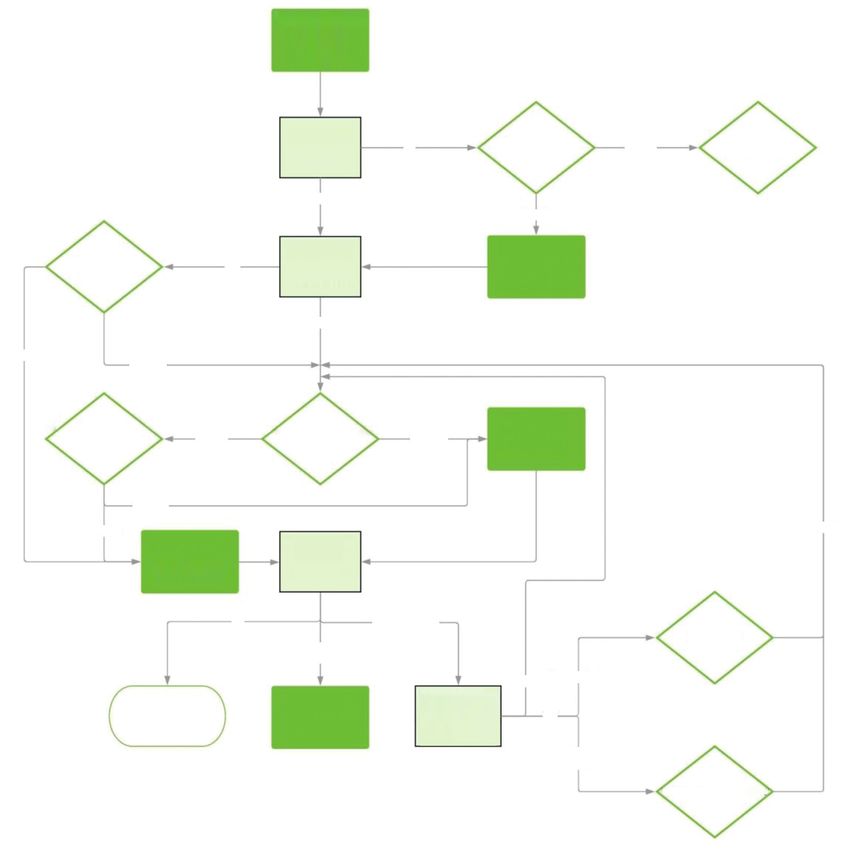
Figure 1 Above is a recommended diagnostic algorithm for obscure gastrointestinal bleeding in patients with chronic kidney disease. Adapted
from “ACG Clinical Guideline: Diagnosis and Management of Small Bowel Bleeding” [8] and “Small-bowel capsule endoscopy and device-assisted
enteroscopy for diagnosis and treatment of small-bowel disorders: European Society of Gastrointestinal Endoscopy (ESGE) Clinical Guideline” [31]
EGD, esophagogastroduodenoscopy; CT, computed tomography
without active bleeding (92.3% vs. 12.9%). In those with prior the general population, but the study itself was limited by the
episodes, the earlier the VCE was done after the cessation of number of patients included [35].
bleeding, the higher the yield [33]. Other factors associated
with increased positive capsule endoscopy results included
overt bleeding, use of non-steroidal anti-inflammatory drugs Device-assisted enteroscopy (DAE)
and a larger transfusion requirement [34].
There is one study on the yield of capsule endoscopy in DAE includes double-balloon (DBE), single-balloon
the evaluation of OGIB in patients with CKD. About half of (SBE) and through-the-scope balloon-assisted enteroscopy,
the patients had positive VCE and 33.3% had angiodysplasia and spiral enteroscopy (SE) [36]. These techniques are used
as the cause of bleeding. The overall yield was lower than in for extensive evaluation of the small bowel (with complete
Annals of Gastroenterology 32Obscure GI bleeding in patients with chronic kidney disease
visualization in about 5-40% of patients) and allow for findings and of these 94.1% were angiodysplasia. All identified
therapeutic intervention [8,37]. bleeding lesions were managed with argon plasma coagulation.
Reported diagnostic yields for DAE are 60-80% for However, the yield of repeat DBE for recurrent bleeding was
DBE [8,38-40], 33-74% for SBE [8,41-43], and 33-75% for only high in those with previously positive DBE. Those with
SE [44-46]. The 2017 guidelines from the American Society initially negative DBE had negative evaluations the second
of Gastrointestinal Endoscopy on the endoscopic evaluation time [54]. There are no studies on the yield of repeat endoscopy
of small-bowel bleeding suggest that all the DAE modalities for recurrent OGIB in patients with CKD.
have similar diagnostic and therapeutic outcomes [9]. Timing
of evaluation improves the yield of these modalities. A 2010
retrospective study found that the overall yield of DBE was
higher if the examination was performed within a month of the
Radiologic evaluation
most recent episode of bleeding (84% vs. 57%) [47]. VCE and
device-assisted enteroscopy have similar diagnostic yields and Radiographic modalities also play a role in the work up of
are typically considered complementary modalities [8,48,49]. OGIB, but in general have more utility in the setting of overt
There are no studies on the use of DAE in evaluating OGIB in bleeding. In those with ongoing bleeding and hemodynamic
those with comorbid CKD. stability, computed tomography angiography (CTA) and
scintigraphy may be useful in identifying the site of bleeding and
allowing for targeted intervention. Patients with brisk bleeding
Recurrent bleeding following negative endoscopic are best evaluated with CTA. A meta-analysis of the use of CTA
evaluation in those presenting with acute GI bleeding showed an overall
sensitivity of 89% and a specificity of 85% [55]. Patients with
It is not uncommon for patients with OGIB and negative positive CTA are also more likely to have positive findings on
enteroscopy [56]. Given the risk of contrast nephropathy, CTA
small-bowel evaluation to present with recurrent bleeding,
has traditionally had a limited role in evaluation of OGIB in
particularly patients with CKD. One study by Curdia Goncalves
CKD and has not been well studied.
et al followed 68 patients with OGIB and negative VCE for
Patients with slower rates of bleeding can be better
32 months. Re-bleeding occurred in 23.5% of patients at an
evaluated with scintigraphy, particularly if a patient presents
average of 15 months following the initial presentation [50].
later after the onset of bleeding [8]. The overall diagnostic
Shinozaki et al followed 42 patients with overt OGIB and
yield varies among reports in the literature (45-73%) [57-61].
negative DBE to evaluate rates of re-bleeding. Re-bleeding
One prospective study assessed the use of scintigraphy in the
occurred in 38% of patients at a mean follow up of 54 years [51].
evaluation of obscure overt bleeding and found an overall yield
Another retrospective study evaluated the rate of recurrent
of 65% with a localization accuracy of 75% [61]. Another study
OGIB after a negative VCE. Re-bleeding occurred in 27.4% of
evaluated the utility of scintigraphy in localizing bleeding from
patients at an average of 15 months from the initial presentation;
angiodysplasia in patients with comorbid CKD compared with
22.6% of the patients who suffered re-bleeding had confirmed
colonoscopy and found that scintigraphy had a much higher
angiodysplasia. Factors associated with re-bleeding included
sensitivity (88.9% vs. 30%) [62]. Scintigraphy is limited by
age >65 years, CKD, aortic stenosis, anticoagulant use and
its ability to characterize the source of bleeding and variable
overt bleeding. Patients with CKD had an increased risk of re- accuracy due to the potential for false localization of bleeding
bleeding on univariate analysis (HR 3.498, 95%CI 1.265-9.671; with rapid transit of labeled red blood cells through the small-
P=0.016) [52]. bowel lumen [8,57,60,61,63,64].
Diagnostic angiography is typically reserved for the
hemodynamically unstable, particularly those with large
Yield of repeat endoscopy transfusion requirements and who present early after the onset
of bleeding, as the yield is highest in this setting [8,65-68].
In the setting of recurrent bleeding, the data on repeat The benefit of this approach is its therapeutic potential. It is
small-bowel evaluation are limited and conflicting. In the otherwise not used often for diagnosis because of its invasive
study by Curdia Goncalves et al, 57.4% of patients who re-bled nature and associated risks [8]. The overall yield of conventional
underwent further diagnostic testing and a cause of bleeding angiography ranges between 20-51% [66,69-71]. Despite the
was only found 13% of the time [50]. Conversely, another utility of angiography in diagnosing the cause of OGIB, VCE
study demonstrated a high yield of repeat VCE. Of those with remains superior and has a higher diagnostic yield [69]. As
negative prior VCE, 75% had findings on repeat VCE and it poses similar risks to those associated with CTA, contrast
62.5% of these patients had a change in management [53]. exposure limits the role of angiography in evaluating OGIB in
In a study by Shinozaki et al that included patients with patients with CKD.
initial negative DBE, of those patients who re-bled, 88% In addition to concerns regarding contrast exposure,
underwent further diagnostic evaluation with a yield of 71%, patients who present with recurrent bleeding are more likely
and of those with a confirmed source, 70% were in the small to get multiple radiographic scans with increased radiation
bowel [51]. In another study that looked at the yield of repeat exposure. One study evaluated the cancer risk associated with
DBE for recurrent OGIB, 53.1% of patients had positive each CTA and found that abdominal CTAs specifically confer
Annals of Gastroenterology 326 M. Muftah et al
the highest cancer risk, estimated to be around 36 cancer risks the later. The patients treated with electrocautery had a
per 1 million procedures [72]. statistically significant reduction in recurrent bleeding and
CT or magnetic resonance enterography (CTE or MRE) can blood transfusions [86]. Similarly, a prospective 2006 study
be used as part of the workup of OGIB. However, in regard to evaluated long-term follow up in 100 patients who underwent
bleeding specifically from vascular lesions and angiodysplasia, argon plasma ablation therapy for colonic angiodysplasia: 85%
VCE has superior yield and higher sensitivity in identifying had stabilized hemoglobin concentration without recurrence
these lesions [73-77]. Thus, CTE or MRE have little utility as of overt bleeding after a 20-month follow up [87]. Lastly, a
part of the initial work up of OGIB in patients with CKD. 2012 prospective study evaluated the long-term outcomes of
61 patients following SE, of whom 45 underwent endoscopic
therapy. There was a statistically significant reduction in
the rate of overt bleeding, transfusion requirements, mean
Endoscopic intervention
hemoglobin and need for iron supplementation over an
average of 25.3-month follow up [88]. There are no studies on
Specific interventions once a culprit lesion is found depends endoscopic intervention for OGIB in patients with CKD.
on the type of lesion itself. While it can be treated with
electrocautery, angiodysplasia is primarily treated with argon
plasma coagulation (APC) via PE or DAE. Other methods of
hemostasis include injection therapy or mechanical hemostasis Non-endoscopic management of GI bleeding
with clips. As mentioned above, DAE is reserved largely for
these therapeutic interventions [9]. Considering the low efficacy of endoscopic management in the
The data regarding long-term outcomes are controversial. long-term prevention of recurrent bleeding from angiodysplasia,
However, the majority of studies report no difference in long- it is important to consider non-endoscopic therapies.
term outcomes in those treated with endoscopy vs. observation
alone [78,79]. One systematic review looked at the long-term
outcomes of patients with bleeding from angiodysplasia Transfusions and iron replacement therapy
managed with endoscopic therapy versus observation. Re-
bleeding rates in the two groups were 42.7% and 49.2% [78].
As discussed above, observation alone with as-needed
Another systematic review found that the pooled re-bleeding
transfusions and iron replacement for anemia are non-inferior
rate of small-bowel angiodysplasia following endoscopic
to endoscopic management. This is potentially a viable option
intervention was 45% [80]. A 2013 study reviewed patients who
for those for whom sedation presents a high risk, or have had
underwent DAE for small-bowel bleeding from angiodysplasia.
multiple prior endoscopic interventions with recurrent or
The overall diagnostic yield was 74% and the therapeutic yield
persistent bleeding. It is important to note, however, that oral
67%; however, there was no difference in long-term outcomes
iron is virtually ineffective at replenishing iron stores in those
between those patients who received endoscopic therapy and
with deficiency and comorbid CKD. It is thought that uremia
those who underwent observation alone: re-bleeding occurred
prevents the GI absorption of iron [14].
in 30% of the former and 20% of the latter [81]. A 2012
A prospective study compared placebo with oral and
retrospective study evaluated re-bleeding rates in patients with
intravenous (IV) iron replacement in patients with iron
OGIB who received endoscopic therapy (primarily APC) for
small-bowel vascular lesions. The re-bleeding rate was 46% at deficiency anemia on HD. Both oral iron and placebo were
36-month follow up [82]. A 2009 survey study evaluated the ineffective in improving the patients’ hemoglobin levels, but
long-term outcomes of 101 patients following DBE for OGIB. IV iron was efficacious (with an average rise in hemoglobin
Of those followed, 40 had angiodysplasia treated with APC; of from below 7 g/dL to above 12 g/dL) [89]. The second study
these 40 patients, 54% had re-bleeding or recurrent need for was a randomized control trial of patients iron replete on
iron replacement and blood transfusions [83]. A 1996 study HD who were initiating erythropoietin for hemoglobin levels
evaluated patients with OGIB who underwent PE. Less than less than 8.5 g/dL. Patients were randomized to placebo, oral
half of patients treated with electrocautery for angiodysplasia iron supplementation or IV iron supplementation. Those
had a good outcome, defined as not having recurrent anemia on IV iron had larger improvements in their hemoglobin
or blood transfusion requirements [84]. Lastly, a 1987 study concentrations and were the only group able to maintain
compared the long-term outcomes of those with angiodysplasia ferritin stores. The other two groups had a drop in their
treated either with surgical resection, endoscopic electrocautery ferritin levels over time [90].
or transfusion alone. No modality was statistically superior in
treating and preventing recurrent bleeding [85].
Conversely, a few studies showed effective reduction of Somatostatin analogs
bleeding with endoscopic therapy. A 1996 study retrospectively
evaluated the long-term follow up of 83 patients found to Increasingly, studies have shown that octreotide is an
have small-bowel angiodysplasia on PE. Of these patients, effective option for the management of bleeding from
55 underwent electrocautery and 28 did not, with a mean angiodysplasia and should be strongly considered in those
follow up of 30 months for the former and 26 months for with recurrent bleeding despite endoscopic intervention [91].
Annals of Gastroenterology 32Obscure GI bleeding in patients with chronic kidney disease
Octreotide is a somatostatin analog whose reported mechanism to treatment, the average hemoglobin was 6.5 g/dL, which
of action includes decreasing splanchnic blood flow, inhibiting increased to 11.3 g/dL at 2 months and 12.1 g/dL at 4 months
angiogenesis, improving platelet aggregation and increasing of treatment. However, 16.7% of patients had to stop treatment
vascular resistance [79,80,92]. early because of side effects [101]. One case report described
A review by Jackson and Gerson found that somatostatin effective treatment of refractory GI bleeding secondary to
analogs were effective in reducing recurrent bleeding episodes diffuse angiodysplasia in a patient on HD. The patient had a
and transfusion requirements with a pooled OR of 14.5 (95%CI significant reduction in transfusion requirements and number
5.9-36) [80]. A 2010 meta-analysis of studies reporting the use of angiodysplastic lesions found on endoscopy [102].
of somatostatin analogs in the treatment of angiodysplasia
determined a clinical response of 76%, with a weighted mean
difference in transfusion requirements before and after treatment Desmopressin
of −2.2 (95%CI −3.9 to −0.5) [93]. Another study followed patients
with recurrent GI bleeding secondary to angiodysplasia after at Given that bleeding in patients with CKD is related to uremic
least 6 months of treatment with a somatostatin analog. Following platelet dysfunction, desmopressin can potentially be used as
treatment, the rate of bleeding episodes was reduced from 73% to a short-term reversal agent for this acquired coagulopathy in
20%, the number of transfusions needed dropped from a median the management of patients presenting with overt or ongoing
of 10 to 2, and the mean hemoglobin increased from a median bleeding [4,18,103,104]. However, only limited relevant data
of 7 to 10 g/dL [94]. Similarly, a prospective study evaluated the are available.
efficacy of 1-year octreotide treatment in patients with chronic
Intranasal or IV desmopressin lasts about 8 h and can be used
bleeding secondary to angiodysplasia. Complete response
for up to two total doses before exhibiting tachyphylaxis [4].
without the need for transfusion or iron supplementation was
The effect of desmopressin peaks at 1-2 h [103]. A double-
achieved in 69% of patients, a partial response in 8% of patients
blind crossover study compared the effect of desmopressin
and no response in 23% of patients [95].
to placebo on bleeding times in patients on HD. Those that
A 2017 study evaluated the use of combination endoscopic
received desmopressin had a reduction in bleeding time from
therapy and the somatostatin analog lanreotide compared
an average of 21.3±8 min to 11.5±6 min; however, there was no
to endoscopic therapy alone in patients with refractory iron
change in hemoglobin or platelet count [105].
deficiency anemia secondary to small-bowel angioectasia.
The proposed mechanism of action is multifactorial and
Combination therapy led to statistically significant reduction
includes an increased number of von Willebrand Factor (vWF)
in transfusion requirements and bleeding episodes with an
and Factor VIII (FVIII) complexes, likely via increased release
increase in hemoglobin compared with the other group [96].
One case series reported 3 patients with CKD and recurrent from storage sites, improved platelet membrane receptor
bleeding secondary to diffuse GI angiodysplasia that responded binding of the vWF: FVIII complexes [103], and direct
to 6 months of octreotide therapy [97]. However, no formal action on the platelet membrane leading to increased platelet
studies have examined the efficacy of octreotide in treating serotonin uptake and subsequent adenosine triphosphate
angiodysplasia in patients with comorbid CKD. release [105, 106].
It is important to note that the effect of desmopressin on
transfusion requirements in those with active bleeding and its
Thalidomide clinical utility in preventing GI bleeding have not been studied.
Additionally, the same effect of desmopressin in reducing
platelet dysfunction is also achieved by HD [18,107]. There
Thalidomide is the most recent addition to the medical
armamentarium in treating bleeding from angiodysplasia. Its is reported utility prior to surgical procedures in preventing
proposed mechanism of action is related to anti-angiogenic bleeding, and it may prove to be useful in patients who are
effects at low doses, as the development of angiodysplasia is about to undergo endoscopic therapy.
suspected to be related to elevated levels of vascular endothelial
growth factor causing increased angiogenesis [80,92,98,99].
A 2011 randomized control trial revealed that thalidomide Hormonal therapy
was effective in reducing the rates of re-bleeding in patients
with recurrent bleeding from angiodysplasia. Patients were In the 1980s and 90s, hormonal therapy gained traction
randomized to treatment or control (treated with iron in the prevention of GI bleeding [108]. However, more recent
therapy) groups and were followed for at least 1 year. Effective studies have proven it to be ineffective, particularly in the
response was defined as a 50% reduction in bleeding episodes. setting of bleeding from angiodysplasia [9,80]. Data on the
Those treated with thalidomide had a response rate of 71.4%, effect of hormonal therapy in preventing bleeding in those with
compared with 3.7% for the control group. Those treated with CKD are controversial and overall lacking [109].
thalidomide also had lower transfusion requirements and fewer A systematic review by Jackson and Gerson of the
hospitalizations. Side effects experienced by patients included management of GI bleeding secondary to angiodysplasia
neuropathy and somnolence [100]. A 2012 prospective study found that hormonal therapy was ineffective overall (OR 1.0,
evaluated patients treated with thalidomide for 4 months 95%CI 0.5-1.96) [80]. A multicenter randomized controlled
for refractory bleeding secondary to angiodysplasia. Prior trial enrolled patients with small-bowel angiodysplasia
Annals of Gastroenterology 328 M. Muftah et al
into a treatment or control group and found that there available evidence concerning the management of patients
was no significant difference between the two groups with with OGIB is ambiguous and systematic reviews suggest no
regard to the number of bleeding episodes and transfusion overall difference in outcomes between those who undergo
requirements [110]. Another cohort study evaluated patients endoscopic versus conservative management for these lesions.
with small-bowel angiodysplasia and found no difference in Therefore, it is important to weigh the risks and the benefits
transfusion requirements between those receiving hormonal prior to endoscopic intervention, especially in patients with
therapy versus observation alone [111]. significant comorbidities. We propose an algorithmic approach
Regarding patients with CKD specifically, there are to evaluating obscure GI bleeding in patients with CKD based
insufficient data on the use of hormonal therapy to prevent on existing guidelines in Figure 1. There are minimal data
bleeding. The above randomized controlled trial was not on the endoscopic evaluation, intervention and outcomes of
sufficiently powered to exclude a benefit in patients with OGIB specifically in patients with CKD, and more studies are
CKD [109,110], in whom it has been shown that hormonal necessary to assess whether their outcomes compare to those
therapy can reduce bleeding times [112-114]. There are case in the general population.
series and case reports suggesting that hormonal therapy
has been effective in the management of bleeding from
angiodysplasia in patients with CKD [115-118]. Given that the
data for the general population show no benefit from using References
hormonal therapy, until there are studies proving otherwise in
the CKD population we would avoid using it in this setting. 1. Gerson LB. Causes of gastrointestinal hemorrhage in patients
with chronic renal failure. Gastroenterology 2013;145:895-897;
discussion 897.
2. Yang JY, Lee TC, Montez-Rath ME, et al. Trends in acute
Arterial embolization nonvariceal upper gastrointestinal bleeding in dialysis patients.
J Am Soc Nephrol 2012;23:495-506.
With advances in technology allowing for super-selective 3. Zuckerman GR, Cornette GL, Clouse RE, Harter HR. Upper
mesenteric embolization, the overall risks of embolization— gastrointestinal bleeding in patients with chronic renal failure.
Ann Intern Med 1985;102:588-592.
particularly bowel infarction—have decreased with time.
4. Kalman RS, Pedrosa MC. Evidence-based review of
However, most data regarding arterial embolization for GI gastrointestinal bleeding in the chronic kidney disease patient.
bleeding are from patients with bleeding sources outside of the Semin Dial 2015;28:68-74.
small bowel [119-122]. Reported clinical success rates range 5. Sakai E, Endo H, Taniguchi L, et al. Factors predicting the
between 63% and 71.4%, with mortality ranging between 9% presence of small bowel lesions in patients with obscure
and 19.3% [119,121,123]. There are no studies on the role of gastrointestinal bleeding. Dig Endosc 2013;25:412-420.
arterial embolization in managing OGIB in patients with CKD. 6. Hágendorn R, Farkas N, Vincze, et al. Chronic kidney disease
severely deteriorates the outcome of gastrointestinal bleeding:
A meta-analysis. World J Gastroenterol 2017;23:8415-8425.
7. Raju GS, Gerson L, Das A, Lewis B; American Gastroenterological
Surgical treatment Association. American Gastroenterological Association (AGA)
Institute technical review on obscure gastrointestinal bleeding.
Surgery is typically reserved for refractory and life- Gastroenterology 2007;133:1697-1717.
threatening cases of bleeding. The primary surgical 8. Gerson LB, Fidler JL, Cave DR, Leighton JA. ACG Clinical
Guideline: diagnosis and management of small bowel bleeding.
intervention is intraoperative enteroscopy with subsequent
Am J Gastroenterol 2015;110:1265-1287.
surgical resection or endoscopic therapy [80]. Surgery is also 9. Gurudu SR, Bruining DH, Acosta RD, et al; ASGE Standards of
useful in the lysis of adhesions that would otherwise preclude Practice Committee. The role of endoscopy in the management
the employment of advanced endoscopic procedures [9]. of suspected small-bowel bleeding. Gastrointest Endosc
Given the high negative predictive value of endoscopic 2017;85:22‑31.
modalities for small-bowel evaluation, there is little to no utility 10. Pennazio M, Arrigoni A, Risio M, Spandre M, Rossini FP. Clinical
for intraoperative enteroscopy in diagnosis. It is primarily evaluation of push-type enteroscopy. Endoscopy 1995;27:164‑170.
11. Chong J, Tagle M, Barkin JS, Reiner DK. Small
reserved for patients who have a lesion identified by other
bowel push-type fiberoptic enteroscopy for patients
endoscopic techniques and need further localization during with occult gastrointestinal bleeding or suspected
surgery prior to an intervention [124,125]. small bowel pathology. Am J Gastroenterol 1994;
89:2143-2146.
12. van Nooten FE, Green J, Brown R, Finkelstein FO, Wish J. Burden
of illness for patients with non-dialysis chronic kidney disease and
Concluding remarks anemia in the United States: review of the literature. J Med Econ 2010;
13:241-256.
13. Stauffer ME, Fan T. Prevalence of anemia in chronic kidney
While peptic ulcer disease is common in CKD, disease in the United States. PLoS One 2014;9:e84943.
angiodysplasias are the most common culprit of OGIB in patients 14. Babitt JL, Lin HY. Mechanisms of anemia in CKD. J Am Soc
with CKD. These patients frequently present with recurrent or Nephrol 2012;23:1631-1634.
OGIB despite endoscopic evaluation and intervention. The 15. Escolar G, Cases A, Bastida E, et al. Uremic platelets have a
Annals of Gastroenterology 32Obscure GI bleeding in patients with chronic kidney disease
functional defect affecting the interaction of von Willebrand bowel capsule endoscopy in patients with chronic kidney disease:
factor with glycoprotein IIb-IIIa. Blood 1990;76:1336-1340. experience from a University Referral Center. Ann Gastroenterol
16. Boccardo P, Remuzzi G, Galbusera M. Platelet dysfunction in 2015;28:99-104.
renal failure. Semin Thromb Hemost 2004;30:579-589. 36. Chauhan SS, Manfredi MA, Abu Dayyeh BK, et al; ASGE
17. Jalal DI, Chonchol M, Targher G. Disorders of hemostasis Technology Committee. Enteroscopy. Gastrointest Endosc 2015;
associated with chronic kidney disease. Semin Thromb Hemost 82:975-990.
2010;36:34-40. 37. Moeschler O, Mueller MK. Deep enteroscopy - indications,
18. Kaw D, Malhotra D. Platelet dysfunction and end-stage renal diagnostic yield and complications. World J Gastroenterol 2015;
disease. Semin Dial 2006;19:317-322. 21:1385-1393.
19. Rosenblatt SG, Drake S, Fadem S, Welch R, Lifschitz MD. 38. May A, Nachbar L, Ell C. Double-balloon enteroscopy (push-
Gastrointestinal blood loss in patients with chronic renal failure. and-pull enteroscopy) of the small bowel: feasibility and
Am J Kidney Dis 1982;1:232-236. diagnostic and therapeutic yield in patients with suspected small
20. Hwang HS, Song YM, Kim EO, et al. Decisive indicator for bowel disease. Gastrointest Endosc 2005;62:62-70.
gastrointestinal workup in anemic patients with nondialysis 39. Gross SA, Stark ME. Initial experience with double-balloon
chronic kidney disease. Int J Med Sci 2012;9:634-641. enteroscopy at a U.S. center. Gastrointest Endosc 2008;67:890‑897.
21. Kuo CC, Kuo HW, Lee IM, Lee CT, Yang CY. The risk of upper 40. Xin L, Liao Z, Jiang YP, Li ZS. Indications, detectability, positive
gastrointestinal bleeding in patients treated with hemodialysis: a findings, total enteroscopy, and complications of diagnostic
population-based cohort study. BMC Nephrol 2013;14:15. double-balloon endoscopy: a systematic review of data over the
22. Laeeq SM, Tasneem AA, Hanif FM, Luck NH, Mandhwani R, first decade of use. Gastrointest Endosc 2011;74:563-570.
Wadhva R. Upper gastrointestinal bleeding in patients with end 41. Tsujikawa T, Saitoh Y, Andoh A, et al. Novel single-balloon
stage renal disease: causes, characteristics and factors associated enteroscopy for diagnosis and treatment of the small intestine:
with need for endoscopic therapeutic intervention. J Transl Int preliminary experiences. Endoscopy 2008;40:11-15.
Med 2017;5:106-111. 42. Prachayakul V, Deesomsak M, Aswakul P, Leelakusolvong S.
23. Chalasani N, Cotsonis G, Wilcox CM. Upper gastrointestinal The utility of single-balloon enteroscopy for the diagnosis and
bleeding in patients with chronic renal failure: role of vascular management of small bowel disorders according to their clinical
ectasia. Am J Gastroenterol 1996;91:2329-2332. manifestations: a retrospective review. BMC Gastroenterol 2013;
24. Holleran G, Hall B, Hussey M, McNamara D. Small bowel 13:103.
angiodysplasia and novel disease associations: a cohort study. 43. Manno M, Riccioni ME, Cannizzaro R, Andreoli A, Marmo R,
Scand J Gastroenterol 2013;48:433-438. Pennazio M. Diagnostic and therapeutic yield of single
25. Ohmori T, Konishi H, Nakamura S, Shiratori K. Abnormalities of balloon enteroscopy in patients with suspected small-bowel
the small intestine detected by capsule endoscopy in hemodialysis disease: results of the Italian multicentre study. Dig Liver Dis
patients. Intern Med 2012;51:1455-1460. 2013;45:211‑215.
26. Hayat M, Axon AT, O’Mahony S. Diagnostic yield and effect on 44. Schembre DB, Ross AS. Spiral enteroscopy: a new twist
clinical outcomes of push enteroscopy in suspected small-bowel on overtube-assisted endoscopy. Gastrointest Endosc 2009;
bleeding. Endoscopy 2000;32:369-372. 69:333‑336.
27. Plotkin E, Imaeda A. Small intestinal angioectasias are not 45. Buscaglia JM, Richards R, Wilkinson MN, et al. Diagnostic
randomly distributed in the small bowel and most may be reached yield of spiral enteroscopy when performed for the evaluation
by push enteroscopy. J Clin Gastroenterol 2016;50:561‑565. of abnormal capsule endoscopy findings. J Clin Gastroenterol
28. Bollinger E, Raines D, Saitta P. Distribution of bleeding 2011;45:342-346.
gastrointestinal angioectasias in a Western population. World J 46. Rahmi G, Samaha E, Vahedi K, et al. Multicenter comparison
Gastroenterol 2012;18:6235-6239. of double-balloon enteroscopy and spiral enteroscopy.
29. Ge ZZ, Hu YB, Xiao SD. Capsule endoscopy and push enteroscopy J Gastroenterol Hepatol 2013;28:992-998.
in the diagnosis of obscure gastrointestinal bleeding. Chin Med J 47. Shinozaki S, Yamamoto H, Yano T, et al. Long-term outcome
(Engl) 2004;117:1045-1049. of patients with obscure gastrointestinal bleeding investigated
30. Triester SL, Leighton JA, Leontiadis GI, et al. A meta-analysis by double-balloon endoscopy. Clin Gastroenterol Hepatol 2010;
of the yield of capsule endoscopy compared to other diagnostic 8:151‑158.
modalities in patients with obscure gastrointestinal bleeding. Am 48. Pasha SF, Leighton JA, Das A, et al. Double-balloon enteroscopy
J Gastroenterol 2005;100:2407-2418. and capsule endoscopy have comparable diagnostic yield in small-
31. PennazioM,SpadaC,EliakimR,etal.Small-bowelcapsuleendoscopy bowel disease: a meta-analysis. Clin Gastroenterol Hepatol 2008;
and device-assisted enteroscopy for diagnosis and treatment 6:671-676.
of small-bowel disorders: European Society of Gastrointestinal 49. Shishido T, Oka S, Tanaka S, et al. Diagnostic yield of capsule
Endoscopy (ESGE) Clinical Guideline. Endoscopy 2015; endoscopy vs. double-balloon endoscopy for patients who
47:352-376. have undergone total enteroscopy with obscure gastrointestinal
32. Bresci G, Parisi G, Bertoni M, Tumino E, Capria A. The role of bleeding. Hepatogastroenterology 2012;59:955-959.
video capsule endoscopy for evaluating obscure gastrointestinal 50. Cúrdia Gonçalves T, Dias de Castro F, Moreira MJ, Rosa B, Cotter J.
bleeding: usefulness of early use. J Gastroenterol 2005;40:256‑259. Small bowel capsule endoscopy in obscure gastrointestinal bleeding:
33. Pennazio M, Santucci R, Rondonotti E, et al. Outcome of normalcy is not reassuring. Eur J Gastroenterol Hepatol 2014;
patients with obscure gastrointestinal bleeding after capsule 26:927-932.
endoscopy: report of 100 consecutive cases. Gastroenterology 51. Shinozaki S, Yano T, Sakamoto H, et al. Long-term outcomes in
2004;126:643‑653. patients with overt obscure gastrointestinal bleeding after negative
34. Ribeiro I, Pinho R, Rodrigues A, Marqués J, Fernandes C, Carvalho J. double-balloon endoscopy. Dig Dis Sci 2015;60:3691‑3696.
Obscure gastrointestinal bleeding: Which factors are associated 52. Magalhães-Costa P, Bispo M, Santos S, Couto G, Matos L,
with positive capsule endoscopy findings? Rev Esp Enferm Dig 2015; Chagas C. Re-bleeding events in patients with obscure
107:334-339. gastrointestinal bleeding after negative capsule endoscopy. World
35. Docherty E, Koulaouzidis A, Douglas S, Plevris JN. Use of small J Gastrointest Endosc 2015;7:403-410.
Annals of Gastroenterology 3210 M. Muftah et al
53. Jones BH, Fleischer DE, Sharma VK, et al. Yield of repeat wireless suspected small bowel disease. Int J Colorectal Dis 2006;21:97‑104.
video capsule endoscopy in patients with obscure gastrointestinal 74. Wiarda BM, Heine DG, Mensink P, et al. Comparison of magnetic
bleeding. Am J Gastroenterol 2005;100:1058-1064. resonance enteroclysis and capsule endoscopy with balloon-
54. Byeon JS, Mann NK, Jamil LH, Lo SK. Is a repeat double balloon assisted enteroscopy in patients with obscure gastrointestinal
endoscopy in the same direction useful in patients with recurrent bleeding. Endoscopy 2012;44:668-673.
obscure gastrointestinal bleeding? J Clin Gastroenterol 2013; 75. Rajesh A, Sandrasegaran K, Jennings SG, et al. Comparison of
47:496‑500. capsule endoscopy with enteroclysis in the investigation of small
55. Wu LM, Xu JR, Yin Y, Qu XH. Usefulness of CT angiography bowel disease. Abdom Imaging 2009;34:459-466.
in diagnosing acute gastrointestinal bleeding: a meta-analysis. 76. Khalife S, Soyer P, Alatawi A, et al. Obscure gastrointestinal
World J Gastroenterol 2010;16:3957-3963. bleeding: preliminary comparison of 64-section CT enteroclysis
56. Tseng CM, Lin IC, Chang CY, et al. Role of computed tomography with video capsule endoscopy. Eur Radiol 2011;21:79-86.
angiography on the management of overt obscure gastrointestinal 77. Wang Z, Chen JQ, Liu JL, Qin XG, Huang Y. CT enterography in
bleeding. PLoS One 2017;12:e0172754. obscure gastrointestinal bleeding: a systematic review and meta-
57. Brunnler T, Klebl F, Mundorff S, et al. Significance of scintigraphy analysis. J Med Imaging Radiat Oncol 2013;57:263-273.
for the localisation of obscure gastrointestinal bleedings. World J 78. Romagnuolo J, Brock AS, Ranney N. Is endoscopic therapy
Gastroenterol 2008;14:5015-5019. effective for angioectasia in obscure gastrointestinal bleeding?:
58. Ohri SK, Desa LA, Lee H, et al. Value of scintigraphic localization a systematic review of the literature. J Clin Gastroenterol
of obscure gastrointestinal bleeding. J R Coll Surg Edinb 1992; 2015;49:823-830.
37:328‑332. 79. Gerson LB, Jackson C. Time to consider medical therapy for small-
59. Howarth DM, Tang K, Lees W. The clinical utility of nuclear bowel angioectasias. Am J Gastroenterol 2012;107:1442‑1443.
medicine imaging for the detection of occult gastrointestinal 80. Jackson CS, Gerson LB. Management of gastrointestinal
haemorrhage. Nucl Med Commun 2002;23:591-594. angiodysplastic lesions (GIADs): a systematic review and meta-
60. Dusold R, Burke K, Carpentier W, Dyck WP. The accuracy of analysis. Am J Gastroenterol 2014;109:474-483; quiz 484.
technetium-99m-labeled red cell scintigraphy in localizing 81. Fan GW, Chen TH, Lin WP, et al. Angiodysplasia and bleeding in
gastrointestinal bleeding. Am J Gastroenterol 1994;89:345-348. the small intestine treated by balloon-assisted enteroscopy. J Dig
61. Dolezal J, Vizda J, Kopacova M. Single-photon emission computed Dis 2013;14:113-116.
tomography enhanced Tc-99m-pertechnetate disodium-labelled 82. Samaha E, Rahmi G, Landi B, et al. Long-term outcome of
red blood cell scintigraphy in the localization of small intestine patients treated with double balloon enteroscopy for small bowel
bleeding: a single-centre twelve-year study. Digestion 2011; vascular lesions. Am J Gastroenterol 2012;107:240-246.
84:207‑211. 83. Gerson LB, Batenic MA, Newsom SL, Ross A, Semrad CE. Long-
62. Oliveras A, Aubia J, Cao H, et al. 99mTc-labelled red blood term outcomes after double-balloon enteroscopy for obscure
cell scintigraphy for localization of gastrointestinal bleeding in gastrointestinal bleeding. Clin Gastroenterol Hepatol 2009;
chronic renal failure. Nephron 1998;80:76-78. 7:664‑669.
63. Wang CS, Tzen KY, Huang MJ, Wang JY, Chen MF. Localization 84. Schmit A, Gay F, Adler M, Cremer M, Van Gossum A. Diagnostic
of obscure gastrointestinal bleeding by technetium 99m-labeled efficacy of push-enteroscopy and long-term follow-up of patients
red blood cell scintigraphy. J Formos Med Assoc 1992;91:63-68. with small bowel angiodysplasias. Dig Dis Sci 1996;41:2348-2352.
64. Howarth DM. The role of nuclear medicine in the detection of 85. Hutcheon DF, Kabelin J, Bulkley GB, Smith GW. Effect of therapy
acute gastrointestinal bleeding. Semin Nucl Med 2006;36:133‑146. on bleeding rates in gastrointestinal angiodysplasia. Am Surg
65. Gerson LB. Is there a role for angiography in patients with 1987;53:6-9.
obscure overt bleeding? Am J Gastroenterol 2012;107:1377-1379. 86. Askin MP, Lewis BS. Push enteroscopic cauterization: long-
66. Abbas SM, Bissett IP, Holden A, Woodfield JC, Parry BR, term follow-up of 83 patients with bleeding small intestinal
Duncan D. Clinical variables associated with positive angiodysplasia. Gastrointest Endosc 1996;43:580-583.
angiographic localization of lower gastrointestinal bleeding. ANZ 87. Olmos JA, Marcolongo M, Pogorelsky V, Herrera L, Tobal F,
J Surg 2005;75:953-957. Dávolos JR. Long-term outcome of argon plasma ablation
67. Whitaker SC, Gregson RH. The role of angiography in the therapy for bleeding in 100 consecutive patients with colonic
investigation of acute or chronic gastrointestinal haemorrhage. angiodysplasia. Dis Colon Rectum 2006;49:1507-1516.
Clin Radiol 1993;47:382-388. 88. Williamson JB, Judah JR, Gaidos JK, et al. Prospective evaluation of
68. Strate LL, Syngal S. Predictors of utilization of early colonoscopy the long-term outcomes after deep small-bowel spiral enteroscopy
vs. radiography for severe lower intestinal bleeding. Gastrointest in patients with obscure GI bleeding. Gastrointest Endosc 2012;
Endosc 2005;61:46-52. 76:771-778.
69. Leung WK, Ho SS, Suen BY, et al. Capsule endoscopy or 89. Fudin R, Jaichenko J, Shostak A, Bennett M, Gotloib L.
angiography in patients with acute overt obscure gastrointestinal Correction of uremic iron deficiency anemia in hemodialyzed
bleeding: a prospective randomized study with long-term follow- patients: a prospective study. Nephron 1998;79:299-305.
up. Am J Gastroenterol 2012;107:1370-1376. 90. Macdougall IC, Tucker B, Thompson J, Tomson CR,
70. Charbonnet P, Toman J, Bühler L, et al. Treatment of Baker LR, Raine AE. A randomized controlled study of iron
gastrointestinal hemorrhage. Abdom Imaging 2005;30:719-726. supplementation in patients treated with erythropoietin. Kidney
71. Rollins ES, Picus D, Hicks ME, Darcy MD, Bower BL, Int 1996;50:1694‑1699.
Kleinhoffer MA. Angiography is useful in detecting the source 91. Iannone A, Principi M, Barone M, Losurdo G, Ierardi E, Di
of chronic gastrointestinal bleeding of obscure origin. AJR Am J Leo A. Gastrointestinal bleeding from vascular malformations: Is
Roentgenol 1991;156:385-388. octreotide effective to rescue difficult-to-treat patients? Clin Res
72. Alkhorayef M, Babikir E, Alrushoud A, Al-Mohammed H, Hepatol Gastroenterol 2016;40:373-377.
Sulieman A. Patient radiation biological risk in computed 92. Szilagyi A, Ghali MP. Pharmacological therapy of vascular
tomography angiography procedure. Saudi J Biol Sci malformations of the gastrointestinal tract. Can J Gastroenterol 2006;
2017;24:235‑240. 20:171-178.
73. Gölder SK, Schreyer AG, Endlicher E, et al. Comparison of 93. Brown C, Subramanian V, Wilcox CM, Peter S. Somatostatin
capsule endoscopy and magnetic resonance (MR) enteroclysis in analogues in the treatment of recurrent bleeding from
Annals of Gastroenterology 32Obscure GI bleeding in patients with chronic kidney disease
gastrointestinal vascular malformations: an overview and systematic 110. Junquera F, Feu F, Papo M, et al. A multicenter, randomized,
review of prospective observational studies. Dig Dis Sci 2010; clinical trial of hormonal therapy in the prevention of rebleeding
55:2129-2134. from gastrointestinal angiodysplasia. Gastroenterology 2001;
94. Bon C, Aparicio T, Vincent M, et al. Long-acting somatostatin 121:1073-1079.
analogues decrease blood transfusion requirements in patients 111. Lewis BS, Salomon P, Rivera-MacMurray S, Kornbluth AA,
with refractory gastrointestinal bleeding associated with Wenger J, Waye JD. Does hormonal therapy have any benefit for
angiodysplasia. Aliment Pharmacol Ther 2012;36:587-593. bleeding angiodysplasia? J Clin Gastroenterol 1992;15:99-103.
95. Scaglione G, Pietrini L, Russo F, Franco MR, Sorrentini I. Long- 112. Livio M, Mannucci PM, Viganò G, et al. Conjugated estrogens for
acting octreotide as rescue therapy in chronic bleeding from the management of bleeding associated with renal failure. N Engl
gastrointestinal angiodysplasia. Aliment Pharmacol Ther 2007; J Med 1986;315:731-735.
26:935-942. 113. Liu YK, Kosfeld RE, Marcum SG. Treatment of uraemic bleeding
96. Chetcuti Zammit S, Sidhu R, Sanders D. Refractory anaemia with conjugated oestrogen. Lancet 1984;2:887-890.
secondary to small bowel angioectasias - comparison between 114. Shemin D, Elnour M, Amarantes B, Abuelo JG, Chazan JA. Oral
endotherapy alone versus combination with somatostatin estrogens decrease bleeding time and improve clinical bleeding
analogues. J Gastrointestin Liver Dis 2017;26:369-374. in patients with renal failure. Am J Med 1990;89:436-440.
97. Rivera M, Lucero J, Guerrero A, et al. [Octreotide in the 115. Manzanera MJ, Gutiérrez E, Domínguez-Gil B, García JA,
treatment of angiodysplasia in patients with advanced chronic González E, Praga M. [Digestive haemorrhage due to
renal failure]. Nefrologia 2005;25:332-335. angiodysplasia in dialysis patients. Treatment with conjugated
98. Tan HH, Ge ZZ, Gao YJ, et al. The role of HIF- 1, angiopoietin-2, Dll4 estrogens]. Nefrologia 2005;25:412-415.
and Notch1 in bleeding gastrointestinal vascular malformations and 116. Bronner MH, Pate MB, Cunningham JT, Marsh WH. Estrogen-
thalidomide-associated actions: a pilot in vivo study. J Dig Dis 2011; progesterone therapy for bleeding gastrointestinal telangiectasias
12:349-356. in chronic renal failure. An uncontrolled trial. Ann Intern Med
99. Bauditz J, Schachschal G, Wedel S, Lochs H. Thalidomide for 1986;105:371-374.
treatment of severe intestinal bleeding. Gut 2004;53:609-612. 117. Richardson JD, Lordon RE. Gastrointestinal bleeding caused by
100. Ge ZZ, Chen HM, Gao YJ, et al. Efficacy of thalidomide for angiodysplasia: a difficult problem in patients with chronic renal
refractory gastrointestinal bleeding from vascular malformation. failure receiving hemodialysis therapy. Am Surg 1993;59:636‑638.
Gastroenterology 2011;141:1629-1637.e1-e4. 118. Granieri R, Mazzulla JP, Yarborough GW. Estrogen-progesterone
101. Garrido A, Sayago M, López J, León R, Bellido F, Márquez JL. therapy for recurrent gastrointestinal bleeding secondary
Thalidomide in refractory bleeding due to gastrointestinal to gastrointestinal angiodysplasia. Am J Gastroenterol 1988;
angiodysplasias. Rev Esp Enferm Dig 2012;104:69-71. 83:556-558.
102. Mimidis K, Kaliontzidou M, Tzimas T, Papadopoulos V. 119. Chan DK, Soong J, Koh F, Tan KK, Lieske B. Predictors for
Thalidomide for treatment of bleeding angiodysplasias during outcomes after super-selective mesenteric embolization for lower
hemodialysis. Ren Fail 2008;30:1040-1041. gastrointestinal tract bleeding. ANZ J Surg 2016;86:459-463.
103. Watson AJ, Whelton A. Therapeutic manipulations in uremic 120. Weldon DT, Burke SJ, Sun S, Mimura H, Golzarian J.
bleeding. J Clin Pharmacol 1985;25:315-317. Interventional management of lower gastrointestinal bleeding.
104. Couch P, Stumpf JL. Management of uremic bleeding. Clin Eur Radiol 2008;18:857-867.
Pharm 1990;9:673-681. 121. Tan KK, Wong D, Sim R. Superselective embolization for lower
105. Malyszko J, Pietraszek M, Buczko W, Mysliwiec M. Study on gastrointestinal hemorrhage: an institutional review over 7 years.
mechanisms of a haemostatic effect of 1 deamino-8-D-arginine World J Surg 2008;32:2707-2715.
vasopressin (desmopressin) in uraemic patients. Folia Haematol 122. Funaki B. Superselective embolization of lower gastrointestinal
Int Mag Klin Morphol Blutforsch 1990;117:319-324. hemorrhage: a new paradigm. Abdom Imaging 2004;29:434-438.
106. Soslau G, Schwartz AB, Putatunda B, et al. Desmopressin-induced 123. Hongsakul K, Pakdeejit S, Tanutit P. Outcome and predictive
improvement in bleeding times in chronic renal failure patients factors of successful transarterial embolization for the treatment
correlates with platelet serotonin uptake and ATP release. Am J of acute gastrointestinal hemorrhage. Acta Radiol 2014;
Med Sci 1990;300:372-379. 55:186‑194.
107. Weigert AL, Schafer AI. Uremic bleeding: pathogenesis and 124. Hartmann D, Schmidt H, Bolz G, et al. A prospective two-center
therapy. Am J Med Sci 1998;316:94-104. study comparing wireless capsule endoscopy with intraoperative
108. van Cutsem E, Rutgeerts P, Vantrappen G. Treatment of bleeding enteroscopy in patients with obscure GI bleeding. Gastrointest
gastrointestinal vascular malformations with oestrogen- Endosc 2005;61:826-832.
progesterone. Lancet 1990;335:953-955. 125. Bonnet S, Douard R, Malamut G, Cellier C, Wind P. Intraoperative
109. Hodgson H. Hormonal therapy for gastrointestinal enteroscopy in the management of obscure gastrointestinal
angiodysplasia. Lancet 2002;359:1630-1631. bleeding. Dig Liver Dis 2013;45:277-284.
Annals of Gastroenterology 32You can also read