High-resolution imaging diagnosis of human fetal membrane by three-dimensional optical coherence tomography
←
→
Page content transcription
If your browser does not render page correctly, please read the page content below
High-resolution imaging diagnosis of
                 human fetal membrane by
                 three-dimensional optical coherence
                 tomography

                 Hugang Ren
                 Cecilia Avila
                 Cynthia Kaplan
                 Yingtian Pan

Downloaded From: https://www.spiedigitallibrary.org/journals/Journal-of-Biomedical-Optics on 09 Feb 2021
Terms of Use: https://www.spiedigitallibrary.org/terms-of-use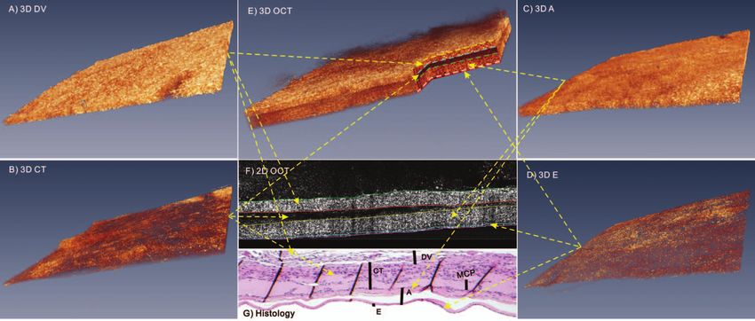
Journal of Biomedical Optics 16(11), 116006 (November 2011)

                High-resolution imaging diagnosis of human fetal
                membrane by three-dimensional optical coherence
                tomography
                Hugang Ren,a Cecilia Avila,b Cynthia Kaplan,c and Yingtian Pana
                a Stony Brook University, Department of Biomedical Engineering, Stony Brook, New York 11794
                b Stony Brook University, Department of Obstetrics, Gynecology, and Reproductive Medicine,
                Stony Brook, New York 11794
                c Stony Brook University, Department of Pathology, Stony Brook, New York 11794

                              Abstract. Microscopic chorionic pseudocyst (MCP) arising in the chorion leave of the human fetal membrane
                              (FM) is a clinical precursor for preeclampsia which may progress to fatal medical conditions (e.g., abortion) if left
                              untreated. To examine the utility of three-dimensional (3D) optical coherence tomography (OCT) for noninvasive
                              delineation of the morphology of human fetal membranes and early clinical detection of MCP, 60 human FM
                              specimens were acquired from 10 different subjects undergoing term cesarean delivery for an ex vivo feasibility
                              study. Our results showed that OCT was able to identify the four-layer architectures of human FMs consisting of
                              high-scattering decidua vera (DV, average thickness dDV ≈ 92 ± 38 μm), low-scattering chorion and trophoblast
                              (CT, dCT ≈ 150 ± 67 μm), high-scattering subepithelial amnion (A, dA ≈ 95 ± 36 μm), and low-scattering epithe-
                              lium (E, dE ≈ 29 ± 8 μm). Importantly, 3D OCT was able to instantaneously detect MCPs (low scattering due to
                              edema, fluid buildup, vasodilatation) and track (staging) their thicknesses dMCP ranging from 24 to 615 μm. It was
                              also shown that high-frequency ultrasound was able to compliment OCT for detecting more advanced thicker
                              MCPs (e.g., dMCP >615 μm) because of its increased imaging depth. C 2011 Society of Photo-Optical Instrumentation Engineers
                              (SPIE). [DOI: 10.1117/1.3646530]

                              Keywords: optical coherence tomography; high-frequency ultrasound; fetal membrane; microscopic chorionic pseudocyst; preeclamp-
                              sia.
                              Paper 11142RRR received Mar. 22, 2011; revised manuscript received Sep. 12, 2011; accepted for publication Sep. 15, 2011; published
                              online Oct. 26, 2011.

                1 Introduction                                                                     leave of the FMs were found to be strongly associated with
                Preeclampsia is a medical disorder associated with increased                       preeclampsia (p ≤ 0.001).6, 7 Although the results were based
                blood pressure and proteinuria during pregnancy or postpartum                      on an ex vivo FM specimen study and artifacts induced by patho-
                period.1 It persists as a major cause of maternal and fetal mortal-                logical preparation (e.g., formalin fixation) could potentially be
                ity and morbidity in the United States and worldwide, affecting                    misinterpreted as MCP due to their similar appearances, the
                5% to 8% of all pregnancies.2 Preeclampsia may progress to                         interesting finding that correlates MCP with preeclampsia may
                eclampsia, which is potentially a fatal medical condition, thus                    provide a new perspective in clinical prediction of preeclampsia.
                rendering it crucial for clinical management.3 Current diagno-                     In this respect, an endoscopic imaging technique that enables in-
                sis of preeclampsia mainly relies on clinical observation of the                   stantaneous, noninvasive, or minimally invasive “optical biopsy”
                symptoms and other unspecific tests4 (e.g., over 140/90 blood                      would be of high clinical relevance in the diagnosis of patho-
                pressure or 300 mg protein in urine). However, these methods                       logical conditions of pregnancy such as preeclampsia.
                become effective only when preeclampsia develops to advanced                           Among several emerging biomedical imaging techniques,
                stages. At that moment, the only treatment option is abortion                      optical coherence tomography (OCT) has shown great promise
                or early delivery, which may lead to various medical compli-                       for noninvasive or minimally invasive optical biopsy of various
                cations to both the preterm newborn and the pregnant woman.                        subsurface tissue owing to its high resolution (e.g., 1 to 12 μm),
                Preeclampsia is a complicated syndrome that may exhibit differ-                    intermediate imaging depth (e.g., 1 to 3 mm), and high detection
                ent symptoms and no definite causative factors are found to be                     sensitivity (e.g., over 100 dB dynamic range). Recent techno-
                responsible for the disease.5 Therefore, studies on specific fea-                  logical advances have enabled real-time two-dimensional (2D)
                tures that have significant correlations with preeclampsia would                   and three-dimensional (3D) OCT imaging, Doppler OCT for
                be of great interest for providing an earlier, more accurate, and                  functional subsurface blood flow imaging, ultrahigh-resolution
                objective clinical diagnosis.                                                      OCT for subcellular imaging, and endoscopic OCT for noninva-
                    A recent study on fetal membranes (FMs) revealed that mi-                      sive imaging of various internal organs.8–10 Meanwhile, preclin-
                croscopic chorionic pseudocysts (MCP) arising in the chorion                       ical and clinical studies have demonstrated the utility of OCT
                                                                                                   in delineating morphological details of biological tissues (e.g.,
                                                                                                   skin, oral cavity, esophagus, colon, bladder, and cervix),11–13
                Address all correspondence to: Yingtian Pan, State University of New York at
                Stony Brook, Department of Biomedical Engineering, Bioengineering Building,
                                                                                                   and thus the potential for detecting cancers in these organs.
                Room G17, Stony Brook, New York 11794-5281. Tel: (631) 632-1519 (Office),
                (631) 632-1750 (Lab); Fax: (631) 632-2322; Email: yingtian.pan@sunysb.edu.         1083-3668/2011/16(11)/116006/6/$25.00
                                                                                                                                         C 2011 SPIE

                Journal of Biomedical Optics                                              116006-1                                        November 2011 r Vol. 16(11)

Downloaded From: https://www.spiedigitallibrary.org/journals/Journal-of-Biomedical-Optics on 09 Feb 2021
Terms of Use: https://www.spiedigitallibrary.org/terms-of-use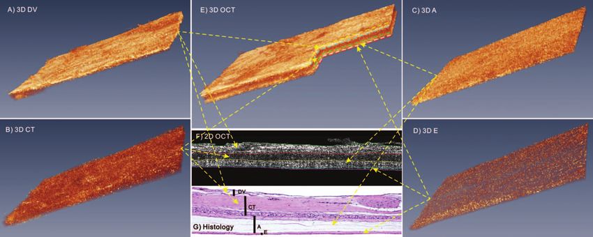
Ren et al.: High-resolution imaging diagnosis of human fetal membrane...

                 While our recent human study showed the clinical feasibility                       (λ = 532 nm) was coupled into the fiber optic system for visual
                 of our endoscopic OCT to significantly enhance early bladder                       guidance of OCT scans. In the reference arm, a prism pair (e.g.,
                 cancer diagnosis,10 here we present a pilot feasibility study on                   using adjustable BK7 and fused silica wedge prisms) were used
                 fresh human fetal membrane specimens from normal controls                          for dispersion compensation and a stationary retroreflective mir-
                 and from patients with MCP to explore the potential of OCT                         ror was used to match the pathlength with the sample arm. The
                 for early detection of pathological changes, which might serve                     sample arm was connected to a handheld stereoscope in which
                 in the prediction of preeclampsia or other diseases associated                     light exiting the monomode fiber was collimated, scanned later-
                 with pregnancy. We compare the image results of OCT and                            ally by 2D servo mirrors (x-y scanners), and then focused by a
                 high-frequency ultrasound (HFUS) with the corresponding his-                       near-infrared objective lens (f 40 mm/NA 0.12) onto the surface
                 tological counterparts (clinical standard), so that the utility and                of the FM specimen under examination. Light from both refer-
                 potential limitations of OCT for high-resolution delineation of                    ence and sample arms was recombined in the detection fiber and
                 the morphology of human FMs and identification of pathologi-                       connected to a spectrometer in which the spectral interferograph
                 cal changes can be examined.                                                       was detected by a line InGaAs camera (1024×1 pixels, up to
                                                                                                    47 kHz line rate) and interfaced via Camlink with a workstation
                                                                                                    for 2D and 3D image acquisition and reconstruction. Recent
                 2 Materials and Methods                                                            system development in detection optics and image acquisition
                 2.1 Sample Preparation                                                             and control resulted in enhanced axial and lateral resolutions
                 A 3D OCT imaging examination was performed on 60 human                             (∼9 μm), large field of view (FOV: 5 × 5 × 2.5 mm3 ) at high
                 FM specimens. These specimens were acquired from 10 dif-                           dynamic range (>110 dB), and fast frame rate (8 to 47 fps).
                 ferent subjects undergoing term cesarean delivery. For each
                 subject, 6 samples were obtained from different sectors of the                     2.3 Imaging Examination
                 FMs, e.g., 4 from the posterior and anterior uterine wall and
                 2 near the cervix. The fresh human specimens were preserved in                     With the FMs properly stretched and the maternal side fac-
                 0.9% saline, rinsed, stretched uniformly to the thickness closely                  ing upwards to mimic in vivo endoscopic imaging diagnosis, a
                 mimicking the anatomic architecture of FMs in vivo, and then                       number of 2D OCT prescans across the entire specimen were
                 mounted on a custom φ20 mm ring holder placed in a modi-                           performed first to quickly locate the regions of interest (ROI).
                 fied ringer’s buffer solution (37◦ C, pH 7.4) to undergo ex vivo                   For each ROI, sequential OCT scans were performed within
                 imaging evaluations. All of the human specimen studies were                        60 s to acquire a 3D OCT image over a cubic volume of 5 × 5
                 approved by the Stony Brook University Institutional Review                        × 2.5 mm3 and displayed in pseudo color to enhance visualiza-
                 Board and with patients’ prior informed consents.                                  tion. By visual guidance with a green laser, the enface FOV for
                                                                                                    each 3D OCT image was landmarked to align the subsequent
                                                                                                    scans for 3D HFUS imaging,14 which was acquired for some
                 2.2 3D OCT                                                                         thickened specimens with advanced preeclampsia. 3D HFUS
                 Figure 1 depicts the schematic of the spectral-domain OCT                          scans were performed using a miniature 40 MHz probe with
                 (SDOCT) used to acquire all of the 3D images of the FM                             an axial resolution of ∼40 μm (Vevo 770, Visualsonics Inc.,
                 specimens in this study. The 3D OCT engine was upgraded                            Toronto, Canada). OCT delineates the morphological details
                 from a high-speed 2D SDOCT setup previously reported,10 in                         (e.g., layers) of human FMs according to their backscattering
                 which a pigtailed broadband laser at central wavelength of λ                       differences. For simplicity, tissue backscattering was expressed
                 = 1310 nm and with a spectral bandwidth of λ = 90 nm (i.e.,                       by back reflectance, defined as the measured OCT intensity nor-
                 coherence length Lc ≈ 8.5 μm) was used to illuminate a fiber                       malized to that of the top layer (i.e., decidua vera). Quantitative
                 optic Michelson interferometer. Green light from a laser diode                     computer segmentation of FMs in both 2D and 3D OCT images
                                                                                                    was performed based on intensity gradient by adapting the algo-
                                                                                                    rithm previously reported,12 and the average thickness (d) and
                                                                                                    back reflectance (r) of each layer were then analyzed. After the
                                                                                                    imaging study, the specimens were preserved in 10% formalin
                                                                                                    fixative together with the ring holders to avoid artifacts such
                                                                                                    as tissue deformation for hematoxylin and eosin (H&E) stained
                                                                                                    histological examination. A double blind histologic evaluation
                                                                                                    was independently performed by a clinical pathologist later to
                                                                                                    compare with the prior OCT and HFUS identifications and di-
                                                                                                    agnoses. The data were presented as mean ± s.t.d.

                                                                                                    3 Results
                                                                                                    Previous studies have demonstrated the utility of OCT to enable
                                                                                                    high-resolution delineation of the morphological features of
                 Fig. 1 A sketch of the 3D OCT setup. BBS: broadband source; GLD:                   biological tissues (e.g., urinary bladder) based on their backscat-
                 green diode laser; FC: fiber optic coupler; PC: polarization controller;
                 CM: collimator; M: mirror; G: grating; LC: linear InGaAs camera; S:                tering differences that attribute to the structural properties.
                 specimen (fetal membrane); X-S, Y-S: X, Y axes of the 2D servo scanner;            Figure 2 exemplifies a typical cross-sectional 2D OCT image
                 L1–L3: lenses.                                                                     [Fig. 2(a)] of a normal FM acquired from the maternal side

                 Journal of Biomedical Optics                                              116006-2                                      November 2011 r Vol. 16(11)

Downloaded From: https://www.spiedigitallibrary.org/journals/Journal-of-Biomedical-Optics on 09 Feb 2021
Terms of Use: https://www.spiedigitallibrary.org/terms-of-use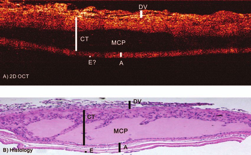
Ren et al.: High-resolution imaging diagnosis of human fetal membrane...
of amnion in some specimens, which is a common artifact
induced by tissue fixation during histological processing.
In addition to the 2D OCT presented in Fig. 2, 3D OCT
image, e.g., by rendering 350 slices of sequential 2D cross-
sectional OCT images, may provide improved image fidelity
and more affirmative identifications of morphological features.
For example, Fig. 3 summarizes the results of normal human
FMs in which Fig. 3(e) shows a pie-cut graph of the 3D OCT
image and Fig. 3(f) illustrates a 2D OCT slice with the four layers
of the FMs automatically segmented based on their backscat-
tering differences. Figures 3(a)–3(d) show the 3D images of the
segmented 4 layers sequentially from DV and CT to A and E
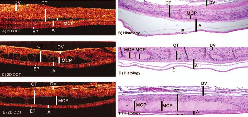
Fig. 2 2D images of a normal human fetal membrane. (a) cross- layers. Compared with 2D OCT in Fig. 2, 3D OCT in Fig. 3,
sectional OCT image; (b) corresponding H&E stained histology. DV: owing to its improved spatial correlation (along the y-axis), pro-
decidua vera (dDV ≈ 92 ± 38 μm), CT: chorion and trophoblast (dCT vides enhanced image quality, which may permit more detailed
≈ 150 ± 67 μm, rCT/DV = 0.51 ± 0.17), A: subepithelial amnion (dA
analysis to characterize the architectural features of individual
≈ 95 ± 36 μm, rA/DV = 0.84 ± 0.45), and E: epithelium (dE ≈ 29
± 8 μm, rE/DV = 0.44 ± 0.20). layers.
In this pilot human specimen study, not only normal human
FMs but also potential pathological human FM specimens were
and the corresponding H&E stained histological evaluation examined to evaluate the utility of OCT for noninvasive and
[Fig. 2(b)]. OCT was able to identify the four layers of the high-resolution imaging diagnosis of FM diseases (e.g., MCP).
FMs according to their backscattering differences. For instance, Figure 4 shows 2D OCT image [Fig. 4(a)] of an FM sample
the outermost layer, decidua vera (DV) was relatively thin with MCP which was characterized by dark holes (low backre-
(dDV ≈ 92 ± 38 μm), heterogeneous, and high scattering. flection) with thickness of dMCP ≈ 320 μm between A and CT
The underlying chorion and trophoblast (CT) layer was thick layers. The low-scattering characteristics of MCP (dark holes
(dCT ≈ 150 ± 67 μm) and relatively low scattering (rCT/DV with rMCP/DV = 0.17 ± 0.06) were caused by fluid buildup (i.e.,
= 0.51 ± 0.17) possibly due to its loose structure and high edema) within the lesions. Figure 4(b) represents the corre-
interstitial fluid content. The subepithelial amnion (A) layer sponding H&E histology which correlated well with the OCT
was slightly thinner (dA ≈ 95 ± 36 μm) than the CT layer identifications of the 4 layers and the cysts (MCP) within the CT
and was high scattering (rA/DV = 0.84 ± 0.45) resulting from and A layers except that the lesions (dMCP ≈ 400 μm) appeared
subepithelial connective tissue. The innermost epithelium larger than those (dMCP ≈ 320 μm) in OCT image [Fig. 4(a)].
(E) was very thin (dE ≈ 29 ± 8 μm or less, with 1 to 2 cell This discrepancy likely resulted from the artifacts induced by
depths), uniform, and low scattering (rE/DV = 0.44 ± 0.20). It tissue fixation and histological processing.
is noteworthy that the thicknesses of the intermediate amnion Similarly, Fig. 5 shows the results of 3D OCT of a human
(A) and chorion (CT) layers and the FMs might vary with FM specimen with “early stage” MCP progression. Despite
the trimester of pregnancy, the extent of tissue stretching, and the fact that the surface image appeared normal, the segmented
the location of OCT scans, which might result in large error 3D OCT images revealed early, minor detachment (dMCP
margins. Overall, the OCT identifications of the four layers ≈ 80 μm, rMCP/DV = 0.14) within the CT and A layers, resulting
within the FMs correlated well with the counterparts in the in drastically increased inhomogeneity within the CT layer
corresponding histological image [Fig. 2(b)] except detachment [Fig. 5(c)]. Interestingly, the innermost epithelial layer E
Fig. 3 3D image of a normal human FM. (a)–(d) 3D OCT images of the segmented DV, CT, A, and E layers; (e) 3D OCT image of the entire human
FMs; (f) 2D OCT image to illustrate the automatic segmentation procedure based on the backscattering differences of each layer; (g) corresponding
H&E histology of OCT image in (f).
Journal of Biomedical Optics 116006-3 November 2011 r Vol. 16(11)
Downloaded From: https://www.spiedigitallibrary.org/journals/Journal-of-Biomedical-Optics on 09 Feb 2021
Terms of Use: https://www.spiedigitallibrary.org/terms-of-use
Ren et al.: High-resolution imaging diagnosis of human fetal membrane...

                                                                                                    [Fig. 5(a)] also became less uniform than the normal coun-
                                                                                                    terpart in Fig. 3(a), which could be associated with the
                                                                                                    inflammatory reactions of MCP. By detecting the size progres-
                                                                                                    sion of MCP lesions, OCT was potentially capable of providing
                                                                                                    noninvasive evaluation (i.e., “staging”) of MCP development
                                                                                                    and severity, in particular by 3D image segmentation to provide
                                                                                                    quantitative assessments of cyst depth (e.g., dMCP ≈ 80 μm in
                                                                                                    Fig. 5, dMCP ≈ 320 μm in Fig. 4) and the resultant inhomogene-
                                                                                                    ity which was associated with fluid buildup, vasodilation, local
                                                                                                    microhemorrhage, macrophage, and mast cell accumulations.
                                                                                                    It should be noted that although the corresponding histological
                                                                                                    image in Fig. 5(g) correlated favorably with the OCT delin-
                                                                                                    eations, the artifacts induced by tissue fixation complicated the
                                                                                                    identification of MCP (cysts) from distortion (fall off) of CT
                 Fig. 4 2D image of a human FM specimen with MCP. (a) 2D OCT im-                    and A layers, which might compromise the utility of histology
                 age; (b) corresponding H&E histology. The MCP lesions characterized                for affirmative staging of MCP growth and spreading.
                 by OCT as dark holes correlated with the cysts in histology. The thick-
                                                                                                        Figure 6 compares three human FM specimens to show
                 ness of MCP lesions in OCT (dMCP ≈ 320 μm) matched the histological
                 counterpart (dMCP ≈ 400 μm).                                                       the capability of OCT to assess the growth of MCP lesions.

                 Fig. 5 3D OCT image of a human FM with MCP. (a)–(d) 3D OCT images of the segmented DV, CT, A, and E layers. (e) 3D OCT image of the intact
                 human FMs with MCP; (f) segmented 2D OCT image to illustrate automatic segmentation based on their backscattering differences; (g) corresponding
                 histology of the OCT image in (f). The CT and E layers were more heterogeneous than the previous normal specimen in Fig. 3.

                 Fig. 6 OCT images of human FM specimens with different-size MCP lesions compared with the corresponding H&E stained histological images.
                 (a), (c), and (e) 2D OCT images with the MCPs automatically segmented as landmarked by green dashed circles. The thicknesses of MCPs were dMCP
                 ≈ 60 μm (a), dMCP ≈ 150 μm (c), and dMCP ≈ 265 μm (e), respectively. The percentage areas of MCP, i.e., the ratios of the area of MCPs against
                 the entire FM cross-section were 3.7% (a), 25.6% (c), and 28.3% (e). (b), (d), (f) The corresponding histological images. The thicknesses of MCPs
                 were dMCP ≈ 53 μm (b), dMCP ≈ 141 μm (d), and dMCP ≈ 251 μm (f), which correlated with the OCT measurements despite artifacts such as tissue
                 detachment induced by histological process.

                 Journal of Biomedical Optics                                              116006-4                                     November 2011 r Vol. 16(11)

Downloaded From: https://www.spiedigitallibrary.org/journals/Journal-of-Biomedical-Optics on 09 Feb 2021
Terms of Use: https://www.spiedigitallibrary.org/terms-of-use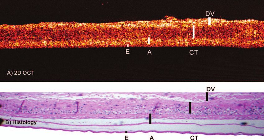
Ren et al.: High-resolution imaging diagnosis of human fetal membrane...

                                                                                                   drastically increased heterogeneity in this FM specimen, OCT
                                                                                                   was unable to delineate the underlying layers within the FMs.

                                                                                                   4 Discussions and Conclusions
                                                                                                   Early diagnosis of preeclampsia, crucial to effective therapeutic
                                                                                                   treatment, remains a clinical challenge due to the multifactorial
                                                                                                   nature of this disease.4, 5 A previous study revealed that MCP
                Fig. 7 3D HFUS image of a human FM with thick MCP. (a) 3D HFUS                     originating from the chorion leave (involving mostly CT and A
                image; (b) a slice of 2D HFUS from image (a); (c) corresponding 2D                 layers) of the FMs was demonstrated to be closely related to
                OCT image. Despite the lower spatial resolution, HFUS was able to                  preeclampsia (p ≤ 0.001),6 thus early diagnosis and evaluation
                penetrate the entirety of the thicker FMs while the image depth of OCT             of the progression of MCP could be of great clinical relevance.
                was limited to layer DV or top CT.
                                                                                                   Current diagnosis is based on ex vivo histopathologic exami-
                                                                                                   nation of the excised tissue specimens, whose clinical value is
                Although the thickness of MCP (dMCP ) may vary with the                            restricted by its invasive and destructive natures. Moreover, little
                point of measurement, the increase of MCP lesions could be                         has been studied about potential pathological misinterpretation
                differentiated by their mean thickness. OCT measurement of                         of MCP as a result of artifacts induced by tissue fixation and
                dMCP ≈ 60 μm, rMCP/DV = 0.13 for the small MCP lesion in                           processing.
                Fig. 6(a) correlated with the histological evaluation dMCP                             In contrast, noninvasive early diagnosis of MCP could
                ≈ 53 μm in Fig. 6(b); the two large lesions dMCP                                   potentially benefit the treatment. Current medical imaging
                ≈ 150 μm, rMCP/DV = 0.18 [Fig. 6(c)] and, dmcp = 265,                              techniques such as MRI and ultrasound may provide limited
                rmcp/dv = 0.19 [Fig. 6(e)] measured by OCT [Fig. 6(c)]                             diagnostic values because of their insufficient spatial resolution
                were correlated favorably with the corresponding histological                      and other technical imperfections (e.g., potential radiation
                measures, dMCP ≈ 141 μm in Fig. 6(d) and dMCP ≈ 251 μm in                          hazard to the fetus). OCT is an emerging optical imaging
                Fig. 6(f), respectively. Alternatively, the percentage area of MCP                 modality that, if integrated with endoscopy, permits noninvasive
                lesion, i.e., SMCP (%) = SMCP /SFM (SMCP and SFM are the cross-                   cross-sectional 2D and 3D imaging of biological tissue at high
                sectional areas of the PCM lesions and FMs) can be employed;                       spatial resolutions (e.g., 1 to 10 μm) and over intermediate
                the results of OCT measures were 3.7%, 25.6%, and 28.3% for                        depths (e.g., 1 to 3 mm). Previous preclinical studies validated
                Figs. 6(a), 6(c), and 6(e), respectively. In addition, we calculated               the capability of OCT for delineating the morphological details
                the statistical result of the average thickness of MCP based on                    of various biological tissues such as oral cavity, bladder, esoph-
                32 lesions. The result showed that the MCP detectable by OCT                       agus, and cervix.11–13 More interestingly, recent in vivo clinical
                varied from dMCP = 24 μm to dMCP = 615 μm with a mean thick-                       studies clearly demonstrated the utility of our endoscopic OCT
                ness of 160 μm and a median thickness of 120 μm. These results                     technique in significantly enhancing current clinical approach
                suggest that endoscopic OCT could potentially be deployed                          (i.e., white-light cystoscopy) for noninvasive diagnosis of early
                to instantaneously diagnose MCPs and quantitatively evaluate                       bladder cancer. Technically, the areas of fetal membranes prone
                (i.e., stage) their progress as well as the treatment effects. It is               to preeclamptic changes can potentially be imaged in vivo using
                noteworthy from the histological images in Figs. 6(b), 6(d), and                   our newly developed miniature (e.g., φ2 mm) flexible OCT
                6(f) that distortion and other artifacts (e.g., formalin infiltration)             catheter during intrauterine examination. We have presented a
                might compromise the evaluation of histological specimens.                         preliminary study based on human tissue specimens, including
                    The limited imaging depth of OCT (e.g., ∼1 to 3 mm) may                        both normal control and diseased FMs. Results in Figs. 2 and
                potentially restrict its utility in the diagnosis and assessment                   3 show that OCT was capable of delineating the morphological
                of later-stage severe MCP lesions. For instance, for a few FM                      details of normal human FMs as the four layers (e.g., DV, CT,
                specimens (e.g., different locations on the FMs) with a thick DV                   A, and E) based on their backscattering differences (e.g., rCT/DV
                layer (e.g., 2 to 4 mm) from the maternal side, OCT was unable                     = 0.51 ± 0.17; rA/DV = 0.84 ± 0.45; rE/DV = 0.44 ± 0.20).
                to fully delineate the layered structures of the FMs, in partic-                   Results in Figs. 4 and 5 further demonstrate the utility of OCT
                ular, the innermost epithelium (E). In cases like this, HFUS                       to affirmatively detect the MCP lesions (cysts) in the CT layer
                might compliment OCT to overcome the imaging-depth limi-                           of human FMs based on their drastically reduced backscattering
                tation. To examine the feasibility, additional 3D HFUS scans                       (e.g., rMCP/DV = 0.17 ± 0.06). More importantly, by applying
                following OCT imaging were performed using a miniature                             post-image processing techniques (e.g., image segmentation and
                40 MHz probe. Figure 7 exemplifies a 3D HFUS image of hu-                          registration) to the original 3D OCT image dataset, OCT mor-
                man FMs. The results show that because of lower resolution than                    phometric placental analysis could potentially be implemented
                OCT, the boundaries between the layers (e.g., DV, CT, and A)                       to provide quantitative, accurate evaluation of MCP progress
                in HFUS images [Figs. 7(a) and 7(b)] were not as well defined                      (i.e., staging of MCP), which is essential to potentially monitor
                than the counterparts in Figs. 2–5 and were unable to resolve                      the preeclampsia development and to evaluate treatment effects.
                the innermost epithelial layer (e.g., dE 1 mm) DV layer and                      limitations of OCT and HFUS in the diagnosis of preeclampsia,

                Journal of Biomedical Optics                                              116006-5                                      November 2011 r Vol. 16(11)

Downloaded From: https://www.spiedigitallibrary.org/journals/Journal-of-Biomedical-Optics on 09 Feb 2021
Terms of Use: https://www.spiedigitallibrary.org/terms-of-use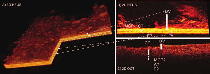
Ren et al.: High-resolution imaging diagnosis of human fetal membrane...

                 further detailed and more quantitative studies should be per-                       2. B. Sibai, G. Dekker, and M. Kupferminc, “Pre-eclampsia,” The Lancet
                 formed in the future, in particular, in vivo imaging evaluations.                      365, 785–799 (2005).
                                                                                                     3. L. Weinstein, “Syndrome of hemolysis, elevated liver enzymes, and low
                     In summary, we performed an in vitro study on fresh human
                                                                                                        platelet count: a severe consequence of hypertension in pregnancy,”
                 FM specimens to examine the efficacy and limitations of OCT                            Obstet. Gynecol. Surv. 142, 159–167 (1982).
                 for future noninvasive or minimally invasive hysteroscopic OCT                      4. B. Sibai, “Diagnosis and management of gestational hypertension and
                 imaging of fetal membranes. Results presented above show that                          preeclampsia,” Obstet. Gynecol. 102, 181–192 (2003).
                 the high resolution and 3D imaging capability of OCT enabled                        5. R. Ness and J. Roberts, “Heterogeneous causes constituting the single
                                                                                                        syndrome of preeclampsia: a hypothesis and its implications,” Am. J.
                 delineation of morphological details of human FMs (e.g., DV,
                                                                                                        Obstet. Gynecol. 175, 1365–1370 (1996).
                 CT, A, and E layers) based on their backscattering differences,                     6. J. Stanek and E. Weng, “Microscopic chorionic pseudocysts in placen-
                 which correlated well with the corresponding histological eval-                        tal membranes: a histologic lesion of in utero hypoxia,” Pediatr. Dev.
                 uations. Additionally, OCT was able to identify early MCPs                             Pathol. 10, 192–198 (2007).
                 and accurately measure the size of these lesions. Further histo-                    7. J. Stanek, “Diagnosing placental membrane hypoxic lesions increases
                                                                                                        the sensitivity of placental examination,” Arch. Pathol. Lab Med. 134,
                 morphometric and immunohistochemical studies are needed to                             989–995 (2010).
                 characterize the heterogeneity increases in these layers with the                   8. Z. Yuan, Z. Luo, H. Ren, C. Du, and Y. Pan, “A digital frequency
                 presence of different cell types and their accumulations so that                       ramping method for enhancing Doppler flow imaging in Fourier-domain
                 more specific diagnosis of the severity and progress of MCP                            optical coherence tomography,” Opt. Express 17, 3951–3963 (2009).
                 can be predicted. In addition, more in vivo animal and human                        9. Z. Yuan, B. Chen, H. Ren, and Y. Pan, “On the possibility of time-lapse
                                                                                                        ultrahigh-resolution optical coherence tomography for bladder cancer
                 studies will be required to fully examine the efficacy, technical                      grading,” J. Biomed. Opt. 1, 050502 (2009).
                 feasibility, and safety of OCT hysteroscopy, as well as HFUS                       10. H. Ren, W. Waltzer, R. Bhalla, J. Liu, Z. Yuan, C. Lee, F. Darras,
                 for potential future clinical application to minimally invasive                        D. Schulsinger, H. Adler, and J. Kim, “Diagnosis of bladder cancer with
                 diagnosis of MCP and staging their progress.                                           microelectromechanical systems-based cystoscopic optical coherence
                                                                                                        tomography,” Urology 74, 1351–1357 (2009).
                                                                                                    11. D. Adler, C. Zhou, T. Tsai, J. Schmitt, Q. Huang, H. Mashimo,
                 Acknowledgments                                                                        and J. Fujimoto, “Three-dimensional endomicroscopy of the human
                                                                                                        colon using optical coherence tomography,” Opt. Express 17, 784–796
                 We acknowledge Jarrett Santorellim, BS at Stony Brook Univer-                          (2009).
                 sity for participating in this study (handling fetal membrane sam-                 12. H. Ren, Z. Yuan, W. Waltzer, K. Shroyer, and Y. Pan, “Enhancing de-
                 ples). This work was supported in part by NIH Grant Nos. 2R01-                         tection of bladder carcinoma in situ by 3-dimensional optical coherence
                 DK059265 (YP), 1RC-1DA028534 (CD, YP), 1R21-DA032228                                   tomography,” J. Urology 184, 1499–1506 (2010).
                                                                                                    13. C. Pitris, P. Hsiung, X. Li, W. Drexler, J. G. Fujimoto, A. Goodman, and
                 (YP) and Fusion Award.                                                                 M. Brezinski, “In vivo cervical imaging with an integrated optical coher-
                                                                                                        ence tomography colposcope,” in Biomedical Optical Spectroscopy and
                                                                                                        Diagnostics, T. Li, ed., Vol. 38 of OSA Trends in Optics and Photonics
                 References                                                                             (Optical Society of America, 2000), paper SuC5.
                  1. R. Gifford, P. August, G. Cunningham, L. Green, M. Lindheimer,                 14. Z. Yuan, Z. Wang, R. Pan, J. Liu, H. Cohen, and Y. Pan, “High-
                     D. McNellis, J. Roberts, B. Sibai, and S. Taler, “Report of the national           resolution imaging diagnosis and staging of bladder cancer: compar-
                     high blood pressure education program working group on high blood                  ison between optical coherence tomography and high-frequency ultra-
                     pressure in pregnancy,” Am. J. Obstet. Gynecol. 183, 1–22 (2000).                  sound,” J. Biomed. Opt. 13, 054007 (2008).

                 Journal of Biomedical Optics                                              116006-6                                           November 2011 r Vol. 16(11)

Downloaded From: https://www.spiedigitallibrary.org/journals/Journal-of-Biomedical-Optics on 09 Feb 2021
Terms of Use: https://www.spiedigitallibrary.org/terms-of-use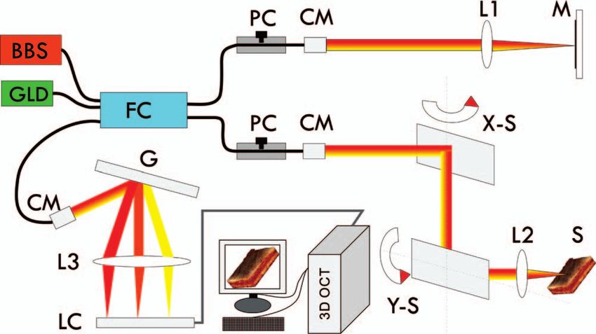
You can also read