Issue One 2017 - National Imaging Facility
←
→
Page content transcription
If your browser does not render page correctly, please read the page content below
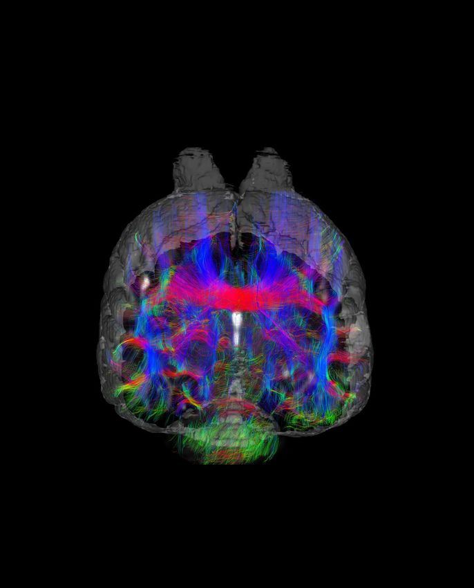
Issue One 2017

Whole brain neuronal connectivity pattern (connectome) of the short-beaked echidna
(spiny anteater) as revealed by Diffusion Tensor Imaging and fibre tracking

Image courtesy of Dr. Andre Bongers - Mark Wainwright Analytical Centre, Biological
Resourcas Imaging Laboratory, The University of New South Wales

Director’s Message

    In This Issue                              We are privileged to be able to share with you
                                               great research stories in every issue of our
                                               newsletter. This issue is no exception. Stories        NIF has been imaging the zebra-fish, building

    Director’s Message                         that describe research that use imaging to reveal
                                               new knowledge for which there is no alternative.
                                                                                                      probabilistic models of the brain, bound to make
                                                                                                      important contributions to the development
                                               Whether it be conservation or preservation,            precision medicine to treat conditions affected by
                                               developing new therapies, or guiding new               brain connectivity.
                                               treatments, imaging has a role.
                                                                                                      If you are not into brains, how about what

    Industry Collaborations                    The Tasmanian Tiger, almost brought back to life.
                                               Who would have thought the latest in imaging
                                                                                                      is happening at the NIF node at the Western
                                                                                                      Sydney University. They are using brain imaging
    •   Elucidating brain structure            technology could be used to learn about brain          techniques, to image colon cancer, and provide
        and connectivity in extinct and        morphology and connectivity in an extinct species.     important information for use in radiotherapy.

        endangered Australian animals          The curators at the Australian Museum, were not
                                               about to allow a valuable sample to be dissected       The use of imaging in research is only limited by
    •   Colon Cancer Characterisation          for conventional histology. So the team at the         your imagination. So, let your imagination go wild,
                                               University of New South Wales node of NIF, non-        think of the most wayout idea of how imaging can
                                               invasively looked inside the brain.                    help your research, and then come and talk to
                                                                                                      the experts across the nation. Or just read about
                                               Equally valuable is the brain of individuals and       the great things that are being done to advance
                                               the varied functions. I am sure that all of us would   knowledge in many disciplines.
    Research Projects                          appreciate that the good parts of the brain are
    •   Assessing regional lateralisation of   preserved, during surgery to remove lesions that
                                               are interfering with proper brain function. That
        language function in the human brain
                                               is what is being done at The Florey node of NIF.
    •   Neuroimaging Phenotypes in Zebrafish   Preserving language, while treating epilepsy.
                                               Talking of epilepsy, the Queensland node of

                                                     T h e u se o f i m a g i n g i n
    News                                             re se a rc h i s o n l y l i m i t e d
    •   2016 in Review
                                                     by your imagination.

                                                               Professor Graham Galloway
                                                                   Director of Operations

2                                                                                                                                                           3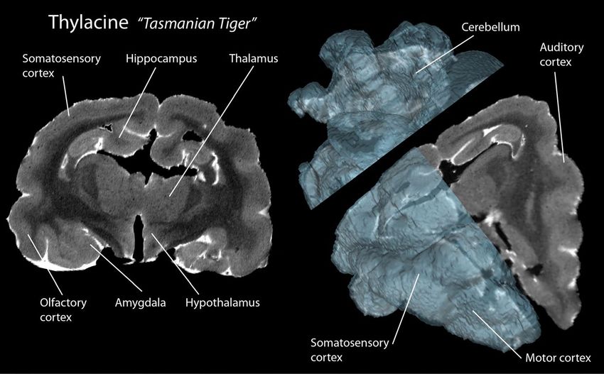
Elucidating brain structure
and connectivity in extinct and
endangered Australian animals
Collaboration
Industry
A
ustralasia has a unique diffusion in the brain and uses this a and connectome of a great diversity of
population of native mammals probe to quantify brain fibre structure. Australian animals. Among them are
and birds. The monotremes These datasets can be used as the the endangered endemic monotremes
(platypus and echidna) are found basis for fibre tracking algorithms to (echidna and platypus), rare marsupials
nowhere else in the world and our reconstruct the structure of the white such as the bilby or numbat as well as
variety of marsupials reflects a matter brain fibres and thus non- animals that are already extinct such as
remarkable evolutionary radiation that destructively elucidate connectivity the thylacine or “Tasmanian Tiger”. The
has produced many exceptional forms. between significant brain regions. project will provide a unique database of
Studying the way that brain structure brain structure for Australia’s exceptional
has evolved in Australian mammals Brain tissues of rare and endangered wildlife that will be invaluable to
can teach many valuable lessons about animals held in museum collections can scientists from around the world.
the way that genes and developmental provide a very rich source of information
mechanisms act in concert to meet the when modern imaging methods such For more information on this project,
requirements of an ecological niche. as DTI and fibre tracking methods are contact Dr. Andre Bongers (andre.
used to map connections between bongers@unsw.edu.au) or Prof. Ken
At the current stage, knowledge about the cerebral cortex and deeper brain Ashwell (k.ashwell@unsw.edu.au).
brain connectivity in many of these structures. This gives us valuable data
interesting Australian endemic species on patterns of connectivity within the Collaborators
is very limited as they are often rare and brain and allows direct comparisons with Prof. Ken Ashwell, Faculty of Medicine,
endangered or even extinct. Ethical and the brains of placental mammals that are Department of Anatomy, School of Medical
technical constraints severely limit the regularly studied by neuroscientists. Sciences, The University of New South
Wales, Sydney
options for brain researchers to access
Dr. Andre Bongers, Mark Wainwright
live animals or to use classical (usually This exciting new approach to studying Analytical Centre, Biological Resourcas
destructive) anatomical methods on brain evolution in rare or extinct animals Imaging Laboratory, The University of New
precious brain samples. is being carried out by an international South Wales, Sydney
team of collaborators at the University Dr. Yamila Gurowich, CONICET y Laboratorio
A very promising approach to non- of New South Wales node of National de Investigaciones en Evolución y
invasively collect missing information Imaging Facility, using the preserved Biodiversidad (LIEB), Universidad Nacional
about the connectome of these brain collection at the Australian de La Patagonia, Argentina
evolutionary significant mammals is Museum in Sydney. Dr. Sandy Ingleby, Mammalogy Collection,
MRI and Diffusion Tensor Imaging (DTI) Australian Museum, Sydney
Prof. Gregory S. Berns, Department of
in preserved brain samples. Structural Using the high field pre-clinical MRI
Psycology, Emory University, Atlanta, USA Coronal T2w MRI slice and 3D volume representation of a brain of the extinct the
MRI yields strong contrast in soft tissues system at UNSW’s Biological Resources Dr. Craig Hardman, , Faculty of Medicine, Thylacine (Tasmanian “Tiger”).
and is able to deliver high resolution and Imaging Laboratory (Mark Department of Anatomy, School of Medical
information about brain regions. DTI Wainwright Analytical Centre) the group Sciences, The University of New South
measures the anisotropy of water studies and analyses the brain structure Wales, Sydney
Case Study: Brain MRI in the Extinct anatomy of its head and neck may have meant it was
“Tasmanian Tiger” (Thylacine) less suited to taking large prey. Its elbow joint suggests
it was more of an ambush than pursuit predator, and
The thylacine “Tasmanian Tiger” was once common an analysis of its teeth hints it was a ‘pounce-pursuit’
across Australia. It vanished from the mainland several predator that hunted preys in 1kg–5kg range. In an
thousand years ago, but persisted in Tasmania until the attempt to better understand what thylacine was capable
early 20th century. A government bounty scheme for of, the team of collaborators scanned two century-old
hunters from 1830–1914 finally drove it extinct there. brains. Although MR Imaging of museum samples is a
Most reports say the last thylacine died in captivity in challenging endeavour (due to long preservation times),
Hobart Zoo in 1936, but it may have survived in the wild the team of researchers at the UNSW node of National
until the 1940s. Imaging Facility could successfully collect precious brain
imaging data of this extinct species. The high resolution
With such few data from living animals, scientists have MRI and DTI studies revealed the relative complexity
turned to the anatomy of museum specimens to make of the thylacine’s brain regions devoted to planning
educated guesses about their behaviour. Though the and decision-making, which would be consistent with a
thylacine had a stronger bite force than the dingo, the predatory ecological niche.
Cortical mappings on representative MRI slices of long-term preserved brain samples of rare or endangered Australian
animals. Upper Row: Brain sample of a short-beaked echidna (Spiny anteater). Lower Row: Three evolutionary related Berns GS, Ashwell KW. Reconstruction of the cortical maps of the Tasmanian Tiger and comparison to the Tasmanian devil. PLoS One. 2017 Jan 18;12(1):e0168993.
marsupials. Left: Southern brown bandicoot (Isoodon), Middle: Bilby (Macrotis), Right: Long-nosed bandicoot (Perameles) Cherupalli S, Hardman, CD, Bongers A, Ashwell KW. Magnetic resonance imaging of the brain of a monotreme, the short-beaked echidna (Tachyglossus aculeatus). Brain, Behavior and Evolution, (Submitted)
4 5
Assessing regional lateralisation
                                     of language function in the human
                                                                               brain
 Project
Research

               W
                          hen human brain surgery is necessary to
                          remove a lesion, it is critical to preserve         Presurgical language mapping can also be undertaken
                          language function. But how do we know which         with a technique that involves anaesthetising one side
               regions of your brain are responsible for language?            of your brain. Invented in the mid- 20th century, this test
               NIF Informatics fellow A/Prof. David Abbott has recently       is known as the Wada test, named after Juhn Wada
               summarised the complexity of the human language                who first described it. The anaesthetic is administered
               system, and how we can map it, in an article for The           through a catheter inserted into one of the main arteries
               Conversation1. Working together with others from the           leading to one side of the brain.
               Florey node of National Imaging Facility including
               Dr. Chris Tailby and Prof. Graeme Jackson, David               Nowadays we can obtain a much better view of brain
               has developed improved neuroimaging methods for                function by using harmless brain imaging techniques,
               quantitative assessment of language laterality. In             especially functional magnetic resonance imaging
               a study just published in the journal NeuroImage:              (fMRI). The fMRI signal changes depending upon
               Clinical2, they reveal the potential variability in            whether blood is carrying oxygen (oxygenated
               lateralisation across different brain regions within an        haemoglobin, which is diamagnetic so slightly
               individual. Here, we look at the critical importance of        reduces any applied magnetic field) or has delivered
               advancing quantitative analysis methods for functional         up its oxygen (deoxygenated haemoglobin, which
               magnetic resonance imaging (fMRI).                             is paramagnetic so slightly increases any applied
                                                                              magnetic field). Changes in this signal closely follow
               Most of the brain activity involved in your language           local neuronal activity (i.e. brain function).
               function is likely occurring in the left side of your brain,
               however some people use a mix of both sides, and,              Neuroimaging has revealed that much more of our
               rarely, some have right dominance for language. How            brain is involved than previously thought. We now know
               do we know this? Before the era of advanced medical            that there are numerous regions in every major lobe of
               imaging, most of our knowledge came from observation           the brain (frontal, parietal, occipital and temporal lobes,
               of unfortunate patients with injuries to particular parts      and the cerebellum) that are involved in our ability to
               of their brain. One could relate the approximate region        produce and comprehend language.                              Variance in subject performance can also influence                                                                           One of the healthy controls was left lateralised
               of damage to their particular symptoms. Language                                                                             activation on fMRI. The method improves robustness                                                                           anteriorly and right lateralised posteriorly. Departures
               function was mapped to several brain regions by                Despite this knowledge, “Which is the dominant                by evaluating laterality over a wide range of statistical                                                                    from normality occurred in ~15-50% of focal epilepsy
               studies undertaken by anatomists Paul Broca, Carl              hemisphere?” is a question that arises frequently in          thresholds. The method was utilised to investigate                                                                           patients across the different regions, with atypicality
               Wernicke and others in the second half of the 19th             patients considered for neurosurgery. The concept             regional lateralisation of language activation in 30                                                                         most common in the Lateral Temporal (Wernicke)
               century and Norman Geschwind and others in the mid-            of the dominant hemisphere implies uniformity of              subjects: 12 healthy controls and 18 focal epilepsy                                                                          region. Across tasks and regions the absolute
               20th century.                                                  language lateralisation throughout the brain. However,        patients. Three different block design language                                                                              magnitude of the laterality estimate increased and its
                                                                              it is increasingly recognised that this is not necessarily    fMRI paradigms were studied in each subject, to                                                                              across participant variance decreased as more cycles
               Weak electrical stimulation of the brain while a patient       the case in a healthy brain, and it is especially not so in   tap different aspects of language processing. This                                                                           of task and rest were included, stabilising at ~4 cycles
               is awake (undertaken for example in some patients              neurological diseases such as epilepsy.                       was done to determine which of the three tasks was                                                                           (~4 minutes of data collection).
               undergoing surgery for epilepsy) can also be used                                                                            most sensitive to laterality in each region, and how
               to cause temporary deficits. In the mid- 20th century          Therefore a method was developed by David and                 the quantity of data collected affected the ability to                                                                       This work highlights the importance of considering
               this helped neurosurgeons including Wilder Penfield            his collaborators to objectively quantitate laterality        robustly estimate laterality across these regions.                                                                           language as a complex task where lateralisation
               to determine functions disturbed by stimulation                of language. The method permits measurement of                                                                                                                                             varies at the sub-hemispheric scale. This is especially
               to particular brain regions. Some of Penfield’s                the laterality of function in various sub-lobar cortical,     In healthy subjects, it was found that lateralisation was                                                                    important for pre-surgical planning of focal resections
               observations shed more light on which side of the brain        subcortical and cerebellar regions of interest. Robust        stronger, and the variance across individuals smaller,                                                                       where the concept of ‘hemispheric dominance’ may
               is most involved in language function. Broca’s work had        (reliable & reproducible) quantitative determination of       in cortical regions, particularly in the Inferior Frontal                                                                    be misleading. The presented method is a precision
               suggested language function arose from the left of the         language laterality is non-trivial due to the inherently      (Broca) region. Lateralisation within temporal regions                                                                       medicine approach that enables objective evaluation
               brain, and indeed Penfield observed this in most people        low signal to noise ratio of fMRI, and confounding            was dependent on the nature of the language task                                                                             of language dominance within specific brain regions
               he studied (including left and right-handers). However         signals that can arise from subject motion and other          employed, highlighting the need to carefully consider                                                                        and can reveal surprising or unexpected anomalies
               in some he observed that language function could be            physiological noise sources.                                  task selection with respect to the particular aims                                                                           that may be clinically important for individual cases.
               largely on the right side of the brain.                                                                                      of a study. Employing more than one task may be
                                                                                                                                            advisable.                                                                                                                   For more information on this project, contact A/Prof.
                                                                                                                                                                                                                                                                         David Abbott (david.abbott@florey.edu.au).

                                                                                                                                                   1. Abbott D. What brain regions control our language? And how do we know this? [Internet]. The Conversation. [cited 2017 Mar 6]. Available from: http://theconversation.com/what-brain-regions-control-our-language-and-how-do-we-know-this-63318
                                                                                                                                                                                                                                 2. Tailby C, Abbott DF, Jackson GD. The diminishing dominance of the dominant hemisphere: Language fMRI in focal epilepsy. NeuroImage: Clinical. 2017;14:141–50.

           6                                                                                                                                                                                                                                                                                                                                                                                      7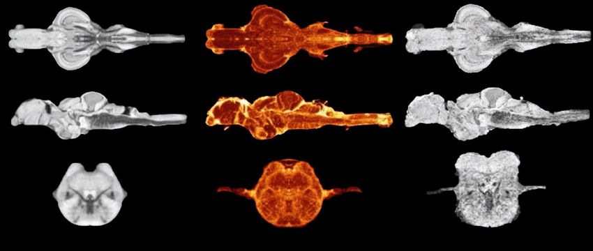
Neuroimaging Phenotypes in Zebrafish

                                                                                                                                                                                                                                                                                                                                                                                         Project
                                                                                                                                                                                                                                                                                                                                                                                         Research
    Comparison of resolution and contrast
          achieved between a minimum
       deformation model (a) and a single
         brain data set (c). The minimum
    deformation model minimizes individual
    differences and only exhibits structures
     present throughout the population. (b)
       Standard deviation map with areas
        in yellow highly variable between
     individual brains and areas in red very
                    consistent.

Z
       ebrafish has become an established model in neuroscience due to the ease with which gene discovery, chemical                  tearing, and variations in labeling are minimized and instead ‘re-slicing’ of the data in any arbitrary orientation is possible. In
       screening, behaviour, and disease modelling can be performed. More recently, neuroimaging, a crucial pre-clinical             zebrafish, MRI was first used in to visualize the entire anatomy of the adult zebrafish, where ex vivo MRI was performed on a
       technique for probing tissue structure, examining volumetric changes, and studying in vivo brain activity has also been       9.4 T magnet with in vivo experiments performed using a flow-through chamber, resulting in images with an inplane resolution
applied to zebrafish. Neuroimaging plays a crucial role in phenotyping research and in studies of neurological diseases. By          of 78 μm. Other studies were able to obtain higher resolution and good contrast to noise ratios in an in situ preparation by
examining the neuroanatomy of animal models, morphological abnormalities can be identified and correlations made with                dissecting the brain out of the skull. The concurrent development of zebrafish-specific fixation and incubation protocols with
behaviour. Magnetic Resonance Imaging (MRI) and Diffusion Weighted Imaging (DWI) are frequently used pre-clinically to               gadolinium-based contrast agents led to the acquisition of 10 μm3 images and the creation of a singlebrain high-resolution atlas.
identify morphological phenotypes in knockout models of neurological diseases. The zebrafish brain is particularly attractive for    Although this singlebrain atlas describes many brain regions in the adult zebrafish brain, a probabilistic atlas (figure above) that
neuroimaging due to its small size, numerous translucent strains, and distinct forebrain organization.                               minimizes individual differences and instead is based upon a large population provides better resolution. The resultant data set
                                                                                                                                     would only exhibit structures present throughout the population and generate mean morphometric measures that represent the
In a book chapter1, published by Dr. Jeremy Ullmann and Dr. Andrew Janke, the Informatics Fellow at The University of                population.
Queensland node of National Imaging Facility, a range of imaging techniques that have been utilized to examine the zebrafish
brain are discussed. Among these are MRI, DWI, Optical projection tomography (OPT), Optical Imaging, and Electron                    In general, neuroimaging modalities for larval zebrafish show great promise for phenotyping. These techniques have primarily
Microscopy. While many of these methods have only begun to be utilized in zebrafish, correlating neuroimaging phenotypes             been used to understand the fundamental workings of the zebrafish brain such as which neurons are responsible for escape
with behaviour in zebrafish has a bright future.                                                                                     behaviour, swimming speed, and swim posture, however similar imaging techniques could be applied to examining the seizure
                                                                                                                                     network in epileptic fish, or functional connectivity in autism models. This would be the first time these networks were examined
MRI visualizes the anatomy of the brain by exploiting differences in the relaxation values of various microstructures. By altering   across the entire brain yet still at the single cell scale! When coupled with microfluidics and automated screening platforms,
repetition times and echo times contrast can be optimized and different neuroanatomical structures visualized. MR imaging            precision medicine at a whole new level becomes possible.
was initially developed for human brain but subsequent improvements in coil design and magnetic field strength have enabled
a range of species including fish to be imaged. MRI permits the acquisition of in-vivo three-dimensional volumes of the whole        For more information on this work, contact Dr. Jeremy Ullmann (j.ullmann@uq.edu.au).
brain eliminating the need for tedious sectioning. By imaging the whole brain many histological artifacts such as shrinkage,

                                                                                                                                                                        1. Ullmann, Jeremy FP, and Andrew L. Janke. “Neuroimaging Phenotypes in Zebrafish.” The rights and wrongs of zebrafish: Behavioral phenotyping of zebrafish. Springer International Publishing, 2017. 273-289.

8                                                                                                                                                                                                                                                                                                                                                                                   9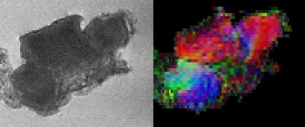
Colon Cancer Characterisation 2016 in Review
T
Industry
Collaboration
News
he Western
University node of the
Sydney the ultrastructure of rectal cancer
and healthy rectal specimens. MRI
Active Users of NIF Collaborations Trainings
National Imaging Facility findings will be correlated with Infrastructure Australian Research Collaborations 400
(NIF) is working with Ingham histopathology. Discovering novel
Institute and Liverpool Hospital MRI biomarkers of bowel cancer Internal External Australian Industry Collaborations
(with Prof. Michael Barton and and developing MRI techniques 300
International Research Collaborations
Dr Trang Pham) on the ex for performing virtual whole
vivo characterisation of bowel tumour biopsies are expected to International Industry Collaborations
200
cancer with high field Magnetic result from this collaboration.
Resonance Imaging (MRI) and 302
specifically diffusion tensor The study on CONCERT (Centre 36% 6% 100
imaging (DTI). High field MRI is for Oncology Education and 14% 105
able to image cancer at exquisite Research Translation) Biobank
0
spatial resolution, allowing for the rectal cancer specimens has been 9%
64%
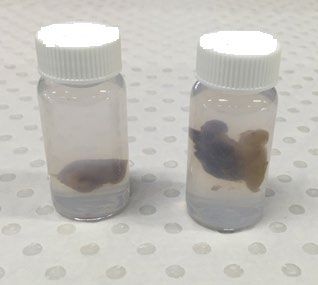
exploration and characterisation commenced. The specimens in Trainings held Users trained
of tumour heterogeneity and Fig. 1 are normal full-thickness 71%
biology. MRI also offers a range rectum on left, and full-thickness
of functional tests that can predict rectum with tumour on the right.
tumour behaviour and treatment They have been fixed in 10%
response. High resolution formalin, and suspended in a
magnetic resonance images and 1% agarose with Magnevist, a
3D diffusion tensor images for fibre commercial MR contrast agent, No. of Publications No. of Grants
tracking will be used to examine solution.
By User Community By User Community
304 65 through accessing 110 74 through accessing
NIF NIF
By Node Members By Node Members
0 200 400 0 100 200
Fig. 1 Specimens of normal Fig. 2 Slices of DT images of a rectal cancer specimen.
Fig 3 (left) a slice from a gradient echo 3D dataset, the
voxels are 100 μm isotropic and (right) the corresponding
NIF N odes :
full-thickness rectum on left, The colours correspond to the components of the principle slice of a diffusion tensor dataset colour coded according
and full-thickness rectum with eigenvectors. to the principle eigenvalue, the voxels are 200 μm isotropic.
tumour and adjacent normal The colour coding clearly reveals the directions of the bowel
rectum on the right. muscle fibres.
3D DTI scans with 200 μm isotropic voxels have been scanned so far MRI-Pathology reveals a correlation between
obtained on the High Field 11.7 T Bruker Avance MRI MRI anisotropy, and tumour heterogeneity and fibrosis. The
located in the Biomedical Magnetic Resonance Facility at the specimens are to be subsequently analysed on the clinical
Western Sydney University node of NIF. The rectal cancer MRI-Simulator (3T), and the Australian MRI-Linac at Liverpool
sample has a heterogeneous anisotropic structure (Fig. 2 and Cancer Therapy Centre to translate high-field findings to low
3). The different colours correspond to different directions of field clinical MRI protocols.
maximum diffusion as is indicated by the coloured sphere at
the top right-hand side of the DT images. Anisotropy of the For more information on this project, contact Dr. Tim Stait-
tissue can be seen very clearly. Diffusion thus provides tissue Gardner (t.stait-gardner@westernsydney.edu.au).
microstructural information at the cellular level which is not
possible on the basis of traditional MRI alone. Collaborators
Biomedical Magnetic Resonance Facility, Western Sydney
The specimens are histologically examined for direct University
correlation of MR DTI findings with histology. Of the specimens Ingham Institute for Applied Medical Research
Liverpool Hospital
For further information regarding the newsletter, please contact Saba Salehi (communications@anif.org.au)
10 11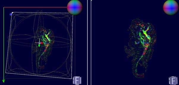

You can also read