3D INSIGHT LIVER MICROTISSUES - HUMAN LIVER RAT LIVER HEPG2 LIVER TOXICOLOGY SERVICES
←
→
Page content transcription
If your browser does not render page correctly, please read the page content below
3D InSight™                                       ▬▬ Human Liver

Liver Microtissues
Organotypic 3D Liver Models for ADME-Tox & DMPK
                                                  ▬▬ Rat Liver
                                                  ▬▬ HepG2
                                                  ▬▬ Liver Toxicology Services
A World of Discovery in Every Hanging Drop
  InSphero is the leading supplier of organotypic, biological in vitro 3D microtissues for highly predictive drug
  testing. Headquartered in Zurich, Switzerland with subsidiaries in the USA and Germany, we currently count
  all of the top 15 global pharmaceutical and cosmetics companies as our customers. Based on our patented,
  fully automatable hanging-drop technology and proprietary processes for cell conditioning, InSphero offers a
  broad range of highly functional, uniform and long-living 3D InSight™ Microtissues.

  Delivering a more biologically relevant model for in vitro applications, we are the first and largest provider of
  assay-ready spheroids and 3D-focused contract research services for toxicology, DMPK, and efficacy studies.
  We have been recognized for our scientific and commercial achievements with a number of national and
  international awards, and are certified to the ISO 9001:2008 standard for our Quality Management System.

  Upgrade Your Cell-based Assays to 3D Today!

                                              Waldshut, DE
                            Zurich, CH                                                      Our Distribution Partners
 Cambridge, MA
                                                                                                  Cambridge Bioscience Ltd.

                                                                                                  BizCom Japan, Inc.

                                                                                                  Handok Biotech Co., Ltd.

2 | Upgrade Your Cell-based Assays to 3D
Contents
Introduction to 3D Liver Microtissues		     4

Advanced Toxicity Assessment in 3D		        5

ADME-Tox Applications: Proof of Principle   6

DMPK Applications: Proof of Principle		     10

Non-liver 3D Toxicity Models			             12

Further Reading					13

3D InSight™ Human Liver Microtissues		      14

3D InSight™ Rat Liver Microtissues		        16

3D InSight™ HepG2 Liver Microtissues		      17

3D InSight™ Toxicology Services			          18

                     www.insphero.com/liver | 3
Filling the Need for More Predictive In Vitro Liver Models
“…conventional (2D) in vitro hepatic model systems…are less than ideal for longer term
toxicity evaluations and elucidation of key cellular and molecular events involved in primary
and secondary adaptation to chemical exposure…”
                                                                          LeCluyse et al. Crit Rev Toxicol. (2012) Jul;42(6):501-48.

           50% of Drug Attrition in
          Development is DILI Related

                                                                                                           Common Failures of
                                                                                                           2D Liver Model Systems
                                                                                                            »» Short-term viability
                                                                                                            »» Rapid loss of liver-specific
                                                                                                               morphology & phenotype
                                                                                                            »» Lack complex extracellular
                                                                                                               matrix

                        3D InSight™ Liver Microtissues
                                                Defining a New Standard

          Increased                                  Complex Organotypic                              Superior Long-lived
      Long-term Viability                              Cytoarchitecture                                Metabolic Activity

3D InSight™ Liver Microtissues are validated          Microtissues consist of polarized           Microtissues display increased metabolic
 for viability and functionality over 5 weeks     hepatocytes with tight cell-cell junctions      enzyme activity compared to 2D culture
                   in culture.                     (H&E staining, top), and an intact bile       persisting over a 4 week incubation period.
                                                canaliculi network expressing bile salt export
                                                   pump (BSEP) protein reflecting that of
                                                       native liver (bottom left/right).

  Day 7      Day 15      Day 19     Day 33

            Viability

                                                       Day 7                  Day 28

            Albumin
                                                   Native Liver           Human Liver
                                                                       Spheroids (Day 10)

4 | Upgrade Your Cell-based Assays to 3D
Advanced Toxicity Assessment in 3D
InSphero 3D culture technology helps you bring far greater assurance to compound de-risking with
3D InSight™ Liver Microtissues and Toxicology Services.

    For Your In-house Evaluation                                                                   Contract Research

Assay-ready 3D InSightTM Liver Microtissues (p14)                                 Tailored 3D InSightTM Toxicology Services (p18)
▬▬ Fixed production schedule for routine experimental timelines                   ▬▬ Identify disconnect between existing in vitro and in vivo toxicity data
▬▬ Reproducibly manufactured and QC‘d to ensure continuity                        ▬▬ Investigate mechanistic toxicities on clinically troublesome compounds
▬▬ Conveniently delivered next-day, ready for dosing                              ▬▬ Rapidly generate supportive data to justify adoption of 3D platform
▬▬ Multiple donor lots available to meet specific profile needs                   ▬▬ Screen for hepatotoxic compounds at an early stage in development

                                                               ENDPOINT
                                                              ASSESSMENT
  Cytotoxicity/                 Kupffer Cell Function             Mitochondrial                   Reactive Metabolite              Metabolic
  Hepatotoxicity                  »» IL-6 secretion               Impairment                      Formation                        Competence
   »»   ATP content               »» TNFα secretion                »» Oxygen consumption            »» Intracellular GSH               »» CYP activity
   »»   LDH                                                           rate & extracellular             content
   »»   AST/ALT                                                       acidification                 »» Malondialdehyde
   »»   α-GST                                                      »» Mitochondrial                    accumulation (IHC)
   »»   Albumin secretion                                             membrane potential
   »»   Urea secretion

 Apoptosis                      Steatosis/                        Cholestasis                     Proliferation                    mRNA & Protein
   »» Caspase-3/7               Phospholipidosis                   »» Transporter protein           »» DNA synthesis               Expression
      activation                  »» Intracellular neutral            expression                                                       »» Transcriptomics
                                     lipid & phospholipid          »» Bile acid secretion                                              »» Proteomics
                                     accumulation

The InSphero Liver Microtissue Pipeline
               CURRENTLY AVAILABLE                           BETA TEST NOW                                  LAUNCH Q3/4 2014
               ▬▬ Human                                      ▬▬ Minipig                                     ▬▬ 384-well microtissues
               ▬▬ Rat                                        ▬▬ Dog                                         ▬▬ Cynomolgus monkey
               ▬▬ HepG2                                      ▬▬ HepaRG                                      ▬▬ Mouse

                                                                                                              www.insphero.com/liver | 5
Multiparametric Analysis for                                                                     Mechanistic Toxicity
                                                                                                 ▬▬ Long-term & chronic toxicity
                                                                                                 ▬▬ Idiosyncratic toxicity
ADME-Tox & DMPK                                                                                  ▬▬ ROS-mediated toxicity
                                                                                                 ▬▬ Apoptosis/Necrosis
No single assay will tell the whole story when it comes to                                       ▬▬ Bile acid transport
predicting toxicity. The long-lived, organotypic nature of InSphero                              ▬▬ Global proteomic analysis
3D InSight™ Liver Microtissues facilitates a multiparametric
approach to characterize toxicity and DMPK that are limited or
unobtainable using conventional 2D culture.

▬▬ Evaluate repeat dose and long-term, low-dose
   exposure in a high-throughput in vitro format                                                 DMPK
▬▬ Use commercially available cell-based assays &
                                                                                                 ▬▬ CYP expression
   immunoassays for mechanism of toxicity analysis
                                                                                                 ▬▬ CYP induction
                                                                                                 ▬▬ Low clearance compounds

Proof of Principle
   Long-term & Chronic Toxicity
   Viability Endpoint Assays: ATP, LDH, AST/ALT, α-GST, Albumin, Urea

          Principle:
          To mimic in vivo hepatotoxic effects in
          vitro, a stable, long-lived liver model
          is required that can be repeatedly
          dosed over prolonged periods to reveal
          subtle, sub-acute toxic effects.

          Proof:
          The ATP content of 3D InSight™ Human
                                                                        3 Day IC50 = N/A
          Liver Microtissues after 3 day or 14
          day treatment with Tolcapone reflects                         14 Day IC50 = 12.45 µM
          the chronic toxicity profile observed
          with the drug in vivo. ATP content
          determined using Promega CellTiter-
          Glo®. Long-term medium exchange/
          repeat dosing was performed every 48
          hours for 14 days.

6 | Upgrade Your Cell-based Assays to 3D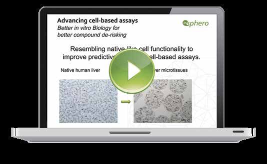
Idiosyncratic Toxicity                                      ROS-mediated Toxicity
Kupffer Function Assays: ATP Assay, IL-6 & TNF-α ELISAs             REDOX Endpoint Assays: GSH Content,
                                                                Oxygen Consumption/Extracellular Acidification

      Principle:                                                Principle:
      To detect idiosyncratic toxicities, an in vitro system    A useful liver model should allow monitoring of cellular
      must include both hepatocytes and non-parenchymal         redox status to reveal excess reactive oxygen species
      Kupffer cells capable of simulating a pro-inflammatory    (ROS) or glutathione (GSH) depletion, such as GSH
      environment.                                              depletion induced by the reactive Acetaminophen
                                                                metabolite NAPQI.
      Proof:
      InSphero’s 3D                                             Proof:
      culture technology                                        Treatment with buthionine-sulfoximine (BSO), a GSH-
      allows co-culture                                         synthesis inhibitor, can be used to mimic decreased
      of hepatocytes                                            GSH levels in liver microtissues, as measured by
      with IL-6 and                                             GSH-GloTM (Promega). BSO treatment depletes GSH
      TNF-α secreting                                           levels (green line) while maintaining cell viability, as
      CD68+ Kupffer cells                                       measured by ATP content (gray line).
      (brown stained
      cells). Co-exposure
      of Human Liver Microtissues with Trovafloxacin in the
      presence of an inflammagen (LPS) reveals toxicity (top
      graph) not seen with Levofloxacin (bottom).

                                                                               www.insphero.com/liver | 7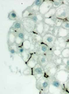
Apoptosis/Necrosis
  Cell Death Assays: Caspase 3/7 activity, ATP

           Principle:
           To pinpoint triggering of programmed cell death in
           response to toxic insult, an in vitro model should
           display responsiveness of intrinsic apoptotic pathways
           (e.g. Caspase 3/7 activation).

           Proof:
           Apoptotic cell death is observed in a dose-dependent
           manner following treatment of Human Liver
           Microtissues with Aflatoxin. The amount of cleaved,
           activated executioner caspases can be measured using
           Promega’s Caspase-Glo® 3/7 Assay (gray dashed line),
           and is mirrored by a corresponding decrease in viability
           (ATP – black solid line).

  Bile Acid Transport
  Bile Acid Profile Analysis: LC-MS Profiling of Microtissue Supernatants

                                                                                                        Cholic Acid (CA)

           Principle:
           To model altered bile acid profiles seen in cholestasis, a liver
           model must contain the cytoarchitecture and specialized
           transporters (i.e. BSEP & MRP2) to support bile acid conjugation
           and secretion into the bile canaliculi and media.

           Proof:
           The intact bile-canalicular network in 3D InSight™ Human                            Glycocholic Acid (GCA)
           Liver Microtissues make them an ideal tool to study effects of
           transporter inhibition. High-sensitivity LC-MS enables profiling
           of up to 13 different bile acids, including cholic acid (top) and
           Glycocholic acid (bottom), in microtissue supernatants.

                                                                               Bile acid transport analysis performed in collaboration with
                                                                                                   Pharmacelsus GmbH

8 | Upgrade Your Cell-based Assays to 3D
Global Proteomic Analysis
Novel Pathway & MOA Identification: HRM Mass Spectroscopy

        Principle:
        A robust microtissue model should facilitate global proteomic        Global Proteomics Identifies
        analysis, providing an unbiased view of mode of action (MOA)       Regulatory Pathways & Drug MOA
        and mechanism of toxicity (MOT).

        Proof:
        Significant changes in global protein expression are observed at
        sub-toxic doses of acetaminophen in Human Liver Microtissues
        using state-of-the-art mass spec to monitor >2400 proteins.
        Unbiased clustering of protein profiles (heatmap, right),
        combined with functional annotation clustering, identify
        upregulation of CYP1A2, CYP2E1 and CYP3A4 enzymes,
        along with changes in more than 160 proteins. Importantly,
        downregulation of icosanoid metabolism and inflammation, both
        known acetaminophen MOAs, are detected.

                                                                             Proteomic analysis performed in collaboration with Biognosys AG

                                                        Don’t Miss New Product Releases!
                                                            Register at www.insphero.com
                                                            ✔✔ New Product Updates
                                                            ✔✔ Latest 3D News
                                                            ✔✔ Webinars/Conferences
                                                            ✔✔ User Group Meetings
                                                            ✔✔ And more…

                                                                                             www.insphero.com/liver | 9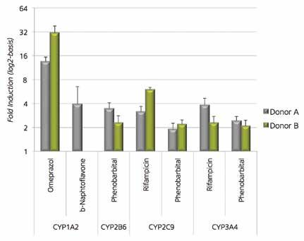
An Improved Organotypic Model for
In Vitro DMPK Analysis
                 Why is 3D > 2D for DMPK?
                          2D Hepatocyte                             3D Liver
                           Monolayers                              Microtissues
  Viability                       Days                                4+ weeks
                                                                                                DMPK
    CYP                 De-differentiation                                                      ▬▬ CYP expression
                                                               Broad & Persistent
 Expression           starting after 8h in 2D                                                   ▬▬ CYP induction
 Uptake/                                                     Transporters & intact              ▬▬ Low clearance compounds
                     Transporters expressed
 Secretion                                                  bile canaliculi network
    Cells
                       1x10⁵ – 4x10⁵ /well                           1x10³/MT
  Required

Proof of Principle
  CYP Expression
  CYP Expression & Activity Assays: Whole Genome Array, LC-MS metabolite detection

           Principle:                                                                         CYP1A2
           To determine if a drug is a substrate, inhibitor, or inducer of
           metabolizing enzymes, an in vitro model should include robust,
           stable, and inducible CYP expression that mimics in vivo
           metabolism. Such profiling can help predict drug-drug interactions.

           Proof:
           3D Human Liver Microtissues display increased, persistent Phase
           I & II enzyme mRNA levels compared to 2D (heat map below, red
           shading indicates ~2-fold change at time point relative to day 0).
           Likewise, microtissues were incubated with CYP substrates, and                      CYP3A4
           metabolites/products from single microtissues were detected in
           supernatants by LC-MS. 3D cultures displayed higher basal levels
           of CYP1A2, CYP3A4, and CYP2D6 activity compared to 2D, and
           activity persisted up to 28 days in culture.

                                                                                               CYP2D6

       CYP Expression performed in collaboration with Karolinska Institute and Biognosys AG

10 | Upgrade Your Cell-based Assays to 3D
CYP Induction
Metabolic Enzyme Induction: LC-MS metabolite detection
Principle:
In addition to displaying CYP
enzyme expression, an in vitro
model should demonstrate CYP
inducibility that mimics in vivo
metabolic responsiveness to known
pharmacological modulators.
Proof:
Responsiveness of CYPs to induction
is a critical parameter for predicting
potential drug-drug interactions.
Human Liver Microtissues from
two individual donors co-cultured
with Kupffer cells were exposed to
prototypic CYP-inducers for 72h
with daily medium exchange. For
the detection of CYP1A2, 2B6, 2C9
and 3A4 activity, a substrate cocktail
was added for 24h to control
tissues and CYP-induced samples,
before analysis of corresponding
metabolites. The fold induction CYP induction analysis performed in collaboration with Pharmacelsus GmbH
above control tissues is depicted.
Low Clearance Compounds
Intrinsic Clearance Assays: LC-MS analysis of supernatants
Principle:
To assist in determining
bioavailability, half-life, and dosing
regimen during discovery and
development, an in vitro model with
an organotypic metabolic profile can
be a powerful tool to predict in vivo
hepatic clearance.
Proof:
Download the Cyprotex poster from
ISSX 2013 at www.insphero.com to
learn more about the ability of 3D
InSightTM Human Liver Microtissues
to predict intrinsic clearance of low
clearance compounds.
Find this poster and more at www.insphero.com
www.insphero.com/liver | 11Beyond 3D Liver Models:
Emerging 3D microtissue models for toxicity testing
Toxicities observed in the heart, pancreas and brain are additional sources of drug attrition, and thus of concern
during the development process. The InSphero pipeline includes a panel of organotypic microtissues for
investigating potential toxic liabilities in non-liver organs.
3D InSight™ Human & Rat Pancreatic Microislets
▬▬ Viability and glucose-stimulated insulin secretion
persist > 4 weeks, enabling long-term studies Immunofluorescent staining
of Human Microislet cross-
▬▬ Homogeneous microislet size sections displaying expression
eliminates tedious hand-picking of insulin (green, beta cells),
glucagon (red, alpha cells), and
▬▬ Uniform ratio of alpha, beta, and delta somatostatin (blue, delta cells)
cells minimizes islet-to-islet variation in various islet cell sub-types.
3D InSight™ Human & Rat Myocardial Microtissues
▬▬ Established from iPS-derived human
or neonatal rat cardiomyocytes
▬▬ Multi-cell type cardiac microtissues display View neonatal rat cardiomyocyte
3D microtissues beating
viability and contractility for >3 weeks
synchronously at
▬▬ Demonstrate temperature- and www.insphero.com
pharmacologically-sensitive
contractile responsiveness
3D InSight™ Rat Neuronal Microtissues
▬▬ Established from newborn rat
brain (cortex) isolates
Immunofluorescent staining of
▬▬ Display stable size and viability for >28 days rat brain microtissues at day 5
of culture for GFAP (green) and
▬▬ Demonstrate organotypic expression β-III tubulin (red). Nuclei stained
of β-III tubulin, Nestin, and GFAP with DAPI (blue).
Interested in beta testing?
Contact us for more information about these and other 3D InSight™ microtissue models.
12 | Upgrade Your Cell-based Assays to 3DFurther Reading
Discover more Applications, Webinars and Technical Notes at www.insphero.com
General References:
Multi-parametric V. Merx (2013). "Cell culture: A better brew" Nature
measurements of ATP International Weekly Journal of Science. 496, 253–258.
and DNA in Microtissues Kelm J.M. and Lichtenberg, J. (2013). "3D Cell Culture for
Compound De-Risking" Innovations in Pharmaceutical
Technology, Zurich.
Oberdanner, C., et al. (2012). "Multiplexed Drug Assessment
in 3D. Combining Luminescence and Fluorescence Genetic
Reporters to Assess Drug Effects." Genetic Engineering &
Biotechnology News.
Messner S, et al. (2011). "An Organotypic Microliver
Assaying 3D Microtissues
Platform for High-Throughput Drug Testing." Society of
with Promega Caspase- Toxicology Annual Meeting, Washington DC, co-publication
Glo® 3/7 with Novartis, Basel.
Human Liver Microtissues:
Messner S., et al. (2013). "Multi-cell type human liver
microtissues for hepatotoxicity testing." In: Archives of
Toxicology. Springer-Verlag. 209-213.
Rat Liver Microtissues:
Kratschmar D.V., et al. (2013). “Characterization of a Rat
Multi-Cell Type 3D-Liver Microtissue System.” J Tissue Sci
Eng 4:130.
HepG2 Liver Microtissues:
Kelm J.M. et al. (2003). “Method for generation of
homogeneous multicellular tumor spheroids applicable to
a wide variety of cell types.” Biotechnol Bioeng. 2003 Jul
20;83(2):173-80.
Mueller D., et al. (2011). “Organotypic Cultures of Hepg2
Cells for In Vitro Toxicity Studies.” J Bioengineer &
Biomedical Sci S2:002.
www.insphero.com/liver | 133D InSight™ Human Liver Microtissues
96 Assay-ready Microtissues
▬▬ Co-culture of primary hepatocytes
& NPCs, including Kupffer cells
▬▬ Suitable for idiosyncratic and long- About this image:
term toxicity with repeated dosing Cytokeratin 8 antibody staining by IHC.
▬▬ Economical and convenient 96-well format
▬▬ Delivered next-day
Organotypic cytoarchitecture
The intact bile canaliculi network (BSEP
expression, brown staining) mirrors
that seen in native human liver.
Native Human Liver 3D InSight™ Human Liver Microtissues (day 10)
Hepatocyte/Kupffer cell co-
culture facilitates idiosyncratic
toxicity testing
IL-6 secretion in control (gray) or
LPS-stimulated (green) Human Liver
Microtissue supernatants, assessed by
ELISA.
Ensure reproducibility withProduct Specifications Catalog # Product
Delivery format GravityTRAPTM MT-02-002-01 3D InSight™ Human Liver Microtissues
from primary hepatocytes (96x)
Microtissues per plate 96, one microtissue per well
MT-02-002-04 3D InSight™ Human Liver Microtissues
Inoculation cell number 1000 hepatocytes per microtissue;
from primary hepatocytes, co-culture
optional: NPCs
with non-parenchymal cells (96x)
Cell source Cryopreserved primary cells
CS-07-001 3D InSight™ Human Liver
Microtissue re-aggregation time 4 days Maintenance Medium, serum-free
(500mL)
Time in culture upon delivery 6 days
Microtissue size upon delivery 220-320 μm
Size distribution (rel.SD) 10 pmol/MT
Maintenance medium CS-07-001; 3D InSightTM Human Liver
Maintenance Medium, serum-free
(500mL)
Medium exchanges 2-3 times a week
Cultivation volume 50-70 μl
Culture life-time >4 weeks
Want to Learn More?
VIEW OUR WEBINAR:
3D liver models for more
predictive toxicology
and drug safety testing
www.insphero.com
www.insphero.com/liver | 153D InSight™ Rat Liver Microtissues
Assay-ready in vitro rat model system
▬▬ Co-culture of fresh primary
hepatocytes & Kupffer cells (NPCs)
About this image:
▬▬ Suitable for idiosyncratic and chronic PAS staining of Rat Liver Microtissue
toxicity with repeated dosing confirms glycogen storage.
▬▬ Economical and convenient 96-well format
▬▬ Delivered next-day
Monitor Inflammation-mediated
toxicity:
Co-exposure of rat liver microtissues
with Trovafloxacin and an inflammagen
(LPS) is identified as toxic, as
evidenced by leakage of LDH into
microtissue supernatants.
Product Specifications Catalog # Product
Delivery format GravityTRAPTM MT-02-001-01 3D InSight™ Rat Liver Microtissues
from primary hepatocytes (96x)
Microtissues per plate 96, one microtissue per well
MT-02-001-04 3D InSight™ Rat Liver Microtissues
Inoculation cell number 1000 hepatocytes per microtissue; optional:
from primary hepatocytes, co-culture
NPCs
with nonparenchymal cells (96x)
Cell source Fresh
CS-07-002 3D InSight Rat Liver Maintenance
Microtissue re-aggregation time 4 days Medium, serum-free (500 mL)
Time in culture upon delivery 6 days
Microtissue size upon delivery 220-320 μm
Size distribution (rel.SD) 5 pmol/MT
Maintenance medium CS-07-002; 3D InSight™ Rat Liver
Maintenance Medium, serum-free (500 mL)
Medium exchanges 2-3 times a week
Cultivation volume 50-70 μl
Culture life-time >3-4 weeks
16 | Upgrade Your Cell-based Assays to 3D3D InSight™ HepG2 Liver Microtissues
Your easiest option for early-stage hepatotoxicity screens
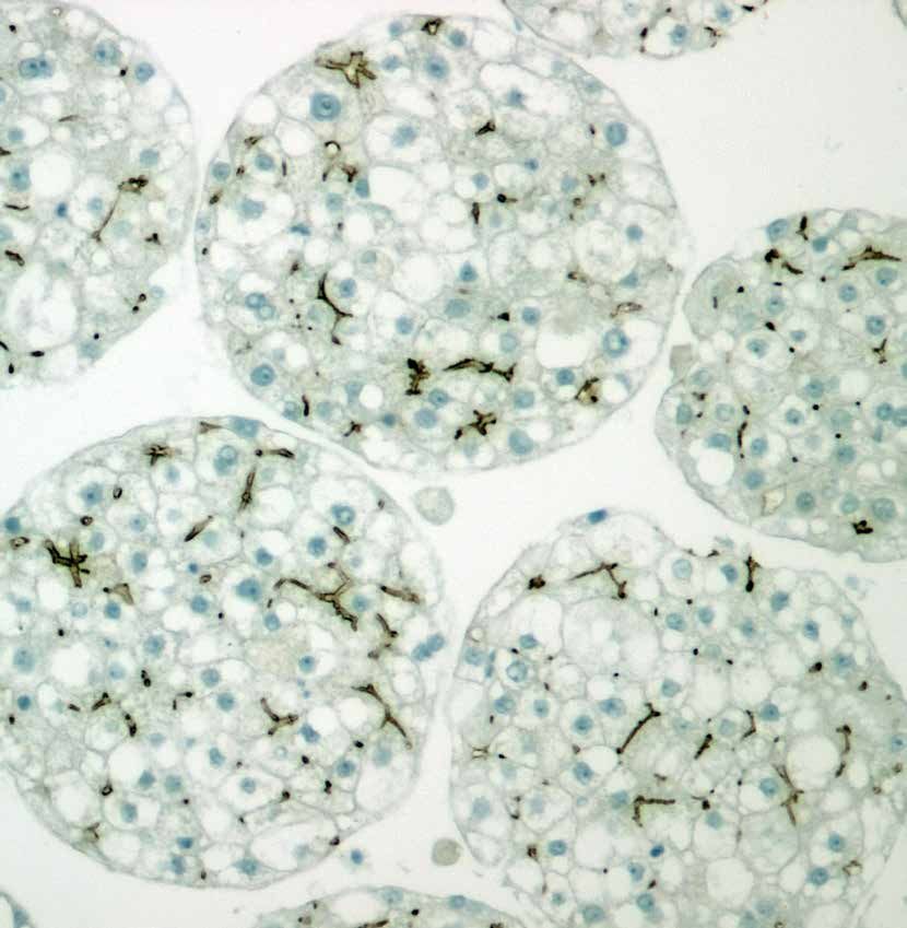
About this image:
▬▬ Compare previous 2D HepG2 Light-sheet microscopy (mDSLM)
test results to 3D data image of HepG2 Microtissue.
Maximum projection of 2000 cells
▬▬ More organotypic functionality in 3D culture based on 310 stacks of 1µm. DRAQ5TM
nuclear staining (red) and MitoViewTM
▬▬ Long-term (>10 days) studies Green mitochondria staining (green).
of drug effects possible Image provided gratefully by Nariman
Ansari, Francesco Pampaloni and Ernst
▬▬ Economical and convenient 96-well 3D format
H. Stelzer, Goethe University Frankfurt.
▬▬ Delivered next-day
Bile canalicular structure
Improved Characteristics
of 3D HepG2 HepG2 microtissue
»» Cuboidal cell shape cytoarchitecture
»» Formation of bile canaliculi Microtissue section, displaying
presence of bile canaliculi and tight
»»h albumin and glutamate cell-cell contacts with desmosomes.
production From J.M Kelm et al. (2003)
Biotechnology and Bioengineering.
»»h CYP450 induction
»»h drug efflux activity
Desmosome
Product Specifications Catalog # Product
Delivery format GravityTRAPTM MT-02-100-01 3D InSight™ HepG2 Liver
Microtissues (96x)
Microtissues per plate 96, one microtissue per well
CS-07-100 3D InSight™ Cell Line Maintenance
Inoculation cell number 200 cells per microtissue; optional: NPCs
Medium (500mL)
Cell source ATCC, cryopreserved
Microtissue re-aggregation time 4 days
Time in culture upon delivery 6 days
Microtissue size upon delivery 150-250 μm
Size distribution (rel.SD) 5 pmol/MT
Maintenance medium CS-07-101, 3D InSight™ Cell Line
Maintenance Medium (500mL)
Medium exchanges 2-3 times a week
Cultivation volume 50-70 μl
Culture life-time >2-3 weeks
www.insphero.com/liver | 173D InSight™ Toxicology Services
The world’s largest 3D-focused contract research organization
✔✔ Tailored screening conducted and interpreted
by our staff of 3D microtissue experts
✔✔ Enable long-term testing for low‑dose
& repeat-dose experiments
✔✔ Choose from a varied menu of
endpoint assays offered in-house, or
through our network of partners
✔✔ Services fully customizable to
meet your requirements
✔✔ Rapid turnaround with strict confidentiality
1 Choose Your Compounds
2 Select Your 3D Model
(Human, Rat, HepG2, Dog, Minipig)
3 Choose Your Endpoints Explore Multiple
Toxicity Mechanisms
4 We Screen
(Multiple dilutions, Repeat dosings)
5 We Deliver
(Web-based presentation, Discussion with our 3D experts)
Visit us at www.insphero.com/services or contact your local branch office
or authorized distributor to discuss a plan that suits your needs.
18 | Upgrade Your Cell-based Assays to 3D3D Compatible Endpoints
A variety of amenable assays
Toxicity Mechanism Cellular Indicator 3D-Compatible Endpoint Assay
Cytotoxicity/Hepatotoxicity ATP-content CellTiter-Glo® (Promega)
LDH (released/intracellular) CytoTox-ONETM (Promega)
AST/ALT (released/intracellular) Fluitest® AST/ALT (Analytikon)
α-GST (released/intracellular) α-GST-ELISA (EKF-Diagnostics)
Albumin secretion Albumin-ELISA (Bethyl Laboratories)
Urea secretion Urea-Assay (Biovision)
Kupffer-cell function IL-6 secretion IL-6 ELISA (Life Technologies)
TNF-α ELISA TNF-α ELISA (Life Technologies)
Mitochondrial impairment Oxygen consumption rate/ XF-Flux (Seahorse Biosciences)
extracellular acidification HCA with TMRM-dye (Life Technologies)
Mitochondrial membrane
potential
Reactive metabolites Intracellular GSH content GSH-Glo® (Promega)
Malondialdehyde generation IHC for MDA (Lipid-peroxidation)
Metabolic competence Cytochrome activation/ induction P450-Glo™ (Promega)
Enzyme activity (Pharmacelsus)
Apoptosis Caspase 3/7 activation Caspase-Glo® 3/7 (Promega)
HCA with CellEvent (Life Technologies)
Steatosis, phospholipidosis Lipid accumulation HCS-LipidTOXTM for Steatosis and Phospholipidosis (Life Technologies)
BODIPY (Life Technologies)
Cholestasis Transporter proteins IHC for BSEP
Bile-acid secretion LC-MS analysis (Pharmacelsus)
Proliferation DNA-synthesis EdU Click-iT® (Life Technologies)
mRNA & Protein Transcriptomics PCR-Arrays (SA-Biosciences); Affymetrix® Arrays
Expression Analysis Proteomics HRM-MS (Biognosys)
3D InSight™ Liver Toxicology Service Packs
Screening simplified
If a custom multi-endpoint project is beyond your needs, our 3D InSight™ Liver Toxicology Service Packs are designed to simplify
the service process. Just send us your compounds, choose your 3D model, and we deliver a report in 2-4 weeks for one
convenient price. Each pack includes toxicity assessment by ATP content for 5 to 20 compounds after 14 days of treatment.
Catalog# Product
SP-02-001-00 3D InSight™ Liver Toxicology Service Pack – 5 Compounds
SP-02-002-00 3D InSight™ Liver Toxicology Service Pack – 10 Compounds
SP-02-003-00 3D InSight™ Liver Toxicology Service Pack – 20 Compounds
www.insphero.com/liver | 19www.insphero.com
Our Strategic Partners
CellTiter-Glo®, GSH-GloTM, P450-GloTM, CytoTox-ONETM and Caspase-Glo® are trademarks of Promega Corp. MitoViewTM is a trademark of Biotium Inc. DRAQ5TM, HCS LipidTOXTM,
and EdU Click-iT® are trademarks of Thermo Fisher Scientific Inc. Fluitest® is a registered trademark of Analyticon AG. Affymetrix® is a registered trademark of Affymetrix, Inc.
InSphero AG InSphero Europe GmbH Authorized Distributors
Schlieren, Switzerland Waldshut, Germany www.insphero.com/distributors
& +41-44-515049-0 & +49-7751-3049665-0
InSphero Inc. InSphero is ISO 9001:2008 certified
Cambridge, MA, USA info@insphero.com All rights reserved, © 2014 InSphero AG. 3D InSight™,
GravityPLUS™ and GravityTRAP™ are trademarks of InSphero AG.
& +1-800-779-7558 www.insphero.com For life science research only. Not for use in diagnostic procedures.You can also read