Immediate lymphatic reconstruction for breast cancer
←
→
Page content transcription
If your browser does not render page correctly, please read the page content below
Review Article
                                                                                                                                           Page 1 of 8

Immediate lymphatic reconstruction for breast cancer
Akhil K. Seth1, Dhruv Singhal2
1
Division of Plastic and Reconstructive Surgery, NorthShore University HealthSystem, Evanston, IL, USA; 2Division of Plastic and Reconstructive
Surgery, Beth Israel Deaconess Medical Center, Boston, MA, USA
Contributions: (I) Conception and design: All authors; (II) Administrative support: D Singhal; (III) Provision of study material or patients: D Singhal;
(IV) Collection and assembly of data: None; (V) Data analysis and interpretation: None; (VI) Manuscript writing: All authors; (VII) Final approval of
manuscript: All authors.
Correspondence to: Dhruv Singhal, MD. Division of Plastic and Reconstructive Surgery, Beth Israel Deaconess Medical Center/Harvard Medical
School, 110 Francis St, Suite 5A, Boston, MA 02215, USA. Email: dsinghal@bidmc.harvard.edu.

                 Abstract: Upper extremity lymphedema remains a significant source of morbidity in breast cancer patients
                 despite significant improvements in breast cancer care. The risk of lymphedema is particularly elevated in
                 patients requiring an axillary lymph node dissection and/or adjuvant radiation to treat their disease. Current
                 treatment options for lymphedema, including conservative management or surgery, are limited and are often
                 aimed at improving symptoms and quality of life rather than curing the disease. In this review we describe
                 immediate lymphatic reconstruction, a novel surgical procedure that is done concurrent with axillary lymph
                 node dissection in an effort to prevent the development of breast cancer-related lymphedema. Based on our
                 growing knowledge of the pathophysiology of lymphedema, microsurgical techniques are used at the time
                 of axillary lymph node dissection to perform a lymphovenous bypass between transected, leaking lymphatic
                 channels and an adjacent, small calibre vein in the axilla. Using several objective metrics for short- and long-
                 term surveillance, patients are monitored for the development of postoperative lymphedema. Early outcomes
                 from using this technique have been promising, both in the literature and within our own institutions,
                 demonstrating significant improvements in rates of postoperative lymphedema. However, future study is still
                 required to better understand the long-term efficacy of immediate lymphatic reconstruction.

                 Keywords: Breast cancer; lymphedema; lymphatic reconstruction

                 Received: 01 September 2020; Accepted: 28 January 2021
                 doi: 10.21037/abs-20-110
                 View this article at: http://dx.doi.org/10.21037/abs-20-110

Background                                                                     of breast cancer care. A 2013 systematic review still placed
                                                                               the overall rate of breast cancer-related lymphedema
Breast cancer remains one of the most commonly diagnosed
                                                                               (BCRL) in breast cancer survivors at 21.4%, including
forms of cancer in women, with 1 in 8 women developing
                                                                               those who received no surgical intervention (7). Risk factors
breast cancer over the course of her lifetime (1). Since the                   for the development BCRL continue to be debated in
advent of the Halsted mastectomy in 1912, treatment for                        the literature, but include axillary lymph node dissection
breast cancer has significantly evolved (2). The use of more                   (ALND), number of nodes removed, number of positive
minimally invasive surgical techniques, combined with                          nodes, radiation, taxane-based chemotherapy, and elevated
neoadjuvant and adjuvant therapy, has improved overall                         BMI (7-13).
mortality, postoperative morbidity, and aesthetic outcomes                        BCRL is a chronic disease that results in asymmetric
(3-6). However, despite more conservative measures for                         swelling of the upper extremities, with the underlying
the management of breast cancer-related axillary disease,                      pathophysiology of the disease defining its clinical
including staging with sentinel lymph node biopsy, upper                       manifestations. An initial inciting insult to the lymphatic
extremity lymphedema remains a significant complication                        system, including surgery, trauma, radiation and/

© Annals of Breast Surgery. All rights reserved.                                        Ann Breast Surg 2021 | http://dx.doi.org/10.21037/abs-20-110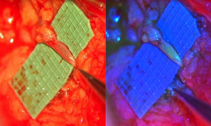
Page 2 of 8                                                                                        Annals of Breast Surgery, 2021

or infection, results in increased resistance within the          outlined, are utilized in a delayed manner after BCRL has
lymphatic channels and decreased flow, leading to the             developed. In contrast, immediate lymphatic reconstruction
accumulation of lymph within the channels (14). The valves        (ILR) is a surgical procedure that aims to prevent the
of the lymphatic system fail, and bidirectional flow of lymph     development of BCRL. By performing lymphovenous
continues to worsen, resulting in dependent edema and             anastomoses at time of axillary nodal dissection, ILR aims
increased rates of extremity cellulitis, which are hallmarks      to promote restoration of physiologic lymphatic flow and
of BCRL. However, the build-up of lymph also triggers             thus prevent the cascade of pathophysiologic events that
a chronic inflammatory response that causes hypertrophy           result in BCRL development. ILR was initially described
of lymphatic channel walls and adjacent smooth muscle             by Boccardo et al. in 2009 and at the time was termed the
cells, accumulation of fibroblasts, adipocytes, keratinocytes     lymphatic microsurgical preventative healing approach or
and mononuclear cells, and ultimately irreversible fat            LYMPHA (19). Divided lymphatics are visualized after
and collagen deposition (14). Although the severity of            completion of ALND and are anastomosed to tributaries of
lymphedema is thought to be progressive in nature, early          an adjacent vein. In the authors seminal study, with 4-year
development of soft tissue edema is most likely occurring         of follow-up, post-operative rates of BCRL were shown to
simultaneously with underlying hypertrophy and smooth             be 4% (20). Similar promising results in reduction of BCRL
muscle changes, which define the chronic nature of the            with ILR have been replicated by other institutions (21-23).
disease.
   Ultimately, BCRL leaves patients with both physical            Anatomic considerations
limitations and a visible aesthetic deformity, which can
significantly impact vocation, social, and sexual interaction     The boundaries of standard level I and II ALND are defined
(15-17). Moreover, the chronicity of the disease can              as the axillary vein superiorly, serratus anterior medially,
result in a dramatic financial burden to both the patient         thoracodorsal vessels posteriorly, and latissimus muscle
and the healthcare system. The mainstay of treatment              laterally (24). Within these boundaries, identification of
for lymphedema has historically relied on conservative            both lymphatics and veins appropriate for anastomosis are
management including compression and decongestive                 critical to the success of ILR. Identification and preservation
therapy. However, these treatments are palliative in nature       of appropriate veins occurs at the time of ALND to
while also being cumbersome and expensive for patients,           prevent inadvertent sacrifice of potential venous targets
further underscoring the chronicity of the disease (16,18).       by the extirpative surgeon. In this way, a coordinated and
Surgical management of BCRL has evolved, and currently            collaborative approach at the time of ALND between the
includes debulking procedures such as liposuction,                oncologic and reconstructive surgeons is important for
vascularized lymph node transfer, and lymphovenous                successful execution of ILR.
bypass (16). To be considered a surgical candidate for               The venous anatomy of the axilla is perhaps the most
these therapies, patients must demonstrate evidence of            varied between patients. A commonly utilized vein for
stability in their disease progression, which may not be          anastomosis is the accessory vein of the axilla, which arises
attainable despite aggressive conservative management.            directly from the axillary vein and generally travels 2-cm
                                                                  anterior and parallel to the thoracodorsal vessels through
Furthermore, although surgical treatment of lymphedema
                                                                  the axillary bed (25,26). Other named secondary venous
can improve quality of life and achieve objective reductions
                                                                  options include the lateral thoracic and thoracodorsal vein,
in limb volume, none of these treatments have proven to
                                                                  which hold an increased risk of injury to the long thoracic
be consistently effective in all patients. Ultimately, the fact
                                                                  nerve and thoracodorsal neurovascular bundle, respectively.
that treatment of BCRL remains limited and outcomes are
                                                                  To minimize these risks, branches off the thoracodorsal
inconsistent underscores the importance of any modality
                                                                  system or venous collaterals running along the chest wall
that may assist in preventing the development of BCRL.
                                                                  or laterally within the soft tissue may also be utilized.
                                                                  Anecdotally, more extensive axillary disease may increase
Immediate lymphatic reconstruction                                the number of venous collaterals available for anastomosis,
                                                                  but may also require a more aggressive extirpative surgery
Introduction
                                                                  that may make these collaterals unavailable. The presence
Current surgical therapies for BCRL, as previously                or absence of valves can often be visualized during venous

© Annals of Breast Surgery. All rights reserved.                         Ann Breast Surg 2021 | http://dx.doi.org/10.21037/abs-20-110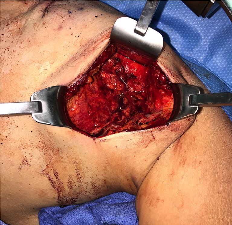
Annals of Breast Surgery, 2021                                                                                                    Page 3 of 8

                     A                                                 B

Figure 1 Demonstration of the two distinct pathways of the upper extremity. Isosulfan blue is injected prior to axillary dissection into the
posterolateral pathway, demonstrating a blue-colored lymphatic. (A) Meanwhile, fluorescein isothiocyanate (FITC) dye was injected into the
medial pathway, demonstrating a green-colored lymphatic under the FITC filter on the microscope (B).

dissection of the vein. Competent valves are critical to                   considered for ILR. While ALND alone elevates the risk
minimize back-bleeding from the vein and are ultimately                    of BCRL development, these patients also frequently
thought to allow for low-pressure flow between the                         undergo neoadjuvant chemotherapy and adjuvant treatment
anastomosed lymphatic channels and veins.                                  including regional lymph node radiation (RLNR), further
    Lymphatic drainage within the upper arm can be divided                 increasing their BCRL risk profile. Moreover, anecdotally,
into two distinct ‘lymphosomes’, medial and posterolateral,                we have noted that a significant percentage of patients
as demonstrated by Suami et al. (27). The medial pathway                   presenting to our Lymphatic Center for BCRL treatment
is the main lymphatic drainage pathway, running along                      have BMIs less than 30. Therefore, elevated patient BMI
the volar aspect of forearm up to the medial upper arm                     is not used as an indication for ILR at our institutions as
and terminating in the axilla. The posterolateral pathway,                 statistical support for its use is lacking. Furthermore, this
or Mascagni-Sappey (M-S) pathway, was first described                      can prevent a specific subset of breast cancer patients from
by Mascagni in 1787 and then Sappey in 1875. The M-S                       undergoing ILR despite still being susceptible to BCRL.
pathway runs alongside the cephalic vein with variable
drainage to the supraclavicular/infraclavicular nodes and/
                                                                           Surgical technique
or axillary basin (28,29). Ultimately, the medial pathway
is thought to be the predominant drainage pathway of the                   Immediately prior to the beginning of ALND, the upper
upper extremity, although draining lymphatics from either                  extremity is injected with dye to allow for identification
pathway are thought to be suitable for ILR lymphovenous                    of draining lymphatics from the upper extremity into the
anastomosis (30) (Figure 1).                                               axilla following ALND. In the case of a modified radical
                                                                           mastectomy, timing of dye injection and ILR must be
                                                                           considered in the context of concurrent mastectomy and
Patient selection
                                                                           possible breast reconstruction. In the case of planned breast
Boccardo et al. proposed that appropriate indications for                  reconstruction, a sequence of mastectomy, dye injection,
LYMPHA or ILR technique included: BMI ≥30 kg/m2 (at                        ALND followed by ILR, then breast reconstruction tends to
highest risk) and a transit index on lymphoscintigraphy of                 be most favorable in our experience. Although the original
≥10 (20). They contend that patients who did not meet these                description of ILR utilized isosulfan blue dye for lymphatic
criteria should not be considered as surgical candidates.                  identification, we have found fluorescein isothiocyanate
At our institutions, all patients undergoing ALND are                      (FITC) dye to be a useful alternative, particularly if the

© Annals of Breast Surgery. All rights reserved.                                  Ann Breast Surg 2021 | http://dx.doi.org/10.21037/abs-20-110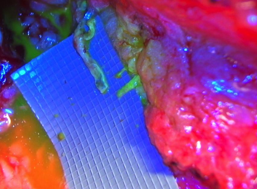
Page 4 of 8 Annals of Breast Surgery, 2021
achieve appropriate magnification of venous and lymphatic
targets, we use a Mitaka MM51 microscope (Mitaka Kohki
Co, Ltd., Tokyo, Japan). The microscope is equipped with
a 560-nm filter, which illuminates FITC-injected lymphatic
channels arising from the arm. Respective size of leaking
lymphatic channels and their relative position with respect
to potential vein targets are noted. The choice of venous
target is dependent on both its location relative to the
desired channels to be bypassed, as well as presence of back-
bleeding following division of the vein. Ideally, there is no
back-bleeding from the vein, although minimal venous
back-bleeding is accepted as the patient is under positive
pressure ventilation and, presumably, the direction of flow
will reverse after extubation. The decision of whether or
Figure 2 Triangulation of the axillary incision to achieve not to bypass to a back-bleeding vein is ultimately at the
appropriate exposure of the axilla using a pediatric Bookwalter discretion of the surgeon.
retractor system prior to immediate axillary lymphatic Adjustments of the microscope, patient positioning,
reconstruction. and axillary retraction are often required prior to
proceeding with lymphovenous anastomosis. In general,
the anastomosis can often be ergonomically challenging,
and can be aided with the use of a microscope foot pedal
and long microsurgical instruments. Our anastomotic
technique mirrors the original description of the procedure
and, specifically, utilizes 9-0 Nylon suture (Ethicon Inc.,
Somerville, NJ, USA) to intussuscept lymphatic channels
into a target vein (Figure 3). Full thickness ‘back-wall’
interrupted sutures are placed between the posterior
aspect of the vein and perilymphatic tissue, allowing for
approximation of the lymphatics to the vein lumen while
also buttressing the ultimate anastomosis. A temporary ‘U’
stitch is then placed through the anterior wall of the vein,
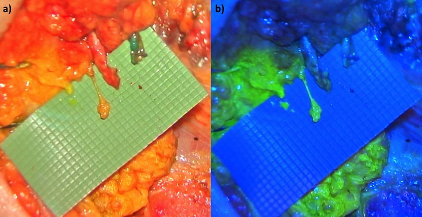
Figure 3 Identification of transected lymphatic channels and an through and through one or more lymphatic channels, and
appropriate venous target for lympho-venous anastomosis. Two then back out the vein, allowing the lymphatic channels to
identified lymphatics are anastomosed to a single adjacent vein. be ‘parachuted’ into the vein (2). The anastomosis is then
completed with additional sutures securing the anterior
vein wall to the perilymphatic tissue. The ‘U’ stitch is then
oncologic surgeon is utilizing blue dye as part of the cut and removed and lymphatic flow visualization through
SLNB (26). A 2% solution of FITC and albumin, allowing the anastomotic site can be confirmed by visualization of
for prolonged retention within the lymphatics, is injected FITC under the microscope (Figure 4). Of note, if the
intradermally into two sites on the volar wrist and in initial U-stitch only captures the adventitia of the lymphatic
muscular fascia of upper medial arm. channel thereby not occluding flow, the surgeon may opt to
Following completion of ALND, ideal exposure of the keep the U-stitch in place. Additional soft tissue can then
axillary basin can be achieved using a self-retaining retractor be approximated around the anastomosis to further buttress
system. We utilize a pediatric Bookwalter retractor set the repair. Lymphatic channels that are not bypassed
(Codman Inc., Raynham, MA), which functions well for are micro-clipped. Between 1–3 lymphatic channels are
both an axillary or mastectomy incision. Initial exposure is generally bypassed in any given patient (20). A #15 Blake
achieved through triangulation of the wound bed, including drain is placed exiting the dependent portion of the axillary
retraction of the pectoralis muscle medially (Figure 2). To bed and the axillary or mastectomy incision is closed in a
© Annals of Breast Surgery. All rights reserved. Ann Breast Surg 2021 | http://dx.doi.org/10.21037/abs-20-110Annals of Breast Surgery, 2021 Page 5 of 8
A B
Figure 4 Completed lympho-venous anastomosis as part of immediate lymphatic reconstruction. Anastomosis is visualized both without (A)
and with (B) the fluorescein isothiocyanate (FITC) filter on the microscope. Note the appearance of FITC within the vein lumen following
completion of the anastomosis.
standard manner. current ILR literature demonstrated rates of BCRL in
patients undergoing ALND alone to be 15.6%, which
increased to 26.5% with the additional of RLNR (21).
Postoperative care and complications
When ILR was performed in these two groups, the rates
The surgically placed drain is left in place until the output is of BCRL decreased to 4.6% and 10.6%, respectively. Our
less than 20cc for two consecutive days. Drains usually meet institutional data has mirrored these promising outcomes,
criteria and are removed by post-operative day 14. We do although longer follow-up is required to better understand
not prophylactically place patients in a compression garment the sustainability of our results. We also to date have not
postoperatively. In general, there is a low complication rate experienced any significant post-operative complications
associated with ILR, with no complications reported in a associated directly with ILR.
meta-analysis by Jørgensen et al. (25). If a venous target Recent literature has also demonstrated that ILR
is chosen with mild back-bleeding, a theoretical risk of can be cost-effective (32). Cost-efficacy was evaluated
hematoma does exist, however this has not been evident and compared amongst two main groups: (I) patients
in our experience thus far. There is also the potential for undergoing ALND alone versus ALND and ILR and (II)
hypersensitivity reactions to the injected dyes, including ALND with RLNR versus ALND with RLNR with ILR.
FITC and isosulfan blue. This risk can be mitigated by pre- Utilizing a previously published cost of one year of life
emptively administering a combination of hydrocortisone, living with BCRL (33), and the estimated cost ILR based on
Benadryl, and Pepcid as has been previously described (31). its associated current procedural terminology (CPT) code,
an incremental cost-utility ratio (ICUR) was calculated for
each patient group. For the ALND versus ALND with ILR
Outcomes
group, the ICUR was $1,587.73/QALY, which decreased
Initial work by Boccardo et al. has reported a rate of further to $699.48/QALY for the ALND with RLNR versus
BCRL of 4% at 4-year follow-up in a high-risk breast ALND with RLNR with ILR group (32). These relatively
cancer population that underwent ALND and RLNR with low ICURs demonstrate the substantial clinical benefit of
ILR (20). Since their seminal study, additional institutions ILR relative to its additional cost. This cost-efficacy was
have replicated these results. A 2019 meta-analysis of the confirmed with even extremely conservative estimates of
© Annals of Breast Surgery. All rights reserved. Ann Breast Surg 2021 | http://dx.doi.org/10.21037/abs-20-110Page 6 of 8 Annals of Breast Surgery, 2021
post-operative BCRL incidence. required for this review article.
Funding: None.
Surveillance and future directions
Footnote
Given the progressive nature in which BCRL develops,
understanding the long-term efficacy of ILR requires Provenance and Peer Review: This article was commissioned
robust patient surveillance. At our institutions, certified by the Guest Editor (Dung Nguyen) for the series “Cutting-
lymphedema therapists obtain baseline data for patients edge of Complex Breast Reconstruction” published in
prior to undergoing ILR, including both quantitative and Annals of Breast Surgery. The article has undergone
qualitative measurements. These same measurements external peer review.
are then repeated at set intervals following completion
of ILR to monitor for early signs of BCRL development. Conflicts of Interest: Both authors have completed the
Quantitative measurements modalities include ICMJE uniform disclosure form (available at http://dx.doi.
circumferential arm measurements at set intervals, which org/10.21037/abs-20-110). The series “Cutting-edge of
can then be converted to volumes using the truncated cone Complex Breast Reconstruction” was commissioned by the
formula (34). Additional modalities include perometry and editorial office without any funding or sponsorship. The
bioimpedance spectroscopy, which can further quantify authors have no other conflicts of interest to declare.
limb volume and the extent of fluid within the limb,
respectively. Qualitative measures include quality of life Ethical Statement: The authors are accountable for all
survey instruments such as the SF-36, LYMQOL, and aspects of the work in ensuring that questions related
DASH. All patient measurements are entered into a clinical to the accuracy or integrity of any part of the work are
quality database to facilitate patient surveillance. Patients appropriately investigated and resolved.
are followed every 3 months for the first 2 years post-
operatively, and then every 6 months the third and fourth Open Access Statement: This is an Open Access article
year assuming all subjective evaluations and objective data distributed in accordance with the Creative Commons
demonstrate no evidence of lymphedema. Attribution-NonCommercial-NoDerivs 4.0 International
Despite the aforementioned standardized metrics used License (CC BY-NC-ND 4.0), which permits the non-
for assessment of BCRL following ILR at our institutions, commercial replication and distribution of the article with
there remains heterogeneity in the modalities used to assess the strict proviso that no changes or edits are made and the
BCRL. This limits the ability for comparison of results original work is properly cited (including links to both the
across different study groups, and thus prevents us from formal publication through the relevant DOI and the license).
having a true understanding of the impact of ILR. As the See: https://creativecommons.org/licenses/by-nc-nd/4.0/.
implementation of ILR grows across institutions nationally
and internationally, standardizing both the surgical
References
approach and the quantitative and qualitative measures used
for monitoring patients will be critical to understanding 1. American Cancer Society. How Common Is Breast
the efficacy of ILR in preventing BCRL. This efficacy data Cancer? Jan. 2020. Available online: https://www.cancer.
will also be important for obtaining consistent coverage of org/cancer/breast-cancer/about/how-common-is-breast-
ILR by insurance payors in the United States, who often cancer.html.
still consider the procedure experimental. As we continue to 2. Freeman MD, Gopman JM, Salzberg CA. The evolution
strive for more preventative and cost-effective modalities of of mastectomy surgical technique: from mutilation to
health care, ILR may emerge as the primary approach for medicine. Gland Surg 2018;7:308-15.
patients at risk for BCRL. 3. Plesca M, Bordea C, El Houcheimi B, et al. Evolution
of radical mastectomy for breast cancer. J Med Life
2016;9:183-6.
Acknowledgments
4. Giuliano AE, Ballman KV, McCall L, et al. Effect of
The authors would like to acknowledge Anna Rose Johnson axillary dissection vs no axillary dissection on 10-year
and Melisa Granoff for helping to put together the data overall survival among women with invasive breast cancer
© Annals of Breast Surgery. All rights reserved. Ann Breast Surg 2021 | http://dx.doi.org/10.21037/abs-20-110Annals of Breast Surgery, 2021 Page 7 of 8
and sentinel node metastasis. JAMA 2017;318:918-26. visualization and restoration of flow. J Surg Oncol
5. Riis M. Modern surgical treatment of breast cancer. Ann 2019;120:160-7.
Med Surg (Lond) 2020;56:95-107. 19. Boccardo F, Casabona F, De Cian F, et al. Lymphedema
6. O’Halloran N, Potter S, Kerin M, et al. Recent microsurgical preventive healing approach: a new
advances and future directions in postmastectomy breast technique for primary prevention of arm lymphedema
reconstruction. Clin Breast Cancer 2018;18:e571-e585. after mastectomy. Ann Surg Oncol 2009;16:703-8.
7. DiSipio T, Rye S, Newman B, et al. Incidence of unilateral 20. Boccardo F, Casabona F, De Cian F, et al. Lymphatic
arm lymphoedema after breast cancer: a systematic review microsurgical preventing healing approach (LYMPHA)
and meta-analysis. Lancet Oncol 2013;14:500-15. for primary surgical prevention of breast cancer-related
8. Rebegea L, Firescu D, Dumitru M, et al. The incidence lymphedema: over 4 years follow-up. Microsurgery
and risk factors for occurrence of arm lymphedema 2014;34:421-4. Corrected in Microsurgery 2015;35:83.
after treatment of breast cancer. Chirurgia (Bucur) 21. Johnson AR, Kimball S, Epstein S, et al. Lymphedema
2015;110:33-7. incidence after axillary lymph node dissection:
9. McLaughlin SA, Wright MJ, Morris KT, et al. Prevalence Quantifying the impact of radiation and the lymphatic
of lymphedema in women with breast cancer 5 years microsurgical preventive healing approach. Ann Plast Surg
after sentinel lymph node biopsy or axillary dissection: 2019;82:S234-41.
Objective measurements. J Clin Oncol 2008;26:5213-9. 22. Hahamoff M, Gupta N, Munoz D, et al. A lymphedema
10. Gillespie TC, Sayegh HE, Brunelle CL, et al. Breast surveillance program for breast cancer patients
cancer-related lymphedema: risk factors, precautionary reveals the promise of surgical prevention. J Surg Res
measures, and treatments. Gland Surg 2018;7:379-403. 2019;244:604-11.
11. Warren LEG, Miller CL, Horick N, et al. The impact 23. Feldman S, Bansil H, Ascherman J, et al. Single institution
of radiation therapy on the risk of lymphedema after experience with lymphatic microsurgical preventive
treatment for breast cancer: A prospective cohort study. healing approach (LYMPHA) for the primary prevention
Int J Radiat Oncol Biol Phys 2014;88:565-71. of lymphedema. Ann Surg Oncol 2015;22:3296-301.
12. Fu MR, Axelrod D, Guth AA, et al. Patterns of obesity 24. Page F, Hamnett N, Chadwick S, et al. Axillary lymph
and lymph fluid level during the first year of breast cancer node dissection: do you know your boundaries? J Plast
treatment: A prospective study. J Pers Med 2015;5:326-40. Reconstr Aesthet Surg 2015;68:597-9.
13. Jammallo LS, Miller CL, Singer M, et al. Impact of body 25. Jørgensen MG, Toyserkani NM, Sørensen JA. The effect
mass index and weight fluctuation on lymphedema risk in of prophylactic lymphovenous anastomosis and shunts
patients treated for breast cancer. Breast Cancer Res Treat for preventing cancer-related lymphedema: a systematic
2013;142:59-67. review and meta-analysis. Microsurgery 2018;38:576-85.
14. Sosin M, Yin C, Poysophon P, Patel KM. Understanding 26. Spiguel L, Shaw C, Katz A, et al. Fluorescein
the concepts and physiologic principles of lymphatic isothiocyanate: A novel application for lymphatic surgery.
microsurgery. J Reconstr Microsurg 2016;32:571-9. Ann Plast Surg 2017;78:S296-8.
15. Johnson AR, Singhal D. Immediate lymphatic 27. Suami H. Lymphosome concept: Anatomical study of the
reconstruction. J Surg Oncol 2018;118:750-7. lymphatic system. J Surg Oncol 2017;115:13-7.
16. Basta MN, Gao LL, Wu LC. Operative treatment of 28. Mascagni P. Vasorum Lymphaticorum Corporis Humani.
peripheral lymphedema: a systematic meta-analysis of Historia & Iconographia. Senis: P. Carli Edit; 1787.
the efficacy and safety of lymphovenous microsurgery 29. Sappey PC. Anatomie, Physiologie, Pathologie de
and tissue transplantation. Plast Reconstr Surg Vaisseaux Lymphatiques. Adrain Delahaye; 1874.
2014;133:905-13. 30. Johnson AR, Bravo MG, James TA, et al. The all but
17. Pusic AL, Cemal Y, Albornoz C, et al. Quality of life forgotten Mascagni-Sappey pathway: Learning from
among breast cancer patients with lymphedema: a immediate lymphatic reconstruction. J Reconstr Microsurg
systematic review of patient-reported outcome instruments 2020;36:28-31.
and outcomes. J Cancer Surviv 2013;7:83-92. 31. Raut CP, Hunt KK, Akins JS, et al. Incidence of
18. Schwarz GS, Grobmyer SR, Djohan RS, et al. Axillary anaphylactoid reactions to isosulfan blue dye during breast
reverse mapping and lymphaticovenous bypass: carcinoma lymphatic mapping in patients treated with
Lymphedema prevention through enhanced lymphatic preoperative prophylaxis: Results of a surgical prospective
© Annals of Breast Surgery. All rights reserved. Ann Breast Surg 2021 | http://dx.doi.org/10.21037/abs-20-110Page 8 of 8 Annals of Breast Surgery, 2021
clinical practice protocol. Cancer 2005;104:692-9. breast cancer and lymphedema. Support Care Cancer
32. Johnson AR, Asban A, Granoff MD, et al. Is 2019;27:1697-708.
immediate lymphatic reconstruction cost-effective? 34. Brorson H, Höijer P. Standardised measurements used to
Ann Surg 2019. [Epub ahead of print]. doi: 10.1097/ order compression garments can be used to calculate arm
SLA.0000000000003746. volumes to evaluate lymphoedema treatment. J Plast Surg
33. Dean LT, Moss SL, Ransome Y, et al. “It still affects our Hand Surg 2012;46:410-5.
economic situation”: long-term economic burden of
doi: 10.21037/abs-20-110
Cite this article as: Seth AK, Singhal D. Immediate lymphatic
reconstruction for breast cancer. Ann Breast Surg 2021.
© Annals of Breast Surgery. All rights reserved. Ann Breast Surg 2021 | http://dx.doi.org/10.21037/abs-20-110You can also read