Mucic Acid Loaded Polyethylenimine@Gold Nanoparticles for the Treatment of Rheumatoid Arthritis
←
→
Page content transcription
If your browser does not render page correctly, please read the page content below
Mucic Acid Loaded Polyethylenimine@Gold
Nanoparticles for the Treatment of Rheumatoid
Arthritis
Yixia Cui
Department of Rheumatism Immunity, Yanan University Affiliated Hospital, No.43 North Street,
BaotaDistrict, Yan'an, 716000, China.
Junwei Zhang
Department of Anesthesiology, Beijing Beiya Orthopaedic Hospital, No.20 Haotian Street, Fangshan
District, Beijing, 102445, China
Yanwu Liu
Department of Orthopaedic, Xijing Hospital of Air Force Medical College, No.127 Changle West Road,
Beilin district, Xi'an, 710032, China
Guolin Meng
Department of Orthopaedic, Xijing Hospital of Air Force Medical College, No.127 Changle West Road,
Beilin district, Xi'an, 710032, China
Changwei Lv ( lucwei76@sina.com )
Department of Orthopaedic, Xi’an No.3 Hospital, The Affiliated Hospital of Northwest University Xi’an
No.3 Hospital, No.10 Fengcheng 3rd Road, Xi’an, 710018, China
Research Article
Keywords: Gold nanoparticles, Arthritis, Polyethylenimine, Mucic acid, HR-TEM
Posted Date: May 20th, 2021
DOI: https://doi.org/10.21203/rs.3.rs-534531/v1
License: This work is licensed under a Creative Commons Attribution 4.0 International License.
Read Full License
Page 1/16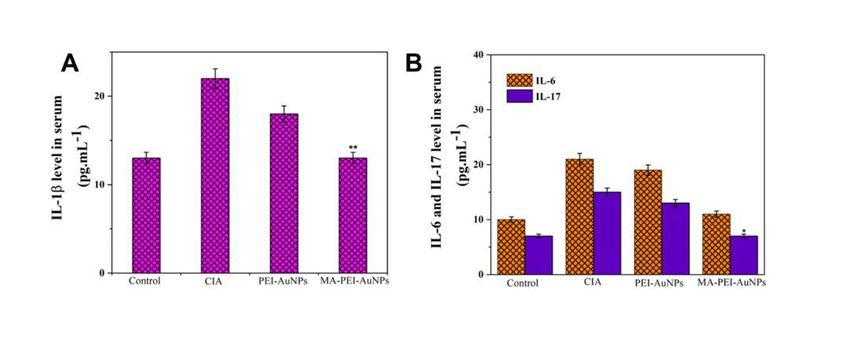
Abstract
Rheumatoid arthritis (RA) could be a common autoimmune disease that involves severe Joint
deformation. The main off-target medicines unable to cure RA, that permits associate in nursing infection
with the unwellness. The nanomaterials-based RA medical aid is a good strategy to enhance the
treatment efficaciousness within the inflammatory region. Specifically, metal-based nanomaterials are a
wonderful choice for a possible delivery vehicle for inflammatory disease agents, attributable to their
novel properties. Herein, we have got developed a small-sized Polyethylenimine (PEI) coated gold
nanoparticles (AuNPs) with an average diameter of 80 nm, that area unit used for top loading of
carboxylic acid (MA). Various microscopic and qualitative analysis tools like high-resolution transmission
microscopy (HR-TEM), Field-emission scanning microscope (FE-SEM), and Fourier-transform infrared
(FTIR) spectroscopic analysis studies were accustomed to make sure as-made PEI-AuNPs. The invented
PEI-coated AuNPs (PEI-AuNPs) exhibited higher contrast with an extended expanse that's promising to
store giant amounts of MA medicine. MA is attributed to deduce the inflammatory response by inhibiting
the pro-inflammatory cytokines and averting undesirable ancient drug aspect effects in Collagen-induced
inflammatory disease (CIA). Numerous organic chemistry parameters like weight, hind paw volume,
protein estimation, anti-serum protein analysis, and microscopic anatomy examination were conducted in
Collagen-induced arthritis mice treated at a dose (10 µg) of MA packed PEI-AuNPs. The obtained results
showed the MA-PEI-AuNPs were used with success within the treatment of Collagen-induced arthritis,
relative to PEI-AuNPs and MA. Therefore, MA loaded PEI-AuNPs as a stimulating candidate in future RA
applications.

1 Introduction
The impaired bone joint chronic autoimmune disorder named rheumatoid arthritis (RA) causes severe
inflammation of tissue, joint degeneration, continual redness, erosive, and destruction of the bone tissue
[1]. Completely different types of a substance cause severe bone destruction of RA, antibodies, and
secretion of various styles of pro-inflammatory cytokine/chemokine like lymphokine (IL)-6, IL-1β, IL-17
and Interferons (IFN), prostaglandin E2, metalloproteinases (MMPs), and tumor necrosis factor (TNF-α)
[2, 3]. The secretion of the pro-inflammatory cytokines is answerable for activating the massive
proportion of invasive macrophages to hurry up and triggers RA progression via regulation of
inflammation micro-environment [4]. Therefore, the RA treatment appears to be entirely dependent on
suppressing the inflammatory reaction. Still, there's no impact of medication on the RA treatment,
because of its expansive cost, long facet effects, and off-target medication [5]. Additionally, various
medication types like biological agents, anti-rheumatic medication, immuno-suppressant steroid
hormones, and anti-inflammatory medication are offered to use as therapeutic agents to deduce
inflammatory and Joint severity in RA [6, 7]. Therefore, the necessity to fabricate anti-arthritis agents can
have fewer facet effects, cheap and longer life, that have the flexibility of transforming bone joint defects,
suppress the secretion of the pro-inflammatory protein, and severe inflammation. Plant-based secondary
metabolites like flavonoids are well-known and most effective therapeutic agents obtained from varied

                                                  Page 2/16
fruits, leaves, roots, etc. It exhibits medicine, immunomodulatory, and inhibitor properties [8, 9]. Earlier
reports powerfully discerned the naturally earned flavonoids will deduce the inflammatory response and
transform bone defects in a short period [10].

Mucic acid (MA) or a pair of, 3,4,5-Tetrahydroxyhexanedioic acid may be a phytocompound obtained
from Phyllanthus Emblica. MA and its derivatives are found to possess antimicrobial, anti-diabetic, and
anti-inflammatory properties. To our information, the osteogenic potential of MA has not been reported
yet. An oversized fraction of phytocompounds is water-insoluble and crystalline, thereby proscribing their
direct application within the flesh. This necessitates the necessity for material-based delivery of
phytocompounds to the wound site since it ensures higher bioavailability. The metal nanoparticle-based
carboxylic acid delivery can greatly impact RA treatment [14, 15]. Attributable to the ability to scavenge
free radicals and lack of harmful, gold nanoparticles (AuNPs) are extensively used nanomaterials in
various applications and RA medical aid [16]. On the opposite hand, the designer is among the foremost
extensively used polycations in drug delivery, because of its high transfection potency [17, 18]. It is
coating on the AuNPs exterior provides most stable in biological fluid and had all-time low toxicity then
clean AuNPs [19]. Therefore, the PEI coated AuNPs as a unique framework to treat RA. This
investigation's key purpose regarding the bone restorative effects and deduce the pro-inflammatory
protein by delivering MA by PEI-AuNPs and exploring its accented regulative mechanisms on in vivo
collagen-induced inflammatory disease (CIA) mice model.

Herein, this study focuses on developing an artificial route accomplished through using HAuCl4 for
manufacturing AuNPs. The PEI was then inserted into the AuNPs (PEI-AuNPs) surface, which is
accustomed to comprehend vital quantities of carboxylic acid (MA). The AuNPs and PEI-AuNPs
developed were generally confirmed by TEM, FE-TEM, and FTIR. The MA encapsulated PEI-AuNPs are
then accustomed to boost the treatment of RA with more effectiveness. The biochemical results have
steered creating the processed MA-PEI-AuNPs a promising candidate for CIA therapy.

2 Materials And Methods
2.1 Materials
Gold(III) chloride hydrate (HAuCl4) (99.9%), MAphinidin chloride (C15H11ClO7), Trisodium citrate
(Na3C6H5O7), polyethylenimine (PEI, Mw2,500) and 70% ethanol, Dulbecco’s modified eagle medium
(DMEM), fetal bovine serum (FBS), penicillin and streptomycin (PS) were purchased from Shanghai,
China.

2.2 Synthesis of PEI coated AuNPs
To fabricate PEI-coated AuNPs, 0.02% HAuCl4 solution (200 mL) and 1.5 mL of 1% (w/w) PEI solution
were added into the beaker containing 50 mL of distilled water (dH2O) and then vortexed at room
temperature for 24 h. Afterward, PEI-coated AuNPs were collected by centrifugation at 10,000 rpm for 15

                                                    Page 3/16
min and washed twice with dH2O and ethanol. The PEI-coated AuNPs solution was kept at 4 ˚C to avoid
aggregation.

2.3 Nanomaterial Characterization
Studies of electron microscopy were used to evaluate the PEI-AuNPs structure, size, and composition.
Transmission electron microscopy (TEM, JEOL JEM-2100) images of the material were taken using HR-
TEM at a wavelength of excitation of 200 kV. The solution PEI-AuNP was mounted on a 200-mesh copper
grid and was vacuum-dried at 37 ° C using a hot air oven. After complete dried, the PEI-AuNP was pictured
at 100 kV. The PEI-AuNP was examined using Field Emission Scanning Electron Microscope (FESEM, 5
kV permitted Quanta 200 FEG to operate) to examine the scale, shape and structure of the produced NPs.
In studying the surface charge and size of PEI-AuNPs, a zeta sizer (Nano-ZS, Malvern Instruments) was
used. For FTIR experiments, the powder form of PEI-AuNPs was combined with KBr powder and
converted into pellets. The spectrum was obtained in transmission mode using Fourier transform infrared
(FTIR, Nicolet 6700 (Thermo Fisher, USA) spectrometer with a measurement range of 4000 − 400 cm− 1.

2.4 Mucic acid entrapment efficiency
Evaluation of MA drug moiety trap efficacy on the surface of PEI-AuNPs was calculated by UV-Visible
spectrometry by measuring the absorption spectrum of collected post-centrifugation supernatant after
stirring the NPs with MA. The absorbances of MA released from the scaffolds were measured at 190 nm,
and the concentration of MA was deduced using its standard graph. Then, the effectiveness of the
trapping was calculated using the formula given below:

% Entrapment Efficiency=[(TMA– FMA)/TMA]×100

Where TMA is measured as the total value of introduced MA and FMA is the sum of free MA in the
supernatant [20].

2.5 Animals
Eight weeks of old male albino rats with 180–210 g of albino strain were acquired and kept inside a
hygienic animal house maintained at 25 ± 3°C condition, and providing proper, limitless access to water
and standard pellet diet, except in conduct experiments. The animal committee of the National Institutes
of Health Guide for Care and Use of Laboratory approved 7 days prior to the initiation of the study and its
guidelines used during animal experiment protocols.

2.6 Collagen-Induced Arthritis in Wistar Rats
Bovine type II collagen (CII) formulated by using well-defined protocol and prepared CII has been
dissolved at 2 mg / mL in 0.05 M acetic acid by slowly vortexed overnight at 4 ° C. Then, CIA was
arousing 16 albino male rats using the footpad method. Each male rat was subcutaneously immunized
in an unfinished Freund's adjuvant (IFA) emulsifying agent with 0.2 mg CII per rat into the left hind paw at
day 0. Immunized male rats were divided into four treatment group (N = 4): I control group (without CIA

                                                  Page 4/16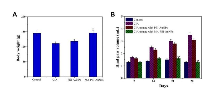
induction), (ii) PEI-AuNP group, (iii) MA-PEI-AuNP-treated CIA group. Protective therapy took place from
day 0 till day 7. Then, the daily intra-articularly (i.a) injection (except weekends) into ankle joints with
different forms of NPs. The treatment was initiate for 7 consecutive days [21].

2.7 Measurement of Body weight and Organ Weight of Rats
with CIA
The male albino rats were weighed using digital balance after finishing the 7, 14, 21, and 28 days of
treatment. The final and initial body volume was taken for each control group and treated rat group. At
the end of the tests, the mice were exposed to anaesthetic with xylazine and ketamine. Following the
treatments, the major internal organs like liver, spleen and thymus and blood were obtained for rapid
measurement and stored for more assessment at − 80 ° C.

2.8 Histological Examination of Rats with CIA
After the Euthanasia performed, each animal's right knee joints have been collected and fixed overnight
with PBS (pH 7.4) in a 4 percent formalin buffer. After the decalcification of extracted knee joints in 10
percent nitric acid (HNO3). Every animal's joint tissue parts were deposited in paraffin which was cut
utilizing microtome device to 4 µm parts. Then, for histological observation under a 400 x magnifying
microscope, the acquired sliced tissue segment stained with hematoxylin-eosine (H&E). Histological
examination of internal sections into joint tissues including such soft periarticular tissues, cartilage and
synovial membrane is observed and distinguish the markedly different features between the control and
MA-PEI-AuNPs was evaluated [22].

2.9 Measurement of biochemical parameters
Blood samples were taken from albino male rats treated with MA-PEI-AuNPs for rapid quantification of
serum pro-inflammatory cytokines, includeIL-6, TNF-α, IL-1β and PGE2 utilizing standard experimental
procedures following the completion of the experimental duration (28 day). Then, the serum anti-CII IgG
antibody was also evaluated using a rat serum anti-CII antibody ELISA kit.

2.10 Statistical Analysis
All the results are expressed as mean ± standard error mean (SEM) (n = 4). Two-way variance analysis
(ANOVA) was used to calculate volume of the hind paw and analyze the biochemical parameters.
Statistical significance as presented in the tables and figures was expected at p < 0.05 and p < 0.01.

3 Result And Discussion
Herein, we have designing well-established mucic acid (MA) packed PEI-AuNP nanomaterial using a
blend of AuNPs and PEI, which are the most noteworthy materials for treating Collagen-Induced arthritis
(CIA).

3.1 Morphological Analysis
                                                   Page 5/16
As depicted in Fig. 1A, the FE-SEM image shows the AuNPs metal oxide nanostructure. The result
suggests that the as-prepared AuNPs well-dispersed small-sized with spherical shape. Figure 1A denotes
HR-TEM images showing that the AuNPs were surrounded by the thinner PEI coated layer (grey). The
high-contrast metal nanostructure and low-contrast coating were observed. The average diameter of
AuNPs was estimated from the HR-TEM image to be 182.5 ± 10 nm. The nanomaterials with tiny sizes
ranging from 1–200 nm gathered are associated with the upper hand of nanoparticles in drug delivery
applications in terms of improved bioavailability, increased discharging of drugs, high cell absorption,
and increased surface-area-to-volume ratio, actuating more space for cellular interactions [23].

3.2 DLS and FTIR Analysis
The corresponding DLS study is shown in Fig. 2A; the approximate size distribution of the AuNPs was
198.5 ± 5 nm. The surface charge of the as-synthesized AuNPs and PEI coated AuNPs were determined
by Zeta potential analyzer. It was determined to be + 26.6 mV in AuNPs, and the noted zeta potential was
+ 34.4 mV due to the presence of PEI coating on the surface of Au NPs, as shown in Fig. 1B. The obtained
zeta potential values displayed plausible stability [25]. FT-IR spectroscopy was demonstrated after AuNPs
manufacturing (Fig. 1C) to attest to functional groups' existence in the AuNPs. The peaks at 2918 and
2855 cm− 1 bands correspond to the C-H bond formation which confirms AuNPs formation. The
characteristic band at 3425 cm− 1 was due to –OH group in the MA [26]. It showed characteristic 2925
and 1630 cm− 1 bands indicating methyl group and amide I for bonding C-O. Broadband at 3430 cm− 1
corresponding to the N-H stretch vibration of PEI.

3.3 Drug Releasing Study
The encapsulating hydrophobic nature of MA onto the surface of AuNPs and PEI-AuNPs is based on the
hydrophobic or electrostatic interactions. The MA loading content of PEI-AuNPs (mass of MA in the PEI-
AuNPs /mass of PEI-AuNPs loaded with MA×100%) and loading efficiency (mass of MA attained in the
PEI-AuNPs /mass of the feeding MA×100) 0.012/0.014×100 = 85.7% of MA in PEI-AuNPs was calculated
to be 18.8 wt%. As depicted in Fig. 3, the in vitro MA discharge profile from PEI-AuNPs was determined by
the dialysis bag analysis at different pH viz pH 7.4 and pH 6.8 at 37°C. It shows the release of MA from
PEI-AuNPs the release rates of flavonoid in solutions of pH 7.4 and pH 6.8 in 24 days were 65% and 74%,
respectively. On the contrary, the gradual degradation of PEI on the surface of the AuNPs depends upon
acidic nature; it improves the loading and sustainable release of MA from PEI-AuNPs. Adding PEI
prevents the burst release of MA from PEI-AuNPs as its carbonyl groups associated with the amine and
hydroxyl groups, ensuring gradual surface erosion [27].

3.4 Behavioral Test
The bodyweight of the different nano-formulation treated mice groups was evaluated shown in Fig. 4A,
showing no difference in the body weight between the control and MA-PEI-AuNPs treated mice group,
except these the rats treated with AuNPs and CIA groups (without any treatment) after the 28 days, which
                                                 Page 6/16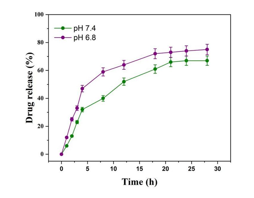
shown significantly decreased body weight observed. Because of the severity of the CIA, a significant loss
of body weight in CIA groups (without any treatment) and no significant effect on the CIA in AuNPs group
is seen. In addition, due to its strong anti-inflammatory and antioxidant effects of MA, the incidence of
CIA was significantly deduced while being treated with MA-PEI-AuNP, as a result of which the severity of
body weight deduction was recovered. No major abnormalities were found in the volume of the hind paw
in the joints of the healthy rats (control group) in Fig. 4B. Twenty-four days later, the hind paw was
noticed its maximum severity in the CIA induction group (saline-treated) due to CIA severity influencing a
significant hind paw swelling as a result of critical inflammation allowing disease to reach its maximum
severity and no significant effect on joint swelling was observed in the AuNPs treated group. In addition,
MA-PEI-AuNPs treated group, suppressed joint swelling up to the day 24 days which show an anti-
inflammatory effect, as a result, the inflammation was gradually resolved.
Figure 5 indicates the relative weight of internal organs of four studied groups of rats. At the end of the
experimental duration, the rat groups caused by the GIA had significantly increased organ weight relative
to control and MA-PEI-AuNPs (P < 0.01). There was no difference in the absolute weight of the spleen,
thymus, and liver between the MA-PEI-AuNPs treated and the control group respectively. The positive
effect of MA-PEI-AuNPs non-toxic nature on spleen, thymus, and liver was observed in rat MA related to
healthy rat. A remarkable impact has been found in the MA-PEI-AuNPs community on the internal organs
due to their beneficial anti-arthritis effects caused by the combination of MA medication and AuNP. In the
case of CIA-induced rats (saline-treated) and AuNPs, an index of undesirable side effect was observed, as
result gradual decrease in the weight of the internal organ after the treatment.
3.5 Histological Examination of Rats with CIA
According to H&E staining results (Fig. 6A), when compared to MA-PEI-AuNPs, the ankle joint region of
the rat with arthritis displays some dashed red or ellipses-shaped structure denoted as a large area of
inflammation. Since it possesses strong metabolism, the CIA-induced rat MA show deduced blue
staining. By comparison, the rat community induced by the CIA treated with groups of MA-PEI-AuNPs
shows much larger blue areas, which means that the antioxidant capacity of MA-PEI-AuNPs could
effectively penetrate the inflammatory region and can maintain it better in the inflammatory region [28].
The group treated with MA-PEI-AuNPs did not show degradation of the bone and erosion with joint space
was found to be quite small. Hence, the H&E staining observation findings showed that ameliorated bone
degeneration in the ankle joint was influenced by subcutaneous administered core-shell MA-PEI-AuNPs
(Fig. 6C).
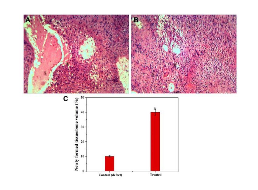
3.6 Measurement of biochemical parameter
Figure 7 and 8 showed the expression profile of pro-inflammatory factors such as the expression
interleukin (IL)-1β, IL-6, IL-17, and PGE2. Arthritis pathogenesis had an important part to play in the
acceleration. Various types of antigen, antibody, and pro-inflammatory cytokine such as interleukin
(IL)-1β, IL-6, interferon (IFN), prostaglandin E2 (PGE2), metalloproteinases (MMPs), and tumor necrosis
Page 7/16factor (TNF-α) are mainly accomplished to participate in the earliest RA systemic autoimmunity growth
incident [29, 30]. Secreting different pro-inflammatory cytokine is responsible for activating a large
number of infiltrating macrophages to speed up and induce RA development through inflammatory
microenvironment regulation. Hence, RA's approach to the treatment is primarily dependent on inhibiting
the inflammatory response. Furthermore, IL-17 was triggered by IL-6 that is a central cytokine in RA
pathogenesis and contributes to both joint damage and extra-articular signs. The high levels of IL-6 and
IL-17 have thus grown in the plasma and synovial fluid of the CIA model, and are participating in the
development of RA. IL‐17 is usually predominantly generated by T‐helper 17 cells (Th17 cells), and IL‐6
[31, 32] causes the differentiation of naive T cells into Th17 cells. The elevated pro-inflammatory rates
are largely responsible for the severity of induced arthritis. The serum from joint tissue was obtained in
the current study after the 28-day treatment of the distinct saline and nano-drug formulations; the
increasing incidence of pro-inflammatory factors such as IL-1β, IL-6 & IL-7, and PGE2 was noted in the
CIA-induced rat group.
In the case of the MA-PEI-AuNPs treated rat group, significant cytokine rates greatly decreased to the CIA-
induced rats. The quantification of serum anti-CII antibody was noted in control in Fig. 8A, CIA-induce rat
group, AuNPs and treated group MA-PEI-AuNPs using ELISA kit. Additionally, severe inflammation and
knee joint damage group influence serum anti-CII antibody CIA induce rat as a result of framework
encourages anti-CII antibody production rate. In the case of treated rats with MA-PEI-AuNPs, the deduced
serum anti-CII antibody rate was observed because of its anti-inflammatory effect. B-lymphocytes are
primarily capable of generating collagen-specific antibodies that can develop immune complexes with
different antigens and associate easily with complementary elements to start inflammation in the joint
tissue. Previous research suggested a possible association between auto-antibody responses and
restorative effects which correlates clinical symptom improvement with auto-antibody degree of
suppression [33, 34].
4 Conclusions
To conclude, AuNPs coated with PEI to generate PEI-AuNPs, we have designed new synthetic method to
create AuNPs that is excellently established. Instead, the PEI-coated AuNPs nanostructure was
encapsulated by electrostatic attraction with mucic acid (MA). PEI-coated AuNPs are notable materials
for the treatment of arthritis. The present study demonstrated that the impacts of saline, AuNPs and MA
loaded PEI-AuNPs were subjected to CIA therapy assessment. Among them, flavonoid loaded PEI-AuNPs
recognized the powerful anti-arthritis agent to deduce CIA in rats as compared to others. In which MA-PEI-
AuNPs reduced significantly the pro-inflammatory component, and mentioned the new bone growth. In
the CIA-induced rat group, because of the maximum severity of the inflammation, the increased risk of the
arthritis index was found to be more important, and AuNPs have a slightly anti-inflammatory activity.
Hence, MA-PEI-AuNPs may represent as an appropriate anti-arthritis in the potential development of
arthritis. Finally, it severely suppressed the formed swelling, erythema, and ankylosis of the hind paw and
ankle joints due to their antioxidant and anti-inflammatory features.
Page 8/16Declarations
Acknowledgement
This research was supported by a grant from the Key Research and Development Program of Shaanxi
Province (No. 2019-SF-194).
References
1. Y. Ma, Y. Song, F. Ma, G. Chen, Journal of Inorganic and Organometallic Polymers and Materials. 1–
10 (2019)
2. P.D. Harvey, J. Plé, Journal of Inorganic and Organometallic Polymers and Materials, 1–42 (2021)
3. B. Han, D. Peng, J. Wang, Z.N. Lu, J. Inorg. Organomet. Polym Mater. 31, 22–31 (2021)
4. S.F. El-Amin, M.S. Kwon, T. Starnes, H.R. Allcock, C.T. Laurencin, J. Inorg. Organomet. Polym Mater.
16, 387–396 (2006)
5. L.D. Quan, G.M. Thiele, J. Tian, D. Wang, Expert Opinion on Therapeutic Patents. 18, 723–738 (2008)
6. P. Li, Y. Zheng, X. Chen, Front. Pharmacol. 8, 460 (2017)
7. T.D. Wilsdon, C.L. Hill, Australian prescriber. 40, p.51 (2017)
8. A.N. Panche, A.D. Diwan, S.R. Chandra, Journal of nutritional science. 5 (2016)
9. A.M. Abdel-Azeem, S.M. Zaki, W.F. Khalil, N.A. Makhlouf, L.M. Farghaly, Frontiers in Microbiology. 7,
1477 (2016)
10. M.H. Pan, C.S. Lai, C.T. Ho, Food & function 1(1), 15–31 (2010)
11. T.C. Wallace, M.M. Giusti, Advances in Nutrition. 6, 620–622 (2015)
12. H.E. Khoo, A. Azlan, S.T. Tang, S.M. Lim, Food & nutrition research 61, 1361779 (2017)
13. M.H. Jeong, H. Ko, H. Jeon, G.J. Sung, S.Y. Park, W.J. Jun, Y.H. Lee, J. Lee, S.W. Lee, H.G. Yoon, K.C.
Choi, Oncotarget. 7(35), 56767 (2016)
14. C.T. Pham,. Wiley Interdisciplinary Reviews: Nanomedicine and Nanobiotechnology. 3, 607–619
(2011)
15. A. Mani, C. Vasanthi, V. Gopal, D. Chellathai, Int. Immunopharmacol. 41, 17–23 (2016)
16. H. Nah, D. Lee, M. Heo, J.S. Lee, S.J. Lee, D.N. Heo, J. Seong, H.N. Lim, Y.H. Lee, H.J. Moon, Y.S.
Hwang, Science and technology of advanced materials. 20, 826–836 (2019)
17. A. Mani, C. Vasanthi, V. Gopal, D. Chellathai, Int. Immunopharmacol. 41, 17–23 (2016)
18. S. Taranejoo, J. Liu, P.K. Verma, Hourigan, Journal of Applied Polymer Science. 132 (2015)
19. C. Hu, Q. Peng, F. Chen, Z. Zhong, R. Zhuo, Bioconjugate chemistry 21, 836–843 (2010)
20. C. Murugan, M. Rajkumar, N. Kanipandian, R. Thangaraj, K. Vimala, S. Kannan, Curr. Sci. 118, 1583
(2020)
Page 9/1621. M.M. Ansari, A. Ahmad, R.K. Mishra, S.S. Raza, R. Khan, ACS Biomaterials Science & Engineering. 5,
3380–3397 (2019)
22. C.Y. Tsai, A.L. Shiau, S.Y. Chen, Y.H. Chen, P.C. Cheng, M.Y. Chang, D.H. Chen, C.H. Chou, C.R. Wang,
C.L. Wu, Arthritis & Rheumatism: Official Journal of the American College of Rheumatology. 56, 544–
554 (2007)
23. R. Singh, J.W. Lillard Jr., Nanoparticle-based targeted drug delivery. Experimental and molecular
pathology. 86, 215–223 (2009)
24. H. Singh, J. Du, P. Singh, T.H. Yi, Artificial cells, nanomedicine, and biotechnology. 46,1163–1170
(2018)
25. D. Jain, R. Banerjee, J. Biomed, Mater. Res., Part B. 86, 105–112 (2008)
26. A.S. Abd Raboh, M.S. El-khooly, M.Y. Hassaan, Journal of Inorganic and Organometallic Polymers
and Materials, 1–12 (2021)
27. P.C. Pandey, G. Pandey, R.J. Narayan, Biointerphases. 12, 011005 (2017)
28. S. Tummala, M.S. Kumar, S.K. Pindiprolu, Improved anti-tumor activity of oxaliplatin by
encapsulating in anti-DR5 targeted gold nanoparticles. Drug Deliv. 23(9), 3505–3519 (2016)
29. S. Ueha, F.H. Shand, K. Matsushima, Frontiers in immunology 3, 71 (2012)
30. R. Domingo-Gonzalez, O. Prince, A. Cooper, S.A. Khader, Tuberculosis and the Tubercle Bacillus. 33–
72 (2017)
31. C. Yang, Z. Daoping, X. Xiaoping, L. Jing, Z. Chenglong, J. Microencapsul. 37, 77–90 (2019)
32. M. Komiyama, T. Mori, K. Ariga, Bull. Chem. Soc. Jpn 91, 1075–1111 (2018)
33. S.D. Vita, F. Zaja, S. Sacco, A.D. Candia, R. Fanin, G. Ferraccioli, Arthr. Rhuem. 46, 2029–2033 (2002)
34. B. Marston, A. Palanichamy, J.H. Anolik, Current opinion in rheumatology. 22, 307 (2010)
Figures
Page 10/16Figure 1
Physical and chemical characterization of PEI coated AuNPs. (A) FE-SEM and (B) HR- TEM image of PEI-
AuNPs clearly showing the small sized spherical shaped nanostructure.
Page 11/16Figure 2
(A) DLS studies on size distribution, (B) zeta potential and (C) FT-IR spectrum of the PEI-AuNPs.
Page 12/16Figure 3
Drug discharging profile of nanoparticles.MA release profile from PEI-AuNPs was demonstrated under pH
7.4 and pH 6.8 at 37 °C for 28 days
Page 13/16Figure 4
(A) body weightof different group of mice such as control (normal mice with saline treated), CIA induced
mice without treatment, CIA induced mice treated with PEI- AuNPs and CIA induced mice treated with MA-
PEI-AuNP after the treatment time (28 days) and (B) Hind paw volume for four different mice group are
considered as a set displayed after the treatment. Four different mice in a set consists a control (Normal
mice with saline treated), CIA induced mice (without any treatment), CIA induced mice treated with PEI-
AuNPs and CIA induced mice treated with MA-PEI-AuNPat different time intervals (7, 14, 21 and 28 days).
Figure 5
(A) Spleen (B) Thymus and (C) liver weight of four different rat groups after the treatment (Control, CIA
induced rat, CIA induced rat treated with AuNPs, and CIA induced rat treated with MA-PEI-AuNPs (n=4).
Page 14/16Figure 6
Histological analysis of (A) CIA induced ratgroup and (B) MA-PEI-AuNPstreated rat group(5 mg/mL) by
treated rats were examined by hematoxylin and eosin (H&E) staining of the ankle joint. The damaged
regions including, synovial inflammation and bone destruction and(C) total volume of new bone tissue
formation after 28 days (40xMagnification).
Page 15/16Figure 7
(A) IL-1β and (B) IL-6 & IL-7 expression profile was assessed by ELISA after the treatment with the four
different groups for 28 days (n=4).
Figure 8
(A) PGE2 and (B) Serum anti-CII antibody expression profile was assessed by ELISA after the treatment
with the four different groups for 28 days (n=4).
Page 16/16You can also read