An unusual type i ribosome inactivating protein from Agrostemma githago L - Refubium
←
→
Page content transcription
If your browser does not render page correctly, please read the page content below
www.nature.com/scientificreports
OPEN An unusual type I
ribosome‑inactivating protein
from Agrostemma githago L.
Christoph Weise1, Achim Schrot2, Leonie T. D. Wuerger2, Jacob Adolf3, Roger Gilabert‑Oriol4,
Simko Sama2, Matthias F. Melzig2 & Alexander Weng2*
Agrostemma githago L. (corn cockle) is an herbaceous plant mainly growing in Europe. The seeds of
the corn cockle are toxic and poisonings were widespread in the past by consuming contaminated
flour. The toxic principle of Agrostemma seeds was attributed to triterpenoid secondary metabolites.
Indeed, this is in part true. However Agrostemma githago L. is also a producer of ribosome-inactivating
proteins (RIPs). RIPs are N-glycosylases that inactivate the ribosomal RNA, a process leading to an
irreversible inhibition of protein synthesis and subsequent cell death. A widely known RIP is ricin from
Ricinus communis L., which was used as a bioweapon in the past. In this study we isolated agrostin, a
27 kDa RIP from the seeds of Agrostemma githago L., and determined its full sequence. The toxicity
of native agrostin was investigated by impedance-based live cell imaging. By RNAseq we identified
7 additional RIPs (agrostins) in the transcriptome of the corn cockle. Agrostin was recombinantly
expressed in E. coli and characterized by MALDI-TOF–MS and adenine releasing assay. This study
provides for the first time a comprehensive analysis of ribosome-inactivating proteins in the corn
cockle and complements the current knowledge about the toxic principles of the plant.
Agrostemma githago L. (corn cockle) is an annual herbaceous waist-height plant from the carnation family (Car-
yophyllaceae). It blooms in splendid pink-purple flowers—the name Agrostemma means “garland of the fields”.
In former times Agrostemma githago L. predominantly grew on corn fields as a troublesome weed. In the
course of grain harvest, Agrostemma seeds were inadvertently processed to flour. According to reports from the
nineteenth century lethal poisonings in humans occurred following the consumption of contaminated fl our1.
The Russian military commissariat allowed the consumption of maximally 5 g Agrostemma seeds per kg bread
for a soldier. This was a dosage, which could already cause severe poisonings1.
With the use of weed killers in the twentieth century and the development of improved seed cleaning tech-
niques the corn cockle gradually disappeared from the fields. Nowadays Agrostemma githago L. is virtually extinct
in the wild in many parts of Europe and is considered as an endangered species.
The seeds of Agrostemma githago L. are known to contain triterpene s aponins2 with gypsogenin as aglycon.
The toxicity of the seeds is usually attributed to the triterpene saponin content in the seeds. It is known that sapo-
nins at higher concentrations are able to lyse eukaryotic cells3–5. In addition to triterpene saponins the seeds of
Agrostemma githago L. contain type I ribosome-inactivating proteins (type I RIPs). These RIPs are N-glycosylases
(EC 3.2.2.22) that remove an essential adenine ( A4324) from the 28S ribosomal RNA. This leads to an irreversible
inhibition of protein synthesis and subsequent cell d eath6.
Type I RIPs consist only of the N-glycosylase domain, whereas type II RIPs such as ricin from Ricinus com-
munis L. additionally contain a lectin domain which binds with high affinity to galactose molecules on the cell
surface. The physiological role of type I RIPs in the plant cell is not completely understood, however they have
been reported to provide protection against h erbivores7,8 and viruses9.
The family of the Caryophyllaceae contains a considerable number of species that biosynthesize type I RIPs.
Prominent examples are saporin from Saponaria officinalis L. or dianthin from Dianthus caryophyllus L10.
In 1983 Stirpe et al. isolated three type I RIPs from Agrostemma githago L., which were termed agrostin
2 (29.2 kDa, IP = 7.7), agrostin 5 (25.5 kDa, IP = 8.7) and agrostin 6 (27 kDa, IP = 8.75)11. According to their
1
Institute of Chemistry and Biochemistry, Freie Universität Berlin, Thielallee 63, 14195 Berlin, Germany. 2Institute
of Pharmacy, Freie Universität Berlin, Königin‑Luise‑Str. 2+4, 14195 Berlin, Germany. 3Tentamus Analytics GmbH,
An der Industriebahn 5, 13088 Berlin, Germany. 4Department of Experimental Therapeutics, British Columbia
Cancer Research Centre, Vancouver, BC V5Z 1L3, Canada. *email: alexander.weng@fu‑berlin.de
Scientific Reports | (2020) 10:15377 | https://doi.org/10.1038/s41598-020-72282-2 1
Vol.:(0123456789)
www.nature.com/scientificreports/
Figure 1. (a) SDS-PAGE (12%) of Agrostin_seed, isolated from the seeds of Agrostemma githago L. by
immuno-affinity chromatography I: Marker; II: Extract (diluted 1:10, PBS); III: Wash fraction after application
of the extract; IV: Agrostin_sigma; V: first fraction of the eluted agrostin; VI second fraction of eluted agrostin.
(b) Intact protein mass as determined by MALDI-TOF MS analysis.
mechanism of action type I ribosome-inactivating proteins contribute significantly to the toxicity of Agrostemma
githago L.
In 2003 Hebestreit et al. showed that triterpene saponins from the seeds of Agrostemma githago L., increased
anner12. The reason for this increase lies in the fact that triterpene
the cytotoxicity of agrostin in a synergistic m
saponins enhance the endosomal escape process of the type I ribosome-inactivating proteins within the c ell13.
The endosomal escape is thus a prerequisite for RIP-related toxicity. This synergistic toxicity of agrostin with
triterpene saponins contributes significantly to the toxicity of the seed material.
Recently we have identified Gypsophila elegans M. Bieb (Caryophyllaceae) as yet another plant able to co-
synthesize triterpene saponins and type I RIPs in s eeds14.
Astonishingly, although a commercial product called “agrostin from Agrostemma githago seeds” (Sigma
A7928) has been available for several years (its distribution was discontinued around 2005), never any molecu-
lar data pertaining to agrostin has been published. This is in contrast to other type I RIPs where such data was
reported at an early stage: The amino-acid composition for two RIPs from Saponaria officinalis L. and a RIP from
the latex of the sandbox tree Hura crepitans (Euphorbiaceae) was published in the very same paper in which the
purification of the three forms of agrostin was originally reported11. The N-terminal sequence of saporin-6, a
RIP from Saponaria officinalis L., was available as early as 1 98515.
In order to fill this lack of knowledge we aimed to isolate, characterize and identify type I RIPs from
Agrostemma githago L.
Results and discussion
Isolation of agrostin from seeds of Agrostemma githago L. Agrostin was isolated by affinity chro-
matography using an anti-agrostin antibody raised against commercially available agrostin. Using this approach
allowed for a direct one-step purification from the aqueous extract from Agrostemma seeds by which agrostin
was obtained in high purity, as shown in Fig. 1a. In comparison with the commercial agrostin from Sigma-
Aldrich a small mass shift was observed in the SDS-gel. This might be due to a glycosylation of agrostin from
Sigma-Aldrich. Glycosylation of agrostin had been reported previously1.
Mass-spectrometric analysis of the intact protein yielded a main peak at 26,962 ± 7 Da, with two side peaks
at a slightly higher total mass. Whether these are due to artificial or physiological protein modifications or rep-
resent very similar protein isoforms could not be established. The isolated agrostin henceforward is referred to
as Agrostin_seed and agrostin obtained from Sigma-Aldrich as Agrostin_sigma.
MALDI‑TOF–MS. The isolated Agrostin_seed was further subjected to in-gel digestion using trypsin and
AspN protease (data not shown) and the resulting peptides were analysed by MALDI-TOF–MS (Fig. 2). A num-
ber of selected peptides (depicted with an asterisk in Fig. 2) were fragmented and their sequences were deter-
mined de novo from the MS/MS spectra. Assuming from its total mass that agrostin consists of approximately
245 amino acids, these peptides represent roughly 40% of its total sequence. A comparison with the tryptic
peptide map of Agrostin_sigma (Fig. 2) showed that the two proteins are essentially identical. Interestingly, one
additional peptide in the Sigma protein at M + H = 1,279 was identified as an O-glycosylated form of the peptide
Scientific Reports | (2020) 10:15377 | https://doi.org/10.1038/s41598-020-72282-2 2
Vol:.(1234567890)
www.nature.com/scientificreports/

                            Figure 2.  Peptide mass fingerprint (trypsin in-gel digestion) of Agrostin_seed (top) and the commercial
                            protein Agrostin_sigma (bottom). The peptides marked by an asterisk were sequenced by MALDI-TOF–MS:
                            [M + H] 794.37; 817.44; 876.51; 1,004.63; 1,117.65; 1,361.78; 1,462.77; 1,488.79; 1565.85; 1693.88; 1997.12 and
                            the underlying peptide sequences are indicated for the each mass. The additional peptide at M + H = 1,279 in
                            Agrostin_sigma was identified as an O-glycosylated form of the peptide AQLFPTATIR (M + H = 1,117) carrying
                            one single hexose residue (∆m = 162).

                            AQLFPTATIR (M + H = 1,117, Pos. 095-104) carrying one single hexose residue. The precise O-glycosylation
                            site was not identified. Supporting the value of this observation, however, the bioinformatic prediction using the
                            NetOGlyc server over the whole length of the agrostin sequence yields the highest O-glycosylation probability
                            for the two threonine residues T100 and T102 contained in precisely this peptide. A higher degree of glycosyla-
                            tion—there might be more glycosylation events that remained undetected—might explain the slightly different
                            migration behaviour in SDS-PAGE seen in Fig. 1.
                                For the determination of the full sequence and to identify further RIPs in the transcriptome of Agrostemma
                            githago L. RNAseq was performed. Prior to RNAseq an expression analysis of agrostin in different developmental
                            stages of Agrostemma githago L. was conducted.

                            Expression analysis of agrostin in Agrostemma githago L. For the RNA extraction (RNAseq)
                            we aimed to identify those developmental stages in which Agrostemma githago L. shows a high expression of
                            agrostin. For this reason Agrostemma githago L. was seeded and grown to different developmental stages (stage
                            a–g, Fig. 3a). The extracts of the plant material derived from the different developmental stages were analysed by
                            western blot using the anti-agrostin antibody. As shown in Fig. 3b agrostin is already expressed in young plants
                            (stage a) but it could also be detected in stage g. The expression of agrostin apparently fluctuates during plant
                            development.
                                For the extraction of RNA (RNAseq) the plant material from stage a was used.

                            Determination of amino‑acid sequences. By transcriptome analysis we identified the sequence
                            Agrostin_RNA3 shown in Fig. 4a, which is very similar to the peptide sequences shown above. However, we
                            found a substantial number of discrepancies between this sequence and the peptide sequences obtained by
                            MALDI-TOF MS-analysis, e.g. while a peptide with the sequence VAITVAFRK (M = 1,003.62) was identified by
                            MS/MS-analysis, the corresponding sequence in Agrostin_RNA3 was VAITVALRK with a clearly different mass
                            (M = 969.63). Similar small differences existed for most of the analysed peptides. We therefore concluded that
                            the Agrostin_RNA3 sequence represents an agrostin isoform, which is present in stage a (Fig. 3a) of the develop-
                            ment, but not exactly the protein purified from the seeds.
                               By combining the results obtained on the peptide level by mass spectrometry and on the level of the nucleotide
                            sequences by transcriptome sequencing, we succeeded in assembling the sequence of Agrostin_seed correspond-
                            ing to the protein isolated from the seeds (Fig. 4a).
                               Agrostin_RNA3 and Agrostin_seed are very similar (92% sequence identity), representing agrostin isoforms.

Scientific Reports |   (2020) 10:15377 |              https://doi.org/10.1038/s41598-020-72282-2                                            3

                                                                                                                                       Vol.:(0123456789)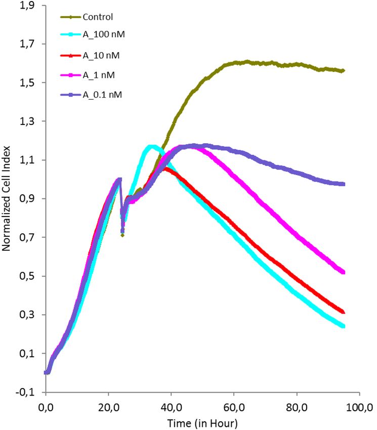
www.nature.com/scientificreports/
Figure 3. (a) Different development stages of Agrostemma githago L. a: Appearance of the sepals; b: Appearance
of the petals; c: Appearance of the seed capsule; d: Petals fully developed and colored; e: Petals parched, growing
seed and seed capsule; f: Maturation of seed and seed capsule, seeds white-yellow colored; g: Loss of sepals, seeds
black colored and fully developed, seed capsule open. (b) Western blot analysis of the extracts from stages a-g
using the anti-agrostin antibody. I: Marker; II: Stage a; III: Stage b; IV: Stage c; V: Stage d; VI: Agrostin_seed;
VII: Marker; VIII: Stage e; IX: Stage f; X: Stage g; XI: Agrostin_seed.
For Agrostin_seed ambiguities remain in three positions between the protein data and the RNA seq data
(V019M, N042S, S228T). In these cases we used the amino acids according to the peptide sequences for the final
sequence presented in Fig. 4, since we assume that they represent more direct evidence for the protein purified
from the plant. The discrepancies might be due to sequencing inaccuracies or might arise from the diversity of the
biological material used in this study. Intriguingly we found one peptide in two versions (R.ANFVANELTAQER,
M = 1,461.7, and R.ANFVANELTPQER, M = 1,487.7) pointing to a certain degree of heterogeneity within the
Agrostin_seed fraction.
Agrostin_seed shows the typical features of a type I RIP such as gypsophilin-S from Gypsophila elegans
M.Bieb14. Its theoretical molecular mass calculated from the sequence is 26,966.0 Da (M + H, average) which
is in good agreement (Δ = 148 ppm) with the experimental value (Fig. 1b). The theoretical isoelectric point as
calculated with the tool ProtParam is 9.43, which is somewhat higher than the experimental values given by
Stirpe for the different agrostin peaks observed in his work (7.7 for peak 2, 8.7 for peak 5 and 8.75 for peak 6)11.
The hypothetical three-dimensional structure of agrostin generated using P hyre217 shows the typical com-
position of other type I RIPs, which consists of an N-terminal β-sheet-rich domain followed by an α-helix-rich
succession (Fig. 4b).
Using the Agrostin_seed amino-acid sequence, a similarity search using the protein basic logic alignment
tool BLASTp yielded the highest percentage identity value of 36% to the type I RIP bouganin from Bougainvillea
spectabilis Willd. This is surprising since Bougainvillea spectabilis Willd belongs to another plant family (Nyctagi-
naceae). The value of 36% is remarkably low; sequence identity is even lower with other RIPs from plants from
the same plant family (Caryophyllaceae), 30% with gypsophilin-S, 27% with saporin-6, and 26% with dianthin.
This finding is even more striking as the similarity between these three proteins is much higher (in the range of
80% sequence identity).
It highlights the exceptional position that Agrostin_seed adopts among the type I RIPs from the carnation
family.
Type I RIPs, especially saporin, are used for the construction of targeted anti-tumor toxins, consisting of
monoclonal antibodies and type I RIPs as toxin portions18. Clinical studies have also been performed with such
kind of c onjugates19,20 and a huge number of saporin-based antibody conjugates, addressing different targets,
are commercially a vailable21. In this context agrostin is a new interesting option for generating conjugates with
potentially lower immunogenicity. Immunogenicity of the RIP portion is a big problem and differs quite a lot
among RIPs22. The conjugation of toxins to monoclonal antibodies is achieved by chemical linkers. Due to their
intrinsic nucleophilicity thiols (cysteines) are well suited for chemical conjugations via disulfide formation or
coupling via m aleimides23. However, in order to take advantage of thiol coupling chemistry the cysteines must
be accessible on the surface of the protein. Agrostin_seed contains the cysteines Cys 32 and Cys 216. Molecular
analysis using Jmol24 shows that the thiol of Cys 32 might be accessible for chemical modification, whereas that
of Cys 216 is rather oriented towards the core of the protein (Fig. 4c). This offers the possibility of a site-specific
modification with chemical linkers such as maleimide cross-linkers and coupling to monoclonal antibodies with
defined coupling stoichiometry.
Scientific Reports | (2020) 10:15377 | https://doi.org/10.1038/s41598-020-72282-2 4
Vol:.(1234567890)www.nature.com/scientificreports/
Hypothetical RIP‑sequences from the transcriptome of Agrostemma githago L. The analyses of the
RNAseq data set revealed 7 different RIP sequences. It is likely that the translation of these transcripts depends on fac-
tors such as development, infections or abiotic s tress25. The derived protein sequences were aligned using C LUSTALW16
and signal sequences were determined by SignalP 5.026 (The alignment is depicted in the supplementary information,
Fig. S1) and the functionally relevant amino acids are present throughout all sequences. There are only very few plants
with such a variety of RIPs in their transcriptomes. Table 1 shows the results of the alignments against Agrostin_seed.
Except for Agrostin_RNA3 all other Agrostin_RNA sequences show rather low percentage identity values. By perform-
ing a BLASTp search bouganin from Bougainvillea spectabilis Willd. was found as best match for most of the agrostin
RNA sequences. This is striking, since Bougainvillea spectabilis Willd. belongs to a different plant family (Nyctaginaceae)
and there is obviously no similarity to other RIPs from more closely related plants of the same family of the Caryophyl-
laceae. However phylogenetic analysis showed an evolutionary relationship of type I RIPs within the C aryophyllales27
and a considerable number of type I RIPs from the Caryophyllaceae such as petroglaucin from Petrocoptis glaucifolia
(Lag.) or pyramidatin from Vaccaria hispanica (Mill.) Rauschert are still not sequenced28,29.
Cytotoxic activity of agrostin. The cytotoxicity of Agrostin_seed was investigated in ECV-304 cells by
impedance-based real-time analysis. In previous studies we have shown that particular triterpene saponins aug-
ment the cytotoxicity of type I RIPs by improving the endosomal escape of internalized type I R IPs13.
Following endocytosis into the cell, type I RIPs need to escape from lysosomes into the cytosol. This is a very
important step in the course of the toxin routing, since the target organelles (ribosomes) are located in the cytosol.
For this reason we combined agrostin with the a non-toxic concentration of the triterpene saponin S O186133
(Fig. 5).
Recombinant expression of Agrostin_seed. Based on the amino-acid sequence of Agrostin_seed, a
codon-optimized nucleic acid sequence including the sequence for an N-terminal 8 × His affinity tag was gener-
ated by gene synthesis. The recombinant Agrostin_seed is henceforward referred to as hisAgrostin. hisAgrostin
was expressed in E. coli. and following the isolation by metal affinity chromatography one prominent band at
around 29 kDa could be seen on the SDS-PAGE (Fig. 6a). The exact mass of hisAgrostin was determined by
MALDI-TOF–MS as 28,117 Da. (Fig. 6b). This value is in very good agreement with the theoretical mass calcu-
lated from the sequence (28,119 Da). The identity of hisAgrostin was further verified by its peptide mass finger-
print (data not shown) and MALDI ISD sequencing (see supplementary information, Fig. S2).
The enzymatic activity of hisAgrostin was determined in a densitometric TLC a ssay34, which is based on the
RIP-catalysed release of adenine molecules from an artificial substrate.
As shown in Fig. 6c hisAgrostin showed enzymatic activity, even though its activity was not as high as the
activity of native Agrostin_seed. This could be due to a partially uncorrect folding of hisAgrostin during expression
in E. coli. In future studies this issue might be solved by optimzing the expression conditions in E. coli. However,
the recombinant type I RIP dianthin from Dianthus caryophyllus L., which was used as positive control, showed
an even a higher activity. This could be also due to a higher substrate specifity of hisAgrostin and native Agrostin
compared to dianthin, DNA not being the natural substrate of RIPs.
Methods
Seed material. Seeds (Agrostemmae semen, AGRO 26/80) from Agrostemma githago L. were obtained
from the Bundesanstalt für Züchtungsforschung und Kulturpflanzen (BAZ) in Gatersleben, Germany. Seeds
(200 g) were grinded and defatted by Soxhlet extraction using petroleum ether overnight. The material was air-
dried and extracted at 4 °C by 500 ml PBS supplemented with protease inhibitor (cOmplete Protease Inhibitor
Cocktail, Roche, Mannheim, Germany). After 12 h the extract was centrifuged at 6,000 g for 20 min and then
subjected to ultracentrifugation (Optima L-90 K, Beckmann Coulter GmbH; 30,000 rpm, 30 min, 4 °C). The
clear supernatant was subjected to affinity chromatography (see below).
Isolation of agrostin. For the isolation of Agrostin_seed an anti-agrostin antibody was generated in rabbits
(Pineda antibody service, Berlin, Germany). For the immunization, commercial agrostin (Sigma-Aldrich, Stein-
heim, Germany) was used. Following ammonium sulfate precipitation of the serum the IgG fraction was isolated
by protein A-based column chromatography (Pierce Protein A Agarose, Thermo Fisher Scientific). The antibod-
ies were eluted by 0.1 M glycine, pH 2.5, 4 °C and neutralized by Tris buffer (1 M, pH 9.0, 4 °C). For the isolation
of anti-agrostin antibodies, 100 µg of commercial agrostin was immobilized on NHS-Activated Agarose Spin
Columns (Pierce, Thermo Fischer Scientific). After applying the IgG fraction and washing (PBS), anti-agrostin
antibodies were eluted by 0.1 M glycine, pH 2.5, 4 °C and neutralized (Tris buffer 1 M, pH 9.0, 4 °C). Fractions
were pooled, dialysed against PBS and analysed by SDS-PAGE (12%).
For the isolation of Agrostin_seed, anti-agrostin antibodies were immobilized on NHS-Activated Agarose
Spin Columns (Pierce, Thermo Fisher Scientific). The Agrostemma seed extract (500 ml) was gradually applied
to the column. After washing (5 ml PBS, 4 °C), bound Agrostin_seed was eluted by adding 5 ml 0.1 M glycine,
pH 2.5, 4 °C. In total 13 fractions (each 0.5 ml) were collected, neutralized (see above) and dialysed against
PBS. Two fractions contained agrostin. Protein concentration was determined by BCA assay and fractions were
analysed by SDS-PAGE (12%), Coomassie Brilliant Blue staining.
Mass spectrometry. Proteins and peptides were analysed by matrix-assisted laser desorption ionization-
time of flight mass spectrometry (MALDI-TOF–MS) using an Ultraflex-II TOF/TOF instrument (Bruker Dal-
tonics, Bremen, Germany) equipped with a 200 Hz solid-state Smart beam laser. The mass spectrometer was
Scientific Reports | (2020) 10:15377 | https://doi.org/10.1038/s41598-020-72282-2 5
Vol.:(0123456789)www.nature.com/scientificreports/
Scientific Reports | (2020) 10:15377 | https://doi.org/10.1038/s41598-020-72282-2 6
Vol:.(1234567890)www.nature.com/scientificreports/
▸ Figure 4. (a) Alignment of gysophilin-S, a type I RIP from Gypsophila elegans M.Bieb, Agrostin_seed, isolated from the seeds and
agrostin (Agrostin_RNA3) from the early development stage a (Fig. 3a) of Agrostemma githago L. Functionally relevant conserved
amino acids are highlighted in yellow. Peptides sequenced by MS/MS analysis, covering the sequence of the Agrostin_seed are shown
in red. Aligment was performed using the Clustal Omega multiple sequence alignment t ool16. (b) Hypothetical tertiary structure of
Agrostin_seed using Phyre217 and Jmol24. The N-terminal region is rich of β-sheets highlighted in yellow, whereas the C-terminal
region is dominated by α-helices. (c) Ball-and-stick model of Agrostin_seed. The amino acids Glu 167 and Arg 170, representing the
active site, are shown in red, Tyr 68, Tyr 114 and Trp 202, representing the substrate binding site are shown in black. Cys 32 and Cys
216, a potential conjugation site to other biomolecules, are depicted in white.
Protein name Query coverage (%) Sequence identity (%) E-value IP Length (amino acid) BLASTp (NCBI*)
Chain A, rRNA N-glyco-
Agrostin_seed 100 100 0 9.4 243 sylase from Bougainvillea
spectabilis (36%)
Agrostin_RNA1 89 35 1e−44 9.4 300 Bouganin (36%)
RIP from Beta vulgaris
Agrostin_RNA2 85 28 5e−19 9.7 273
(33%)
Agrostin_RNA3 91 91 2e−169 6.8 265 Bouganin (36%)
Agrostin_RNA4 95 35 1e−46 9.4 300 Bouganin (35%)
RIP from Atriplex patens
Agrostin_RNA5 83 27 1e−16 8.8 287
(35%)
Agrostin_RNA6 90 37 1e−44 9.2 296 Bouganin (36%)
RIP from Bougainvillea
Agrostin_RNA7 75 53 7e−82 6.9 312
spectabilis (40%)
Table 1. Alignment using BLASTp30,31 of sequences obtained from transcriptome sequencing against
Agrostin_seed and BLASTp database. The isoelectric point (IP) was determined using the ExPASy ProtParam
tool32. *Database: All non-redundant GenBank CDS translations + PDB + SwissProt + PIR + PRF excluding
environmental samples from WGS projects.
Figure 5. Impedance-based live cell imaging of ECV-304 cells. After an incubation period of 24 h Agrostin_
seed (A) was added at different concentrations (0.1–100 nM) with SO1861, which is a triterpene saponin
isolated from Saponaria officinalis L. Cells were continuously monitored for 96 h. SO1861 enhanced the
cytotoxicity of Agrostin_seed by improving the delivery of the protein to the ribosomes.
Scientific Reports | (2020) 10:15377 | https://doi.org/10.1038/s41598-020-72282-2 7
Vol.:(0123456789)www.nature.com/scientificreports/
Figure 6. Recombinant expression of hisAgrostin in E. coli. (a) SDS-PAGE of hisAgrostin, Coomassie Brilliant
Blue stain. hisAgrostin appeared at ~ 29 kDa. (b) MALDI-TOF–MS spectrum of intact hisAgrostin. The mass
of hisAgrostin was determined as 28,117 Da. (c) TLC-based adenine releasing assay of hisAgrostin and native
Agrostin. hisAgrostin showed N-glycosylase activity against an oligo (A) substrate. However native Agrostin
(1 µg), that was isolated from the seeds (Agrostin_seed) exhibited significantly higher activity compared to
his
Agrostin (1 µg). Recombinant dianthin (0.25 µg) was used as positive control for a type I RIP and single
adenine (0.25 µg) was used as chromatographic control. *significant to hisAgrostin, t test, p ≤ 0.05.
operated in positive mode. Samples were spotted using the dried-droplet technique. Intact protein mass was
determined in linear mode (LP_ProtMix) using sinapinic acid as the matrix (saturated solution in 33% acetoni-
trile/0.1% trifluoroacetic acid) and spectra were acquired over an m/z range of 3,000–40,000. The mass accuracy
obtained in linear mode measurements in the higher mass range (> 10 kDa) was estimated as ± 1 ‰.
Peptides generated by in-gel trypsin digestion (modified from Shevchenko et al.35) were measured in reflec-
tor mode (RP_PepMix) using α-cyano-4-hydroxycinnamic acid (saturated solution in 33% acetonitrile/0.1%
trifluoroacetic acid) as the matrix and spectra were typically acquired over an m/z range of 600–4,000. Data was
analysed using FlexAnalysis 2.4. software. MS/MS spectra of selected tryptic peptides were acquired in the LIFT
mode36 and de novo interpretation of the fragment spectra was performed manually. In-source decay (ISD) was
used to generate N-terminal c ions and C-terminal (z + 2) ions from the intact purified and acetone-precipitated
recombinant protein using 1,5-diaminonaphthalene (1,5-DAN) as matrix. Spectra were recorded in the positive
reflector mode (RP_PepMix) in the m/z range 800–4,000. Mass accuracy here was < 100 ppm.
Agrostin expression in different development stages. In order to identify the right time point for
RNA isolation for the transcriptome sequencing different maturation states of growing Agrostemma githago L.
plants were analysed for agrostin expression. For this purpose 7 development states of the plant were selected:
Stadium a: 3 months after seeding, appearance of the sepals, stadium b: appearance of the petals, stadium c:
appearance of the seed capsule, stadium d: Petals fully developed and colored, stadium e: Petals parched, grow-
ing seed and seed capsule, stadium f: Maturation of seed and seed capsule, seeds white-yellow, stadium g: Loss
of sepals, seeds black and fully developed, seed capsule open.
The fresh plant material was snap-frozen in liquid nitrogen, grinded and defatted. The material was extracted
by PBS (cOmplete Protease Inhibitor Cocktail; Roche, Mannheim, Germany) at a concentration of 100 mg/ml and
analysed by western blot using the anti-agrostin antibody (1:7,500) as primary and a goat anti-rabbit antibody
(IgG, H and L Chain Specific Peroxidase Conjugate, Merck, 1:2,000) as secondary antibody. Amersham Hybond
ECL, (GE Healthcare Lifesciences), ECL (Enhanced chemoluminiscence)-reagent and an Optimax TR (M&S
Laborgeräte, Heidelberg, Germany) were used for development.
Transcriptome sequencing (RNAseq). Total RNA was isolated from plants in stadium a (see above).
For this purpose, the frozen plant material was grinded in liquid nitrogen. RNA was extracted from 102 mg
plant material using TriSure and Direct-zol RNA Miniprep kits (Zymo Research, Freiburg, Germany). Extracted
RNA was stored at − 80 °C. The sample was analysed by agarose gel (1%) electrophoresis. Concentration was
determined to 2.50 µg/µl (NanoDrop 1,000, Thermo Fischer Scientific). The RNAseq was performed using an
Illumina MiSeq V3 (LGC Genomics GmbH, Berlin, Germany).
The raw results were demultiplexed with Illumina’s data analysis software CASAVA and then cleaned of
adapter sequences. Forward and reverse reads were combined u sing37 BBMerge 34.48.
The resulting sequences were deconcatemerised and quality trimmed to include only reads with an average
P. hred quality score of at least 30. Based on these 12,115,767 reads, a de novo assembly was performed using
Scientific Reports | (2020) 10:15377 | https://doi.org/10.1038/s41598-020-72282-2 8
Vol:.(1234567890)www.nature.com/scientificreports/
Newbler v 2.9 in cDNA mode, and putative ORF identification was done by Transdecoder. Trinotate was used
to annotate the resulting transcontigs and predicted peptides to identify those sequences with a high similarity
to known RIPs.
Besides using Newbler, we performed another assembly with M ira38. This assembly was based on all quality
trimmed reads with a sequence that could be translated into either of the peptide fragments obtained by MALDI-
TOF MS and all other reads similar to these originally filtered reads.
Impedance‑based real‑time measurements. The toxicity of the isolated Agrostin_seed was inves-
tigated by impedance-based real time imaging. For this purpose ECV-304 cells (ACC 310, Leibniz Institut,
DMSZ, Braunschweig, Germany) were seeded in 100 µl (5,000/well) DMEM medium, supplemented with 10%
FBS in 96-well E-Plates (xCELLigence RTCA System, ACEA Biosciences)13,39. After 24 h, Agrostin_seed was
added (final conc. 0.1–100 nM). In order to scrutinize a potential synergistic toxicity with triterpene s aponins13
SO186133 was added at a final concentration of 1 µg/ml. Cells were continuously imaged for 96 h.
Recombinant protein expression. The codon-optimized coding sequence was established by gene
synthesis (General Biosystems, Inc., Morrisville, USA) and cloned into the expression vector pET11d (Merck,
Darmstadt, Germany). The coding sequence contained an N-terminal 8 × His tag for metal affinity chromatog-
raphy. The construct hisAgrostin_pET11d was transformed into competent Escherichia coli Rosetta 2 (DE3) cells
(Merck, Darmstadt, Germany). The bacterial culture was expanded to 3.2 l using LB medium containing 50 µg/
µl ampicillin and incubated until an optical density at 600 nm (OD600) between 0.9 and 1.2 was reached. Protein
expression was induced using 1 mM isopropyl β-D-1-thiogalactopyranoside (AppliChem, Darmstadt, Germany)
for 3 h at 37 °C and 200 rpm. The expression was stopped by centrifugation for 10 min at 5,000 g and 4 °C. Sub-
sequently, the bacterial pellets were resuspended in 20 ml PBS and stored at − 20 °C. The bacterial suspensions
were thawed and lysed by sonication (Branson Sonifier 250, G. Heinemann, Schwäbisch Gmünd, Germany). The
lysates were centrifuged at 15,800 g and 4 °C for 10 min and imidazole was added to the supernatant to a final
concentration of 20 mM. hisAgrostin was purified using Ni-nitrilotriacetic acid agarose affinity chromatography
(Protino Ni–NTA agarose, Macherey–Nagel, Düren, Germany). The bound protein was eluted using increasing
imidazole concentrations (20, 50, 75, 125 and 250 mM, 5 ml for each concentration) and analysed by SDS-PAGE
[12% acrylamide (w/v) gel]. The protein was dialysed against 2 l PBS and protein concentration was determined
using the bicinchoninic acid assay (Pierce BCA Protein Assay, Thermo Scientific, Waltham, MA, USA).
N‑glycosidase assay. The N-glycosidase activity was determined using an adenine releasing assay with an
artificial substrate. The assay is described in detail elsewhere34.
Briefly, the substrate consists of the DNA oligonucleotide 5′-A30-3′ (A30). Once the N-glycosidic bond is
cleaved, released adenine is separated from the reaction mixture by Thin Layer Chromatography (TLC) on
silica gel 60 glass plates. The glass plates are then scanned by a TLC-densitometer (TLC Scanner 4, CAMAG,
Berlin, Germany) at 260 nm. The RIP-mediated release of adenine is determined by calculating the Area Under
the Curve (AUC).
Received: 3 March 2020; Accepted: 28 August 2020
References
1. Kruskal, N. in Arbeiten des Pharmakologischen Instituts zu Dorpat Vol. VI (ed R. Kobert) 89–149 (Verlag von Ferdinand Enke,
Stuttgart, 1891).
2. Siepmann, C. et al. New saponins from the seeds of Agrostemma githago var. githago. Planta Med. 64, 159–164. https://doi.
org/10.1055/s-2006-957395 (1998).
3. Seeman, P. Ultrastructure of membrane lesions in immune lysis, osmotic lysis and drug-induced lysis. Fed. Proc. 33, 2116–2124
(1974).
4. Shany, S., Bernheimer, A. W., Grushoff, P. S. & Kim, K. S. Evidence for membrane cholesterol as the common binding site for
cereolysin, streptolysin O and saponin. Mol. Cell Biochem. 3, 179–186 (1974).
5. Bottger, S., Hofmann, K. & Melzig, M. F. Saponins can perturb biologic membranes and reduce the surface tension of aqueous
solutions: a correlation?. Bioorg. Med. Chem. 20, 2822–2828. https://doi.org/10.1016/j.bmc.2012.03.032 (2012).
6. Endo, Y., Mitsui, K., Motizuki, M. & Tsurugi, K. The mechanism of action of ricin and related toxic lectins on eukaryotic ribosomes.
The site and the characteristics of the modification in 28 S ribosomal RNA caused by the toxins. J. Biol. Chem. 262, 5908–5912
(1987).
7. Barbieri, L., Battelli, M. G. & Stirpe, F. Ribosome-inactivating proteins from plants. Biochim. Biophys. Acta 1154, 237–282 (1993).
8. Stirpe, F. Ribosome-inactivating proteins. Toxicon 44, 371–383. https://doi.org/10.1016/j.toxicon.2004.05.004 (2004).
9. Barbier, J. & Gillet, D. Ribosome inactivating proteins: from plant defense to treatments against human misuse or diseases. Toxins
(Basel) https://doi.org/10.3390/toxins10040160 (2018).
10. Schrot, J., Weng, A. & Melzig, M. F. Ribosome-inactivating and related proteins. Toxins (Basel) 7, 1556–1615. https://doi.
org/10.3390/toxins7051556 (2015).
11. Stirpe, F. et al. Ribosome-inactivating proteins from the seeds of Saponaria officinalis L. (soapwort), of Agrostemma githago L.
(corn cockle) and of Asparagus officinalis L. (asparagus), and from the latex of Hura crepitans L. (sandbox tree). Biochem. J. 216,
617–625. https://doi.org/10.1042/bj2160617 (1983).
12. Hebestreit, P. & Melzig, M. F. Cytotoxic activity of the seeds from Agrostemma githago var. githago. Planta Med. 69, 921–925. https
://doi.org/10.1055/s-2003-45101(2003).
13. Weng, A. et al. Saponins modulate the intracellular trafficking of protein toxins. J. Control Release 164, 74–86. https://doi.
org/10.1016/j.jconrel.2012.10.002 (2012).
14. Kokorin, A., Weise, C., Sama, S. & Weng, A. A new type 1 ribosome-inactivating protein from the seeds of Gypsophila elegans
M.Bieb. Phytochemistry 157, 121–127. https://doi.org/10.1016/j.phytochem.2018.10.024 (2019).
Scientific Reports | (2020) 10:15377 | https://doi.org/10.1038/s41598-020-72282-2 9
Vol.:(0123456789)www.nature.com/scientificreports/
15. Lappi, D. A., Esch, F. S., Barbieri, L., Stirpe, F. & Soria, M. Characterization of a Saponaria officinalis seed ribosome-inactivating
protein: immunoreactivity and sequence homologies. Biochem. Biophys. Res. Commun. 129, 934–942. https: //doi.org/10.1016/0006-
291x(85)91981-3 (1985).
16. Sievers, F. et al. Fast, scalable generation of high-quality protein multiple sequence alignments using Clustal Omega. Mol. Syst.
Biol. 7, 539. https://doi.org/10.1038/msb.2011.75 (2011).
17. Kelley, L. A., Mezulis, S., Yates, C. M., Wass, M. N. & Sternberg, M. J. E. The Phyre2 web portal for protein modeling, prediction
and analysis. Nat. Protoc. 10, 845. https://doi.org/10.1038/nprot.2015.053 (2015).
18. Giansanti, F., Flavell, D. J., Angelucci, F., Fabbrini, M. S. & Ippoliti, R. Strategies to improve the clinical utility of saporin-based
targeted toxins. Toxins (Basel) https://doi.org/10.3390/toxins10020082 (2018).
19. French, R. R., Bell, A. J., Hamblin, T. J., Tutt, A. L. & Glennie, M. J. Response of B-cell lymphoma to a combination of bispecific
antibodies and saporin. Leuk. Res. 20, 607–617. https://doi.org/10.1016/0145-2126(96)00007-0 (1996).
20. Schindler, J. et al. A phase I study of a combination of anti-CD19 and anti-CD22 immunotoxins (Combotox) in adult patients with
refractory B-lineage acute lymphoblastic leukaemia. Br. J. Haematol. 154, 471–476. https: //doi.org/10.1111/j.1365-2141.2011.08762
.x (2011).
21. Advanced Targeting Systems, (2019).
22. Strocchi, P., Barbieri, L. & Stirpe, F. Immunological properties of ribosome-inactivating proteins and a saporin immunotoxin. J.
Immunol. Methods 155, 57–63. https://doi.org/10.1016/0022-1759(92)90271-t (1992).
23. Spicer, C. D. & Davis, B. G. Selective chemical protein modification. Nat. Commun. 5, 4740. https://doi.org/10.1038/ncomms5740
(2014).
24. Herraez, A. Biomolecules in the computer: Jmol to the rescue. Biochem. Mol. Biol. Educ. 34, 255–261. https://doi.org/10.1002/
bmb.2006.494034042644 (2006).
25. Girbes, T., Ferreras, J. M., Arias, F. J. & Stirpe, F. Description, distribution, activity and phylogenetic relationship of ribosome-
inactivating proteins in plants, fungi and bacteria. Mini. Rev. Med. Chem. 4, 461–476. https://doi.org/10.2174/138955704340389
1 (2004).
26. Almagro Armenteros, J. J. et al. SignalP 5.0 improves signal peptide predictions using deep neural networks. Nat. Biotechnol. 37,
420–423. https://doi.org/10.1038/s41587-019-0036-z (2019).
27. Di Maro, A., Citores, L., Russo, R., Iglesias, R. & Ferreras, J. M. Sequence comparison and phylogenetic analysis by the maximum
likelihood method of ribosome-inactivating proteins from angiosperms. Plant Mol. Biol. 85, 575–588. https://doi.org/10.1007/
s11103-014-0204-y (2014).
28. Arias, F. J. et al. Isolation and partial characterization of a new ribosome-inactivating protein from Petrocoptis glaucifolia (Lag.)
Boiss. Planta 186, 532–540. https://doi.org/10.1007/BF00198033 (1992).
29. Bolognesi, A. et al. Ribosome-inactivating proteins (RNA N-glycosidases) from the seeds of Saponaria ocymoides and Vaccaria
pyramidata. Eur. J. Biochem. 228, 935–940. https://doi.org/10.1111/j.1432-1033.1995.tb20343.x (1995).
30. NCBI Research coordinators. Database resources of the National Center for Biotechnology Information. Nucl. Acids Res. 46,
D8–D13. https://doi.org/10.1093/nar/gkx1095 (2018).
31. McGinnis, S. & Madden, T. L. BLAST: at the core of a powerful and diverse set of sequence analysis tools. Nucl. Acids Res. 32,
W20-25. https://doi.org/10.1093/nar/gkh435 (2004).
32. Gasteiger E., H. C., Gattiker A., Duvaud S., Wilkins M.R., Appel R.D., Bairoch A. in The Proteomics Protocols Handbook, Humana
Press (2005). (ed John M. Walker) Ch. 52, 571–607 (Springer, 2005).
33. Sama, S. et al. Sapofectosid—ensuring non-toxic and effective DNA and RNA delivery. Int. J. Pharm. 534, 195–205. https://doi.
org/10.1016/j.ijpharm.2017.10.016 (2017).
34. Weng, A. A novel adenine-releasing assay for ribosome-inactivating proteins. J. Chromatogr. B Analyt. Technol. Biomed. Life Sci.
1072, 300–304. https://doi.org/10.1016/j.jchromb.2017.11.038 (2018).
35. Shevchenko, A., Wilm, M., Vorm, O. & Mann, M. Mass spectrometric sequencing of proteins silver-stained polyacrylamide gels.
Anal. Chem. 68, 850–858. https://doi.org/10.1021/ac950914h (1996).
36. Suckau, D. et al. A novel MALDI LIFT-TOF/TOF mass spectrometer for proteomics. Anal. Bioanal. Chem. 376, 952–965. https://
doi.org/10.1007/s00216-003-2057-0 (2003).
37. Bushnell, B., Rood, J. & Singer, E. BBMerge—accurate paired shotgun read merging via overlap. PLoS ONE 12, e0185056. https://
doi.org/10.1371/journal.pone.0185056 (2017).
38. Kumar, S. & Blaxter, M. L. Comparing de novo assemblers for 454 transcriptome data. BMC Genomics 11, 571. https://doi.
org/10.1186/1471-2164-11-571 (2010).
39. Gilabert-Oriol, R. et al. Real-time analysis of membrane permeabilizing effects of oleanane saponins. Bioorg. Med. Chem. 21,
2387–2395. https://doi.org/10.1016/j.bmc.2013.01.061 (2013).
Acknowledgements
For mass spectrometry (C.W.), we would like to acknowledge the assistance of the Core Facility BioSupraMol
supported by the Deutsche Forschungsgemeinschaft (DFG). We acknowledge support by the German Research
Foundation and the Open Access Publication Fund of the Freie Universität Berlin.
Author contributions
C.W. designed and performed experiments, analysed data and wrote parts of the manuscript; A.S. and L.T.D.W.
performed experiments; J.A. designed and performed parts of the bioinformatic analyses, R.GO. performed
experiments; S.S. read and edited the manuscript, M.F.M. designed research, read and edited the manuscript;
A.W. designed research, analysed data and wrote the main manuscript.
Funding
Open Access funding provided by Projekt DEAL.
Competing interests
The authors declare no competing interests.
Additional information
Supplementary information is available for this paper at https://doi.org/10.1038/s41598-020-72282-2.
Correspondence and requests for materials should be addressed to A.W.
Reprints and permissions information is available at www.nature.com/reprints.
Scientific Reports | (2020) 10:15377 | https://doi.org/10.1038/s41598-020-72282-2 10
Vol:.(1234567890)www.nature.com/scientificreports/
Publisher’s note Springer Nature remains neutral with regard to jurisdictional claims in published maps and
institutional affiliations.
Open Access This article is licensed under a Creative Commons Attribution 4.0 International
License, which permits use, sharing, adaptation, distribution and reproduction in any medium or
format, as long as you give appropriate credit to the original author(s) and the source, provide a link to the
Creative Commons licence, and indicate if changes were made. The images or other third party material in this
article are included in the article’s Creative Commons licence, unless indicated otherwise in a credit line to the
material. If material is not included in the article’s Creative Commons licence and your intended use is not
permitted by statutory regulation or exceeds the permitted use, you will need to obtain permission directly from
the copyright holder. To view a copy of this licence, visit http://creativecommons.org/licenses/by/4.0/.
© The Author(s) 2020
Scientific Reports | (2020) 10:15377 | https://doi.org/10.1038/s41598-020-72282-2 11
Vol.:(0123456789)You can also read