Arthropod entombment in weathering formed opal: new horizons for recording life in rocks - Mount St ...
←
→
Page content transcription
If your browser does not render page correctly, please read the page content below
www.nature.com/scientificreports
OPEN Arthropod entombment
in weathering‑formed opal: new
horizons for recording life in rocks
Boris Chauviré1*, Mickal Houadria2, Aline Donini3, Brian T. Berger4, Benjamin Rondeau5,
Gene Kritsky6 & Pierre Lhuissier7
Animal fossils preserved in various geological materials, such as limestone, claystone, or amber,
provide detailed information on extinct species that is indispensable for retracing the evolution
of terrestrial life. Here, we present the first record of an animal fossil preserved in opal formed by
weathering with such high-resolution details that even individual cuticle hairs are observed. The fossil
consists of the exoskeleton of a nymphal insect belonging to the order Hemiptera and either the
family Tettigarctidae or the Cicadidae. This identification is based on anatomical details such as the
tibial and femoral morphology of the forelegs. The exoskeleton of the insect was primarily zeolitized
during the alteration of the host rocks and later sealed in opal deposited by silica-rich fluids derived
from the continental weathering of the volcanic host rocks. Organic matter is preserved in the form of
amorphous carbon. This finding makes opal formed by rocks weathering a new, complementary source
of animal fossils, offering new prospects for the search for ancient life in the early history of Earth and
possibly other terrestrial planets such as Mars, where weathering-formed opal occurs.
Most invertebrate fossil records where anatomical details are well preserved are from amber or lacustrine or
fluvial sedimentary rocks1. These fossils have provided evolutionary evidence of ancestral traits and extinct spe-
cies, retracing the evolution of life. Here, we describe an insect fossil in opal—a variety of silica—in which the
degree of preservation is comparable (Fig. 1)2.
Silica minerals exist in various species ranging in structure from amorphous to crystalline. Opal is the main
species of amorphous silica that precipitates from saturated or near-saturated silica-rich aqueous fluids pro-
duced by the alteration of the earth’s crust. One of the main sources of silica for opal formation (and hence the
eventual silicification of fossils) are volcanic rocks, especially volcanic ashes due to their inherent porosity and
glass abundance3–8.
Among opals, the most sought-after type is precious opal, which differs from common opal only by the pres-
ence of colored patches (called play-of-color) that appear to move as the stone is viewed from different angles,
an optical phenomenon attributed to the diffraction of visible light by a regular arrangement of silica spheres9,10.
Both precious and common opal form mainly in two geological settings: (1) hydrothermal alteration occurring
when hot water circulates underground and precipitates opal when the temperature drops, e.g., as siliceous sin-
ter (a hard incrustation around hot springs); and (2) continental weathering that occurs when meteoric waters
percolate, hydrolyzing silicate while transforming the rock into a soil. In both contexts, the dissolved silica exists
as silicic acid ( H4SiO2) in aqueous solution. Silicic acid will polymerize into silica during a variation in physi-
cal or chemical conditions, primarily pH and t emperature11. The degradation of organic matter may favor the
precipitation of silica by providing functional groups (hydroxyl groups), making amorphous silica an important
preservation agent of fossils11,12.
Fossilization involving silica results from permineralization, replacement, or entombment. Permineralization
is especially documented for wood and bone when a silica-rich fluid fills the cavities between organ tissues12–17.
1
CNRS, IRD, IFSTTAR, ISTerre, Université Grenoble Alpes, Université Savoie Mont Blanc, 38000 Grenoble,
France. 2Biology Centre of Academy of Sciences, Institute of Entomology, Branisovska 31, České Budějovice,
Czech Republic. 3Actias, 44260 Savenay, France. 4@VelvetBoxSociety, Timberbrook Capital, Philadelphia,
PA, USA. 5Laboratoire de Planétologie Et Géodynamique, CNRS UMR 6112, Université de Nantes, BP 92208,
44322 Nantes, France. 6School of Behavioral and Natural Sciences, Mount St. Joseph University, Cincinnati, OH,
USA. 7Univ. Grenoble Alpes, CNRS, Grenoble INP, SIMAP, 38000 Grenoble, France. *email: boris.chauvire@
gmail.com
Scientific Reports | (2020) 10:10575 | https://doi.org/10.1038/s41598-020-67412-9 1
Vol.:(0123456789)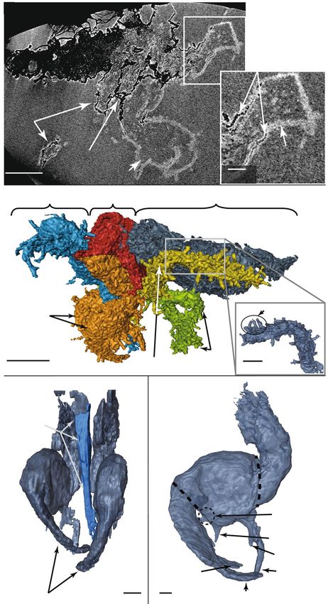
www.nature.com/scientificreports/
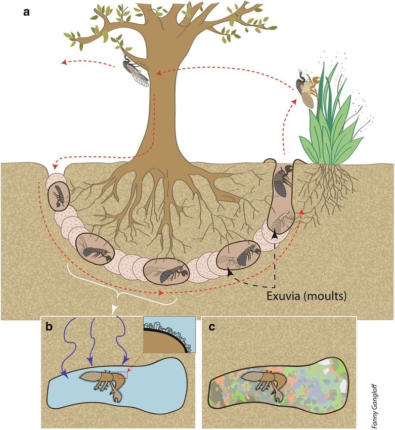
Figure 1. Insect entombed in the opal studied here. 3D surface reconstruction (top) and a microphotograph
(bottom). The 3D reconstruction has been performed by X-ray microtomography. The fossil is clearly visible
within the near-transparent opal (named “Beverly”). Photo by B. Rondeau and 3D reconstruction by P.
Lhuissier.
Australian wood and dinosaur bone fossilized in precious opal are an impressive manifestation of perminer-
alization by s ilica18,19. Replacement results from the dissolution of an original organism, creating a mold that
can be later filled by the precipitation of silica7,8,20. The dissolution or degradation of the original material and
precipitation of silica can be concurrent, leading to the exceptional preservation observed in arthropods and
brachiopods in the Barstow Formation (California) or the Glass Mountains (Texas)21–23. By contrast, entomb-
ment is the trapping of an organism in silica that occurs by a rapid silica precipitation on the external surface.
This is mainly documented in hydrothermal settings24–26, where silica may preserve an entire ecosystem, as
observed in the 407-million-year-old Rhynie Chert (Scotland), the oldest fossil terrestrial environment with a
comprehensive preservation of flora and f auna27. During diagenesis (the hardening of sediments into rocks due
to burial), the most amorphous phase of silica (opal-A: amorphous) will transform into more crystalline phases
(opal-CT, constituted of nano-domains of cristobalite and tridymite) until it becomes quartz. The transformation
of opal into quartz leads to the formation of chert, a sedimentary rock that contains the oldest evidence of life
on Earth28. Silica entombment associated with hot springs is thus highly studied for its similarity to early Earth
environments, and the same process is suspected to have occurred in some locations on M ars25,26,29.
Entombment also occurs in the context of continental weathering, where opal develops through the weather-
ing of rocks, mostly of volcanic origin. However, until now, plant roots were the only organisms documented
as being preserved through entombment in continental weathering opal in Ethiopia4,5. Here, we present novel
data on a sample nicknamed “Beverly,” the first occurrence of an animal fossil entombed in gem opal formed
by continental weathering. This provides new prospects for life evolution studies by demonstrating that silica
entombment can occur during opal formation through the weathering process, which is more widespread than
highly localized hydrothermal settings. We discuss the processes that led to the preservation of an insect in opal,
and we consider the rarity of such a phenomenon and its implications for science.
Fossil and host material
Geological context. Co-author B.T.B. acquired the sample, which came from a precious opal deposit
mined in the Genteng Formation, Western Java, Indonesia. The Genteng Formation, dated to the Early Pliocene
(5–10 million years old)30,31, is located 100 km southwest of Jakarta and covers approximately 1,300 km2. This
geological unit consists of a series of pumice tuff, tuffaceous sandstone, and claystone deposited from air-fall
and/or pyroclastic flow in a terrestrial to littoral environment31–33. Each layer has been highly weathered into
a paleosoil33, and the upper part contains a significant number of silicified wood and other plant remains. The
opal-bearing layer is a tuffaceous claystone composed of feldspar, smectite, and volcanic glass associated with
zeolite (clinoptilolite)32. The presence of zeolite is interpreted by Einfalt34 as evidence of limited burial and for-
mation in an open freshwater system, whereas A nsori32 proposes a burial diagenesis taking place at a depth
of several hundred meters, with temperatures reaching 45 °C. The geochemistry of Javanese opals from the
Genteng formation exhibits a signature characteristic of silica produced by continental weathering and leaching
of volcanic tuff32,34. According to these studies, Javanese opal originates from the weathering of volcanoclastic
rocks, especially volcanic glass32,34. Large amounts of silica are released to percolating fluids, which precipitate
into opal during circulation through porous rocks32,34. Ansori32 proposed that this silica-rich fluid has been
trapped in clay-rich layers, where large cavities may have originated from the degradation of plant remains.
Scientific Reports | (2020) 10:10575 | https://doi.org/10.1038/s41598-020-67412-9 2
Vol:.(1234567890)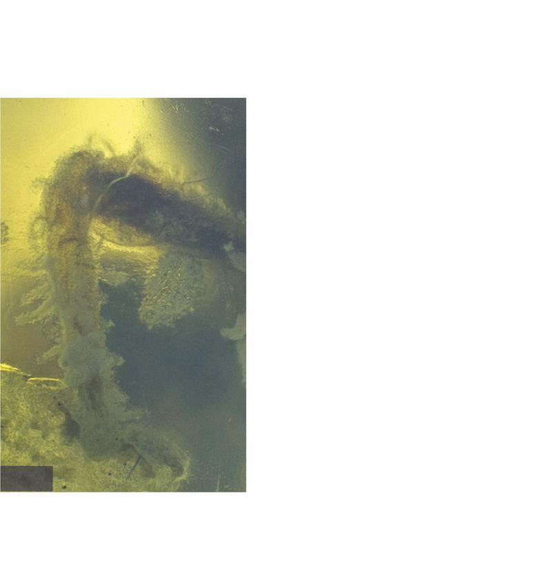
www.nature.com/scientificreports/
b
335 Opal-CT
Intensity
a
780
100 300 500 700 900 1100 1300
Wavenumber (cm-1)
c Carbon-rich material
Intensity
1200 1300 1400 1500 1600 1700 1800
Wavenumber (cm-1)
d 408 Zeolite Coating
Acquired spectrum
Reference spectrum
480
Intensity
100 µm
Op
150 265 624
100 300 500 700 900 1100 1300
Wavenumber (cm-1)
Figure 2. Microphotographs showing the locations of Raman spectra: (a) microphotographs of a middle leg of
the specimen showing the thin layer coating the fossil. Photo by B. Rondeau. (b) Raman spectrum of the matrix
with peaks characteristic of opal-CT35. (c) Raman spectrum of the black carbon-rich material inside the coating.
(d) Raman spectrum of the thin layer showing features of clinoptilolite–heulandite zeolite. Our Raman spectra
are shown in black; in grey—a reference sample38. (Op opal contribution).
Material characterization. The broad band near 335 cm−1 in the Raman spectra (Fig. 2b) of the mate-
rial that enclosed the fossil clearly indicates opal-CT35. The fossil also contains black material that exhibits a
weak Raman signal consisting of an asymmetrical broad band near 1,600 cm−1, with a weaker band at about
1,360 cm−1 (Fig. 2c). These broad bands are typical of G and D bands (respectively) of amorphous c arbon36,37.
A transparent layer 50–100 µm thick coats the surface of the fossil, preserving even the hairs on the cuticle. The
Raman spectra of this coating is typical of zeolites from the clinoptilolite–heulandite s eries38: two main bands
at 408 and 480 cm−1, with additional bands near 150, 265, and 624 cm−1 and broad bands near 797 cm−1 and
1,130 cm−1 (Fig. 2d).
Taxonomy of the fossil
Taxonomic identification was performed on the basis of scans obtained through non-destructive X-ray com-
puted microtomography (details in “Methods” section). The X-ray computed tomography scans show both the
exterior and the interior of the fossil outlined by the coating of lighter zeolite material (Fig. 3a). The interior of
the fossil does not exhibit any internal structure and may be composed of the same material as the host (opal).
The 5.5 mm long specimen includes the head, the thorax with three pairs of legs, and part of the abdo-
men (Fig. 3b) and is undoubtedly from a nymphal insect. Both microphotography (Fig. 2) and high-resolution
tomography (Fig. 3b) allowed us to distinguish individual cuticle hairs, highlighting the excellent preservation.
The tomography and 3D reconstruction provide vital information used to identify the insect superfamily. The
prominent rostrum (sucking mouthparts; Fig. 3c) identifies the specimen as an insect of the order Hemiptera39.
To reveal greater detail on the forelegs, the interior of the zeolite coating was reconstructed, and the exterior was
Scientific Reports | (2020) 10:10575 | https://doi.org/10.1038/s41598-020-67412-9 3
Vol.:(0123456789)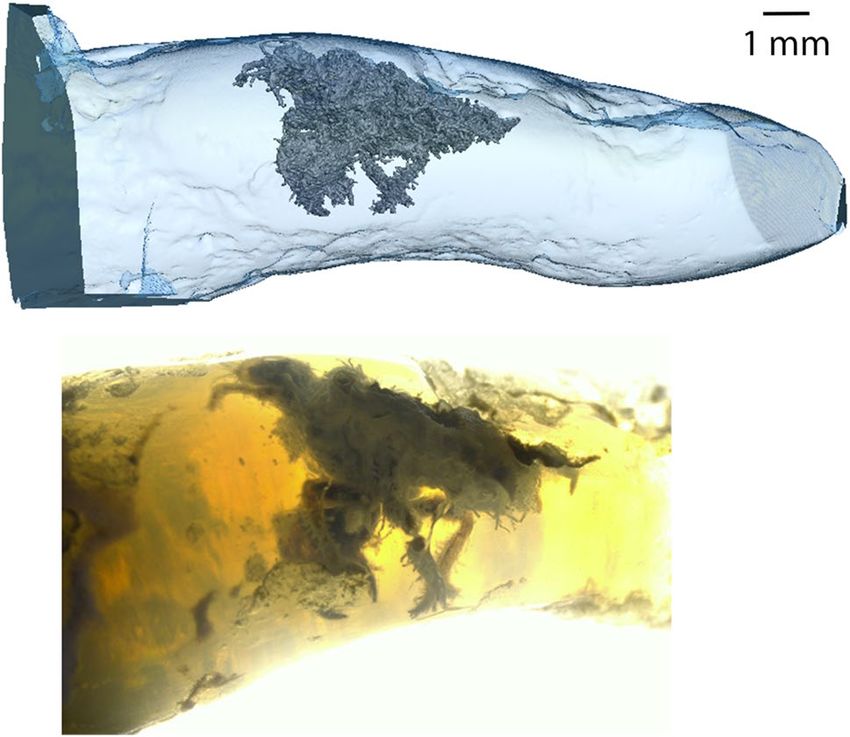
www.nature.com/scientificreports/
a
Carbon-rich
material
Back
leg
Zeolite
Middle
coating
leg
1 mm Foreleg 0,5 mm
b HEAD THORAX ABDOMEN
Forelegs Cuticule hairs
Back
1 mm Middle leg legs 100 µm
c TROCHANTER d
Rostrum
FEMUR
Femoral comb
area
Intermediate tooth
TIBIA of femur
Accessory tooth
of femur
Point of blade
of tibia Posterior tooth
of femur
Forelegs Apical tooth of tibia
200 µm 200 µm
Figure 3. Anatomical details of the fossil. X-ray computed microtomography projections and 3D views. (a)
X-ray projection of the fossil showing the zeolite coating and the carbon-rich material. This image demonstrates
that the material inside and outside the fossil is identical. (b) 3D side view of the whole insect exuvia with
colored anatomical parts; the inset (bottom right) highlights cuticle hairs. (c) Frontal view of the forelegs with
rostrum highlighted in blue. (d) Internal side view of the right foreleg, after numerically removing the zeolite
coating. Animated images corresponding to (b), (c) and (d) are available in Supplementary Information 1.
numerically removed. This reveals the tibial and femoral morphology of the forelegs (Fig. 3d) and identifies the
specimen as a nymph (immature form) of a hairy cicada or a true c icada40.
Discussion
X-ray computed microtomography shows that both the interior and the exterior of the fossil are coated by a thin
layer of lighter material, identified as zeolite by Raman spectroscopy (Fig. 3a). No internal structure has been
observed and the interior is occupied by the same opal as that outside the fossil (based on Raman spectroscopy
and X-ray). These observations indicate that the insect integument was hollow, suggesting that it may be an
exuvia (the shed exoskeleton of an insect). An alternative explanation may be that the insect died in the soil, and
the fluids dissolved the soft interior parts of the insect. The more resistant cuticle would have been preserved
and subsequently fossilized.
Scientific Reports | (2020) 10:10575 | https://doi.org/10.1038/s41598-020-67412-9 4
Vol:.(1234567890)www.nature.com/scientificreports/
Morphological details (especially the rostrum and the prominent forelegs) identify the fossil as a cicada
nymph belonging to either the Tettigarctidae (known as hairy cicadas) or the Cicadidae (otherwise called true
cicadas) families. According to the existing literature, fossils of immature cicadas are rare. To date, only four
have been discovered, all in amber. Of these cicada fossils, two are new records: one in the Dominican Republic
(Dominicicada youngi) and the other in Myanmar (Burmacicada protera)41. A third cicada specimen was found
in Baltic amber, but no taxonomic description has been published so f ar42. Fragments of another nymph have
also been reported in Cretaceous amber from New Jersey43.
Nymphs of the two families can only be distinguished metamorphically using features such as the number of
femoral spines or the presence or absence of the tarsi, which only develop during the last nymphal stage (instar)44.
The size of the nymph (5 mm) and the shape of the slightly swollen zeolite layer on the femoral comb (Fig. 3d)
lead us to believe that it is an early instar44, rendering identification to one of the families impossible. In terms
of phylogeographical context, the presence of the two families on both sides of the Wallace line45–50 (a faunal
boundary separating two different ecozones in southeast Asia) prevents any clear assertion of this specimen
belonging to the Cicadidae rather than the Tettigarctidae.
The amorphous carbon composing the black, carbon-rich material (according to the Raman s ignature36,37)
could result from the transformation of organic material composing the fossil. This material is coated by clinop-
tilolite–heulandite zeolites (Fig. 2). The zeolitic coating is consistent with the clinoptilolite identified by E infalt34
using X-ray diffraction. As zeolites are observed exclusively at the interface between the organic material and
the host opal, we propose that zeolitization occurred before the precipitation of opal. Therefore, opal precipita-
tion may not have occurred immediately or concurrently after the zeolitization, although opal precipitation
ultimately preserves the fossil.
Clinoptilolite–heulandite are the most common zeolitic series found in sedimentary and igneous rocks; they
form in various environments, including hydrological open and closed systems, deep marine sediments, soils,
through hydrothermal alteration, and during diagenesis and burial metamorphism, especially from volcanic
material38,51,52. The presence of clinoptilolite–heulandite zeolites has been discussed by E infalt34 and Ansori32,
both concluding that they originate from the destabilization of volcanic glass contained in the opal-bearing or
overlying layers. However, the conditions of formation proposed by each author differs; E infalt34 proposed that
no burial diagenesis occurred, in contradiction to A nsori32. The fine details preserved in our sample could be
an evidence of a limited burial, as higher pressure might have obliterated them. Zeolitization may have played a
significant role in preventing alteration of the fossil in the aqueous environment required for opal precipitation.
Unfortunately, the zeolite coating the insect cuticle partially hides the morphological details required for a more
thorough taxonomic identification.
The identification of the fossil as a true or hairy cicadas provides information on the genesis of our sample.
Indeed, in extant species of cicada, after eggs laid in branches have hatched, the first instar nymphs fall to the
ground53,54 (Fig. 4). They burrow into the soil with their strong front legs, using the other two pairs of legs to
move through a narrow burrow54, searching for roots from which they suck xylem fluid. The many cuticle hairs
observed on our specimen (Fig. 2b) play an important role in aggregating loosened earth behind the tunneling
nymph. Cicada nymphs are usually found between 5 and 80 cm deep in the soil, but they have been reported
from up to 1.2 m below g round54,55. The nymphs feed on tree roots, usually going through four instars under-
ground before the final instar nymph emerges from the soil to transform into an a dult53. The time required for
this subterranean development varies among different species from 1 to 17 years56.
Opal is a common product of the weathering of volcanic g lass57,58 and precipitates from an aqueous g el9.
Opal precipitation could originate from the weathering of volcanic rocks deposited later, rather than occurring
simultaneously with zeolite crystallization, as demonstrated for both precious opal f ormation3–6 and fossils
replaced by silica in various l ocation7,8,20.
Because cicada nymphs live in shallow s oils53–55 (Fig. 4) and paleosoils have been documented in the Genteng
formation31–33, we consider that a shallow soil is the most probable environment for the genesis of our sample.
Moreover, the excellent preservation of the fossil, particularly the conservation of cuticle hairs (Fig. 3b), is incon-
sistent with exhumation and transport that would have damaged or even obliterated these delicate features. We
therefore propose that the process of zeolitization occurred shortly after the shedding of the nymphal skin. This
first stage of zeolitization has been observed in plant fossils preserved in Ethiopian opals4–6.
The encrustation of insects in opal is a rare phenomenon. The few previously documented examples have orig-
inated in hydrothermal settings, where silicification rapidly entombs organisms24–27,59. Opal formation through
continental weathering is a much more widespread phenomenon, but is also slower. Animals such as insects
would normally crawl away before the process could begin—in contrast to fossilization in amber, where insects
are literally glued into the sap that forms the amber. In addition, insect cuticles are usually hydrophobic, which
tends to isolate the insect from silica-rich w ater60. Only immobile, attached invertebrates (dead or alive) may be
entrapped by the silica-rich fluids, which gradually turn into a gel and finally into opal. Hence, it is much more
probable that plants rather than animals would be preserved as fossils in opal. This supports the idea that this
fossil is of a lifeless, immobile cicada exuvia. Specific geological and environmental conditions were necessary
to preserve our sample and a detailed geological investigation of the opal mine is required to determine these
conditions more fully.
Implications
In this study, we demonstrate that opal forming through continental weathering can preserve insects and traces
of organic compounds nearly as efficiently as hydrothermal siliceous sinters or amber. We also demonstrate that
X-ray computed tomography is an efficient technique to detect and characterize fossils preserved in opal. Even
if opals may be opaque, making visual examination of the interior impossible, tomography could still be used to
Scientific Reports | (2020) 10:10575 | https://doi.org/10.1038/s41598-020-67412-9 5
Vol.:(0123456789)www.nature.com/scientificreports/
Figure 4. Scenario of sample formation. (a) Life cycle of an extant cicada. The white bracket identifies the
instars corresponding to our sample compare with Fig. 3. (b) Fossilization of cicada exuvia. Percolating fluid
(represented by blue arrows) fills the cavity, and zeolites crystallize on exuvia. (c) The precipitation of opal sealed
the exuvia in place, forming the well-preserved fossil. Drawing by F. Gangloff.
reveal entombed specimens and hence uncover new paleontological or evolutionary clues of ancient life. Opal
precipitates from fluids enriched in silica through the weathering of silicate crust, a process that has operated
since the Archean times. Preservation in weathering-formed opal is therefore a potential source of information
about the evolution of life in the earliest periods of Earth’s history. Moreover, preservation in amber overrepre-
sents insect fossils from forest habitats, due to its formation from tree resin, and insect fossils in sedimentary
rocks mainly sample communities from aqueous e nvironments61. The rarity of cicada nymphs compared to
other insects62 could be related to their underground habitat, where fossilization is scarcer. Therefore, weath-
ering-formed opal here demonstrates its potential for a better understanding of the evolution of species that
live underground. Opal’s capacity to preserve fossils provides new opportunities in the search for ancient and/
or extraterrestrial life. Amorphous silica is present on the surface of M ars29,63,64, and is already targeted as the
most promising source for the search for extraterrestrial life. The fossil-preserving potential of amorphous silica
has been mostly observed in samples that formed in hydrothermal e nvironments25,26,65, whereas opaline silica
deposits have been identified as originating from the weathering of its silicate c rust66,67. The future landing site
of NASA’s next rover missions on Mars (Mars 2020) includes opaline silica that is thought to be of weathering
origin68. Even if opaline silica is already the most encouraging mineral for exploring the traces of extraterrestrial
life, its potential is now widened to a larger range of geological settings, thus offering a new possibilities for a
record of life forms on Mars.
Scientific Reports | (2020) 10:10575 | https://doi.org/10.1038/s41598-020-67412-9 6
Vol:.(1234567890)www.nature.com/scientificreports/
Methods
Microphotography. Microphotographs have been acquired using a Keyence VHX-2000 numerical micro-
scope allowing Z-stack to enhance field depth. We used a VH-Z20T zoom lens allowing magnification from × 20
to × 200, operated in transmitted light without polarizing filters. To improve the exposition of some parts, addi-
tional light has been added by external optical fibers.
X‑ray microtomography. 3D images have been acquired by x-ray microtomography on RX-Solutions
EasyTom XL Nano equipment using a Hamamatsu L8121 sealed tube and a Paxscan 2520DX flat panel from
VAREX Imaging. A source tension of 100 kV with a current of 80 µA was combined with a 0.25 mm thick alu-
minum filter. The magnification was set to 25.4 resulting in a voxel size of 5 µm. 1984 projections were acquired
over one turn. Each project is the average of 6 images exposed during 0.5 s each. A classical filtered back projec-
tion algorithm was used for 3D reconstruction.
In order to reveal fine details on forelegs, the reconstruction has been segmented slice by slice by manual
border selection using the open software ImageJ. 3D renderings are obtained with the ThermoFicher Scientific
Avizo software.
Raman spectroscopy. Raman spectra were acquired using a Labram Jobin–Yvon Raman spectrometer
(2 cm–1 resolution) equipped with an Ar laser emitting at 514 nm, 50 mW power, combined with a microscope
with a 50 × objective.
Received: 5 December 2019; Accepted: 4 June 2020
References
1. Penney, D. & Jepson, J. E. Fossil Insects: An Introduction to Palaeoentomology (Siri Scientific Press, Castleton, 2014). https://doi.
org/10.1038/032053c0.
2. Smith, T. & Renfro, N. Fossil insect in opal. Gems Gemol. 55, 113–114 (2019).
3. Rey, P. F. Opalisation of the Great Artesian Basin (central Australia): an Australian story with a Martian twist. Aust. J. Earth Sci.
60, 291–314 (2013). https://doi.org/10.1080/08120099.2013.784219.
4. Rondeau, B. et al. Geochemical and petrological characterization of gem opals from Wegel Tena, Wollo, Ethiopia: opal formation
in an Oligocene soil. Geochem. Explor. Environ. Anal. 12, 93–104 (2012). https://doi.org/10.1144/1467-7873/10-MINDEP-058.
5. Chauviré, B., Rondeau, B., Mazzero, F. & Ayalew, D. The precious opal deposit at Wegel Tena, Ethiopia: formation via successive
pedogenesis events. Can. Mineral. 55, 701–723 (2017). https://doi.org/10.3749/canmin.1700010.
6. Chauviré, B. et al. Pedogenic origin of precious opals from Wegel Tena (Ethiopia): evidence from trace elements and oxygen
isotopes. Appl. Geochem. 101, 127–139 (2019). https://doi.org/10.1016/j.apgeochem.2018.12.028.
7. Laufeld, S. & Jeppsson, L. Silicification and bentonites in the Silurian of Gotland. Geol. Föreningen i Stock. Förh. 98, 31–44 (1976).
https://doi.org/10.1080/11035897609454336.
8. Butts, S. H. Silicification. In Reading and Writing of the Fossil Record: Preservational Pathways to Exceptional Fossilization (eds
Laflamme, M. et al.) (Paleontological Society, Cambridge, 2014).
9. Darragh, P. J. & Gaskin, A. J. The nature and origin of opal. Aust. Gemol. 8, 5–9 (1966).
10. Sanders, J. V. Diffraction of light by opals. Acta Crystallogr. Sect. A 24, 427–434 (1968). https://doi.org/10.1107/S05677394680008
60.
11. Iler, R. K. The occurrence, dissolution, and deposition of silica. In The Chemistry of Silica: Solubility, Polymerization, Colloid and
Surface Properties and Biochemistry of Silica (ed. Sons, J. W.) 3–93 (Wiley, New York, 1979).
12. Martin, R. E. Taphonomy. Climate Change 2013—The Physical Science Basis Vol. 53 (Cambridge University Press, Cambridge,
2013).
13. Leo, R. F. & Barghoorn, E. S. Botanical museum leaflets volume XXV. Bot. Mus. Leafl. 25, 1–289 (1976).
14. Scurfield, G. & Segnit, E. R. R. Petrifaction of wood by silica minerals. Sediment. Geol. 39, 149–167 (1984). https://doi.
org/10.1016/0037-0738(84)90048-4.
15. Ballhaus, C. et al. The silicification of trees in volcanic ash—an experimental study. Geochim. Cosmochim. Acta 84, 62–74 (2012).
https://doi.org/10.1016/j.gca.2012.01.018.
16. Mustoe, G. E. Wood petrifaction: a new view of permineralization and replacement. Geoscience 7, 119 (2017). https://doi.
org/10.3390/geosciences7040119.
17. Liesegang, M. & Gee, C. T. Silica entry and accumulation in standing trees in a hot-spring environment: cellular pathways, rapid
pace and fossilization potential. Palaeontology https://doi.org/10.1111/pala.12480 (2020).
18. Pewkliang, B., Pring, A. & Brugger, J. The formation of precious opal: clues from the opalization of bone. Can. Mineral. 46, 139–149
(2008). https://doi.org/10.3749/canmin.46.1.139.
19. Bell, P. R. et al. Revised geology, age, and vertebrate diversity of the dinosaur-bearing Griman Creek Formation (Cenoma-
nian), Lightning Ridge, New South Wales Australia. Palaeogeogr. Palaeoclimatol. Palaeoecol. 514, 655–671 (2019). https://doi.
org/10.1016/j.palaeo.2018.11.020.
20. Daley, R. L. & Boyd, D. W. The role of skeletal microstructure during selective silicification of brachiopods. SEPM J. Sediment. Res.
66, 155–162 (1996). https://doi.org/10.1306/D42682E3-2B26-11D7-8648000102C1865D.
21. Cooper, G. A. & Grant, R. E. Permian brachiopods of west Texas, VI. Smithson. Contrib. Paleobiol https://doi.org/10.5479/si.00810
266.32.1 (1977).
22. Park, L. E. & Downing, K. F. Paleoecology of an exceptionally preserved arthropod fauna from lake deposits of the Miocene Barstow
Formation, Southern California, U.S.A.. Palaios 16, 175 (2001).
23. Olszewski, T. D. & Erwin, D. H. Change and stability in Permian brachiopod communities from western Texas. Palaios 24, 27–40
(2009). https://doi.org/10.2110/palo.2008.p08-061r.
24. Guido, D. M., Campbell, K. A., Foucher, F. & Westall, F. Life is everywhere in sinters: examples from jurassic hot-spring environ-
ments of argentine patagonia. Geol. Mag. 156, 1631–1638 (2019). https://doi.org/10.1017/S0016756819000815.
25. Des Marais, D. J. & Walter, M. R. Terrestrial hot spring systems: introduction. Astrobiology 19, 1419–1432 (2019). https://doi.
org/10.1089/ast.2018.1976.
26. Teece, B. L. et al. Biomolecules from fossilized hot spring sinters: implications for the search for life on Mars. Astrobiology 20,
537–551 (2020). https://doi.org/10.1089/ast.2018.2018.
Scientific Reports | (2020) 10:10575 | https://doi.org/10.1038/s41598-020-67412-9 7
Vol.:(0123456789)www.nature.com/scientificreports/
27. Edwards, D., Kenrick, P. & Dolan, L. History and contemporary significance of the Rhynie cherts—our earliest preserved terrestrial
ecosystem. Philos. Trans. R. Soc. B Biol. Sci. 373, 20160489 (2018). https://doi.org/10.1089/ast.2018.2018.
28. Djokic, T., Van Kranendonk, M. J., Campbell, K. A., Walter, M. R. & Ward, C. R. Earliest signs of life on land preserved in ca. 3.5
Ga hot spring deposits. Nat. Commun. 8, 15263 (2017). https://doi.org/10.1038/ncomms15263.
29. Ruff, S. W. & Farmer, J. D. Silica deposits on Mars with features resembling hot spring biosignatures at El Tatio in Chile. Nat.
Commun. 7, 13554 (2016).
30. Van Bremmelen, R. W. The Geology of Indonesia. (Governement Printing, The Hague, 1949).
31. Sujatmiko, H. & Santosa S. Geological map of the Leuwidamar Quadrangle, Java 1:100000.
32. Ansori, C. Model mineralisasi pembentukan opal banten. Indones. J. Geosci. 5, 151–170 (2010). https://doi.org/10.17014/ijog.
v5i3.100.
33. Oktariani, H., Winantris, W. & Hamzah, A. Dryobalanoxylon sp.: ilicified fossil wood from Lebak Regency, Banten Province.
Indones. J. Geol. Sumberd. Miner. 20, 93 (2019). https://doi.org/10.33332/jgsm.geologi.20.2.93-99.
34. Einfalt, H. C. Some observations on the composition and origin of opals from Java. J. Gemol. 30, 383–398 (2007).
35. Ostrooumov, M., Fritsch, E., Lasnier, B. & Lefrant, S. Spectres Raman des opales: aspect diagnostique et aide à la classification.
Eur. J. Mineral. 11, 899–908 (1999). https://doi.org/10.1127/ejm/11/5/0899.
36. Wang, Y., Alsmeyer, D. C. & McCreery, R. L. Raman spectroscopy of carbon materials: structural basis of observed spectra. Chem.
Mater. 2, 557–563 (1990). https://doi.org/10.1021/cm00011a018.
37. Ferrari, A. C. & Robertson, J. Interpretation of Raman spectra of disordered and amorphous carbon. Phys. Rev. B 61, 14095–14107
(2000). https://doi.org/10.1103/PhysRevB.61.14095.
38. Deer, W. A., Howie, R. A., Wise, W. S. & Zussman, J. Rock-Forming Minerals. Volume 4B. Second Edition. Framework Silicates: Silica
Minerals, Felspathoids and the Zeolites. (The Geological Society, London, 2004).
39. Doyle, S., Carter, S. & Hammond, N. Concise Insect Guide. (Bloomsbury Natural History, 2014).
40. Resh, V. H. & Cardé, R. T. Encyclopedia of insects (Elsevier, Amsterdam, 2009). https: //doi.org/10.1016/B978-0-12-374144 -8.X0001
-X.
41. Poinar, G. & Kritsky, G. Morphological conservatism in the foreleg structure of cicada hatchlings, Burmacicada protera n. gen.,
n. sp. in Burmese amber, Dominicicada youngi n. gen., n. sp. in Dominican amber and the extant Magicicada septendecim (L.)
(Hemipte). Hist. Biol. 24, 461–466 (2011). https://doi.org/10.1080/08912963.2011.603421.
42. Shcherbakov, D. E. The 270 million year history of Auchenorrhyncha (Homoptera). Denisia 176, 29–36 (2002).
43. Grimaldi, D. A. & Engel, M. S. Evolution of the insects. Choice Rev. Online 43, 2563 (2005).
44. Maccagnan, D. H. B. & Martinelli, N. M. Systematics, morphology and physiology description and key to the fifth-instars of
some cicadas (hemiptera: Cicadidae) associated with coffee plants in brazil. Neotrop. Entomol. 40, 445–451 (2011). https://doi.
org/10.1590/S1519-566X2011000400006.
45. Moulds, M. S. Cicada fossils (Cicadoidea: Tettigarctidae and Cicadidae) with a review of the named fossilised Cicadidae. Zootaxa
4438, 443–470 (2018). https://doi.org/10.11646/zootaxa.4438.3.2.
46. Wang, B. & Zhang, H. Tettigarctidae (Insecta: Hemiptera: Cicadoidea) from the Middle Jurassic of Inner Mongolia, China. Geobios
42, 243–253 (2009). https://doi.org/10.1016/j.geobios.2008.09.003.
47. Fu, Y., Cai, C. & Huang, D. First hairy cicadas in mid-Cretaceous amber from northern Myanmar (Hemiptera: Cicadoidea: Tet-
tigarctidae). Cretac. Res. 93, 285–291 (2019). https://doi.org/10.1016/j.cretres.2018.09.022.
48. Zheng, Y., Chen, J. & Wang, X. A new genus and species of tettigarctidae from the mesozoic of northeastern china (Insecta,
Hemiptera, Cicadoidea). Zookeys 2016, 47–55 (2016). https://doi.org/10.3897/zookeys.632.10076.
49. Shcherbakov, D. E. Review of the fossil and extant genera of the cicada family Tettigarctidae (Hemiptera: Cicadoidea). Russ.
Entomol. J. 17, 343–348 (2008).
50. Kaulfuss, U. & Moulds, M. A new genus and species of tettigarctid cicada from the early miocene of New Zealand: Paratettigarcta
zealandica (Hemiptera, Auchenorrhyncha, Tettigarctidae). Zookeys 484, 83–94 (2015). https://doi.org/10.3897/zookeys.484.8883.
51. Hay, R. L. & Sheppard, R. A. Occurrence of zeolites in sedimentary rocks: an overview. Rev. Mineral. Geochem. 45, 217–234 (2001).
https://doi.org/10.2138/rmg.2001.45.6.
52. Ming, D. W. & Boettinger, J. L. Zeolites in soil environments. Rev. Mineral. Geochem. 45, 323–345 (2001). https://doi.org/10.2138/
rmg.2001.45.11.
53. White, J. & Strehl, C. E. Xylem feeding by periodical cicada nymphs on tree roots. Ecol. Entomol. 3, 323–327 (1978). https://doi.
org/10.1111/j.1365-2311.1978.tb00933.x.
54. Smith, J. J. & Hasiotis, S. T. Traces and burrowing behaviors of the cicada nymph Cicadetta calliope: neoichnology and paleoeco-
logical significance of extant soil-dwelling insects. Palaios 23, 503–513 (2008). https://doi.org/10.2110/palo.2007.p07-063r.
55. O’Geen, A. T. & Busacca, A. J. Faunal burrows as indicators of paleo-vegetation in eastern Washington, USA. Palaeogeogr. Palaeo-
climatol. Palaeoecol. 169, 23–37 (2001). https://doi.org/10.1016/S0031-0182(01)00213-9.
56. Llyod, M. & Dybas, H. S. The periodical cicada problem. I. Population ecology. Evolution 20, 133–149 (1966).
57. Ugolini, F. & Dahlgren, R. Soil development in volcanic ash. Glob. Environ. Res. 6, 69–81 (2002).
58. Gutiérrez-Castorena, M. C. Pedogenic Siliceous Features. Interpretation of Micromorphological Features of Soils and Regoliths (Else-
vier B.V., Amsterdam, 2018). https://doi.org/10.1016/b978-0-444-63522-8.00006-1.
59. Campbell, K. A. et al. Geyserite in hot-spring siliceous sinter: window on Earth’s hottest terrestrial (paleo)environment and its
extreme life. Earth-Sci. Rev. 148, 44–64 (2015). https://doi.org/10.1016/j.earscirev.2015.05.009.
60. Hillerton, J. E. & Vincent, J. F. V. Consideration of the importance of hydrophobic interactions in stabilizing insect cuticle. Int. J.
Biol. Macromol. 5, 163–166 (1983). https://doi.org/10.1016/0141-8130(83)90032-6.
61. Martínez-Delclòs, X., Briggs, D. E. G. & Peñalver, E. Taphonomy of insects in carbonates and amber. Palaeogeogr. Palaeoclimatol.
Palaeoecol. 203, 19–64 (2004). https://doi.org/10.1016/S0031-0182(03)00643-6.
62. Labandeira, C. C. Amber. In Reading and Writing of the Fossil Record: Preservationnal Pathways to Exceptionnal Fossilization, The
Paleontological Society Papers, vol. 20, (The Paleontological Society Short Course, 2014).
63. Milliken, R. E. et al. Opaline silica in young deposits on Mars. Geology 36, 847 (2008). https://doi.org/10.1130/G24967A.1.
64. Squyres, S. W. et al. Detection of silica-rich deposits on Mars. Science 80(320), 1063–1067 (2008). https://doi.org/10.1126/scien
ce.1155429.
65. Ruff, S. W., Campbell, K. A., Van Kranendonk, M. J., Rice, M. S. & Farmer, J. D. The case for ancient hot springs in Gusev crater,
Mars. Astrobiology 19, 1–25 (2019). https://doi.org/10.1089/ast.2019.2044.
66. Pan, L. & Ehlmann, B. L. Phyllosilicate and hydrated silica detections in the knobby terrains of Acidalia Planitia, northern plains,
Mars. Geophys. Res. Lett. 41, 1890–1898 (2014). https://doi.org/10.1002/2014GL059423.
67. Pineau, M. et al. Toward the geological significance of hydrated silica detected by near infrared spectroscopy on Mars based on
terrestrial reference samples. Icarus 347, 113706 (2020). https://doi.org/10.1016/j.icarus.2020.113706.
68. Tarnas, J. D. et al. Orbital identification of hydrated silica in Jezero crater, Mars. Geophys. Res. Lett. 46(22), 12771–12782. https://
doi.org/10.1029/2019GL085584 (2019).
Scientific Reports | (2020) 10:10575 | https://doi.org/10.1038/s41598-020-67412-9 8
Vol:.(1234567890)www.nature.com/scientificreports/
Acknowledgements
Authors thank Jessee J. Smith for copy-editing and reviewing the manuscript before submission. The assistance
of Nathan Renfro (GIA) has been appreciated. Authors are thankful to the team “Minéralogie et environement”
of ISTerre, with a special thanks to Laurent Truche (head of team) for providing funding and assistance for this
research. We thank Fanny Gangloff for designing and drawing the final figure. Authors are thankful to Nick
Arndt for his useful comments.
Author contributions
The datasets generated during and/or analysed during the current study, whether not included in this published
article, are available from the corresponding author on reasonable request. Correspondence and requests should
be addressed to boris.chauvire@gmail.com. B.C. led the research. B.C. designed the research with significant
contribution of M.H. and A.D. B.C. designed most of figures and participate at each step of the writing. M.H. and
A.D. led the identification of the fossil and wrote the corresponding part. B.T.B. made the sample available for
the research and participated to each acquisition. B.R. has led the acquisition and contribute to the interpreta-
tion of Raman spectra, and contribute to the understanding of opal formation. G.K. provided his expertise for
the identification of the fossil. P.L. has designed and acquired the x-ray microtomography data and contributed
to the 3D reconstruction.
Competing interests
The authors declare no competing interests.
Additional information
Supplementary information is available for this paper at https://doi.org/10.1038/s41598-020-67412-9.
Correspondence and requests for materials should be addressed to B.C.
Reprints and permissions information is available at www.nature.com/reprints.
Publisher’s note Springer Nature remains neutral with regard to jurisdictional claims in published maps and
institutional affiliations.
Open Access This article is licensed under a Creative Commons Attribution 4.0 International
License, which permits use, sharing, adaptation, distribution and reproduction in any medium or
format, as long as you give appropriate credit to the original author(s) and the source, provide a link to the
Creative Commons license, and indicate if changes were made. The images or other third party material in this
article are included in the article’s Creative Commons license, unless indicated otherwise in a credit line to the
material. If material is not included in the article’s Creative Commons license and your intended use is not
permitted by statutory regulation or exceeds the permitted use, you will need to obtain permission directly from
the copyright holder. To view a copy of this license, visit http://creativecommons.org/licenses/by/4.0/.
© The Author(s) 2020
Scientific Reports | (2020) 10:10575 | https://doi.org/10.1038/s41598-020-67412-9 9
Vol.:(0123456789)You can also read