Photosynthetic textile biocomposites: Using laboratory testing and digital fabrication to develop flexible living building materials
←
→
Page content transcription
If your browser does not render page correctly, please read the page content below
Science and Engineering of Composite Materials 2021; 28: 223–236
Regular Article
Assia Stefanova*, Pichaya In-na, Gary Stephen Caldwell, Ben Bridgens, and
Rachel Armstrong
Photosynthetic textile biocomposites: Using
laboratory testing and digital fabrication to
develop flexible living building materials
https://doi.org/10.1515/secm-2021-0023 is tested using imaging pulse amplitude modulation fluoro-
received November 09, 2020; accepted January 29, 2021 metry (Imaging-PAM) to investigate changes in microalgae
Abstract: Urban development and the construction industry chlorophyll fluorescence over a 14 day period.
account for a considerable proportion of global carbon Keywords: flexible biocomposites, living materials, addi-
dioxide (CO2) emissions. Emerging biological materials, tive manufacture, bio-fabrication, photosynthetic textiles
such as those proposed in this paper, seek to utilize the
metabolic functions of living microorganisms to reduce
some of the negative impacts of humans on the environ-
ment. The material explorations demonstrated in this paper 1 Introduction
propose a living photosynthetic carbon capture textile for
the built environment. We demonstrate making practices Modern construction practices and large-scale urban devel-
that integrate living microorganisms within experimental opment have significantly impacted the ecology of the
methods of digital fabrication; specifically, harnessing planet, from pollution and resource depletion to global
photosynthetic microalgae that feed on waste and are cap- warming and decreased biodiversity. Urban areas account
able of sequestering CO2 from internal building settings. for 70% of energy consumption worldwide, with buildings
These new biocomposites incorporate flexible textile sub- being responsible for the majority of carbon emissions [1]. It
strates, i.e. cotton, hessian, polyester, and canvas, which is in this context that designers and scientists look to nature
provide a range of algae laden matrices that continue to to provide alternatives and models for symbiotic networks.
develop and change during the useful part of the material’s Within bio-design, solutions are aimed at harnessing, and
lifecycle. This paper explores biological 3D printing fabrica- ideally enhancing, the natural metabolic functions of living
tion processes and studies the development of mixtures that organisms to produce sustainable end-products or to pro-
are compatible with the fabrication method and support vide tangible ecosystem services. In the examples demon-
microalgae (Chlorella vulgaris) metabolic processes. A range strated in this paper, single cell photosynthetic microalgae
of incubation methods are assessed, highlighting the need are used as they utilize CO2 and produce oxygen as a by-
for a support environment. The biocomposites’ performance product. As living organisms, they come with their own set
of requirements, including the need for light, suitable envir-
onmental pH and temperatures, and access to nutrients [2].
The work demonstrates how these conditions can be met so
that built environments will benefit from their integration.
* Corresponding author: Assia Stefanova, School of Architecture, These new ‘living materials’ require a rethinking of
Planning and Landscape, Newcastle University, Newcastle Upon
building as well as fabrication practices, for example,
Tyne, NE1 7RU, United Kingdom,
e-mail: a.stefanova@newcastle.ac.uk printing using bio–gels has come to the forefront of mate-
Pichaya In-na: School of Engineering, Newcastle University, rials research with applications within the pharmaceuticals
Newcastle Upon Tyne, NE1 7RU, United Kingdom and food industries driving research into the integration
Gary Stephen Caldwell: School of Natural and Environmental of living cells within digital fabrication processes [3,4].
Sciences, Newcastle University, Newcastle Upon Tyne, NE1 7RU,
Such applications typically focus on restoring human tissue
United Kingdom
Ben Bridgens, Rachel Armstrong: School of Architecture, Planning
or growing living cells in vitro and manufacturing food sub-
and Landscape, Newcastle University, Newcastle Upon Tyne, stitutes such as tissue-cultured meat for human consump-
NE1 7RU, United Kingdom tion [5]. Within the field of bio-design, 3D printing with
Open Access. © 2021 Assia Stefanova et al., published by De Gruyter. This work is licensed under the Creative Commons Attribution 4.0
International License.
224 Assia Stefanova et al.
microorganism laden mixtures has become an area of However, many biofilms can be short lived and are not
exploration which includes the use of living mycelium for mechanically robust [23]. In this paper, low moisture
the manufacture of furniture [6], 3D printing bacterial cellu- matrices (biocomposites) are developed that go beyond
lose as a building component [7], various algae-based pro- natural biofilms by integrating biocoatings with textiles.
jects that include experimental ink jet printing techniques The flexible nature of the materials (cotton, hessian, poly-
[8], and algae-based 3D filaments for product manufacture ester, and canvas) enables a wide range of applications
[9], including self-healing properties [10]. This paper builds that can be adapted by optimizing the morphology of the
upon existing studies on algae ink development and 3D individual component in a lightweight system that acts as a
printing techniques for gel extrusion as part of digital fabri- breathing skin. The textiles were chosen based on consid-
cation [11–13], aiming to demonstrate how the developed erations regarding sustainability, availability, and potential
bio-matrices can be used in conjunction with 3D printing. future design application. The textiles vary in texture,
Methods of cultivating algae in gels form part of alternative absorbency, density, and strength. This paper seeks to
practices for growing microalgae within low moisture con- establish if the textile’s properties impact the chlorophyll
tent environments. Low moisture methods of cultivating fluorescence of the microalgae (used as a proxy for algae
microorganisms have previously been used in applications health) over a period of two weeks, providing an adequate
such as microbial fuel cells [14,15] and microalgae and cyano- timeframe to infer if the substrate and matrix combinations
bacteria biocomposites using loofah [16] and ceramic [17] can support cell viability and growth.
substrates. These approaches enable a greater density of
cells to be grown within a smaller area, thereby signifi-
cantly reducing the need for water and space [18]. 2 Method
Potential architectural applications of flexible photo-
synthetic materials include internal environment finishes The methods utilized in this paper are based on existing
such as screens, wallpaper, and signage. Office environ- protocols used to develop algae-based biocomposites
ments and large public buildings provide appropriate [16]. The study comprises two parts; the first seeks to
conditions for such organisms to thrive by offering con- establish the compatibility between four textile types
stant temperatures, a regular light cycle with high light (cotton, hessian, polyester, and canvas) and a range of
intensity, and large surface areas. The eukaryotic green matrices that contain living algae; the second part out-
(Chlorophyta) microalga, Chlorella vulgaris (C. vulgaris), lines the 3D printing process and matrix adjustment for
was used due to its compatibility with such environments extrusion compatibility. The bio–gel matrices incorporate
and its resilience compared to many other microalgae the use of kappa–carrageenan (a hydrocolloid polymer
and cyanobacteria. Microalgae are preferred organisms extracted from red seaweeds), chitosan (a polymer com-
for CO2 fixation in such environments as they capture monly extracted from the exoskeletons of marine crusta-
inorganic carbon from low atmospheric CO2 concentra- ceans), Aloe vera (extracted from the succulent plant Aloe
tions [19] and grow in a variety of settings difficult to barbadensis), and a clay-based paint binder (Auro 331)
populate by terrestrial plants [20]. In nature, such low (ingredient information provided in Table 1). The study
moisture growth environments are referred to as biofilms was split into three sequential stages; (1) closed testing of
[21] that contain a range of substances including living textile compatibility, (2) open testing of textile compati-
cells, secreted polymers, absorbed nutrients, metabolites, bility, and (3) 3D printing with bio–gels. Each stage of
cell lysis products, and particles from the environment [22]. testing informed the next set of experiments.
Table 1: List of primary bio–gel components and their composition
Ingredient Product information
Kappa–carrageenan Linear sulfated polysaccharides extracted from edible red seaweeds [24]
Auro clay paint Water, clay, mineral fillers, Replebin®, titanium dioxide, cellulose, surfactants made from rapeseed oil and
castor oil, potassium, silicate, silicates, mineral pigments [25]
Aloe vera gel Aloe barbadensis leaf juice, aqua, Cyamopsis tetragonoloba (guar) gum, xanthan gum, glycerin, sodium
levulinate, sodium anisate, sodium phytate [26]
Chitosan A linear polysaccharide composed of randomly distributed β-linked D-glucosamine and N-acetyl-D-glucosamine
made by treating chitin shells of shrimp and other crustaceans with an alkaline substance, such as sodium
hydroxide [27]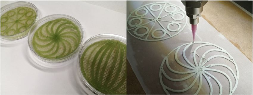
Photosynthetic textile biocomposites 225
2.1 Microalgae and bio–gel matrix 2.2 Textile substrate characterization
preparation
The absorbency of the textiles was measured (n = 3) by
The inoculating liquid algae culture was grown in full weighing dry 1 cm2 samples of each textile that had been
strength Blue–Green medium (BG11) comprising 1.5 g/L immersed in 3 mL of deionised water (dH2O) for 1 h. Each
NaNO3, 0.036 g/L CaCl2·2H2O, 0.075 g/L MgSO4·7H2O, textile sample (1 cm2) was placed on a glass slide with
0.04 g/L K2HPO4, and 0.02 g/L Na2CO3 [28], at 18 ± 2°C transparent tape to prevent the sample from slipping and
with a 16:8 h light to dark photoperiod, at a light intensity imaged using a Leica DMi 8 microscope with LasX soft-
of 2,500 lux (≈30.5 μmol m−2 s−1; [29]) provided by 30 W ware, and thread size was measured. The pH of the tex-
daylight-type fluorescent tubes (Sylvania Luxline Plus, tiles was determined using a standard water extraction
n = 6). The cultures were placed in 50 mL Falcon tubes method [30] by boiling 0.4 g of finely cut pieces (n = 3) in
and centrifuged at 1,620 RCF (relative centrifugal force) 75 mL of dH2O for 10 min. After cooling to room tempera-
for 10 min to produce a dense algae slurry. The algae ture, the samples were drained and the pH of the water
slurry was mixed with the tested gel matrix at a ratio of was tested using a pH meter (Mettler Toledo Seven Com-
0.05 mL algae slurry per 1 mL of gel. pact) relative to a control of dH2O that had undergone the
Each bio–gel matrix was prepared in 20 mL batches same process.
in 50 mL beakers. For the baseline kappa–carrageenan
treatment (Table 2), 0.8 g of kappa–carrageenan (from
>99.9% pure powder; Sigma Aldrich, UK) was added to
20 mL (0.04 g/mL) of a dilution series of BG11 (full strength, 2.3 Closed lid testing: textile and bio–gel
75, 50, and 25% dilution) and stirred at room temperature. compatibility
Once a gel-like consistency was achieved, 0.4 mL of C. vulgaris
slurry was added and stirred until homogeneous. The quan- The purpose of this assay was to develop a viscous matrix
tity of kappa–carrageenan was reduced for later experiments that was compatible with C. vulgaris and to assess the
which tested the performance of lower strength kappa– performance of this bio–gel once applied to the different
carrageenan gels. Aloe vera and Auro 331 Clay Paint binder textiles, forming biocomposites. The experimental treat-
(Auro Paint Company, UK) were tested as additives to the ments are outlined in Table 2. Textile samples (1 cm2)
kappa–carrageenan baseline (Table 2). Ten grams of Aloe were placed in 24-well plates with 0.2 mL of BG11 (100,
vera was added to the mixture. For the Auro paint, 0.64 g of 75, 50, and 25% strength). The following treatments were
kappa–carrageenan was dissolved in 16 mL of BG11 (0.04 g/mL) run: (1) textile-gels containing algae, i.e. the biocompo-
across the previous dilution series, to which 4 mL of Auro site, (2) textile-gels without algae, i.e. non-biological con-
331 was added (equivalent to 20% w/w Auro content). This trols, (3) algae suspension controls, (4) gels with algae,
process was repeated for all Auro paint mixtures, changing and (5) gels only. Each treatment was in triplicate (Figure 1).
the quantities of kappa–carrageenan and Auro paint to pro- The gel coatings were deposited using a spatula, by applying
duce the desired ratios and consistency. Chitosan treatments 0.3 g of gel per sample. The well plates were sealed with a
were prepared by dissolving food grade chitosan powder in transparent lid to minimize any effect of evaporation or bio-
a dilution series of BG11 following the addition of acetic acid logical contamination and incubated at 18 ± 2°C with a
(Table 2). The solution was stirred until an even viscous 16:8 h light:dark cycle with 2,500 lux. Cell viability was
consistency was achieved prior to adding the algae slurry. assessed using an imaging pulse amplitude-modulated
Table 2: Bio–gel mixtures within a closed setup and method of incubation
Bio–gel mixture Method of incubation BG11 nutrient Textiles
dilution (%)
Kappa–carrageenan 0.4 g/10 mL, BG11, C. vulgaris slurry Closed lid, 24-well plate, 100, 75, 50, 25 Cotton, polyester,
0.2 mL/10 mL gel 1 cm2 textile samples hessian, canvas
Kappa–carrageenan 0.4 g/10 mL, 80% w/w BG11, 20% w/w Closed lid, 24-well plate, 100, 75, 50, 25 Cotton, polyester,
Auro Clay Paint, C. vulgaris slurry 0.2 mL/10 mL gel 1 cm2 textile samples hessian, canvas
Kappa–carrageenan 0.4 g/10 mL, 50% w/w BG11, 50% w/w Closed lid, 24-well plate, 100, 75, 50, 25 Cotton, polyester,
Aloe vera gel, C. vulgaris slurry 0.2 mL/10 mL gel 1 cm2 textile samples hessian, canvas
Chitosan 0.6 g/10 mL, acetic acid 0.3 mL/10 mL, BG11, Closed lid, 24-well plate, 100, 75, 50, 25 Cotton, polyester,
C. vulgaris slurry 0.2 mL/10 mL gel 1 cm2 textile samples hessian, canvas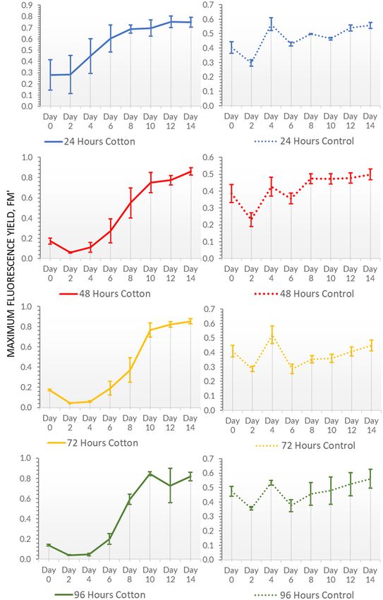
226 Assia Stefanova et al.
Figure 1: (Left) Closed lid incubations in 24-well plates with four BG11 nutrient dilutions, (right) open lid incubations in 6-well plates with full
strength BG11.
fluorometer (Imaging-PAM M-Series; Walz GmbH) which to stresses such as prolonged drying. The same treatments
quantifies the level of chlorophyll fluorescence using intense were run as per the closed testing trial. The gel coatings
light pulses to stimulate photosystem II [31]. Data were col- (Table 3) were deposited using a spatula, by applying
lected every 2 days for 14 days by opening each well plate 0.8 g of gel per sample. Textile samples (2 cm2, n = 3) were
and applying 1 μs pulses of 660 nm (pulsed LED, 0.5 μmol placed in 6-well-plates with 1 mL of BG11. The well plates
quanta m−2 s−1) to incite photosynthesis using 16 red LEDs were left open and incubated under the same conditions
(660 nm) and 16 NIR LEDs (780 nm) [32]. as the closed test (14 days and assessed using imaging-
PAM). The samples were rehydrated with dH2O using a spray
bottle delivering 1 mL per sample every 24 h. Separately,
2.4 Open testing: textile and bio–gel a combination of kappa–carrageenan and Auro Clay Paint
compatibility in an uncontrolled biocomposites was further tested for performance over
environment a range of drying times (24, 48, 72, and 96 h; Table 3).
The drying periods enabled the point whereby drying
This stage investigated the behaviour of the biocompo- may begin to detrimentally affect cell health and thus
sites during an open lid incubation, as well as their response biocomposite performance.
Table 3: Bio–gel mixtures within open setup and method of incubation
Bio–gel mixture Method of incubation
Kappa–carrageenan 0.4 g/10 mL, BG11, C. vulgaris slurry 0.2 mL/ Open lid, 6-well plate, 2 cm2 textile samples
10 mL gel
Kappa–carrageenan 0.4 g/10 mL, 80% w/w BG11, 20% w/w Auro clay Open lid, 6-well plate, 2 cm2 textile samples
paint, C. vulgaris slurry 0.2 mL/10 mL gel
Kappa–carrageenan 0.4 g/10 mL, 50% w/w BG11, 50% w/w Auro clay Dried for 24 h prior to rehydration with 3 mL BG11, open
paint, C. vulgaris slurry 0.2 mL/10 mL gel lid, 6-well plate, 2 cm2 textile samples
Kappa–carrageenan 0.4 g/10 mL, 50% w/w BG11, 50% w/w Auro clay Dried for 48 h prior to rehydration with 3 mL BG11, open
paint, C. vulgaris slurry 0.2 mL/10 mL gel lid, 6-well plate, 2 cm2 textile samples
Kappa–carrageenan 0.4 g/10 mL, 50% w/w BG11, 50% w/w Auro clay Dried for 72 h prior to rehydration with 3 mL BG11, open lid,
paint, C. vulgaris slurry 0.2 mL/10 mL gel 6-well plate, 2 cm2 textile samples
Kappa–carrageenan 0.4 g/10 mL, 50% w/w BG11, 50% w/w Auro clay Dried for 96 h prior to rehydration with 3 mL BG11, open
paint, C. vulgaris slurry 0.2 mL/10 mL gel lid, 6-well plate, 2 cm2 textile samples
Chitosan 0.7 g/10 mL, acetic acid 0.2 mL/10 mL, BG11, C. vulgaris slurry Open lid, 6-well plate, 2 cm2 textile samples
0.2 mL/10 mL gel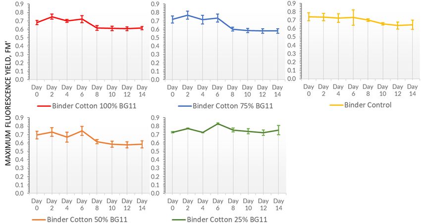
Photosynthetic textile biocomposites 227
Figure 2: (Left) 3D printed algae cells in a kappa–carrageenan mixture on hessian during closed lid incubation, (right) 3D printing with
0.6 mm nozzle on wet cotton using living algae in Auro Clay Paint.
2.5 Gel development for 3D printing and repeated with control samples without living cells. Sam-
extrusion tests ples were incubated in open 90 mm Petri dishes under the
same incubation conditions as the compatibility studies.
During this stage of testing, two mixtures that had suc- Following 3D printing with the Auro binder and kappa–
cessfully formed a paste-like consistency and did not carrageenan mixtures, the samples were dried at 20°C for
present issues of biological contamination were further 24 h prior to rehydrating with 5 mL of dH2O via spraying.
developed to match the requirements of the 3D printing Noticeable shrinkage occurred during the drying process,
equipment to enable controlled extrusion. The 3D printer resulting in flaking and peeling from the textile substrate.
was an air pressure-based extrusion printer (Lutum 4.5; The experiment was repeated without drying the mixture
VormVrij) as shown in Figure 2. The pressure-based post-printing and the textile substrates were immediately
system required a minimum pressure of 10 psi to be main- hydrated with 5 mL of dH2O after printing and incubated
tained, which required a lower viscosity than the pre- with a closed lid in 90 mm diameter Petri dishes. The sam-
viously tested consistency. This presented a challenge ples were sprayed with 3 mL of dH2O every other day to
of decreasing the viscosity of the mixtures, while main- reduce evaporation. In the case of the kappa–carrageenan
taining a favourable growth environment for the algae. mixture, the samples were sprayed with 1 M potassium
This part of the study compared fabrication and subsequent chloride solution immediately after printing to help stabi-
incubation of two different gels including a kappa– lize the gel and to prevent distortion.
carrageenan and full strength BG11 mixture as well as
the Auro 331 Clay Paint mixed with BG11 and kappa–car-
rageenan, both of which remained wet for the duration of 2.6 Statistical analysis
incubation (Table 4). Three bio–gel patterns were printed
onto 30 cm × 10 cm samples of each of the four textiles The Anderson–Darling test was used to test whether the
(cotton, hessian, polyester, and canvas). The process was data were normally distributed [33]. All data were non-
Table 4: 3D printing mixtures tested, extrusion conditions, and matrix behaviour during mechanical extrusion
Bio–gel mixture Extrusion pressure (psi)
Kappa–carrageenan 0.6 g/10 mL, BG11, C. vulgaris slurry 0.2 mL/10 mL gel 10
Kappa–carrageenan 0.8 g/10 mL, BG11, C. vulgaris slurry 0.2 mL/10 mL gel 10
Kappa–carrageenan 1 g/10 mL, BG11, C. vulgaris slurry 0.2 mL/10 mL gel 20
Kappa–carrageenan 0.6 g/10 mL, 50% w/w BG11, 50% w/w Auro clay paint, C. vulgaris slurry 0.2 mL/ 10
10 mL gel
Kappa–carrageenan 0.8 g/10 mL, 50% w/w BG11, 50% w/w Auro clay paint, C. vulgaris slurry 0.2 mL/ 10
10 mL gel
Kappa–carrageenan 1 g/10 mL, 50% w/w BG11, 50% w/w Auro clay paint, C. vulgaris slurry 0.2 mL/10 mL gel 20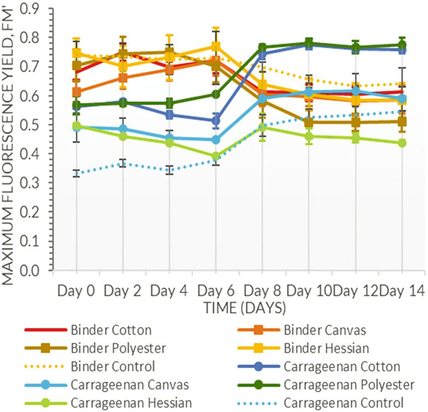
228 Assia Stefanova et al.
normally distributed and therefore two tests were applied 3 Results
for single and two factor datasets. The non-parametric
Scheirer–Ray–Hare test was used for datasets with two
3.1 Material characterization
factors – this is equivalent to two-way Analysis of Var-
iance. The test assesses variance between two factors,
With the exception of canvas, a smaller thread size
which determines whether or not the interaction between
resulted in lower absorbency (Table 5). There was no
the two has a bearing, with P-value of ≤0.05 indicating
relationship between pH and thread size or absorbency.
that the interaction between the two is not significant and
therefore does not affect the outcome [34]. The non-para-
metric Kruskal–Wallis test was applied to datasets with a
single factor, which assesses the statistical significance of 3.2 Closed testing: textile and bio–gel
three or more independent sets by comparing the median compatibility
values [35].
Anderson–Darling tests and Kruskal–Wallis tests were Most of the biocomposites supported either a gradual
conducted using Minitab 18, while Scheirer–Ray–Hare tests increase in chlorophyll fluorescence with time or enabled
were performed using RealStatistics add-in for Microsoft Excel. C. vulgaris to maintain the initial level of chlorophyll
Table 5: List of textiles and material properties
Textile type Microscopic images of textile thread (×5 Thread diameter (µm) pH Absorption (mL/1 cm2)
magnification)
213 (mean StDev = 7.66 (mean 0.0429 (mean StDev =
Cotton
1.960) StDev = 0.213) 0.0021)
322 (mean StDev = 8.36 (mean 0.1269 (mean StDev =
Polyester
22.360) StDev = 0.247) 0.0080)
Canvas 367 (mean StDev = 7.40 (mean 0.0930 (mean StDev =
(300 gsm) 43.848) StDev = 0.139) 0.0076)
1,115 (mean StDev = 7.32 (mean 0.1680 (mean StDev =
Hessian
157.849) StDev = 0.062) 0.0138)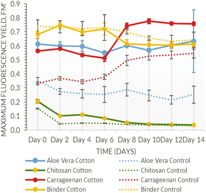
Photosynthetic textile biocomposites 229
Figure 3: Closed incubation cotton samples with the following Figure 4: Closed incubation of 20% w/w binder on textiles: cotton
matrices: Aloe vera (mean StDev = 0.049) and control (mean (mean StDev = 0.032), canvas (mean StDev = 0.030), polyester
StDev = 0.038), chitosan (mean StDev = 0.004) and control (mean StDev = 0.047), hessian (mean StDev = 0.047), control (mean
(mean StDev = 0.002), binder (mean StDev = 0.027) and control StDev = 0.057), and carrageenan samples: cotton (mean StDev =
(mean StDev = 0.045), carrageenan (mean StDev = 0.014) and 0.014), canvas (mean StDev = 0.029), polyester (mean StDev =
control (mean StDev = 0.022) in full strength BG11 media. 0.026), hessian (mean StDev = 0.028), control (mean StDev =
0.025) in full strength BG11 media.
fluorescence (Figures 3 and 4). The exception was the
nutrient dilution (Figure 5) was not (Scheirer–Ray–Hare,
chitosan containing biocomposite, which experienced a
n = 160, d.f. = 3, H = 3.94, P = 0.267); there was a sig-
rapid decline in fluorescence by day 2; this was matched
nificant interaction between textile type and nutrient
by the chitosan control indicating that the inclusion of
dilution (Scheirer–Ray–Hare, n = 160, d.f. = 12, H =
acetic acid created an acidic pH that was incompatible
25.65, P ≤ 0.05). A similar outcome was attained with
with the algae (Figure 3). Textile type was a significant
the kappa–carrageenan biocomposites, with textiles type
factor for those biocomposites that did support the algae
significantly affecting chlorophyll fluorescence (Scheirer–
for the duration of the trial (Scheirer–Ray–Hare, n = 160,
Ray–Hare, n = 160, d.f. = 4, H = 90.25, P ≤ 0.001), but not
d.f. = 4, H = 20.82, P ≤ 0.001), whereas nutrient dilution
nutrient levels (Scheirer–Ray–Hare, n = 160, d.f. = 3, H =
was not significant (Scheirer–Ray–Hare, n = 160, d.f. = 3,
0.57, P = 0.901); however, in this case any interaction was
H = 5.91, P = 0.116) and there was no significant interac-
not significant (Scheirer–Ray–Hare, n = 160, d.f. = 12, H =
tion between the two (Scheirer–Ray–Hare, n = 160, d.f. = 4,
3.17, P = 0.994).
H = 0.66, P = 1.000). Although biocomposites with the
Aloe vera additive did support the algae over the duration
of the experiment, these biocomposites experienced severe
bacterial contamination that inhibited algae growth in the 3.3 Open samples testing: uncontrolled
affected areas. Despite this, textile type was a significant environment
factor (Scheirer–Ray–Hare, n = 160, d.f. = 4, H = 131.50,
P ≤ 0.001), unlike nutrient dilution (Scheirer–Ray–Hare, Textile type was a significant factor for the 20% w/w Auro
n = 160, d.f. = 3, H = 0.369, P = 0.947) and there was no paint bio–gels incubated in the open setup (Kruskal–Wallis,
interaction between the factors (Scheirer–Ray–Hare, n = n = 40, d.f. = 4, H = 14.51, P = 0.006); it was also significant
160, d.f. = 12, H = 3.082, P = 0.995). for the 50% w/w Auro bio–gels that had been dried over
The most promising biocomposites were kappa– 24 h (Kruskal–Wallis, n = 40, d.f. = 3, H = 12.00, P = 0.007)
carrageenan and full strength BG11 with the Auro Clay (Figure 6). Biocomposites with a shorter drying time exhib-
Paint additive. These biocomposites exhibited increased ited more consistent fluorescence levels that increased
fluorescence in combination with cotton and polyester throughout the 14 days period, whereas samples rehy-
(Figure 5), with textile type a significant factor (Scheirer– drated after a longer drying time exhibited a sharp decline
Ray–Hare, n = 160, d.f. = 4, H = 16.77, P ≤ 0.005), whereas of chlorophyll fluorescence in the initial days following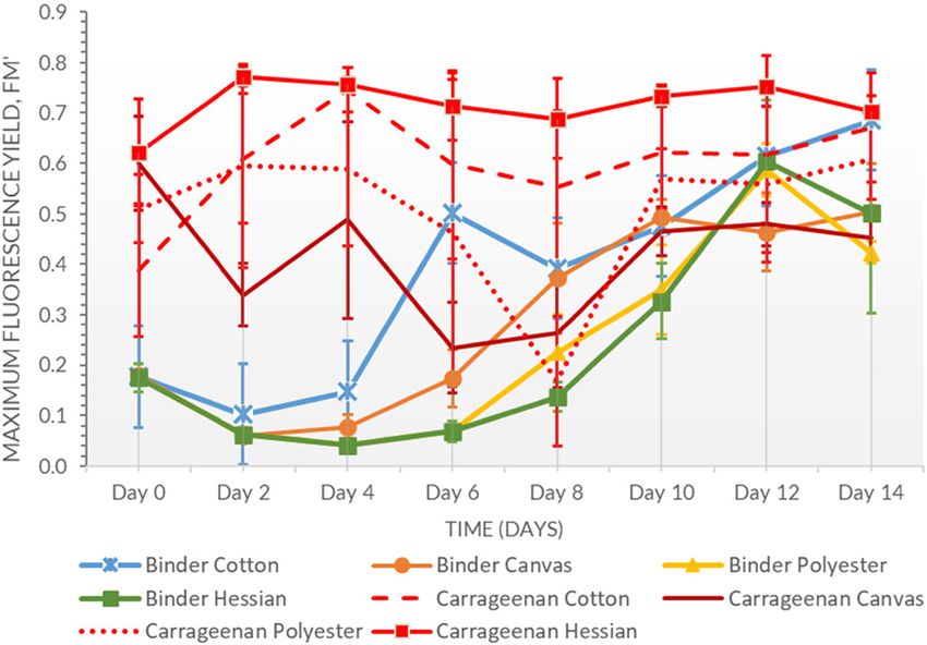
230 Assia Stefanova et al.
Figure 5: Closed incubation of 20% w/w binder on cotton with various nutrient media dilutions: 100% BG11 (mean StDev = 0.027), 75% BG11
(mean StDev = 0.036), 50% BG11 (mean StDev = 0.044), 25% BG11 (mean StDev = 0.020).
rehydration and a rapid recovery thereafter (Figure 7). similar viscosity, the quantity of chitosan was increased.
Despite this, drying time was not statistically significant This resulted in some bacterial contamination. Textile type
(Kruskal–Wallis, n = 32, d.f. = 3, H = 0.70, P = 0.873). In the was not significant (Kruskal–Wallis, n = 40, d.f. = 4, H =
chitosan treatment, the acetic acid content was reduced to 3.17, P = 0.530). The kappa–carrageenan and BG11 biocom-
make the environment less acidic; however, to achieve a posites were not detrimentally affected by open air incu-
bation and substantial shrinkage or severe drying were not
observed. In this case, textile type was statistically signifi-
cant (Kruskal–Wallis, n = 40, d.f. = 4, H = 14.51, P ≤ 0.05).
3.4 3D Printing with bio–gels
When extruding the kappa–carrageenan and paint mix-
tures, the decreased viscosity affected the ability to
extrude a consistent layer even when using higher pres-
sure. Air pockets and a separation of water content due
to the pressure caused more noticeable distortion in
the lower viscosity mixtures; therefore, the kappa–carra-
geenan content had to be increased while maintaining a
consistency that allowed for smooth extrusion. A higher
kappa–carrageenan content also resulted in crumbling
Figure 6: Open incubation of carrageenan cotton (mean StDev =
and issues of adhesion to the substrate post-printing.
0.038) and control (mean StDev = 0.014), 20% binder cotton (mean
StDev = 0.039) and control (mean StDev = 0.013), 50% binder
The mixture variations and pressures used in each instance
cotton (mean StDev = 0.098) and control (mean StDev = 0.022), are presented in Table 6.
chitosan cotton (mean StDev = 0.012) and control (mean During the printing process, the texture of the textiles
StDev = 0.006). was of particular interest with smoother textiles such as
Photosynthetic textile biocomposites 231 Figure 7: Open incubation of 50% w/w binder cotton samples following drying times of 24 h (mean StDev = 0.098), 48 h (mean StDev = 0.067), 72 h (mean StDev = 0.042), and 96 h (mean StDev = 0.045). cotton and canvas presenting more favourable surfaces The 3D printed kappa–carrageenan samples con- for printing, whereas the larger thread size of hessian tinued to support cell metabolism during the 14 days produced better adhesion but caused issues during printing testing period, with an increase in chlorophyll fluores- with the nozzle being prone to catching on the threads. cence recorded for cotton. Textile type was significant
232 Assia Stefanova et al.
Table 6: 3D printing mixtures tested, extrusion conditions, and matrix behaviour during mechanical extrusion
Bio–gel mixture Extrusion Nozzle size Comments
pressure (psi) (Diameter in mm)
Kappa–carrageenan 0.6 g/10 mL, BG11, 10 0.6 Continuous flow from nozzle prior to
C. vulgaris slurry 0.2 mL/10 mL gel printing, occurring due to pressurization of
canister
Kappa–carrageenan 0.8 g/10 mL, BG11, 10 0.6 Steady flow upon extrusion, gel
C. vulgaris slurry 0.2 mL/10 mL gel maintaining its structure without crumbling
or uncontrolled distortion
Kappa–carrageenan 1 g/10 mL, BG11, C. vulgaris 20 0.8 Inconsistent follow upon extrusion,
slurry 0.2/10 mL gel crumbling of extruded filament, and poor
adhesion to textile surface
Kappa–carrageenan 0.6 g/10 mL, 50% w/w BG11, 10 0.6 Continuous flow from nozzle prior to
50% w/w Auro clay paint, C. vulgaris slurry printing, occurring due to pressurization of
0.2 mL/10 mL gel canister
Kappa–carrageenan 0.8 g/10 mL, 50% w/w BG11, 10 0.6 Mousse–like consistency, steady flow,
50% w/w Auro clay paint, C. vulgaris slurry good adhesion to textiles
0.2 mL/10 mL gel
Kappa–carrageenan 1 g/10 mL, 50% w/w BG11, 20 0.8 Inconsistent flow, crumbling of filament,
50% w/w Auro clay paint, C. vulgaris slurry and poor adhesion
0.2 mL/10 mL gel
(Kruskal–Wallis, n = 32, d.f. = 3, H = 21.17, P ≤ 0.001) for 4 Discussion
the closed lid kappa–carrageenan samples. Canvas and
polyester samples exhibited greater fluctuations, although This study aimed to develop and test a range of flexible,
all combinations still imply supported microalgae survival textile-based microalgae biocomposites that could be
(Figure 8). integrated into the internal fabric of the built environ-
There was a slight decline in chlorophyll fluores- ment. A fundamental driver for this work was to provide
cence of the Auro paint set in the initial days; however, an environment that supported microbial life (in this
this was followed by a gradual increase in chlorophyll instance, the microalga Chlorella vulgaris), which would
fluorescence over the remaining 12 days (Figure 9). Tex- subsequently become part of the metabolism of the building,
tile type was significant (Kruskal–Wallis, n = 32, d.f. = 3, i.e. by sequestering CO2 whilst releasing O2, and potentially
H = 21.17, P ≤ 0.001). Visible changes were observed in remediating wastewater. Aside from the functional aspect,
the mixture colour, with more saturated green areas the studies also sought to address the aesthetics of deploying
emerging. There was less cell migration (movement biocomposites using extrusion 3D printing. Chlorella was
of living algae cells) from the mixture onto the empty chosen for its ability to flourish in temperatures similar to
substrate regions compared to the carrageenan samples those offered by interior building environments (19–24°C),
(Figure 8), with the exception of the canvas samples for its resilience to desiccation, and its efficient photosyn-
that exhibited a greater level of cell migration and thetic rate (some Chlorella species can reach efficiencies of
the resolution of the printed pattern under I-PAM was more than 20% compared to the typical 1% efficiency of
severely affected, although visible pattern distortion did terrestrial plants [36]).
not occur. This study utilized imaging PAM fluorometry as a
The kappa–carrageenan and BG11 combination had a means to non-destructively measure in situ chlorophyll
more scattered distribution of cells when viewed in I-PAM; florescence. While being particularly helpful in visualizing
however, unlike the Auro samples, they did not initially go cell migration and pattern distortion as well as pattern
through a consistent period of decline, although sudden of growth, it does not measure the amount of carbon cap-
fluctuations were evident in the cotton and canvas sam- tured; this can only be inferred based on the cell’s chloro-
ples. When comparing the two sets of samples, bio–gel phyll fluorescence levels that indicate a greater amount
type was significant (Scheirer–Ray–Hare, n = 64, d.f. = 1, of photosynthetic cells and therefore more agents for CO2
H = 22.60, P ≤ 0.001), whereas textile type was not sequestration. Further research is necessary, for instance,
(Scheirer–Ray–Hare, n = 64, d.f. = 3, H = 7.26, P = 0.064). testing the biocomposites as part of an airtight CO2Photosynthetic textile biocomposites 233
Figure 8: 3D printed patterns on cotton using kappa–carrageenan (top) and Auro Clay Paint (bottom), image showing cell chlorophyll
fluorescence in I-PAM, red and yellow indicate low levels of fluorescence of living photosynthetic cells, green and blue indicate higher
levels, and black indicates a lack of living photosynthetic cells. The dense green areas on day 14 indicate cell migration outside of the
printed area.
absorption system to quantify the captured carbon. One of matrix; therefore it is only capturing chlorophyll fluores-
the drawbacks of PAM fluorometry is that it is not able to cence on the surface of an opaque matrix or parts of a
measure the chlorophyll fluorescence inside an opaque translucent matrix. A chlorophyll extraction technique
can potentially be used at the end of the test to compare
with the data obtained from the imaging PAM.
The first set of tests helped establish differences in
biocomposite performance (as measured by in situ chloro-
phyll fluorescence) based on the type of textile substrate.
The initial findings indicated that textile type had a sig-
nificant bearing on the development of the living cells.
The nutrient levels built into the bio–gels indicated that
full strength BG11 produced the most favourable results;
however, all other dilutions ably supported the algae
development throughout the test period.
During the two weeks of closed incubation tests,
C. vulgaris embedded in the Auro binder with cotton had
the best overall performance followed by the carrageenan-
based matrices; [16] also used Auro binders to produce
Figure 9: Chlorophyll fluorescence levels of the kappa–carrageenan cyanobacteria loofah-based biocomposites and reported
on the following textiles: cotton (mean StDev = 0.134), canvas increased CO2 absorption rates compared to suspension
(mean StDev = 0.086), polyester (mean StDev = 0.144), hessian
controls. For carrageenan-based matrices, enhanced bio-
(mean StDev = 0.043), and Auro paint 3D printed samples on tex-
tiles: cotton (mean StDev = 0.056), canvas (mean StDev = 0.053),
logical performances have been widely reported in many
polyester (mean StDev = 0.037), hessian (mean StDev = 0.060) over studies particularly in wastewater treatment [37]. The less
a 14 day period. favourable gel matrices such as chitosan-based mixtures234 Assia Stefanova et al.
need further study to overcome the low pH of acetic acid In the final set of experiments, the consistency of the
and promote biocompatibility [38]. Tığlı et al. provide an bio–gels was adapted to suit the limitations of the extru-
alternative where the chitosan-based scaffolds were formed sion 3D printer, with high viscosity mixtures resulting
using a glycerol phosphate disodium salt as an ionic cross- in uncontrolled expulsion of material prior to initiating
linker while adding NaOH to increase the pH up to 7.0 after the printing cycle and lower viscosity mixtures causing
dissolving the chitosan in acetic acid; such an environment blockages and crumbling of the matrix with poor adhe-
may provide a more favourable pH for algae growth [39]. sion to the textile substrate. Although detailed structural
The second set of tests demonstrated the resilience of analysis of the printed biocomposites was not under-
the biomaterials, suggesting that they can be deployed in taken in the study, prior analysis using latex binders
interior settings where it would be exposed to the air. [16] suggests that dry cracked binders can be a source
However, this presents a risk of contamination or cell of adhesion and cell retention failure. An aspect of the
damage if other agents are present such as aerosols, 3D printed process that needs to be assessed is the effect
dust particles, or large numbers of other microorganisms. of the nozzle size on cell viability as a larger nozzle, as
During the rehydration process, a level of flaking was observed by Unagolla and Jayasuriya, requires less pres-
observed due to shrinkage caused by the kappa–carra- sure and is likely to result in less cell damage during
geenan content. The kappa–carrageenan was added to fabrication and therefore better cell viability following
aid the creation of a paste-like consistency which would extrusion [38]. The 3D printing process has created chal-
be suitable for 3D extrusion; however, an alternative lenges, and issues of speed, cost, and gel pattern preser-
application that accommodates distribution of a less vis- vation have been identified as limiting factors for large-
cous solution, such as painting with a brush or a roller, scale mass production and implementation [43]. Further
would negate the need for kappa–carrageenan in the testing is necessary to assess if the matrix would permit
mixture and would reduce flaking. 3D extrusion of a geometry at a height as studied by Wang
This paper builds upon early research into bio-ink et al. [44] and its effect on the quality of adhesion to the
rehydration and mimicry of natural biofilm growth, by substrate. The integration of the textile substrates into
engineering thin layers of nontoxic materials that are design needs to be studied as the samples of this study
capable of supporting a high cell density [40,41]. Non- were incubated flat, with vertical incubation taken into
toxic binders, for example in latex [16,42], provide a more account of gravity or twisting and tensioning of the mate-
appropriate matrix for cell cultivation, as hydrogels (although rial presenting areas of interest for future investigation.
easily formed) possess a larger pore structure that is prone
to the physical release of cells. Hydrogels also present a
less stable environment for long-term storage of dormant 5 Conclusion
cells as outlined by Flickinger et al. [42].
The incubation studies also provided insight into This paper has demonstrated that C. vulgaris can be cul-
the potential distribution and storage of a living material tivated in a minimal moisture environment in a range of
in a dormant state. The dry Auro samples that were rehy- matrices and on a variety of textiles that lend themselves
drated after a range of timeframes demonstrate the to an array of potential applications within the building
potential for prefabricating such elements off-site and fabric. The aim of the study was to develop a new type of
transporting at a distance, aiding the distribution pro- living material that can be fabricated using digital 3D
cess. However, cultivation in an open interior environ- printing methods and to study the subsequent develop-
ment is also prone to sudden changes in moisture levels ment of such materials in terms of cell development
where the substrate could dry out, causing cell stress and and migration demonstrating live behaviour that is not
matrix flaking. This suggests the need for an automated traditionally associated with materials used within the
irrigation system that could detect such changes and construction industry. The results indicate that living tex-
maintain a minimal moisture level at all times, which tile materials would need some form of maintenance
may raise issues of needing an energy source that may schedule to prevent sudden moisture evaporation which
negate the benefits of biological CO2 sequestration. An could result in cell death and flaking of the matrix.
alternative may be to utilize a protective cover and to Further investigation is necessary to design an artificial
develop a human maintenance protocol that may place support environment and to develop ways of overcoming
a burden upon inhabitants. Both options need to be challenges of matrix shrinkage and flaking as well as to
explored further, with the current study indicating that develop viable design solutions for integration within the
evaporation is a limiting factor. built environment.Photosynthetic textile biocomposites 235
Acknowledgments: This research was funded by Research [15] Mohammadifar M, Zhang J, Yazgan I, Sadik O, Choi S. Power-
England as part of the Hub for Biotechnology in the Built on-paper: origami-inspired fabrication of 3-D microbial fuel
Environment. cells. Renew Energy. 2018 Apr;118:695–700.
[16] In-na P, Umar AA, Wallace AD, Flickinger MC, Caldwell GS,
Lee JGM. Loofah-based microalgae and cyanobacteria bio-
Conflict of interest: Authors state no conflict of interest. composites for intensifying carbon dioxide capture. J CO2 Util.
2020 Dec;42:101348.
[17] Stefanova A, Bridgens B, Armstrong R, In-Na P, Caldwell GS.
Engineering a living building realm: development of protective
References coatings for photosynthetic ceramic biocomposite materials.
The 7th International Conference on Architecture and Built
[1] UIA. UIA Durban declaration 2050 imperative – news & media; Environment with Architecture AWARDs. Tokyo: Get It
2014. [Online]. Available: https://wp.architecture.com.au/ Published Verlag; 2020. p. 362–72.
news-media/uia-durban-declaration-2050-imperative/. [18] Stefanova A, Bridgens B, In-na P, Caldwell G, Armstrong R.
Accessed 24 December 2019. Architectural laboratory practice for the development of clay
[2] Reynolds C. Ecology of phytoplankton. 1st edn. Cambridge: and ceramic-based photosynthetic biocomposites. Technol +
Cambridge University Press; 2006. Des. 2020 Jul;4(2):200–10.
[3] Mark Co JR, Culaba AB. 3D printing: challenges and opportu- [19] Whitton B. Ecology of cyanobacteria II: their diversity in space
nities of an emerging disruptive technology. In 2019 IEEE 11th and time. Dordrecht: Springer Science & Business Media; 2012.
International Conference on Humanoid, Nanotechnology, [20] Searchinger T, Heimlich R, Houghton RA, Dong F, Elobeid A,
Information Technology, Communication and Control, Fabiosa J, et al. Use of US croplands for biofuels increases
Environment, and Management (HNICEM). Laoag: IEEE; 2019. greenhouse gases through emissions from land-use change.
p. 1–6. Science (80–). 2008 Feb;319(5867):1238–40.
[4] Poomathi N, Singh S, Prakash C, Subramanian A, Sahay R, [21] Costerton JW, Stewart PS, Greenberg EP. Bacterial biofilms:
Cinappan A, et al. 3D printing in tissue engineering: a state of a common cause of persistent infections. Science (80–).
the art review of technologies and biomaterials. Rapid May 1999;284(5418):1318–22.
Prototyp J. Jun. 2020;26(7):1313–34. [22] Flemming H. Sorption sites in biofilms. Water Sci Technol.
[5] Dick A, Bhandari B, Prakash S. 3D printing of meat. Meat Sci. 1995;32(8):27–33.
2019 Jul;153:35–44. [23] Mantzorou A, Ververidis F. Microalgal biofilms: a further step
[6] Fairs M. Mycelium chair by Eric Klarenbeek is 3D-printed with over current microalgal cultivation techniques. Sci Total
living fungus. Dezeen. 2013 Oct 20 [Online]. Available: Environ. Feb. 2019;651:3187–201.
https://www.dezeen.com/2013/10/20/mycelium-chair-by- [24] McHugh DJ. A guide to the seaweed industry. Rome: Food and
eric-klarenbeek-is-3d-printed-with-living-fungus/. Accessed Agriculture Organization of the United Nations; 2003.
23 October 2020. [25] Auro. Clay paint, white | auro. Auro [Online]. Available:
[7] Chiujdea RS, Nicholas P. Design and 3D printing methodolo- https://auropaint.co.uk/product/high-grade-clay-paint-no-
gies for cellulose-based composite materials. 331/. Accessed 29 September 2020.
Anthropologic – architecture and fabrication in the cognitive [26] “Holland & Barrett Aloe Vera Gel,” Holland & Barrett; 2020.
age. Berlin: eCAADe; 2020. p. 547–58. [Online]. Available: https://www.hollandandbarrett.com/
[8] Sawa M. Algaerium. In: Myers W, editor. Bio design: nature: shop/product/holland-barrett-aloe-vera-gel-60033725.
science creativity. London: Thames Hudson; 2012. p. 88–91. Accessed 29 September 2020.
[9] Morris A. Dutch designers convert algae into bioplastic for 3D [27] Shahidi F, Synowiecki J. Isolation and characterization of
printing. Dezeen. 2017 Dec 4. [Online]. Available: https:// nutrients and value-added products from snow crab
www.dezeen.com/2017/12/04/dutch-designers-eric- (Chinoecetes opilio) and Shrimp (Pandalus borealis) proces-
klarenbeek-maartje-dros-convert-algae-biopolymer-3d- sing discards. J Agric Food Chem. Aug. 1991;39(8):1527–32.
printing-good-design-bad-world/. Accessed 23 October 2020. [28] Stanier RY, Kunisawa R, Mandel M, Cohen-Bazire G.
[10] Yu K, Feng Z, Du H, Xin A, Lee KH, Li K, et al. Photosynthesis- Purification and properties of unicellular blue-green algae
assisted remodeling of three-dimensional printed structures. (order Chroococcales). Bacteriol Rev. Jun. 1971;35(2):171–205.
Proc Natl Acad Sci. 2021 Jan;118(3):e2016524118. [29] Thimijan RW, Heins RD. Photometric, radiometric, and
[11] Ali S, Majeed S. Advancement of bio inks in three dimensional quantum light units of measure: a review of procedures for
bioprinting. Biomed J Sci Tech Res. 2018 Dec;11(4):1–4. interconversion. HortScience. 1983;18(6):818–22.
[12] Malik S, Hagopian J, Mohite S, Lintong C, Stoffels L, [30] AATCC C. Test method for pH of the water-extract from wet
Giannakopoulos S, et al. Robotic extrusion of Algae–Laden processed textiles. AATCC Technical Manual. American
hydrogels for large‐scale applications. Glob Chall. 2020 Association of Textile Chemists and Colorists; 2007.
Jan;4(1):1900064. [31] Schreiber U. Pulse-amplitude-modulation (PAM) fluorometry
[13] Shaklova I, Cruz M. Living screen: robotic fabrication of algae based and saturation pulse method: an overview. Chlorophyll a
gels. MaterialAbility. 2015 [Online]. Available: http://materiability. Fluorescence. Dordrecht: Springer Netherlands; 2004.
com/portfolio/living-screen/. Accessed 27 October 2020. p. 279–319.
[14] Pang S, Gao Y, Choi S. Flexible and stretchable biobatteries: [32] “Specifications,” Heinz Walz GmbH; 2021. [Online]. Available:
monolithic integration of membrane-free microbial fuel cells in https://www.walz.com/products/chl_p700/imaging-pam_
a single textile layer. Adv Energy Mater. 2018;8(7):1702261. ms/spec_3d_version.html. Accessed 20 January 2021.236 Assia Stefanova et al.
[33] McNeese B. Anderson–Darling test for normality. BPI con- [39] Tığlı RS, Karakeçili A, Gümüşderelioğlu M. In vitro characterization
sulting; Jun-2011 [Online]. Available: https://www. of chitosan scaffolds: influence of composition and deacetylation
spcforexcel.com/knowledge/basic-statistics/anderson- degree. J Mater Sci Mater Med. 2007;18(9):1665–74.
darling-test-for-normality. Accessed 18 January 2021. [40] Flickinger MC, Bernal OI, Schulte MJ, Broglie JJ, Duran CJ,
[34] Mangiafico SS. R handbook: Scheirer–Ray–Hare test. Wallace A, et al. Biocoatings: challenges to expanding the
Summary and Analysis of Extension Program Evaluation in functionality of waterborne latex coatings by incorporating
RCompanion; 2016 [Online]. Available: https://rcompanion. concentrated living microorganisms. J Coat Technol Res. 2017
org/handbook/F_14.html. Accessed 19 January 2021. July;14(4):791–808.
[35] Zach. Kruskal–Wallis test: definition, formula, and [41] Lawton CW, Klei HE, Sunstrom DV, Voronka PJ, Scott CD.
example. Statology; 2019 Jan 18 [Online]. Available: Immobilization of whole cells using polymeric coatings.
https://www.statology.org/kruskal-wallis-test/. Accessed 19 Biotechnol Bioeng Symp. 1986;17:507–17.
January 2021. [42] Flickinger MC, Schottel JL, Bond DR, Aksan A, Scriven LE.
[36] Adamczyk M, Lasek J, Skawińska A. CO2 biofixation and Painting and printing living bacteria: engineering nanoporous
growth kinetics of Chlorella vulgaris and Nannochloropsis biocatalytic coatings to preserve microbial viability and
gaditana. Appl Biochem Biotechnol. 2016 intensify reactivity. Biotechnol Prog. 2007 Feb;23(1):2–17.
Aug;179(7):1248–61. [43] Ngo TD, Kashani A, Imbalzano G, Nguyen KTQ, Hui D. Additive
[37] Kesaano M, Sims RC. Algal biofilm based technology for manufacturing (3D printing): a review of materials, methods, appli-
wastewater treatment. Algal Res. Jul. 2014;5:231–40. cations and challenges. Compos Part B Eng. 2018 Jun;143:172–96.
[38] Unagolla JM, Jayasuriya AC. Hydrogel-based 3D bioprinting: [44] Wang W, Yao L, Cheng C-Y, Zhang T, Atsumi H, Wang L, et al.
a comprehensive review on cell-laden hydrogels, bioink for- Harnessing the hygroscopic and biofluorescent behaviors of
mulations, and future perspectives. Appl Mater Today. 2020 genetically tractable microbial cells to design biohybrid
Mar 1;18:100479 (Elsevier Ltd.). wearables. Sci Adv. 2017;3(5):e1601984.You can also read