Hemophagocytic lymphohistiocytosis: a review inspired by the COVID 19 pandemic
←
→
Page content transcription
If your browser does not render page correctly, please read the page content below
Rheumatology International (2021) 41:7–18
https://doi.org/10.1007/s00296-020-04636-y
 Rheumatology
 INTERNATIONAL

 REVIEW

Hemophagocytic lymphohistiocytosis: a review inspired
by the COVID‑19 pandemic
Mehmet Soy1 · Pamir Atagündüz2 · Işık Atagündüz3 · Gülsan Türköz Sucak4

Received: 26 April 2020 / Accepted: 19 June 2020 / Published online: 25 June 2020
© Springer-Verlag GmbH Germany, part of Springer Nature 2020

Abstract
Hemophagocytic syndrome (HPS) or hemophagocytic lymphohistiocytosis (HLH) is an acute and rapidly progressive sys-
temic inflammatory disorder characterized by cytopenia, excessive cytokine production, and hyperferritinemia. Common
clinical manifestations of HLH are acute unremitting fever, lymphadenopathy, hepatosplenomegaly, and multiorgan failure.
Due to a massive cytokine release, this clinical condition is considered as a cytokine storm syndrome. HPS has primary and
acquired (secondary, reactive) forms. Its primary form is mostly seen in childhood and caused by various mutations with
genetic inheritance and, therefore, is called familial HLH. Secondary HLH may be caused in the presence of an underlying
disorder, that is, secondary to a malignant, infectious, or autoimmune/autoinflammatory stimulus. This paper aims to review
the pathogenesis and the clinical picture of HLH, and its severe complication, the cytokine storm, with a special emphasis on
the developed classification criteria sets for rheumatologists, since COVID-19 infection has clinical symptoms resembling
those of the common rheumatologic conditions and possibly triggers HLH. MED-LINE/Pubmed was searched from incep-
tion to April 2020, and the following terms were used for data searching: “hemophagocytic syndrome” OR “macrophage
activation syndrome” OR “hemophagocytic lymphohistiocytosis”, OR “cytokine storm”. Finally, AND “COVID-19” was
included in this algorithm. The selection is restricted to the past 5 years and limited numbers of earlier key references were
manually selected. Only full-text manuscripts, published in an English language peer-reviewed journal were included.
Manuscript selection procedure and numbers are given in Fig. 2. Briefly, the database search with the following terms of
“Hemophagocytic syndrome” OR “Macrophage activation syndrome” OR “Hemophagocytic lymphohistiocytosis” OR
“Cytokine storm” yielded 6744 results from inception to April 2020. The selection is restricted to the past 5 years and only
limited numbers of earlier key references were selected, and this algorithm resulted in 3080 manuscripts. The addition of
(AND “COVID-19”) resulted in 115 publications of which 47 studies, together with four sections of an online book were
used in the final review. No statistical method was used. HLH is triggered by genetic conditions, infections, malignancies,
autoimmune-autoinflammatory diseases, and some drugs. In COVID-19 patients, secondary HLH and cytokine storm may
be responsible for unexplained progressive fever, cytopenia, ARDS, neurological and renal impairment. Differentiation
between the primary and secondary forms of HLH is utterly important, since primary form of HLH requires complicated
treatments such as hematopoietic stem cell transplantation. Further studies addressing the performance of HScore and other
recommendations in the classification of these patients is necessary.

Keywords Hemophagocytic syndrome · Hemophagocytic lymphohistiocytosis · Macrophage activation syndrome ·
Cytokine storm syndrome

 Introduction

 Hemophagocytic lymphohistiocytosis (HLH) comprises
 two different conditions that may be difficult to distinguish
 from one another: A primary form that occurs due to genetic
 disorders and a secondary form that is triggered by vari-
* Pamir Atagündüz
 pamir.atagunduz@gmail.com ous infections, autoimmune/autoinflammatory diseases, or
 chemicals [1, 2]. Recent reports suggest that the cytokine
Extended author information available on the last page of the article

 13
 Vol.:(0123456789)
8 Rheumatology International (2021) 41:7–18

storm caused by the novel Coronavirus infection, COVID- pronounced histiocytic erythro-phagocytosis in the bone
19, has significant similarities with the clinical and labora- marrow and cellular proliferation in the lymph nodes,
tory findings of this disorder. Due to the accelerated deterio- medulla of the spleen and the periportal area. Later, Scott
ration of patients’ general status, hemophagocytic syndrome et al. named this clinical entity as histiocytic medullary
(HPS) necessitates a timely diagnosis for the initiation of reticulocytosis (HMR) because of the pronounced phago-
life-saving treatment. cytosis with intense hyperplasia in medullary areas. In
 HPS or HLH is an acute and rapidly progressive sys- the light of the data obtained, it was accepted as a well-
temic inflammatory disorder characterized by cytopenia, differentiated reticulum cell sarcoma, a malignant process
excessive cytokine production, and hyperferritinemia. until 1973. In 1991, it was designated as Hemophagocytic
Common clinical manifestations of HLH are acute unre- Lymphohistiocytosis The term of Macrophage Activation
mitting fever, lymphadenopathy, hepatosplenomegaly, and Syndrome (MAS) was used first in 1993. In 1997, the HLH
multiorgan failure. Due to a massive cytokine release, this working group classified the HPS into primary and sec-
clinical condition is considered as a cytokine storm syn- ondary HLH [4–6].
drome. HLH has primary and acquired (secondary, reac- In this review, we aim to contribute to the rheumatolo-
tive) forms. Its primary form is mostly seen in childhood gists’ awareness of the life-threatening rare complication
and caused by various mutations with genetic inheritance of HLH, the cytokine storm, to prevent a possible misdi-
and, therefore, is called familial HLH (fHLH) (Fig. 1). agnosis in the presence of the clinical and laboratory fea-
Secondary HLH (sHLH) may be caused in the presence of tures of COVID-19 resembling or mimicking to that of an
an underlying disorder, that is, secondary to a malignant, underlying or a new-onset rheumatological condition. We
infectious, or autoimmune/autoinflammatory stimulus hope that this review will support the collaboration of the
[1–3]. This clinical condition was first reported by Scott rheumatologist and hematologists with their colleagues,
et al. in 1939. The first reported four cases were identi- who treat COVID-19 patients in the first place, such as the
fied as atypical Hodgkin’s Lymphoma, characterized by infectious disease specialists, pneumologists, or intensive
 care physicians.

Fig. 1  Classification

13
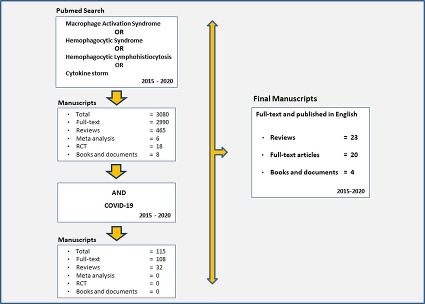
Rheumatology International (2021) 41:7–18 9 Methods reviewed the article for both immunologic and rheuma- tologic aspects, prepared tables, translated into English, Study selection and gave the manuscript’s final form. No statistical method was used. Manuscript selection procedure and numbers are given in Fig. 2. Briefly, MED-LINE/Pubmed was searched from inception to April 2020, and the following terms were used for data searching: “Hemophagocytic syndrome” OR “Mac- Results rophage activation syndrome” OR “Hemophagocytic lym- phohistiocytosis”, OR “Cytokine storm”. To increase the The database search with the following terms of weight of the recent publications, the selection is restricted “Hemophagocytic syndrome” OR “Macrophage activation to the past 5 years and only limited numbers of earlier key syndrome” OR “Hemophagocytic lymphohistiocytosis” references were manually selected. Finally, (AND “COVID- OR “Cytokine storm” yielded 6744 results from inception 19”) was applied in this algorithm. Only full-text manu- to April 2020. When the selection is restricted to the past scripts, published in an English language peer-reviewed 5 years and only limited numbers of earlier key references journal were included. Free full-text articles available at were selected, this algorithm resulted in 3080 manuscripts https://pubmed.ncbi.nlm.nih and selected full-text articles and the addition of AND “COVID-19” resulted in 115 indexed in Web of Science or Scopus are downloaded using publications of which 47 studies, together with four sec- the EBSCO Discovery services. tions of an online book were used in the final review. Only The author MS collected data and wrote the manuscript. full-text manuscripts, published in an English language The authors GTS and IA carefully reviewed the data from peer-reviewed journal were included. We used data only the perspective of a hematologist. And the author PA from the index papers for table creation and figures. Fig. 2 Article selection process 13
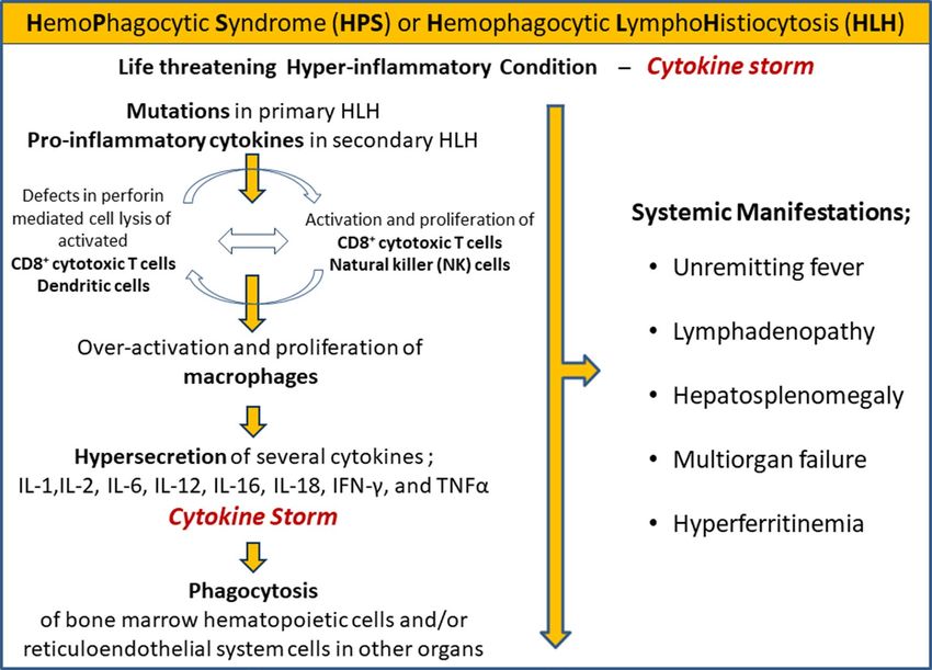
10 Rheumatology International (2021) 41:7–18 Discussion The most important immunological abnormality in pri- mary or reactive HLH is the impaired cytotoxic cell func- Primary hemophagocytic lymphohistiocytosis tion. Under normal physiological conditions, when a virus- (pHLH) infected cell or tumor cell is encountered, CD8+ cytotoxic T cells, NK and NKT cells release perforin (a tetrameric pro- About a quarter of total pHLH cases have a genetic tran- tein that creates pores in the target cell, facilitates the entry sition, and therefore, it is advisable to question a history of granzymes, and disrupts the target cell’s membrane) con- of consanguineous marriage. Infections, especially viral, taining cytolytic granules, and granzymes (proteins involved frequently trigger an inappropriate cytotoxic immune in triggering apoptosis in the target cell) initiate cytolytic response in the presence of some genetic conditions. destruction of the target cell. For this process to proceed Mutations in C D8+T cells and natural killer (NK) cells normally, perforin and granzymes must be structurally nor- (homozygous, compound heterozygous, or digenic (with mal and properly distributed in the cell and packaged into heterozygous mutations in two different genes) result in a granules. Then the content of these granules should enter the loss of performance in the cytolytic pathway proteins such target cell via exocytosis through the immunological synapse as PRF1, STX11, UNC13D, and UNC18-2. The pathogen- between the cytotoxic cell and its target. Disruption of this esis of this syndrome is underlined by the activation and process with genetic mutations leads to the development of proliferation of T lymphocytes (Mainly CD8+cytotoxic T primary or familial HLH. Under normal conditions, perforin cells), and natural killer (NK) cells leading to over-acti- and granzymes contribute to the destruction of target cells vation and proliferation of macrophages resulting in the and the elimination of the immune-activating stimulus; this phagocytosis of bone marrow hematopoietic cells and/or eventually results in apoptosis of cytotoxic cells, together reticuloendothelial system cells in other organs with the with reduced antigen stimulation. This physiological down- hypersecretion of several cytokines including interferon- regulation, called activation-induced cell death, is critical gamma (IFN-γ), interleukin (IL)-1, IL-6, IL-18, and tumor for the control of the immune response. Failure to clear the necrosis factor-alpha (TNFα) [5, 7] (Fig. 3). antigenic stimulus for any reason results in persistence and strengthening of the immune response. Immune activation Fig. 3 Pathogenesis and clinical findings 13
Rheumatology International (2021) 41:7–18 11 is further driven by the high levels of macrophage activa- interesting finding that the prevalence of some mutations tion, with proinflammatory cytokines released by activated differs in certain populations such as those who are linked to immune cells, resulting in hemophagocytosis, tissue dam- perforin mutations that are more common in Turks [15, 16]. age, organ failure, and other inflammatory symptoms of the On the other hand, unlike primary HLH patients, syndrome [2, 4, 8, 9]. Numerous serum proinflammatory observed differences in the pattern of T lymphocyte acti- cytokine levels, including IL-1, IL-2, IL-6, IL-12, IL-16, vation and differentiation in sHLH patients may imply dif- IL-18, TNF- , and IFN-γ, were found to be significantly high ferences in the pathogenesis of these two conditions. For in HLH patients sera. The patient’s prognosis worsens as example, Amman et al. demonstrated that the expression of the levels of these cytokines increase. Inability to clear anti- HLA DR4 and perforin in activated CD8 T cells were higher genic stimulation of certain infections, malignant or autoim- in genetically predisposed HLH patients [17]. It has been mune/autoinflammatory processes leads to an inappropriate shown that lymphomas resulting in sHLH produce proin- immune stimulation and a self-sustaining hyper-inflamma- flammatory cytokines that initiate and permanently stimulate tory condition known as the cytokine storm, characterized the activation of cytotoxic T lymphocytes and NK cells as by excessive and persistent high cytokines levels listed above the triggers of this syndrome. Epstein–Barr virus (EBV) is [9]. HLH may be classified as a cytokine storm syndrome the most common infectious trigger for both primary and in the presence of the intense and rapid cytokine elevation. secondary HLH. An interesting finding in this association Cytokine storm has emerged as an important, under-recog- is that EBV, which normally infects B lymphocytes, can also nized cause of death in COVID-19 [10, 11]. High levels infect CD8+ cytotoxic T lymphocytes, resulting in EBV- of anti-inflammatory cytokines have also been observed in associated HLH [4]. HLH, especially the overproduction of IL-10, indicating a mechanism, albeit insufficient, to suppress the hyperactiva- Secondary HLH and MAS tion of Th1 cells and monocyte/macrophage functions in patients with this disease [9]. The source of IL-10 probably Secondary forms of HLH can be triggered by infections, is the hemophagocytes and this may imply that hemophago- malignancies, autoimmune/autoinflammatory diseases, and cytosis is a compensatory reaction to HLH [12, 13]. In medications. Most frequently, sHLH is triggered by infec- pHLH and related immunodeficiency syndromes, the inher- tions (termed as HLH-associated with infection). In some itance of defective genes involved in the control of cytoly- cases, a history of traveling is present. In particular, infec- sis (e.g., PRF1, STX11, UNC13D, UNC18-2) leads to the tious of especially EBV and other members of the Herpes- uncontrolled proliferation and survival of C D8+ T Cells by virus family, HIV, bacteria, and fungi are the triggers of impairing the cytolytic activity of cytotoxic T Lymphocytes this clinical picture. Malignancies, especially leukemia or [5, 7]. Children with certain immunodeficiency syndromes, lymphoma (termed as HLH-associated with malignancy) such as Chédiak-Higashi syndrome, Hermansky-Pudlak are among common triggers [14]. HLH, which develops in syndrome Type II, Griscelli syndrome (partial albinism), association with an autoimmune/autoinflammatory rheuma- X-linked lymphoproliferative disease, XMEN disease, ITK tological disease, is often referred to as MAS and will be (interleukine-2-inducible T cell kinase deficiency), Lysinuric discussed in detail below. However, it is worth pointing out protein intolerance, CD27 deficiency, and chronic granu- that some authors prefer to use the term MAS for secondary loma also possess genetic defects associated with impaired HLH. Interestingly, secondary HLH may arise iatrogenically cytolysis and are at risk of developing familial HLH. Spe- due to drugs used in the treatment of the primary disease. cific X-linked immune deficiencies such as signaling lym- In 2014, Ramos-Callas et al. reported in a meta-analysis phocytic activation molecule associated protein (SAP) and of 775 HLH cases that the disease was more frequent in X-linked apoptosis inhibitor (XIAP) deficiency, are also Japan, France, China, South Korea, Taiwan, Italy, Spain, associated with HLH triggered by the Epstein–Barr virus and Turkey [1]. Infections and drugs were the most com- (EBV) [3, 5, 14]. pHLH is a rare (1/50,000 of all live births) mon reported causes in adults, while at least 30% had three condition and usually develops in the very first years of life. or more possible triggering factors. The distribution of the It should be kept in mind that pHLH as accepted widely as causes varied between countries. Malignancies were the a pediatric disease, may develop its first symptoms in adult- leading cause in Japan, S. Korea, Taiwan, China, France, hood. Patients with sHLH may have cytolytic genes with and Italy, whereas autoimmune diseases were more frequent underlying hypomorphic defects found in pHLH, as well. in Spain and the USA. Indeed, up to 40% of sHLH and MAS patients are reported to carry heterozygous (some dominant-negative) mutations Macrophage activation syndrome of known familial HLH genes. Therefore, some researchers suggest that MAS, sHLH, and familial HLH may be con- Secondary HLH, which develops in the context of rheumato- sidered as members of a disease spectrum [3, 5, 7]. It is an logical diseases, is often referred to as MAS and is reported 13
12 Rheumatology International (2021) 41:7–18

most frequently in systemic Juvenile Idiopathic Arthritis may develop acute respiratory distress syndrome (ARDS),
(sJIA, Still Disease) patients [5]. Strippoli et al. suggested a which requires mechanical ventilation. ARDS has a poor
trilayer mechanism for the HLH that is secondary to rheu- prognosis and is more common in severe sHLH requiring
matic diseases: Firstly, a genetic tendency (such as defects intensive care [29]. Neurological involvement is a sign
in the cytotoxic activity of CD8 and NK cells and a genetic of poor prognosis, as well, and is usually a sign of estab-
predisposition to TLR hyper-response), followed by a high lished sHLH. Several clinical signs and symptoms of CNS
disease activity caused by the high proinflammatory state involvement ranging from mood and personality changes to
mediated by the high levels of cytokines such as IL-6, and seizures, muscle weakness in the extremities, cranial nerve
finally the activation of immune cells such as the T cells and palsy, decreased consciousness, and coma, may develop.
macrophages triggered by a viral infection that leads to the Acute renal failure necessitating chronic renal replacement
cytokine storm [19]. may be seen and worsens the outcome [30–33]. In AOSD
 Up to 10% of sJIA cases develop MAS. In adults, it is and MAS cases, leukocyte and platelet count, hemoglobin,
reported in association with cases of adult onset still disease albumin, and fibrinogen levels are significantly lower than
(AOSD), and to a lesser extent in Systemic Lupus Erythema- non-MAS AOSD cases. However, ferritin, LDH, and tri-
tosus (SLE) cases. MAS is a not well known but life-threat- glyceride levels (which develops due to TNFα suppression
ening complication of these rheumatic diseases. Similar to of lipoprotein lipase) are significantly higher in patients
sJIA, 10-15% of AOSD patients develop MAS. The activa- with MAS [5, 27]. Here, serum ferritin levels are of criti-
tion of shared innate immunopathogenic pathways, including cal value in diagnosis. The probability of MAS at levels
IL-1 and IL-18, leads to a similar systemic inflammation below 500 ng/ml is rather weak. While the probability of
in both AOSD and sJIA. Therefore, it is not surprising that MAS increases for values between 500 and 1000, a value of
MAS is a common complication of both diseases [20, 21]. 10,000 ng/ml (10,000 μ/L) or higher has a 96% specificity
This hypothesis is supported by the observation that gain-of- and 90% sensitivity for the presence of MAS. Ferritin is the
function mutations in the gene encoding the innate immune most dynamic test that rises rapidly and extensively in the
protein NLRC4 promote the spontaneous formation of the early inflammatory period and decreases significantly with
NLRC4 inflammasome, production of IL-1 family cytokines active treatment and cessation of the inflammation. The ratio
(IL-1β and IL-18) and inflammatory cell death in autoin- between baseline and recovery may reach 1–800. High levels
flammatory diseases complicated by MAS [22]. Similar to persisting despite active treatment or a decline of less than
sJIA, MAS in AOSD may either occur at onset ora later %50 is associated with a poor prognosis. Pre- and posttreat-
stage, mostly triggered by viral infections [23]. In some ment levels of the laboratory parameters such as D-dimer,
patients, MAS may lead to ARDS and death in the course triglycerides, platelet levels, LDH, and transaminases differ
of COVID-19 infection [10, 24, 25]. Therefore, MAS should as much as about %50 and may be useful in the diagnosis.
be kept in mind in a COVID-19 patient with signs of rapid The presence of hyperferritinemia in association with cyto-
clinical and laboratory deterioration. Every rheumatologist penia in various series, low levels of fibrinogen (due to the
should be alert because of the cytokine storm, a severe com- procoagulant effects of TNFα or increased lysis), and the
plication of COVID-19, and symptoms of COVID-19 such absence of a significant increase in sedimentation despite
as fever, arthralgia, and myalgia resembling those seen dur- the increased CRP may be used to support the diagnosis of
ing several rheumatologic diseases [26]. MAS [34]. Thrombocytopenia and hypofibrinogenemia can
 MAS occurring in the early stages of AOSD is character- lead to ecchymoses and bleeding, especially in critical cases.
ized by neutrophilic leucocytosis and this laboratory find- Since hemophagocytosis is a late-stage finding, it may not be
ing may be used in the differential diagnosis. Later in the detected in early bone marrow examinations. Bone marrow
course of AOSD MAS may coincide with disease flares, findings may not correlate with clinical symptoms or high
infections, or the use of anti-inflammatory drugs such as sul- ferritin levels and are, therefore, not essential for diagnosis
fasalazine, methotrexate, hydroxychloroquine, and anti-TNF [30, 34]. One should not wait for hemophagocytosis to be
agents. Secondary HLH (MAS) is characterized by fever, proven to initiate treatment, but a bone marrow biopsy, when
pulmonary insufficiency, liver failure, neurological symp- available, is necessary, since hemophagocytosis in the bone
toms, and signs of pancytopenia and coagulopathy that could marrow may support the therapy when present.sCD25 as a
not be explained for any other reason. Instead of a fever marker of T cell activation, and sCD163 as the marker of
that rises typically in the afternoons with chills in AOSD, hemophagocytosis, are specific markers for HLH. However,
unremitting fever is more common in the presence of MAS. the aforementioned parameters are not included in the MAS
Therefore, persistent fever despite the treatment of infec- classification criteria made for sJIA due to difficulties in
tions that develops in the course of sJIA or AOSD should their routine use [35]. But, the HLH-2004 criteria include
raise the suspicion of MAS [27, 28]. Pulmonary involve- sCD25 (sIL2Rα) ≥ 2400 U/ml [34].
ment is found in about half of sHLH patients and patients

13
Rheumatology International (2021) 41:7–18 13 In summary, it is crucial to exclude HLH/MAS in sJIA, Diagnostic tools: HLH‑2004 score, HScore, MH score AOSD, and when patients with COVID-19 suddenly dete- riorate in the presence often remitting high fever, addi- A timely diagnosis of HLH is of particular importance, tional pulmonary, and/or neurological symptoms, cyto- because patients may be critically ill and delay in diagnosis penia, and abnormal ferritin or LDH levels [24, 25, 36]. contributes only to a poor outcome. The diagnosis is based mainly on clinical and laboratory criteria. There is no single laboratory test or clinical finding that is pathognomonic. The widely used HLH-2004 criteria set is developed by the Histiocyte Society (Table 1) [37]. Table 1 Diagnostic guidelines for hemophagocytic syndrome (HPS) or hemophagocytic lymphohistiocytosis (HLH) Clinical/laboratory findings HLH-2004 HScorec MH scorec,d sJIA/MAS Fever (°C) ≥ 38.5 < 38.4: 0 points − + 38.4–39.4: 33 points > 39.4: 49 points Organomegaly Splenomegaly Absent: 0 points Splenomegaly: − Hepatomegaly or splenomeg- Absent: 0 points aly: 23 points Hepatomegaly Present: 12 points and splenomegaly: 38 points Cytopenia Cytopenia of ≥ 2 series Single series: 0 points Neutrophil count Platelet count Hemoglobin < 9 g/dL Two series: 24 points > 1.4: 0 points ≤ 181 × 109/L Platelet count < 100 × 109/L Three series: 34 points ≤ 1.4, ×109/L: 37 points Absolute neutrophil count Platelet: < 1×10⁹/L > 78: 0 points ≤ 78, ×109/L: 11 points Hemoglobin > 8.3: 0 points ≤ 8.3, g/dL: 11 points Triglyceridesa (mmol/L) Hypertriglycemia and/or < 1.5: 0 points > 156 mg/dl hypofibrinogenemia 1.5–4. 0: 44 points Triglycerides ≥ 3,0 (65 mg/dL) > 4.0: 64 points Fibrinogen (mg/dL)a Fibrinogen ≤ 150 mg/dl, > 2.5: 0 points > 131: 0 points ≤ 360 mg/dl (1.5 g/l) ≤ 2.5: 30 points ≤ 131 mg/dl: 15 points (1.31 g/l) Ferritin (ng/L) ≥ 500 μg/L < 2000: 0 points > 684 ng/ml 2000–6000: 35 points > 6000: 50 points Serum AST (IU/L) − 48 U/L ≥ 30: 19 points Hemophagocytosis in the bone Hemophagocytosis in the bone Absent: 0 points marrow marrow, spleen or the lymph Present: 35 points nodes Immunosupressionb − Absent: 0 points Present: 18 points NK cell activitiy Low or absent − sCD25 (sIL2Rα) ≥ 2400 U/ml − Age at onset (years) − − > 1.6: 0 points − ≤ 1.6: 37 points a Are used as single parameters for HLH-2004 scoring b The presence of human immune deficiency virus (HIV) infection or long term treatment with immunosuppressive drugs such as glucocorti- coids, cyclosporine or azathioprine c A scoring is applied. An HScore of ≥ 169 has 93% sensitivity and 86% specificity for HLH. A cut-off value of ≥ 60 for MH score discriminated best between pHLH and MAS d In a sJIA patient with fever and Ferritin > 684 ng/ml, the presence of two additional parameters has a sensitivity of 73% and specificity of 99% for this score 13
14 Rheumatology International (2021) 41:7–18 HScore it is extremely important to start the HLH-2004 treatment protocol containing etoposide in patients with pHLH imme- This score is developed to estimate the individual’s risk of diately. Yet, most patients with pHLH need hematopoietic reactive HPS for both primary and secondary HLH. Clinical stem cell transplantation as the final treatment [3]. In con- and laboratory features scored are given in Table 1 [28]. For trast, the treatment of sHLH/MAS depends on the clinical this scoring, the following nine variables are used: Three severity and may either be treated with a moderate increase clinical variables (underlying immunosuppression, high in the corticosteroid dosage or aggressive immunosuppres- fever, organomegaly), five biochemical variables (triglyc- sives drugs may be favored as the first therapeutic approach erides, ferritin, serum transaminases, fibrinogen, presence [8]. of cytopenia) and one cytological (findings of hemophago- For these reasons, it is important to differentiate pHLH cytosis in the bone marrow) (Table 1). The median score from pHLH/MAS as early as possible. However, the diag- for patients with a positive pHLH diagnosis was 230 [inter- nostic work-up required for pHLH takes time and may not be quartile range (IQR) 203–257 and 125 (IQR) 91–150] for readily performed everywhere. In this instance, some clini- patients with a negative diagnosis. cal and laboratory features gain importance. In a known sJIA The probability of suffering from HPS was less than 1% patient, the clinical diagnosis of MAS is relatively simple for an HS score of ≤ 90 and 99% for an HScore of ≥ 250. in the presence of typical clinical features, but when MAS A cutoff of 169 had 93% sensitivity and 86% specificity for coincides with the onset of sJIA it may be substantially HLH. Note that bone marrow hemophagocytosis is not man- more difficult to distinguish MAS from pHLH. According datory for an HLH diagnosis. This scoring system is avail- to data from a large series of international MAS cases in able online for general use (http://saintantoine.aphp.fr/score sJIA patients, approximately 23% of reported MAS episodes /). Ferritin values have prognostic value in septic patients, occurred at the onset of sJIA and mimicked the character- additionally [29]. istics of the underlying disease [40]. The first attempt to determine the laboratory properties that will separate MAS How to decide between HScore and HLH‑2004 score from pHLH was performed by Lehmberg et al. [18]. An important issue of this report is that in 70% of MAS patients A Belgian group addressed this issue in detail. At the onset included in this study, clinical manifestations of MAS of the disease, HScore was more effective than the HLH- appeared before the diagnosis of sJIA. Neutrophil counts 2004 criteria in accurately identifying HLH for both children and CRP were significantly higher in MAS/sJIA patients, and adults; Diagnostic sensitivity and specificity were 100 and high levels of sIL2Ra were observed more frequently and 80% for children and 90 and 79% for adults, respectively. in pHLH. Minoia et al. proposed a more sophisticated HScore’s performance dropped to levels similar to the HLH- composite score to distinguish better between pHLH and 2004 criteria, with 73% accuracy for the same specificity MAS: The macrophage activation syndrome/Hemophago- once the patient’s clinical condition worsens. The authors cytic Lymphohistiocytosis (MH) score. The creation of the concluded that HScore for children is generally more useful MH score is based on a much larger data set (362 patients than the HLH-2004 criteria, and for adults, HScore is most with MAS and 258 patients with HLH). The MH score con- useful only at the patient’s initial presentation. The authors sists of six demographic, clinical, and laboratory variables: also concluded that the originally published cut-off value Age of onset, neutrophil count, fibrinogen level, presence of 169 should optimally be adapted according to the target of splenomegaly, platelet count, and hemoglobin level. A patient group of interest [38]. Another group addressed this multivariate analysis was done to assess the weight of each issue in rheumatological diseases. And proposed an adopted variable for its contribution to the diagnosis of pHLH. The cut-off value for a target population with various rheumato- MH score was initially developed based on the data of 80% logical diseases. In a cohort of 94 patients with rheumato- of patients and then validated using the data of the remaining logical diseases (30 with HLH and 64 controls), the optimal 20% of the patients. MH score performed extremely well in cut-off value was found to be 190.5 (sensitivity 96.7% and the validation. In this study, the MH score ranged from 0 to specificity 98.4%) [39]. 123, and the median value was 97 (1st–3rd quartile 75–123) and 12 (1st–3rd quartile 11–34) in pHLH and MAS, respec- MH scoring tively. The probability of receiving a diagnosis of pHLH was less than one percent for a score of less than one and Although both sHLH/MAS and pHLH are life-threatening 99% for a score of ≥ 123. The cut-off value of ≥ 60 had conditions, the mortality rate in patients with pHLH is much the best performance in separating pHLH from MAS [41]. higher. Before the era of treatment protocols involving However, for cases other than sJIA such as SLE or Kawasaki aggressive chemotherapy and immunosuppression, only 5% disease, where cytopenia and liver enzyme elevations may of pHLH patients survived 1 year after diagnosis. Therefore, also be related to the activity of the underlying diseases, 13
Rheumatology International (2021) 41:7–18 15

the discriminatory value may be lower. In this case, it is fail to control the disease for most of the time only in com-
advisable to make a decision based on the patient’s history, bination with corticosteroids [43]. It is encouraging that the
clinical complaints, and response to treatment. use of Anakinra is associated with an improved outcome in
 patients with sepsis and MAS features, and gives an appro-
sJIA‑MAS classification criteria priate signal for safe use in the context of sHLH, even when
 triggered by infection [44]. In refractory MAS cases, doses
These are the criteria developed by Ravelli et al. to over- of 100 mg anakinra four times a day may be required to
come some difficulties encountered in the diagnosis of MAS achieve remission [45]. Anakinra is safely used in the treat-
developing in sJIA cases (Table 1). An important difference ment of autoinflammatory diseases, such as sJIA and FMF
from HLH-2004 is the absence of sCD25 measurement. The in rheumatology outpatient clinics.
calculated sensitivity and specificity of this classification set Some studies of anakinra, canakinumab, and rilonacept
was 73 and 99%, respectively [35]. reported that IL-1 blockade alone is not sufficient to prevent
 The HLH2004 criteria set is a reliable tool in the diag- MAS in the course of sJIA. In these studies, corticoster-
nosis of HLH. The HScore estimates best the risk of HLH, oids, cyclosporin, and/or other drugs had to be added to treat
and the MH score differentiates reliably between MAS and MAS patients [23]. Interestingly, tocilizumab is useful in the
pHLH in pediatric cases. HScore is more accurate when treatment of HLH. However, MAS has also been reported in
used at the onset of the disease in adult patients, but cut-off patients receiving tocilizumab. In these cases, clinical and
values recommended for HScore may vary depending on the laboratory findings of MAS such as lower ferritin levels,
underlying disease. It is advisable to use the 2016 classifica- frequency of hepatomegaly, and normal CRP may become
tion criteria to assess MAS in the course of sJIA. less subtle under tocilizumab treatment [46]. The cytokine
 As described above, HLH is a life-threatening medical storm seen in the course of COVID-19 is effectively treated
condition. Early diagnosis and rapid intervention are life- with tocilizumab or anakinra [11, 47].
saving. It should be noted that the sHLH-cytokine storm Rituximab may have a place in the treatment in combina-
may be the main reason for the rapid deterioration seen in a tion with other drugs and has been shown to reduce EBV
COVID-19 patient. viral load, serum ferritin levels, and to improve the overall
 clinical outcome in patients with EBV-related HLH [18].
Treatment Already, the first cases of MAS in association with COVID-
 19 cases treated successfully with JAK inhibitors such as
Despite treatment with the HLA-2004 protocol, most Baricitinib and IL-1 or IL-6 blockers are accumulating [47].
patients with pHLH need an allogeneic stem cell transplant.
Secondary forms and MAS rarely necessitate transplanta-
tion. In these cases, triggering factors such as infection, Conclusion
malignancy, and medication should be searched for. Remis-
sion is possible with a combined immunosuppressive treat- HLH may be triggered by genetic conditions, infections
ment in sHLH/MAS cases. Early use of high-dose steroids including COVID-19, malignancies, autoimmune-auto-
can be successful alone, but more than half of reported adult inflammatory diseases, and some drugs. HLH should be
cases are steroid-resistant. Unlike pHLH, patients rarely suspected in patients with unexplained progressive fever,
need hemopoietic stem cell transplantation due to MAS cytopenia, ARDS, neurological and renal impairment. Dif-
developing as a result of severe sJIA [42]. Carter et al. favor ferentiation between the primary and secondary forms of
an immediate treatment with 1 g of intravenous methylpred- HLH is utterly important, since primary forms of HLH
nisolone per day for 3–5 days combined with IVIG 1 g/kg for require complicated treatments such as hematopoietic stem
2 days (consider repeating every 14 days due to its half-life cell transplantation. As the COVID-19 pandemic devel-
of 14–21 days) as the first-line treatment of sHLH [23]. The ops, large numbers of patients with rare complications of
emergence of established HLH features or clinical deterio- viral infections such as HLH accumulate. Increasing cases
ration despite treatment may necessitate the early initiation of COVID-19 and its severe complications such as the
of the IL-1 blocker, Anakinra [23]. In our personal expe- cytokine storm and HLH may necessitate a closer collabo-
rience, when caught early in the course, the patient often ration of rheumatologists and hematologists with intensive
relieves with the elimination of the triggering factors (such care physicians, infectious disease specialists, and pneu-
as medication] and a medium dose of corticosteroid therapy. mologists, who are more likely to treat these patients in
Evidence in sJIA-related MAS suggests that early use of the first place. Early recognition of Hemophagocytic lym-
anakinra and IL-1 blockade would also be beneficial in the phohistiocytosis and its severe complication of cytokine
adult MAS population. Anakinra is effective in MAS treat- storm is possible only using the diagnostic sets of crite-
ment when corticosteroids, IVIG, cyclosporin, and etoposide ria and by knowing their shortcomings and advantages

 13
16 Rheumatology International (2021) 41:7–18 in specific patient groups. Further studies addressing the 9. Osugi Y, Hara J, Tagawa S, Takai K, Hosoi G, Matsuda Y et al performance of these criteria sets in the diagnosis in the (1997) Cytokine production regulating Th1 and Th2 cytokines in hemophagocytic lymphohistiocytosis. Blood 89:4100–4103 setting of COVID-19 is necessary. 10. Mehta P, McAuley D, Brown M, Sanchez E, Tattersall RS, Man- son J (2020) COVID-19: consider cytokine storm syndrome and immunosuppression. Lancet 395(10229):1033–1034. https Author contributions The author MS collected data and wrote the man- ://doi.org/10.1016/s0140-6736(20)30628-0 uscript. The authors GTS and IA carefully reviewed the data from the 11. Sarzi-Puttini P, Giorgi V, Sirotti S, Marotto D, Ardizzone S, perspective of a hematologist. And the author PA reviewed the article Rizzardini G, Antinori S, Galli M (2020) COVID-19, cytokines for both immunologic and rheumatologic aspects, prepared tables and and immunosuppression: what can we learn from severe acute figures, translated manuscript into English, and gave the manuscript’s respiratory syndrome? Clin Exp Rheumatol 38(2):337–342 final form. 12. Crayne CB, Albeituni S, Nichols KE, Cron RQ (2019) The immunology of macrophage activation syndrome. Front Immu- Funding No funding is used for the preparation of the manuscript. nol 10:119. https://doi.org/10.3389/fimmu.2019.00119 (eCol- lection 2019) 13. Weaver LK, Behrens EM (2014) Hyperinflammation, rather than Compliance with ethical standards hemophagocytosis, is the common link between macrophage activation syndrome and hemophagocytic lymphohistiocytosis. Conflict of interest Authors SM, AP, AI, and GTS declare that they Curr Opin Rheumatol 26(5):562–569. https://doi.org/10.1097/ have no conflict of interest. BOR.0000000000000093 14. Cetica V, Sieni E, Pende D, Danesino C, De Fusco C, Loca- Ethics approval Not applicable. telli F, Micalizzi C et al (2016) Genetic predisposition to hemophagocytic lymphohistiocytosis: report on 500 patients Consent to participate Not applicable. from the Italian registry. J Allergy Clin Immunol 137(1):188– 196.e4. https://doi.org/10.1016/j.jaci.2015.06.048 Consent for publication Not applicable. 15. Ericson KG, Fadeel B, Andersson M, Gudmundsson GH, Gür- gey A, Yalman V, Janka G et al (2003) Sequence analysis of the Availability of data and material The search algorithm for cited articles granulysin and granzyme B genes in familial hemophagocytic is given in Fig. 2. lymphohistiocytosis. Hum Genet 112(1):98–99. https: //doi. org/10.1007/s00439-002-0841-0 Code availability Not applicable. 16. ZurStadt U, Beutel K, Kolberg S, Schneppenheim R, Kabisch H, Janka G, Hennies HC (2006) Mutation spectrum in children Disclaimer No part of the review is copied or published elsewhere. with primary hemophagocytic lymphohistiocytosis: molecu- lar and functional analyses of PRF1, UNC13D, STX11, and RAB27A. Hum Mutat 27(1):62–68. https: //doi.org/10.1002/ humu.20274 17. Ammann S, Lehmberg K, Zur Stadt U, Janka G, Rensing-Ehl A, Klemann C et al (2017) Primary and secondary hemophagocytic References lymphohistiocytosis have different patterns of T-cell activation, differentiation, and repertoire. Eur J Immunol 47:364–373 1. Ramos-Casals M, Brito-Zerón P, López-Guillermo A, Khamashta 18. Lehmberg K, Pink I, Eulenburg C, Beutel K, Maul-Pavicic A, MA, Bosch X (2014) Adult haemophagocytic syndrome. Lan- Janka G (2013) Differentiating macrophage activation syndrome cet 26 383(9927):1503–1516. https: //doi.org/10.1016/s0140 in systemic juvenile idiopathic arthritis from other forms of -6736(13)61048-x hemophagocytic lymphohistiocytosis. J Pediatr 162:1245–1251. 2. Al-Samkari H, Berliner N (2018) Hemophagocytic lymphohistio- https://doi.org/10.1016/j.jpeds.2012.11.081 cytosis. Annu Rev Pathol 13:27–49. https: //doi.org/10.1146/annur 19. Strippoli R, Caiello De Benedetti IF (2013) Reaching the thresh- ev-pathol-020117-043625 old: a multilayer pathogenesis of macrophage activation syndrome 3. Jordan MB, Allen CE, Weitzman S, Filipovich AH, McClain KL http://www.jrheum.org/content/40/6/761. J Rheumatol 40:761– (2011) How I treat hemophagocytic lymphohistiocytosis. Blood. 767. https://doi.org/10.3899/jrheum.121233 118:4041–4052. https://doi.org/10.1182/blood-2011-03-278127 20. Fukaya S, Yasuda S, Hashimoto T, Kataoka H, Horita T, Atsumi 4. Morimoto A, Nakazawa Y, Ishii E (2016) Hemophagocytic lym- T, Koike T (2008) Clinical features of hemophagocytic syn- phohistiocytosis: pathogenesis, diagnosis, and management. Pedi- drome in patients with systemic autoimmune diseases: analysis atr Int 58:817–825. https://doi.org/10.1111/ped.13064 of 30 cases. Rheumatology (Oxford) 47:1686–1691. https://doi. 5. Janka GE (2019) History of hemophagocytic lymphohistiocytosis. org/10.1093/rheumatology/ken342 In: Cronand RQ, Behrens EM (eds) Cytokine storm syndrome. 21. Arlet JB, Le TH, Marinho A, Amoura Z, Wechsler B, Papo T, Springer Nature, Switzerland, pp 3–16 Piette JC (2006) Reactive haemophagocytic syndrome in adult- 6. Silverman DE (2019) The history of macrophage activation syn- onset Still’s disease: a report of six patients and a review of the drome in autoimmune diseases. In: Cronand RQ, Behrens EM literature. Ann Rheum Dis 65:1596–1601 (eds) Cytokine storm syndrome. Springer Nature, Switzerland, 22. Canna SW, de Jesus AA, Gouni S, Brooks SR, Marrero B, Liu pp 17–30 Y et al (2014) An activating NLRC4 inflammasome mutation 7. Schimuzu M (2019) Clinical features of cytokine storm syndrome. causes autoinflammation with recurrent macrophage activation In: Cronand RQ, Behrens EM (eds) cytokine storm syndrome. syndrome. Nat Genet 46:11406. https://doi.org/10.1038/ng.3089 Springer Nature, Switzerland, pp 31–42 23. Carter SJ, Tattersall RS, Ramanan AV (2019) Macrophage acti- 8. Rosado FG, Gopal P (2019) Laboratory features and pathology of vation syndrome in adults: recent advances in pathophysiology, the cytokine storm syndrome. In: Behrens EM (ed) Cronand RQ. diagnosis, and treatment. Rheumatology 58:5–17. https://doi. Springer Nature, Switzerland, pp 43–60 org/10.1093/rheumatology/key006 13
Rheumatology International (2021) 41:7–18 17 24. Paules CI, Marston HD, Fauci AS (2020) Coronavirus infec- 37. Henter JI, HorneA Maurizio A, Egeler RM, Filipovich AH, tions—more than just the common cold. JAMA 323(8):707–708. Imashuku S et al (2007) HLH-2004: diagnostic and therapeu- https://doi.org/10.1001/jama.2020.0757 tic guidelines for hemophagocytic lymphohistiocytosis. Pediatr 25. Huang C, Wang Y, Li X, Ren L, Zhao J, Hu Y, Zhang L, Fan G, Blood Cancer 48(2):124–131. https://doi.org/10.1002/pbc.21039 Xu J, Gu X (2020) Clinical features of patients infected with 2019 38. Debaugnies F, Mahadeb B, Ferster A, Meuleman N, Rozen L, novel coronavirus in Wuhan, China. Lancet 395(10223):497–506. Demulder A, Francis Corazza F (2016) Performances of the https://doi.org/10.1016/S0140-6736(20)30183-5 H-score for diagnosis of hemophagocytic lymphohistiocytosis in 26. Misra DP, Agarwal V, Gasparyan AY, Zimba O (2020) Rheu- adult and pediatric patients. Am J Clin Pathol 145:862–870. https matologists’ perspective on coronavirus disease 19 (COVID-19) ://doi.org/10.1093/ajcp/aqw076 and potential therapeutic targets. Clin Rheumatol. https://doi. 39. Batu ED, Erden A, Seyhoglu E, Kilic L, Buyukasik Y, Karadag O org/10.1007/s10067-020-05073-9 et al (2017) Assessment of the HScore for reactive haemophago- 27. Ravelli A, Grom AA, Behrens E, Cron RQ (2012) Macrophage cytic syndrome in patients with rheumatic diseases. Scand J Rheu- activation syndrome as part of systemic juvenile idiopathic arthri- matol 46:44–48. https: //doi.org/10.3109/030097 42.2016.116795 1 tis: diagnosis, genetics, pathophysiology, and treatment. Genes 40. Kostik MM, Dubko MF, Masalova VV, Snegireva LS, Kor- Immun 13:289–298. https://doi.org/10.1038/gene.2012.3 nishina TL, Chikova IA et al (2015) Identification of the best 28. Fardet L, Galicier L, Lambotte O, Marzac C, Aumont C, Chahwan cutoff points and clinical signs specific for early recognition of D, Coppo P, Hejblum G (2014) Development and validation of macrophage activation syndrome in active systemic juvenile idi- the HScore, a score for the diagnosis of reactive hemophagocytic opathic arthritis. Semin Arthritis Rheum 44(4):417–422. https:// syndrome. Arthritis Rheumatol 66(9):2613–2620. https://doi. doi.org/10.1016/j.semarthrit.2014.09.004 org/10.1002/art.38690 41. Minoia F, Bovis F, Davì S, Insalaco A, Lehmberg K, Shenoi K 29. Grangé S, Buchonnet G, Besnier E, Artaud-Macari E, Beduneau et al (2017) Development and initial validation of the MH score a G et al (2016) The use of ferritin to identify critically ill patients diagnostic tool that differentiates primary hemophagocytic lym- with secondary hemophagocytic lymphohistiocytosis. Crit Care phohistiocytosis from macrophage activation syndrome. J Pediatr Med. 44(11):e1045–e1053. https://doi.org/10.1097/CCM.00000 189:72–78.e3. https://doi.org/10.1016/j.jpeds.2017.06.005 00000001878 42. Wulffraat NM, Rijkers GT, Elst E, Brooimans R, Kuis W (2003) 30. Minoia F, Davì S, Horne A, Demirkaya E, Bovis F, Li C, Lehm- Reduced perforin expression in systemic juvenile idiopathic berg K et al (2014) Clinical features, treatment, and outcome of arthritis is restored by autologous stem-cell transplantation. Rheu- macrophage activation syndrome complicating systemic juve- matology (Oxford) 42:3759. https://doi.org/10.1093/rheumatolo nile idiopathic arthritis: a multinational, multicenter study of gy/keg074 362 patients. Arthritis Rheumatol 66:3160–3169. https://doi. 43. NHS England Clinical Commissioning Policy Statement: Bio- org/10.1002/art.38802 logic Therapies for the Treatment of Juvenile Idiopathic Arthritis 31. Kim M-M, Yum M-S, Choi H-W, Ko TS, Im HJ, Seo JJ et al (2012) (JIA). In https: //www.englan d.nhs.uk/commis sioni ng/wp-conten t/ Central nervous system (CNS) involvement is a critical prognostic uploads/sites/12/2015/10/e03pd-bio-therapies-jia-oct15.pdf. Last factor for hemophagocytic lymphohistiocytosis. Korean J Hematol accessed 14th Apr 2020 47:27380. https://doi.org/10.5045/kjh.2012.47.4.273 44. Shakoory B, Carcillo JA, Chatham WW, Amdur RL, Zhao H, Din- 32. Aulagnon F, Lapidus N, Canet E, Galicier L, Boutboul D, Peraldi arello CA et al (2016) Interleukin1 receptor blockade is associated MN et al (2015) Acute kidney injury in adults with hemophago- with reduced mortality in sepsis patients with features of mac- cytic lymphohistiocytosis. Am J Kidney Dis 65:851–859. https:// rophage activation syndrome: reanalysis of a prior phase III trial. doi.org/10.1053/j.ajkd.2014.10.012 Crit Care Med 44:275–281. https://doi.org/10.1097/CCM.00000 33. Hadchouel M, Prieur AM, Griscelli C (1985) Acute hemorrhagic, 00000001402 hepatic, and neurologic manifestations in juvenile rheumatoid 45. Kahn PJ, Cron RQ (2013) Higher-dose Anakinra is effective in a arthritis: possible relationship to drugs or infection. J Pediatr case of medically refractory macrophage activation syndrome. J 106:561–566. https://doi.org/10.1016/s0022-3476(85)80072-x Rheumatol 40:743–744. https://doi.org/10.3899/jrheum.121098 34. Ravelli A, Minoia F, Davi S, Horne A, Bovis F, Pistorio A et al 46. Yokota S, Itoh Y, Morio T, Sumitomo N, Daimaru K, Minota S (2016) Expert consensus on dynamics of laboratory tests for diag- (2015) Macrophage activation syndrome in patients with systemic nosis of macrophage activation syndrome complicating systemic juvenile idiopathic arthritis under treatment with tocilizumab. J juvenile idiopathic arthritis. RMD Open 2:e000161. https://doi. Rheumatol 42(4):712–722. https://doi.org/10.3899/jrheum.14028 org/10.1136/rmdopen-2015-000161 (eCollection 2016) 8 (Epub 2015 Feb 15) 35. Ravelli A, Minoia F, Davi S, Horne A, Bovis F, Pistorio A et al 47. Ferro F, Elefante E, Baldini C, Bartoloni E, Puxeddu I, Talarico (2016) 2016 Classification criteria for macrophage activation syn- R, Mosca M, Bombardieri S (2020) COVID-19: the new challenge drome complicating systemic juvenile idiopathic arthritis: a Euro- for rheumatologists. Clin Exp Rheumatol 38:175–180 (Epub 2020 pean League Against Rheumatism/American College of Rheuma- Mar 24) tology/Paediatric Rheumatology International Trials Organisation collaborative initiative. Ann Rheum Dis 75:481–489. https://doi. Publisher’s Note Springer Nature remains neutral with regard to org/10.1136/annrheumdis-2015-208982 jurisdictional claims in published maps and institutional affiliations. 36. Li X, Geng M, Peng Y, Meng L, Lu S (2020) Molecular immune pathogenesis and diagnosis of COVID19. J Pharm Anal. https:// doi.org/10.1016/j.jpha.2020.03.001 13
18 Rheumatology International (2021) 41:7–18

Affiliations

Mehmet Soy1 · Pamir Atagündüz2 · Işık Atagündüz3 · Gülsan Türköz Sucak4
 2
 Mehmet Soy Internal Medicine and Rheumatology, Division
 mhmtsoy@gmail.com of Rheumatology, Department of Internal Medicine, Faculty
 of Medicine, Marmara University, Istanbul, Turkey
 Işık Atagündüz
 3
 ikatagunduz@gmail.com Internal Medicine and Hematology, Division of Hematology,
 Department of Internal Medicine, Faculty of Medicine,
 Gülsan Türköz Sucak
 Marmara University, Istanbul, Turkey
 gulsan.sucak@medicalpark.com.tr
 4
 Internal Medicine and Hematology, Division of Hematology
1
 Internal Medicine and Rheumatology, Division and Bone Marrow Transplantation Unit, Bahcelievler
 of Rheumatology, Department of Internal Medicine, Faculty MedicalPark Hospital, Istanbul, Turkey
 of Medicine, Bahcelievler MedicalPark Hospital, Altınbas
 University, Istanbul, Turkey

13
You can also read