Cardiac Magnetic Resonance and amyloidosis: Review - SciELO
←
→
Page content transcription
If your browser does not render page correctly, please read the page content below
International Journal of Cardiovascular Sciences. 2019;32(2)177-189
177

      REVIEW ARTICLE

      Cardiac Magnetic Resonance and amyloidosis: Review
      Vaneza Ferreira Ribeiro,1 Diogo Costa Leandro de Oliveira,1 Daniel Gama das Neves,1,2 Nágela Simão Vinhosa
      Nunes,2 Humberto Villacorta Junior,1 Marcelo Souto Nacif1,2
      Universidade Federal Fluminense,1 Niterói, RJ - Brazil
      Complexo Hospitalar de Niterói,2 Niterói, RJ - Brazil

      Abstract                                                                        Immunoglobulin light chain amyloidosis is the most
                                                                                   common form of amyloidosis in the USA, with 4,500
          Amyloidosis is a disease caused by extracellular                         new cases diagnosed per year. It affects mostly older
      deposition of insoluble protein fibrils, that results                        individuals, aged between 50 and 80 years, especially
      in changes in tissue architecture and consequently                           men (who account for one third of the cases). In the
      modification of the organ structure. Cardiac involvement
                                                                                   USA, wild-type transthyretin amyloidosis is more
      is common in amyloidosis. Two major types of systemic
                                                                                   frequently diagnosed in older, African American
      amyloidosis affect the myocardium – immunoglobulin
                                                                                   patients, whereas patients with hereditary systemic
      light chain and transthyretin amyloidosis – each leading
                                                                                   amyloidosis are generally younger white women.
      to different prognosis. Early detection and diagnosis
                                                                                   Survival of transthyretin amyloidosis patients is higher
      of cardiac amyloidosis are the main objectives in the
                                                                                   than that of light-chain amyloidosis patients.3
      assessment of the disease. New techniques of magnetic
      resonance imaging have minimized the need for biopsies                          Several non-invasive methods have been used to predict
      for the diagnosis. Late gadolinium enhancement                               the presence of amyloid deposits in myocardial tissue,
      technique, and more recently T1 mapping, have                                including electrocardiogram (ECG), echocardiogram
      allowed a simplified evaluation of amyloid deposits                          (ECHO), cardiac biomarkers, scintigraphy (SPECT) and
      and extracellular volume. The aim of this review                             cardiac magnetic resonance (CMR) imaging. Among
      was to describe basic concepts and updates of the                            these techniques, CMR has been considered the reference
      use of magnetic resonance imaging for the diagnosis                          standard for assessment of global and regional myocardial
      amyloidosis and evaluation of its severity.                                  function and for detection and quantification of fibrosis
                                                                                   areas and expansion of myocardial extracellular volume.4
      Introduction                                                                 Cardiac involvement is decisive for the prognosis and
                                                                                   treatment of systemic amyloidosis. Although ECG,
         Amyloidosis is a group of diseases characterized                          ECHO and scintigraphy are the main imaging tests
      by deposition of insoluble fibrils formed from soluble                       used in cardiology, CMR can provide a new perspective,
      molecules that had suffered structural changes and                           especially on the analysis of amyloid deposits. The use
      become relatively insoluble.1 Amyloid fibrils deposit                        of the late gadolinium enhancement technique reveals
      either locally or systemically. Symptoms vary according                      more specific, and sometimes pathognomonic imaging
      to the content of amyloid deposits, which is directly                        features. Also, CMR enables the assessment of the
      related to the type of protein. Cardiac involvement is                       extension of the cardiac area affected by amyloidosis.
      determinant for the choice of the therapy.2                                  T1 mapping technique measures myocardial amyloid
                                                                                   load and myocyte response to infiltration, thereby
                                                                                   allowing monitoring and eventual change of therapy,
      Keywords                                                                     even when cardiac function is normal. These techniques
         Amyloidosis; Myocarditis/pathology; Plaque, Amyloid/                      are very promising for the development of treatment and
      diagnostic imaging; Magnetic Resonance Imaging.                              prognosis of this condition.5-8

      Mailing Address: Marcelo Souto Nacif
      Departamento de Radiologia (MRD), Universidade Federal Fluminense - UFF, Hospital Universitária Antonio Pedro - HUAP, Rua Marques de Paraná, 303.
      Postal Code: 24033-900, 2º andar, Centro, Niterói, Rio de Janeiro - Brazil.
      E-mail: msnacif@gmail.com

      DOI: 10.5935/2359-4802.20190005                                               Manuscript received December 20, 2017, revised manuscript March 27, 2018, accepted May 07, 2018.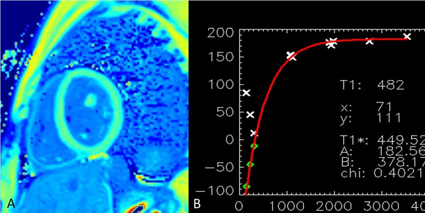
Int J Cardiovasc Sci. 2019;32(2)177-189                                                                               Ribeiro et al.
Review Article                                                                                                CMR and amyloidosis      178

   Based on these considerations, this study aimed to            Hereditary Systemic Amyloidosis
present current concepts of amyloidosis and the use of
                                                                 Hereditary systemic amyloidosis is a dominant
CMR in the diagnosis and follow-up of these patients.
                                                              autosomal disease caused by deposition of amyloid
We conducted a bibliographic review on Pubmed
                                                              fibrils resulting from mutations in genes that encode
(National Library of Medicine) database, using the terms
                                                              transthyretin and apolipoprotein A-I, A-II, fibrinogen,
“amyloidosis”, “disease, myocardial” and “magnetic
                                                              gelsolin, cystatin C and lysozyme. Transthyretin is
resonance imaging” for the search.
                                                              mainly produced by the liver and participates in the
                                                              transport of thyroxine and retinol. Hereditary systemic
Results                                                       amyloidosis is caused by 100 different mutations in
                                                              the DNA of transthyretin, generating a heterogeneity
   A total of 135 articles were retrieved, and 60 were
                                                              of penetrance. Clinical presentation of the disease
selected for being published in higher impact journals and
                                                              includes cardiomyopathy, nephropathy and neuropathy.
consensus of the authors. We summarized current concepts
                                                              Although transthyretin-related cardiac amyloidosis
of amyloidosis types, diagnostic methods, prognosis and
                                                              is less aggressive than immunoglobulin light chain
treatment of the disease in the following text.
                                                              amyloidosis, hereditary systemic amyloidosis is also a
                                                              cause of significant symptoms of heart failure.13,14
Discussion

Main types of amyloidosis                                        Senile systemic amyloidosis
                                                                 Senile systemic amyloidosis, also related to transthyretin,
   Two types of amyloidosis can affect the myocardial
                                                              is not hereditary and generally occurs after the seventh
ventricle: immunoglobulin light chain amyloidosis
                                                              decade of life. This presentation of amyloidosis can
and transthyretin amyloidosis, which, in turn, has two
                                                              be called as senile systemic or wild type amyloidosis.
forms of presentation – wild type and genetically variant
                                                              Similar to other types of the disease, in senile systemic
transthyretin amyloidosis.
                                                              amyloidosis, amyloid deposition can occur in several
                                                              tissues such as cardiac, hepatic, and pancreatic tissues.
    Immunoglobulin light chain                                Senile systemic amyloidosis is usually preceded by heart
   Light chain amyloidosis, also known as primary             failure approximately 3-5 years before. Patients are mostly
systemic amyloidosis, is the most common type                 men, older than 70 years. The diagnosis of the disease
of amyloidosis. This is a plasma cell dyscrasia               had been probably underestimated; with advances in
characterized by deposition of amyloidogenic chains           the diagnosis provided by CMR, from 2000 to 2009, the
in the extracellular space, causing a lesion in this tissue   number of patients seen at amyloidosis centers in the
affected. Cardiac dysfunction is seen in up to 50% of         United Kingdom increased by approximately 6.5%.5-9,15
patients with amyloidosis,9 and this type of amyloidosis         Recent reports have described cases of senile
can cause restrictive cardiomyopathy. Congestive              amyloidosis in patients with heart failure with increased
heart failure is quite common, caused by thickening           ejection fraction, highlighting the importance of clinical
and lack of dilatation of the left ventricle, detected by     cardiologists considering the disease in suspected cases.
echocardiography.10 Myocardial dysfunction caused             Clinical suspicion is higher in cases of heart failure with
by amyloidosis can be evaluated by measurement of             increased ejection fraction associated with infiltrative
brain natriuretic peptide (BNP) and troponin levels.          cardiomyopathy revealed by cardiac imaging tests.16
Right heart failure is frequently aggravated by the
concomitant presence of nephrotic syndrome, observed
                                                              Other diagnostic methods
in 30-50% of the cases. Cardiomyopathy is the most
aggressive manifestation of amyloidosis, and the main            Electrocardiography
cause of poor prognosis and death. Cardiac amyloidosis          In amyloidosis patients, particularly in those with light
has also been associated with multiple myeloma, which         chain amyloidosis, electrocardiography usually reveals
can also be evaluated.2,9,11,12                               low voltage of the QRS complexes which, together with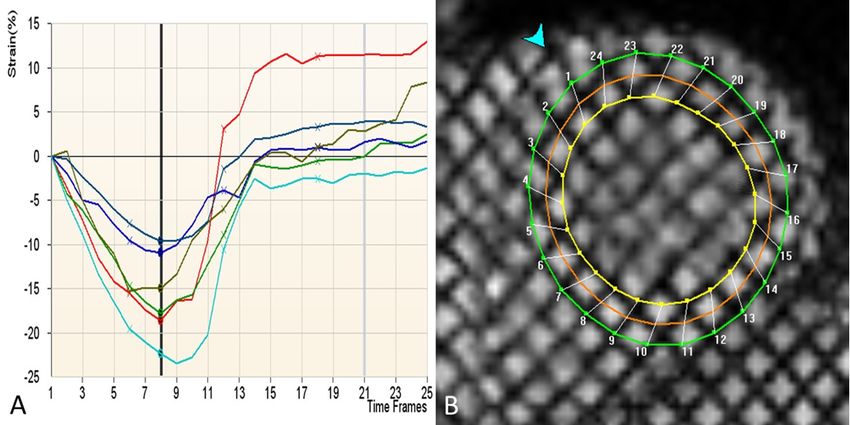
Ribeiro et al.                                                                                     Int J Cardiovasc Sci. 2019;32(2)177-189
179   CMR and amyloidosis                                                                                                       Review Article

      myocardial thickening increases the suspicion of cardiac       hand, the use of other markers – 99m Technetium labelled
      amyloidosis. One hypothesis of voltage decrease is             3,3-diphosphono-1,2-propanodicarboxylic acid (99mTc-
      related with amyloid infiltration, which would cause a         DPD) – has been suggested to specifically locate cardiac
      decrease in cardiac muscle cells. Atrioventricular block,      amyloid in transthyretin amyloidosis and considered
      atrial flutter and ventricular tachycardia have been also      the gold standard for the diagnosis of this condition.25-28
      reported in these patients.9,14,17,18
                                                                     Cardiac magnetic resonance
           Echocardiography                                             CMR has been widely used as the gold standard
         When cardiac amyloidosis is suspected,                      for the assessment of myocardial function and
      echocardiography is usually the first imaging test ordered     characterization of myocardial tissue. The technique
      by the physician. The use of two-dimensional speckle           is more precise than echocardiography, allowing the
      tracking technique has been used to detect subclinical         detection of earlier changes.4
      or initial changes of the disease, which may help in the          CMR without contrast can also be used to provide
      treatment of the patients.19 At initial stages, diagnosis      a precise quantification of amyloid infiltration
      of amyloidosis may be confounded with parietal                 associated with morphological changes (left ventricular
      hypertrophy, as in hypertrophic cardiomyopathy.                hypertrophy, decreased chamber size and atrial
      However, characteristic findings are more frequently           dilation). The method can be used for patients with
      seen at more advanced stages of the disease and                contraindication to contrast, such as patients with renal
      are observer-dependent. Common findings include                failure, using the T1 mapping technique.15,29
      increased ventricular parietal thickness, frequently
                                                                        A CMR test for evaluation of diseases characterized by
      involving the right ventricle, decreased ejection
                                                                     abnormal protein deposit, such as amyloidosis, should
      fraction 20,21 and thickening of valve and interatrial
                                                                     have an average duration of 35 minutes. Although
      septum. Echocardiography may show wall thickening,
                                                                     routine sequences are cine and delayed enhancement,
      especially basal, with high birefringence in the apical area
                                                                     we believe that, when T1 mapping technique is available,
      (apical sparing) of the amyloid deposits, in addition to
                                                                     it should be chosen for routine use. Other techniques
      involvement of valves and papillary muscles.18
                                                                     such as anatomy, perfusion and flow techniques may be
                                                                     included or not in amyloidosis protocol depending on
           Cardiac biomarkers                                        patients’ clinical status and information obtained from
         Immunoglobulin light chain amyloidosis is commonly          the addition of these sequences to the test.
      associated with increased levels of troponin and
      BNP and N-terminal pro-B-type natriuretic peptide                Cine MR
      (NT-proBNP), which is not observed in transthyretin
                                                                        In the analysis of ventricular, segmental and total
      amyloidosis. This is important for patients with some
                                                                     activity, gradient-recalled echo (GRE) and balanced
      renal dysfunction, since natriuretic peptides require
                                                                     steady-state free precession (SSFP) imaging is the
      renal excretion. Increased NT-proBNP in patients with
                                                                     most used sequence. The sequence is added to the
      systemic amyloidosis is predictive of cardiac disease.22
                                                                     electrocardiographic tracings by segmented K-space
      More recently, high-sensitivity troponin has been used
                                                                     technique, eliminating motion artifacts.30
      as a marker of morbidity and mortality.23 At diagnosis,
                                                                        Segmented K-space GRE sequences allow acquisition
      measurements of natriuretic peptide and troponin are
                                                                     of many frames during the cardiac cycle in a dissection
      used for risk stratification of patients.
                                                                     plane. Presentation of the frames in sequence allows
                                                                     dynamic, motion visualization of the heart during cardiac
           Scintigraphy                                              cycle, due to real-time interpolation of the R-R interval
         Scintigraphy is performed with intravenous injection        in the ECG. Strengths of the technique include optimal
      of iodine-123 (123I) for locating amyloid deposit in           temporal resolution, the clear definition of endocardial
      different organs, including liver, kidney, spleen, adrenal     and epicardial borders and acquisition of bright-blood
      glands, and bones. Thus, this is not an adequate technique     cine images. Also, images can be obtained in any
      for cardiac evaluation in amyloidosis.24 On the other          geometric plan.31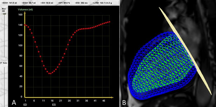
Int J Cardiovasc Sci. 2019;32(2)177-189                                                                                                 Ribeiro et al.
Review Article                                                                                                                  CMR and amyloidosis      180

   Similarly, fast-GRE images, previously used for                           Myocardial tagging uses a fast gradient echo sequence
functional evaluation, are more sensitive to turbulent                    with saturation lines, creating a grid on the images
flow, and are valuable for assessment of stenosis and                     that moves with cardiac motion, allowing an objective
valve insufficiency. Nevertheless, the method produces                    quantification of myocardial contraction during cardiac
worse specification between the myocardium and                            cycle (myocardial strain).34-36
blood in cardiac cavities when compared with SSFP.32,33                      Myocardial tagging (Figure 2) should be used for
A new sequencing technique, known as “real time”                          the diagnosis of initial or subclinical amyloidosis. In
sequencing has high diagnostic quality and can be                         addition, the technique may detect longitudinal or
used for patients with arrhythmias and patients with                      circumferential functional changes in individuals with
inability to sustain apnea.4                                              mutations but without a clear phenotype determined by
   In the assessment of cardiac amyloidosis,                              other techniques.37,38 We understand that one limitation
characterization of morphology and function of the                        for the use of myocardial tagging in many centers is the
disease by cine-MR (Figure 1) is crucial, especially in                   lack of specific softwares for analysis, but we believe in
established disease. This is a widely used technique and                  the benefits of the implementation of the technology.
is used in all protocols of cardiac study by MR.

                                                                              Anatomy
    Tagging                                                                   In the analysis of the anatomy the heart and large
   Myocardial tagging by CMR provides a noninvasive,                      vessels, double-inversion fast spin-echo is the most used
powerful method to quantify segmental and diastolic                       sequence. It is based on the acquisition of fast spin-echo
functions. The development of the technology,                             combined with double inversion preparation pulse - the
particularly of the sequences, type of devices and analysis               first applied to the whole tissue (nonselective) and the
software have facilitated the use of this technique. This                 second, slice-selective. This technique has high spatial
will be very useful to assess patients’ conditions and                    resolution (selective); blood is dark in the images (null
course of the disease.                                                    signal) and, because of that, the technique is known as

      Figure 1 - Evaluation of systolic mass and systolic function by three-dimensional and Simpson’s technique time-volume curve.
      (A) Left ventricular time-volume curve; (B) cine-magnetic resonance imaging during diastole. Left ventricular mass (blue) and final
      diastolic volume (green).
Ribeiro et al. Int J Cardiovasc Sci. 2019;32(2)177-189
181 CMR and amyloidosis Review Article
Figure 2 - Myocardial tagging for quantification of left ventricular systolic and diastolic function; (A) Circumferential strain curves
over a cardiac cycle; (B) subendocardial, mesocardial and epicardial tracings for quantification of myocardial deformation using
cine-tagging.
“black-blood” imaging. This occurs because fast moving cavities and then into the myocardium. One of the most
tissues, similarly to blood, when moving outside the slice of frequently used methods constitutes a hybrid of fast
interest, do not produce any signal, while stable tissues or gradient-echo and ultra-fast echo- planar imaging preceded
slow-moving tissues, like the myocardium, produce a high by a saturation pulse from the tissue signal. This allows
signal. However, endocardial borders (between the blood the acquisition of images in several planes every one-two
and the myocardium) are well defined. This sequence has heartbeats, repeatedly over a 60-second time period,
a segmented acquisition, obtained from final expiratory along with the contrast passage. Myocardial perfusion
apnea, synchronized with the electrocardiogram (ECG). can be used at rest or with pharmacological stress using
An acceptable change, known as triple inversion recovery, adenosine, dipyridamole or regadenoson. The method is
is the addition of a third saturation pulse, to suppress considered adequate to identify myocardial ischemia and
the signal from the adipose tissue (fat saturation), which has also been widely used in the identification of cardiac
may help in the diagnosis in certain situations, such as tumors. Progress has been made in the velocity of data
characterization of cardiac tumors.30,31,34 acquisition, yielding perfusion images with high spatial and
These techniques add little to the assessment of temporal resolution, as well as motion correction methods
amyloidosis, and often are not included in the protocols that positively contribute to the quality of the data.35,39-41
to optimize the time of test execution. Nevertheless, in In a protocol for cardiac amyloidosis investigation, this
some situations, differential diagnosis is important, and sequence will be probably unnecessary, since a suspicion
situations that require evaluation of the pericardium, of myocardial ischemia is not considered for differential
better tissue or even morphological characterization can diagnosis in almost all cases.
be added to the basic protocol.
Myocardial delayed enhancement
Perfusion Myocardial delayed enhancement is based on a T1-
Myocardial perfusion imaging using CMR is obtained weighted fast gradient echo sequence, with an inversion
by the first pass of contrast (gadolinium) into ventricular recovery pre-pulse and inversion time (TI) adjustedInt J Cardiovasc Sci. 2019;32(2)177-189 Ribeiro et al.
Review Article CMR and amyloidosis 182
to null the normal myocardial signal after infusion myocardial extracellular volume. Such increase in
of gadolinium-based contrast (0.02 – 0.04 mmol/kg). extracellular space results from expansion of amyloid
Gadolinium does not penetrate intact cell membranes deposits to the extracellular space. Several studies
and hence is distributed in the extracellular space; in using the delayed enhancement technique have enabled
case of myocyte membrane rupture (e.g., infarction), the classification of amyloid deposition patterns –
gadolinium shows a larger volume of distribution.41,42 subendocardial (Figure 3), mesocardial (Figure 4) and
In addition, kinetics of the contrast distribution is altered, transmural (Figure 5).15 The most common patterns
with a slower washout.40 of amyloid distribution in immunoglobulin light
Consequently, gadolinium concentrations are much chain amyloidosis and transthyretin amyloidosis are
higher in regions of greater extracellular volume, and subendocardial and transmural, respectively. 43 The
areas of membrane rupture and communication between treatment of amyloidosis depends on the disease
intra and extracellular space as compared with normal subtype; the use of delayed enhancement CMR is hence
myocardial tissue approximately 10 minutes after the paramount as it serves as a screening test that lead to
contrast administration. 39 These areas are white in other test, such as biopsy, for establishment of disease
delayed enhancement images (hypersignal), whereas subtype and definition of therapy.
normal myocardium appears black (low signal - null).
Thus, it is important to reinforce that CMR with late
Recent technological progresses have allowed enhancement can be used not only for the diagnosis of
acquisition of three-dimensional late gadolinium
cardiac amyloidosis, but also can influence the type of
enhancement, with respiratory navigator during free
therapy and follow-up of patients, and hence be decisive
breathing, one respiratory pause, in real time and
for the prognosis of these individuals.
without manual adjustment of TI (self-viability or phase
sensitivity inversion recovery – PSIR – technique).7,35
Therefore, in cardiac amyloidosis, tissue definition T1 mapping and extracellular volume (ECV)
after contrast administration is obtained from gadolinium An important advance in cardiac amyloidosis
deposition and accumulation in areas with increased treatment is the increasing use of quantitative analysis
Figure 3 - Cardiac magnetic resonance from the four-chamber axis view showing (A) increased interventricular septal thickness.
(B) Delayed myocardial enhancement in right atrium (white arrow), left atrium (white arrows) and tricuspid valve system (white
dashed elliptical circle).Ribeiro et al. Int J Cardiovasc Sci. 2019;32(2)177-189
183 CMR and amyloidosis Review Article
Figure 4 - Cardiac magnetic resonance from the four-chamber axis view showing (A) left ventricular hypertrophy. (B) Delayed
myocardial enhancement in interatrial septum (white dashed elliptical circle) and mesocardial interventricular septum (white arrow).
Figure 5 - Cardiac magnetic resonance from the long axis two-chamber (A) and three-chamber (B) view showing mid-apical
transmural delayed myocardial enhancement (white arrows).Int J Cardiovasc Sci. 2019;32(2)177-189 Ribeiro et al.
Review Article CMR and amyloidosis 184
by magnetic resonance, including measurements of the It has demonstrated that T1 mapping without
T2 (edema) and T2* (iron deposition) and T1 mapping, contrast can identify amyloid deposits with a T1
focusing on non-invasive quantification of diffuse of approximately 1,140 ± 61 ms. 29 In post-contrast
myocardial fibrosis. The most used sequence for T1 studies, the cut-off value demonstrated to have a worse
mapping quantification is the modified lock-locker prognosis was 565 ms when combined with a pre-
inversion recovery (MOLLI) (Figure 6).44-51 contrast value greater than 1,044 ms.52
With technological advances in the context of In addition, the use of pre- and post-contrast T1
amyloidosis or even infiltrative diseases, the use of mapping enables the calculation of the ECV fraction
ECV expansion and T1 mapping has been consolidated by the relationship between relaxation fractions of the
in the diagnosis and follow-up of patients with cardiac muscle and the blood, corrected by hematocrit:
myocardial interstitial disease caused by increased
(ΔR1myocardium / ΔR1blood)*(1 – Ht)
amyloid deposition or fibrosis. T1 mapping transforms
Normal ECV in healthy volunteers is 27 ± 3%.46,49
a physical principle of magnetic resonance into a
quantifiable number (expressed in milliseconds) in These made the assessment of interstitial fibrosis or
an image. T1 relaxation time is the measure of how subclinical amyloid deposition by T1 mapping possible,
quickly the longitudinal magnetization component even in situations of negative delayed enhancement
recovers to its equilibrium state. In the T1 mapping magnetic resonance imaging. T1 time is decreased in
technique, measurements of T1 relaxation with and the presence of fibrosis (Figure 7), making this map
without contrast can be obtained by a simple software and powerful tool for quantification of ECV expansion
that directly defines a region of interest within the and fibrosis. T1 mapping has been validated by
myocardium, generating pre- and post-contrast endomyocardial biopsies in patients with non-ischemic
administration values in milliseconds. Amyloid heart diseases, referred for cardiac transplantation.51
deposition increases T1 in the pre-contrast phase and The measurement of the ECV fraction determines, in
reduces T1 in the post-contrast phase due to increased percentage, areas of possible fibrosis not yet detected by
extracellular space by the amyloid infiltration.44-51 delayed enhancement technique.52-54
Figure 6 - (A) Modified lock-locker inversion recovery (MOLLI) T1 mapping on the short axis showing the myocardium (green) and
the blood (blue). (B) T1 relaxation curve (482 ms for the myocardium).Ribeiro et al. Int J Cardiovasc Sci. 2019;32(2)177-189
185 CMR and amyloidosis Review Article
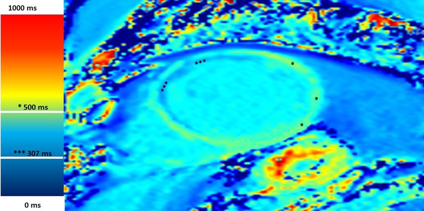
Figure 7 - Postcontrast T1 mapping short axis, showing the myocardium with high T1 value (approximately 500 ms) in green (*).
Inferoseptal, anteroseptal and anterior subendocardial fibrosis, (approximately 307 ms) in blue (***).
Prognosis CMR can also help in the characterization of severe
patients by assessment of left ventricular function, as
In 2008, Maceira et al.,11 have evaluated the capacity of
proposed by Mohty et al.56
CMR in characterizing mortality, survival with therapeutic
response and development of diastolic function. Recently, Martinez-Naharro et al., 57 in a study
published in JACC in 2017, demonstrated the role
In 2015, the study by Fontana et al., 7 made clear
of the ECV as an independent factor of survival in
the importance of detecting delayed enhancement
amyloidosis patients. We believe that, in combination
in immunoglobulin light chain and transthyretin
with technological updates of imaging systems, ECV can
amyloidosis and differentiating subendocardial from
provide incremental prognostic information in patients
transmural pattern. Regardless of the type of amyloidosis,
with amyloidosis over myocardial delayed enhancement.
transmural delayed enhancement is an important
indicator of mortality, which seems to occur earlier in
immunoglobulin light chain amyloidosis. Treatment
In 2016, Boynton et al.,55 have demonstrated that In general, cardiac amyloidosis has a poor prognosis,
delayed enhancement technique provides additional depending on the type of amyloidosis, therapy
prognostic information to serum biomarkers in availability and response to treatment. The treatment can
patients with cardiac amyloidosis. An important be classified as – supportive therapy (specific treatment
finding was the definition of disease severity by the for heart failure, including pacemakers and automated
method in case of inability to null the myocardium defibrillators); therapies to suppress the synthesis
by delayed enhancement or in case of diffuse or of amyloid precursor protein (e.g. chemotherapy in
transmural enhancement. This study had a long immunoglobulin light chain amyloidosis); and new
editorial highlighting the importance of the delayed strategies to inhibit the formation of amyloid fibrils,
enhancement technique in patients with amyloidosis,5 as those targeting amyloid deposits or those including
corroborated by the study by Raina et al.43 promising medications (such as tafamidis and diflunisal)Int J Cardiovasc Sci. 2019;32(2)177-189 Ribeiro et al.
Review Article CMR and amyloidosis 186
to stabilize amyloid precursor protein. Despite its low transplantation. Most chemotherapeutic regimens
availability, heart transplantation can be a very successful include dexamethasone combined with an alkylating
approach in carefully selected patients.17 agent (melphalan or others), which, although they are
Standard supportive therapy for heart failure is of very useful in the treatment of immunoglobulin light
limited benefit and occasionally deleterious to cardiac chain amyloidosis, they should be used with caution
amyloidosis patients. Since cardiac amyloidosis leads by patients with cardiac disease due to the high risk
to a classical phenotype of restrictive cardiomyopathy, of volume overload. New therapeutic strategies
cardiac output can be dependent on heart rate. In this case, include bortezomib, a proteasome inhibitor, and novel
patients can be intolerant to beta-blockers, in contrast immunomodulators (lenalidomide and pomalidomide).
to heart failure patients with reduced or preserved Regarding transthyretin amyloidosis, based on the fact
ejection fraction. There is scarce evidence for the use that transthyretin is a transport protein mostly produced
of angiotensin converting enzyme inhibitors (ACEI) or in the liver, scientists have developed studies on RNA
angiotensin receptor blockers (ARBs) in patients with interference therapies, two recently investigated in phase
myocardial amyloid deposition, and in dysautonomic III trials,61,62 which showed that small interfering RNA
patients, these drugs can even aggravate the symptoms (patisiran) or “antisense” (inotersen) constructs, can
of delayed orthostatic hypotension. Medications such as reduce levels of TTR messenger RNA, the amount of
calcium channel blockers and digitalis should be avoided transthyretin synthesized, the serum concentrations of
as they can selectively concentrate in myocardial tissue transthyretin, and the amount of misfolded monomer
with amyloid deposition, causing increased toxicity. available to aggregate and form deposits. In both trials,
The use of anticoagulants should be considered in atrial the patients who received the active drug had a lower
fibrillation or intracavitary thrombus, which is not mean rate of progression of the manifestations of the
neuropathy (as determined by the modified Neuropathy
infrequent in these patients, even in sinus rhythm.16,17
Impairment Score+7 [NIS+7]) than did the patients who
Preservation of adequate filling pressures is vital
received placebo.3,13,17
because of the restrictive physiology of this heart
Liver transplantation has been used in some cases as
disease, to achieve the balance between the treatment
a strategy to eliminate transthyretin variants from the
of peripheral edema and the development of prerenal
circulation. However, although the therapy has been
kidney failure caused by water-salt depletion and careful
shown to be effective for the amyloidogenic variant
use of diuretics. In contrast to other types of heart failure,
Val30Met, disappointing results have been reported with
maintenance of blood pressure, often requiring the use
other genetic variants, which frequently involves the heart,
of alpha-agonists such as Midodrine, can make the use
since cardiac disease continues to progress with continuous
of high doses of diuretics possible, especially in patients
deposition of wild-type transthyretin amyloid.58
with autonomic neuropathy.3,17,58
Strategies for the inhibition of amyloid fibril formation
Pacemakers and implantable defibrillators may not
assume a massive change of precursor protein into a
prevent sudden death, since it is known that the mechanism
completely different form. The key stage of transthyretin
of death in these patients involves electromechanical
amyloid fibril formation is the transthyretin tetramer
dissociation. Given the lack of robust evidence, indication
dissociation into monomers prone to self-aggregation.
of these devices is similar to that in other heart diseases.
Diflunisal, a nonsteroidal anti-inflammatory drug rarely
High defibrillation threshold may be present in cardiac
used nowadays, binds to plasma transthyretin, increasing
amyloidosis patients and the benefit of these devices
the structural stability of this soluble protein. Studies
remain uncertain.33 Biventricular pacemakers seem to involving diflunisal are in progress. Tafamidis, a novel
be the ideal therapeutic option to prevent worsening of drug with no analgesic or anti-inflammatory properties
cardiac function resulting from desynchrony caused by but with similar mechanisms of action, can selectively
excessive stimulation of the right ventricle.59 bind to thyroxine binding sites, and is currently the most
Currently, amyloidosis treatment is based on promising drug for amyloidosis treatment. The drug
reducing the provision of amyloid precursor protein. was approved for use by ANVISA in November 2016
In immunoglobulin light chain amyloidosis, therapy for the treatment of transthyretin familial amyloidotic
is focused on plasma cell clones, and combines polyneuropathy. Although not worldwide approved
chemotherapy with autologous bone marrow yet, the recently published the ATTR-ACT trial showedRibeiro et al. Int J Cardiovasc Sci. 2019;32(2)177-189
187 CMR and amyloidosis Review Article
that tafamidis was associated with reductions in all- Conclusion
cause mortality, cardiovascular-related hospitalizations
and the decline in functional capacity and quality of life CMR can evaluate amyloid deposits in cardiac tissues
as compared with placebo, in patients with exclusively by delayed enhancement technique, even when cardiac
transthyretin amyloid cardiomyopathy.63 This will soon function is preserved. CMR is rewriting the knowledge
extend the range of indications of the use of tafamidis about cardiac amyloidosis, leading to the development
in hereditary amyloidosis, previously restricted only to of new classification models and therapies as well as
patients with polyneuropathy. change in the prognosis of the patients.
Although amyloid deposits are very stable, the body
has limited capacity to eliminate them, particularly Potential Conflict of Interest
from myocardial deposits. The concept of passive There is no potential conflict of interest to declare.
immunotherapy to increase amyloid deposit clearance
has been proven effective in experimental models and
has been widely developed.17 Experimental studies have Sources of funding
shown that doxycycline, a widely used antimicrobial There was no external funding for the study.
agent, affects the synthesis of amyloid fibrils, and
when combined with biliary salts (taurodeoxycholic Study association
acid), shows a synergic effect in removing transthyretin
This report is part of the “scientific initiation” research
deposits from tissues; these findings have led to studies
project of Vaneza Ferreira Ribeiro at the Department of
in humans. In addition, the presence of a flavonoid
Radiology of Fluminense Federal University Medical
abundant in green tea, has motivated studies on cardiac
School, Niterói-RJ, Brazil.
amyloidosis, since it has been demonstrated that this
compound can inhibit amyloid fibril formation.12
Ethics approval and consent to participate
Cardiac transplantation has yielded disappointing
results because of the multisystemic nature of amyloidosis; This article does not contain any studies with human
nevertheless, in highly selective cases undergoing participants or animals performed by any of the authors.
suppression of light chain, and after treatment of
extracardiac disease, prognosis may be good.17,58
References
1. Bulluck H, White SK, Rosmini S, Bhuva A, Treibel TA, Fontana M, et al. 7. Fontana M, Pica S, Reant P, Abdel-Gadir A, Treibel TA, Banypersad SM, et al.
T1 mapping and T2 mapping at 3T for quantifying the area-at-risk in Prognostic value of late gadolinium enhancement cardiovascular magnetic
reperfused STEMI patients. J Cardiovasc Magn Reson. 2015 Aug 12;17:73. resonance in cardiac amyloidosis. Circulation. 2015;132(16):1570-9.
2. Meier-Ewert HK, Sanchorawala V, Berk JL, Ruberg FL. Cardiac 8. Fontana M, Pica S, Reant P, Abdel-Gadir A, Treibel TA, Banypersad
amyloidosis: evolving approach to diagnosis and management. Curr SM, et al. Response to letters regarding article, "Prognostic value of late
Treat Options Cardiovasc Med. 2011;13(6):528-42. gadolinium enhancement cardiovascular magnetic resonance in cardiac
amyloidosis". Circulation. 2016;133(12):e450-1.
3. Ritts AJ, Cornell RF, Swiger K, Singh J, Goodman S, Lenihan DJ. Current
Concepts of Cardiac Amyloidosis: Diagnosis, Clinical Management, and 9. Falk RH, Dubrey SW. Amyloid heart disease. Prog Cardiovasc Dis.
the Need for Collaboration. Heart Fail Clin. 2017;13(2):409-16. 2010;52(4):347-61. Erratum in: Prog Cardiovasc Dis. 2010;52(5):445-7.
4. Sara L, Szarf G, Tachibana A, Shiozaki AA, Villa AV, de Oliveira AC, 10. Sher T, Gertz MA. Recent advances in the diagnosis and management
et al., Sociedade Brasileira de Cardiologia and Colegio Brasileiro de of cardiac amyloidosis. Future Cardiol. 2014;10(1):131-46.
Radiologia. [II Guidelines on Cardiovascular Magnetic Resonance and 11. Maceira AM, Prasad SK, Hawkins PN, Roughton M, Pennell DJ.
Computed Tomography of the Brazilian Society of Cardiology and the Cardiovascular magnetic resonance and prognosis in cardiac
Brazilian College of Radiology]. Arq Bras Cardiol. 2014;103(6 Suppl amyloidosis. J Cardiovasc Magn Reson. 2008 Nov 25;10:54.
3):1-86.
12. Maurer MS, Elliott P, Comenzo R, Semigran M, Rapezzi C. Addressing
5. Fontana M. Prognosis in cardiac amyloidosis by LGE: ready for prime Common Questions Encountered in the Diagnosis and Management of
time? JACC Cardiovasc Imaging. 2016;9(6):687-9. Cardiac Amyloidosis. Circulation. 2017;135(14):1357-77.
6. Fontana M, Martinez-Naharro A, Hawkins PN. Staging cardiac 13. Perfetto F, Cappelli F, Bergesio F, Ciuti G, Porciani MC, Padeletti L,
amyloidosis with CMR: understanding the different phenotypes. JACC et al. Cardiac amyloidosis: the heart of the matter. Intern Emerg Med.
Cardiovasc Imaging. 2016;9(11):1278-9. 2013;8(3):191-203.Int J Cardiovasc Sci. 2019;32(2)177-189 Ribeiro et al.
Review Article CMR and amyloidosis 188
14. Desai HV, Aronow WS, Peterson SJ, Frishman WH. Cardiac amyloidosis: 33. Tyler DJ, Hudsmith LE, Petersen SE, Francis JM, Weale P, Neubauer S, et
approaches to diagnosis and management. Cardiol Rev. 2010;18(1):1-11. al. Cardiac cine MR-imaging at 3T: FLASH vs SSFP. J Cardiovasc Magn
Reson. 2006;8(5):709-15.
15. Narotsky DL, Castano A, Weinsaft JW, Bokhari S, Maurer MS. Wild-
type transthyretin cardiac amyloidosis: novel insights from advanced 34. Axel L, Dougherty L. MR imaging of motion with spatial modulation of
imaging. Can J Cardiol. 2016;32(9):1166.e1-1166.e10. magnetization. Radiology. 1989;171(3):841-5.
16. Mesquita ET, Jorge AJ, Souza CV Junior, Andrade TR. Cardiac 35. Biglands JD, Radjenovic A, Ridgway JP. Cardiovascular magnetic
amyloidosis and its new clinical phenotype: heart failure with preserved resonance physics for clinicians: Part II. J Cardiovasc Magn Reson. 2012
ejection fraction. Arq Bras Cardiol. 2017;109(1):71-80. Sep 20;14:66.
17. Banypersad SM, Moon JC, Whelan C, Hawkins PN, Wechalekar 36. Zerhouni EA, Parish DM, Rogers WJ, Yang A, Shapiro EP. Human heart:
AD. Updates in cardiac amyloidosis: a review. J Am Heart Assoc. tagging with MR imaging--a method for noninvasive assessment of
2012;1(2):e000364. myocardial motion. Radiology. 1988;169(1):59-63.
18. Esplin BL, Gertz MA. Current trends in diagnosis and management of 37. Kuetting DL, Homsi R, Sprinkart AM, Luetkens J, Thomas DK, Schild
cardiac amyloidosis. Curr Probl Cardiol. 2013;38(2):53-96. HH, et al. Quantitative assessment of systolic and diastolic function
in patients with LGE negative systemic amyloidosis using CMR. Int J
19. Rocha AM, Ferreira SG, Nacif MS, Ribeiro ML, Freitas MR, Mesquita
Cardiol. 2017 Apr 1;232:336-41.
CT. Speckle tracking and transthyretin amyloid cardiomyopathy. Arq
Bras Cardiol. 2017;108(1):21-30. 38. Oda S, Utsunomiya D, Nakaura T, Yuki H, Kidoh M, Morita K, et al.
Identification and assessment of cardiac amyloidosis by myocardial
20. Patel AR, Dubrey SW, Mendes LA, Skinner M, Cupples A, Falk RH, et
strain analysis of cardiac magnetic resonance imaging. Circ J.
al. Right ventricular dilation in primary amyloidosis: an independent
2017;81(7):1014-21.
predictor of survival. Am J Cardiol. 1997;80(4):486-92.
39. Diesbourg LD, Prato FS, Wisenberg G, Drost DJ, Marshall TP, Carroll SE,
21. Porciani MC, Lilli A, Perfetto F, Cappelli F, Massimiliano Rao C, Del
et al. Quantification of myocardial blood flow and extracellular volumes
Pace S, et al. Tissue Doppler and strain imaging: a new tool for early
using a bolus injection of Gd-DTPA: kinetic modeling in canine ischemic
detection of cardiac amyloidosis. Amyloid. 2009;16(2):63-70.
disease. Magn Reson Med. 1992;23(2):239-53.
22. Wechalekar AD, Gillmore JD, Wassef N, Lachmann HJ, Whelan C,
Hawkins PN. Abnormal N-terminal fragment of brain natriuretic peptide 40. Kim RJ, Chen EL, Lima JA, Judd RM. Myocardial Gd-DTPA kinetics
in patients with light chain amyloidosis without cardiac involvement determine MRI contrast enhancement and reflect the extent and severity
at presentation is a risk factor for development of cardiac amyloidosis. of myocardial injury after acute reperfused infarction. Circulation.
Haematologica. 2011;96(7):1079-80. 1996;94(12):3318-26.
23. Dispenzieri A, Kyle RA, Gertz MA, Therneau TM, Miller WL, 41. Saeed M, Wendland MF, Masui T, Higgins CB. Reperfused myocardial
Chandrasekaran K, et al. Survival in patients with primary infarctions on T1- and susceptibility-enhanced MRI: evidence for loss of
systemic amyloidosis and raised serum cardiac troponins. Lancet. compartmentalization of contrast media. Magn Reson Med. 1994;31(1):31-9.
2003;361(9371):1787-9. 42. Simonetti OP, Kim RJ, Fieno DS, Hillenbrand HB, Wu E, Bundy JM, et al.
24. Hawkins PN, Lavender JP, Pepys MB. Evaluation of systemic amyloidosis An improved MR imaging technique for the visualization of myocardial
by scintigraphy with 123I-labeled serum amyloid P component. N Engl infarction. Radiology. 2001;218(1):215-23.
J Med. 1990;323(8):508-13. 43. Raina S, Lensing SY, Nairooz RS, Pothineni NV, Hakeem A, Bhatti S, et
25. de Miguel C, Llorente L, de Haro-Del Moral FJ, Garcia-Pavia P, Gonzalez- al. Prognostic value of late gadolinium enhancement CMR in systemic
Lopez E, Segovia J, et al. Myocardial uptake of (99m)Tc-DPD in patients amyloidosis. JACC Cardiovasc Imaging. 2016;9(11):1267-77.
with AL amyloidosis. Amyloid. 2017;24(supl1):48-9. 44. Kawel N, Nacif M, Zavodni A, Jones J, Liu S, Sibley CT, et al. T1 mapping
26. Hutt DF, Fontana M, Burniston M, Quigley AM, Petrie A, Ross JC, et al. of the myocardium: intra-individual assessment of the effect of field
Prognostic utility of the Perugini grading of 99mTc-DPD scintigraphy strength, cardiac cycle and variation by myocardial region. J Cardiovasc
in transthyretin (ATTR) amyloidosis and its relationship with skeletal Magn Reson. 2012 May 1;14:27.
muscle and soft tissue amyloid. Eur Heart J Cardiovasc Imaging.
45. Kawel N, Nacif M, Zavodni A, Jones J, Liu S, Sibley CT, et al. T1 mapping
2017;18(12):1344-50.
of the myocardium: intra-individual assessment of post-contrast T1 time
27. Moore PT, Burrage MK, Mackenzie E, Law WP, Korczyk D, Mollee evolution and extracellular volume fraction at 3T for Gd-DTPA and Gd-
P. The utility of (99m)Tc-DPD scintigraphy in the diagnosis of BOPTA. J Cardiovasc Magn Reson. 2012 Apr 28;14:26.
cardiac amyloidosis: an Australian experience. Heart Lung Circ.
46. Lee JJ, Liu S, Nacif MS, Ugander M, Han J, Kawel N, et al. Myocardial
2017;26(11):1183-90.
T1 and extracellular volume fraction mapping at 3 tesla. J Cardiovasc
28. Sachchithanantham S, Hutt DF, Quigley AM, Hawkins P, Wechalekar AD. Magn Reson. 2011 Nov 28;13:75.
Role of (99m) Tc-DPD scintigraphy in imaging extra-cardiac light chain
47. Liu CY, Liu YC, Wu C, Armstrong A, Volpe GJ, van der Geest RJ, et al.
(AL) amyloidosis. Br J Haematol. 2017 Oct 30. [Epub ahead of print].
Evaluation of age-related interstitial myocardial fibrosis with cardiac
29. Karamitsos TD, Piechnik SK, Banypersad SM, Fontana M, Ntusi NB, magnetic resonance contrast-enhanced T1 mapping: MESA (Multi-Ethnic
Ferreira VM, et al. Noncontrast T1 mapping for the diagnosis of cardiac Study of Atherosclerosis). J Am Coll Cardiol. 2013;62(14):1280-7.
amyloidosis. JACC Cardiovasc Imaging. 2013;6(4):488-97.
48. Liu S, Han J, Nacif MS, Jones J, Kawel N, Kellman P, et al. Diffuse
30. Bluemke DA, Boxerman JL, Mosher T, Lima JA. Segmented K-space cine myocardial fibrosis evaluation using cardiac magnetic resonance T1
breath-hold cardiovascular MR imaging: Part 2. Evaluation of aortic mapping: sample size considerations for clinical trials. J Cardiovasc
vasculopathy. AJR Am J Roentgenol. 1997;169(2):401-7. Magn Reson. 2012 Dec 28;14:90.
31. Bluemke DA, Boxerman JL, Atalar E, McVeigh ER. Segmented K-space 49. Nacif MS, Raman FS, Gai N, Jones J, van der Geest RJ, T Sibley C, et al.
cine breath-hold cardiovascular MR imaging: Part 1. Principles and Myocardial T1 mapping and determination of partition coefficients at 3
technique. AJR Am J Roentgenol. 1997;169(2):395-400. tesla: comparison between gadobenate dimeglumine and gadofosveset
trisodium. Radiol Bras. 2018;51(1):13-9.
32. Barkhausen J, Ruehm SG, Goyen M, Buck T, Laub G, Debatin JF. MR
evaluation of ventricular function: true fast imaging with steady-state 50. Nacif MS, Turkbey EB, Gai N, Nazarian S, van der Geest RJ, Noureldin
precession versus fast low-angle shot cine MR imaging: feasibility study. RA, et al. Myocardial T1 mapping with MRI: comparison of look-locker
Radiology. 2001;219(1):264-9. and MOLLI sequences. J Magn Reson Imaging. 2011;34(6):1367-73.Ribeiro et al. Int J Cardiovasc Sci. 2019;32(2)177-189
189 CMR and amyloidosis Review Article
51. Sibley CT, Noureldin RA, Gai N, Nacif MS, Liu S, Turkbey EB, et 57. Martinez-Naharro A, Treibel TA, Abdel-Gadir A, Bulluck H, Zumbo G,
al. T1 Mapping in cardiomyopathy at cardiac MR: comparison with Knight DS, et al. Magnetic resonance in transthyretin cardiac amyloidosis.
endomyocardial biopsy. Radiology. 2012;265(3):724-32. J Am Coll Cardiol. 2017;70(4):466-77.
52. Banypersad SM, Fontana M, Maestrini V, Sado DM, Captur G, Petrie A, 58. Tuzovic M, Yang EH, Baas AS, Depasquale EC, Deng MC, Cruz D, et
et al. T1 mapping and survival in systemic light-chain amyloidosis. Eur al. Cardiac amyloidosis: diagnosis and treatment strategies. Curr Oncol
Heart J. 2015;36(4):244-51. Rep. 2017;19(7):46.
53. Piechnik SK, Ferreira VM, Dall'Armellina E, Cochlin LE, Greiser A, 59. Bellavia D, Pellikka PA, Abraham TP, Al-Zahrani GB, Dispenzieri A, Oh
Neubauer S, et al. Shortened Modified Look-Locker Inversion recovery JK, et al. 'Hypersynchronisation' by tissue velocity imaging in patients
(ShMOLLI) for clinical myocardial T1-mapping at 1.5 and 3 T within a with cardiac amyloidosis. Heart. 2009;95(3):234-40.
9 heartbeat breathhold. J Cardiovasc Magn Reson. 2010 Nov 19;12:69. 60. Zumbo G, Sadeghi-Alavijeh O, Hawkins PN, Fontana M. New and
54. Schelbert EB, Testa SM, Meier CG, Ceyrolles WJ, Levenson JE, Blair AJ, et developing therapies for AL amyloidosis. Expert Opin Pharmacother.
al. Myocardial extravascular extracellular volume fraction measurement 2017;18(2):139-49.
by gadolinium cardiovascular magnetic resonance in humans: slow 61. Adams D, Gonzalez-Duarte A, O,Riordan WD, Yang CC, Ueda M, Kristen
infusion versus bolus. J Cardiovasc Magn Reson. 2011 Mar 4;13:16. AV, et al. Patisiran, an a RNAi therapeutic, for hereditary transthyretin
amyloidosis. N Engl J Med. 2018;379(1):11-21.
55. Boynton SJ, Geske JB, Dispenzieri A, Syed IS, Hanson TJ, Grogan M,
et al. LGE immunoglobulin light chain. JACC Cardiovasc Imaging. 62. Benson MD, Waddington-Cruz M, Berk JL, Polydefkis M, Dyck PJ, Wang
2016;9(6):680-6. AK, et al. Inotersen treatment for patients with hereditary transthyretin
amyloidosis. N Engl J Med. 2018;379(1):22-31.
56. Mohty D, Boulogne C, Magne J, Varroud-Vial N, Martin S, Ettaif H,
et al. Prognostic value of left atrial function in systemic light-chain 63. Maurer MS, Schwartz JH, Gundapaneni B, Elliott PM, Merlini G,
amyloidosis: a cardiac magnetic resonance study. Eur Heart J Cardiovasc Waddington-Cruz M, et al. Tafamidis treatment for patients with
Imaging. 2016;17(9):961-9. transthyretin amyloid cardiomyopathy. N Engl J Med. 2018;379(1):1007-16.
This is an open-access article distributed under the terms of the Creative Commons Attribution LicenseYou can also read