COMMITTEE ON TOXICITY OF CHEMICALS IN FOOD, CONSUMER PRODUCTS AND THE ENVIRONMENT
←
→
Page content transcription
If your browser does not render page correctly, please read the page content below
This is a draft statement for discussion. It does not reflect the final views of
                        the Committee and should not be cited.

                                                                       TOX/2021/38

COMMITTEE ON TOXICITY OF CHEMICALS IN FOOD,
CONSUMER PRODUCTS AND THE ENVIRONMENT

Sub-statement on the potential risks from exposure to
microplastics: Oral route (First draft)

Background

1.     In 2019, as part of horizon scanning, the Committee on Toxicity of
Chemicals in Food, Consumer Products and the Environment (COT) identified
the potential risks from microplastics as a topic it should consider
(TOX/2019/08) 1. In 2021, the COT published an overarching statement on the
potential risks from exposure to microplastics (COT Statement 2021/02) 2. This
document provided a high-level overview of the current state of knowledge,
data gaps and research requirements with regards to this topic.

2.     The purpose of this sub-statement is to provide supplementary material
to the overarching statement (COT Statement 2021/02) and consider in detail
the potential toxicological risks of exposure from microplastics via the oral
route (i.e. resulting from the presence of microplastics in food, drinking water
and bottled drinks). It is based on current available literature and data from
internal tools at the UK Food Standards Agency (FSA).

United Kingdom (UK) food safety alerts on the presence of plastic
particles on food items

3.     Computational tools currently being developed internally within the UK
FSA provide up to date information on various facets of plastics including
presence, hazards, and trends. The output of these tools will be monitored
internally, reviewed and when necessary presented to the COT on an ad hoc
basis.

4.      Outputs from this internal tool show that in the past 12 months (June
2020 – June 2021), twelve incidents have been reported for the presence of
plastic foreign bodies across several food categories in the UK. This included:
cereals and bakery products, dietetic foods, poultry meat, beef and pork meat,
and milk and milk products. Each of these incidents have had product recalls
on the affected batches.

1
  TOX/2019/08 is available on the COT website.
2
  COT Statement 2021/02 is available on the COT website. The lay summary
is also available on the COT website.
                                                                                       1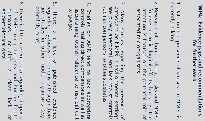
This is a draft statement for discussion. It does not reflect the final views of
                         the Committee and should not be cited.

5.     It should be noted that the food incident alert often lacks granularity,
where the basic properties (e.g. size and shape) of the plastic pieces are not
recorded in full detail. Food incident alerts typically have the following
reporting format: “[Company X] recalls [food product] because it (may) contain
(small) pieces of plastic.”

6.      It is hypothesised that the presence of plastic in these food products
are introduced during the later stage of the food manufacturing process, most
likely during packaging. In UK legislation, the Food Safety Act 1990 3 and
General Food Regulations 2004 4, state that manufacturers have due diligence
and if foreign bodies (e.g. plastic) are present in the product, they are able to
provide evidence that they have done as much as is reasonably practicable to
prevent the contamination.

7.      Although the number of food safety reports in the system were low, this
value may be underreported due to late reporting, lack of post-market
surveillance, and because not all food product types will be
screened/monitored for the presence of micro- and nanoplastics on a regular
basis.

Update on literature

8.    A short update on the emerging literature is provided in the following
paragraphs.

Reviews

9.       The COT Members have been previously informed of the ongoing
research efforts by the UK Food Standards Agency (FSA) on performing a
critical literature review on the microbiological colonisation of nano- and
microplastics (NMPs) and their significance to the food chain (FS307021) 5,
which was contracted to the Centre for Environment Fisheries & Aquaculture
Science. Preliminary outputs from this review were first presented by Bakir et
al., (2021) during the European Food Safety Authority’s (EFSA) Scientific
Colloquium 25 titled, “A coordinated approach to assess the human health
risks of micro- and nanoplastics in food” on May 2021 6.

10.   The four research areas as part of this work package (WP) include:
NMPs in the environment (WP1); pathways of colonised NMPs into food
chains (WP2); interactions between NMPs and microorganisms (WP3); and
NMP-specific microbial risks to consumers (WP4).

3
  The Food Safety Act 1990 is available on the UK legislation website here.
4
  The General Food Regulations 2004 is available on the UK legislation
website here.
5
  Further details concerning this research project (FS307021) are available on
the FSA website.
6
  Further information on EFSA’s scientific colloquium is available on the EFSA
website.
                                                                                        2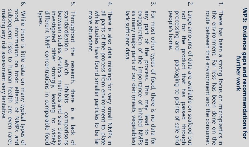
This is a draft statement for discussion. It does not reflect the final views of
the Committee and should not be cited.
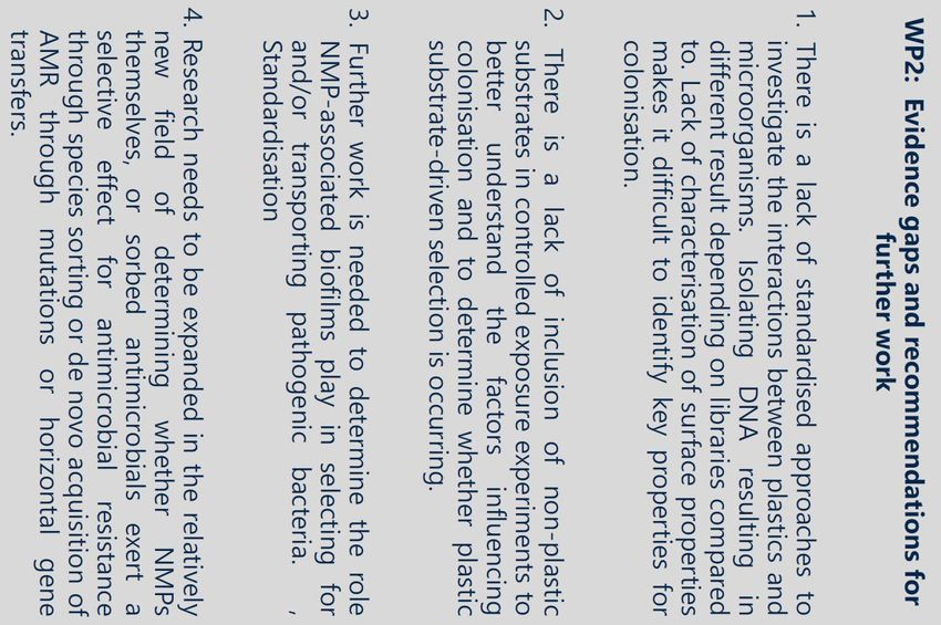
11. The evidence gaps and recommendations for further work in each WP
is shown in Figures 1-2.
12. An authoritative full synthesis report and special report document will
be published later in the year to provide a collated and impartial summary of
the scientific evidence on the impacts of microplastics and human health,
utilizing the most relevant and contemporary scientific data available.
13. Paul et al., (2020) published a review on the current state of knowledge
of micro- and nanoplastics with the focus on oral uptake and toxicity. They
concluded that risk assessment of micro- and nanoplastics is still not possible,
due to various data gaps in terms of exposure, biodistribution and related
effects. Data from the literature suggests that, passage though the
gastrointestinal barrier is possible for a low value of ingested particles
(particularly those at the nanoscale). Cellular toxicity-related effects have
been detected; however, these occur at high-doses and often without dose-
response relationships. Overall, the authors considered that the number of
available studies were still very limited.
3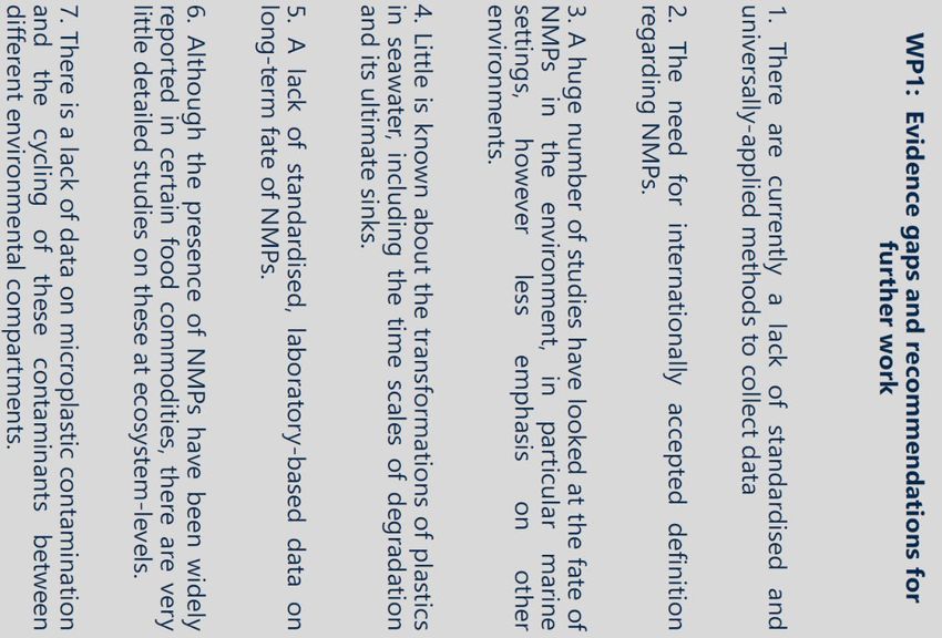
This is a draft statement for discussion. It does not reflect the final views of the
                      Committee and should not be cited.

Figure 1 – Provides a list of the evidence gaps and recommendations for
further work for Work Package 1 (WP1) and Work Package 2 (WP2); nano-
and microplastics (NMPs) in the environment and pathways of colonised
NMPs into food chains, respectively (reproduced from Bakir et al., 2021).

                                                                                  4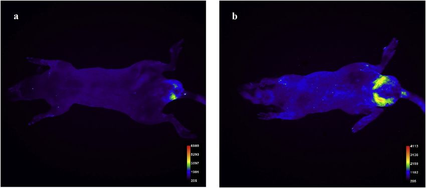
This is a draft statement for discussion. It does not reflect the final views of the
                      Committee and should not be cited.

Figure 2 - Provides a list of the evidence gaps and recommendations for
further work for Work Package 3 (WP3) and Work Package 4 (WP4);
interactions between nano- and microplastics (NMPs) and microorganisms,
and NMP-specific microbial risks to consumers, respectively (reproduced from
Bakir et al., 2021).

                                                                                  5
This is a draft statement for discussion. It does not reflect the final views of the
Committee and should not be cited.
Toxicological data
14. A limited search (i.e non-systematic search) was carried out to identify
any new literature since the publication of COT overarching statement on the
potential risks from exposure to microplastics (COT Statement 2021/02) in
February 2021 – current. Below provide brief summaries of toxicological
studies which investigated the effects of micro- and nanoplastics following oral
exposure.
Reproductive toxicity
In vivo (animal models)
15. Deng et al., (2021) investigated whether the distribution of virgin
polyethylene microplastic (PE-MPs) spheres would affect the bioaccumulation
of phthalate esters (PAEs), as well as investigating effects on reproductive
toxicity from exposure to PAE-contaminated PE-MPs in 5-week old male CD-1
mice (n=120; 12/group). Four PAEs were tested, these were di-2-ethylhexyl
phthalate (DEHP), dibutyl phthalate (DBP), diethyl phthalate (DEP) and
dimethyl phthalate (DMP). The composition of the PE-MP spheres was
confirmed by Fourier Transform Infrared Spectroscopy (FT-IR). Spheres were
40-50 µm in size, as confirmed by Scanning Electron Microscopy (SEM) and
laser scattering particle size distribution analysis.
16. Mice were split into 10 treatment groups. One was treated as a control.
Another was dosed with virgin PE-MPs. DEHP-contaminated PE-MPs were
prepared by mixing 0.2 g/L of PE-MPs with either 5 or 50 µg/L DEHP. A
mixture of all four PAEs was prepared in equimolar proportions to 5 or 50 µg/L
and was mixed with 0.2 g/L of PE-MPs. The virgin PE-MPs and PAE-
contaminated PE-MPs (100 mg/kg bw) were administered to mice via oral
gavage for 30 days. The remaining four groups were exposed to DEHP and
the PAE mixture alone; the concentration was based on the adsorption of
PAEs on PE-MPs and the MPs administered for mice each day. The carrier
was unclear; however, it is believed to be distilled water. Post-treatment all
animals were anaesthetised. The epididymis, liver and gut were collected for
histopathological and biochemical analyses. The testes were also collected for
transcriptomic analyses.
17. Irrespective of whether the PAEs were carried by PE-MPs or not, the
accumulation of PAE was gut > liver > testis. The maximum accumulation of
DEHP and PAE mixture in the gut was ~470 ng/g dry weight, which was ~2
times higher than that in the liver and testis.
18. The number of sperm was significantly reduced in all PAE-
contaminated PE-MPs treatment groups when compared with the PAE alone
treatment groups. Acid phosphatase (ACP), succinate dehydrogenase (SDH)
and lactate dehydrogenase (LDH) levels were used as biomarkers for
disturbance in spermatogenesis. ACP and LDH were increased, and SDH
was significantly decreased in in all PAE-MPs groups compared with PAEs
6This is a draft statement for discussion. It does not reflect the final views of the
Committee and should not be cited.
alone. When compared to the virgin PE-MPs alone, the same observation was
found for the PAE-MPs groups at higher concentrations. Oxidative stress was
also induced in the testis, as higher levels of superoxide dismutase (SOD) and
malonaldehyde (MDA) were measured.
19. Transcriptomic analysis of the left testis showed that higher
concentrations of DEHP-MPs and PAE mix-MPs induced transcriptomic
changes (~180 upregulated and downregulated genes; and ~51 upregulated
and 90 downregulated genes, respectively). Differentially expressed genes
that were of statistical significance were involved with transport and
catabolism, energy metabolism and amino acid metabolism.
20. The authors concluded that exposure to PAE contaminated
microplastics increased the accumulation of PAEs in the gut and liver of mice,
when compared to virgin PE-MPs and PAE alone. No statistically significant
increase in accumulation of PAEs was observed in the testis. Although, PAE-
contaminated PE-MPs were seen to enhance reproductive toxicity, as
changes in sperm parameters and oxidative stress were observed.
21. Hou et al., (2021) evaluated the effect(s) of 5 µm polystyrene
microplastics (PS-MPs) on spermatogenesis in 4-5-week-old male ICR mice
(n=40; 10/group). The particle size (shape undescribed) was determined by
Kurt particle size analysis. The PS-MPs concentrations were 100 μg/L, 1,000
μg/L, and 10 mg/L. The mice in the control group were given distilled water
only. All solutions (tests and control) were provided as drinking water on an ad
libitum basis. The average daily PS-MP exposure dose of each group was
calculated to be 0.6–0.7 μg/day, 6–7 μg/day, and 60–70 μg/day, for each
respective solution concentrations. Mice were exposed to PS-MPs for 35
days.
22. Post-treatment all animals were anaesthetised. The testes and
epididymal tissues were collected and weighed prior to histopathological
analyses. Gene expression of inflammatory (interleukins (IL-1β, IL-6) 7, TNFα 8,
NF-Kβ 9 and Nrf2/HO-1 10) proteins were analysed by Western blot and
7
Interleukin (IL)-1β and IL-6 are pro-inflammatory cytokines for host-defence
responses.
8
Tumour necrosis factor-α (TNF- α) is a mediator of inflammatory and
immune functions.
9
Nuclear factor kappa-light-chain-enhancer of activated B cells (NF-Kβ) is a
protein complex that controls transcription of DNA, cytokine production and
cell survival.
10
Nuclear factor erythroid 2-related factor 2 (Nrf2) regulates the expression of
antioxidant proteins that protect against oxidative damage, of which, heme
oxygenase (HO-1) is one. The Nrf2/HO-1 signalling pathway regulates anti-
inflammation and antioxidation.
7This is a draft statement for discussion. It does not reflect the final views of the
Committee and should not be cited.
qPCR 11. Apoptosis-related proteins (Bax and Bcl2) were also analysed by
Western blot.
23. No deaths or significant changes in body weight were observed.
Although, testicular weight decreased in the low and high treatment groups
when compared to the control. The ratio of live sperm in the epididymis to the
total number of sperm in the treatment groups was significantly lower when
compared to the control group; however, no obvious dose-response
relationship was observed. Sperm deformities were observed such as two-
tailed, hookless or swollen neck, particularly in the mid and high dose ranges.
With regards to the testicular tissue structure, when compared to the control,
the treatment groups had decreased number of spermatids and were
disorderly arranged in the testicular seminiferous tubules. A decrease in
number of spermatozoa, as we all as; pyknosis, nucleus rupture and cell
detachment were also observed. Outputs from the TUNEL 12 staining of the
testis showed a positive correlation between the dose administered and the
number of apoptotic cells.
24. The expression of all inflammatory factors (IL-1β and IL-6) were
increased for all treatment groups, whereas expression of TNF-α was only
significantly increased in the high dose group. When compared to the control
and mid-dose group, the expression of Bcl2 and Bax in the high dose group
was significantly decreased and increased, respectively. Results from the
qPCR analyses showed that expression of NF-κB, IL-1β, and IL-6 increased
significantly, whereas that of the anti-inflammatory molecule Nrf2/HO-1
decreased.
25. Based on the authors’ observations, they considered that the abnormal
sperm quality in ICR mice caused by PS-MP exposure is closely related to the
Nrf2/ HO-1/NF-κB pathway.
26. Xie et al., (2020) investigated the impact of 5 - 5.9 µm PS-MPs on the
reproductive system of 5-6-week-old male Balb-C mice (n=80; 10/group).
They first investigated the effects of PS-MP exposure on testicular injury in
mice and then attempted to confirm the effects of PS-MP exposure on
oxidative stress and MAPK 13 signalling pathways, as well as its effects on
triggering proinflammatory cytokine production. N-acetyl cysteine (NAS) was
used as the antioxidant and SB203580 was used as the p38 MAPK inhibitor.
11
Quantitative polymerase chain reaction (qPCR) is a method by which the
amount of the PCR product can be determined, in real-time, and is very useful
for investigating gene expression
12
Terminal deoxynucleotidyl transferase dUTP nick end labelling (TUNEL)
staining is an assay used to detect DNA breaks formed during the final phase
of apoptosis.
13
Mitogen-activated protein kinase (MAPK) cascades are key signalling
pathways that regulate a wide variety of cellular processes including
proliferation, differentiation, apoptosis, and stress responses.
8This is a draft statement for discussion. It does not reflect the final views of the
Committee and should not be cited.
27. The particle size and shape were not confirmed by separate analyses.
The PS-MPs were mixed with ultrapure water. The mice were split into 8
groups: (1) saline group (control); (2) 100 mg/kg/d NAC; (3) 5 mg/kg
SB203580; (4) 0.01 mg/d PS-MPs; (5) 0.1 mg/d PS-MPs; (6) 1 mg/d PS-MPs;
(7) 1 mg/d PS-MPs + NAC; (8) 1 mg/d PS-MPs + SB203580. The mice in
groups (4), (5), (6), (7), and (8) were given 0.25 mL of the different
concentrations of PS-MPs by oral gavage, once a day for 42 days.
28. Post-treatment all mice were anesthetised. Blood, testes, and
epididymis samples were collected for biochemistry and histopathology
analyses (e.g. haematoxylin and eosin; H & E staining). Reactive oxygen
species (ROS) content was determined by DCFH-DA method 14. The
concentrations of IL-1β, IL-6, TNF-α and Casp-3 15 in the supernatant of the
tissue homogenate, and the concentration of testosterone in the serum were
measured using commercial ELISA 16 kits.
29. No deaths were observed. Although, significant decrease in the body
weight (post-6 weeks of treatment) was observed in PS-MP treatment groups
when compared to the control group. H & E staining showed decrease in the
number of spermatogenic cells which were loosely arranged, and some blank
cavities were also observed in the testicular tissue of PS-MP treatment
groups. The number of sperms in the PS-MP treatment groups was
significantly lower than that in the control group and the sperm deformity rate
gradually increased with increasing exposure concentrations of PS-MPs.
30. LDH and SDH enzyme activities, as well as testosterone concentration
levels were significantly reduced in PS-MP treatment groups when compared
to the control. Administration of NAC and SB203580 inhibitors significantly
attenuated the decrease in testosterone levels and SDH enzyme activity in
group 7 mice compared to group 6. Exposure to PS-MPs caused a significant
increase in ROS and MDA levels compared to the saline group (group 1).
These results indicate that exposure to PS-MPs adversely affects the sperm
count and quality of mice.
31. To determine the effect of PS-MP exposure on the p38 MAPK
signalling pathways, the authors measured the extent of p38 phosphorylation
using ELISA. The intensity of the p38 MAPK immunofluorescence signals in
group 6 and group 7, was significantly alleviated by NAC treatment. This
indicates that MAPK activation occurs after oxidative stress is induced by PS-
MP exposure. When compared with the control (group 1), levels of Casp-3,
TNF-α, IL-1β and IL-6 were significantly increased in the PS-MP treatment
14
Dichloro-dihydro-fluorescein diacetate (DCFH-DA) is a quantitative method
for oxidative stress assessment. It detects levels of intracellular hydrogen
peroxide (H2O2).
15
Caspase-3 (Casp-3) is a lysosomal enzyme involved in the apoptotic
pathway. It interacts with Casp-8 and Casp-9.
16
Enzyme-linked immunosorbent assay (ELISA) is an immunological assay
commonly used to measure antibodies, antigens,
proteins, and glycoproteins in biological samples.
9This is a draft statement for discussion. It does not reflect the final views of the
Committee and should not be cited.
groups. The levels of Casp-3 and IL-1β increased with increasing exposure
concentrations of PS-MP.
32. Based on the authors’ observations, they considered that exposure to
PS-MPs induces oxidative stress and activates the p38 and JNK MAPK
signalling pathways, resulting in poorer sperm quality in mice, decreased
testosterone production, and decreased SDH and LDH enzyme activity.
33. Amereh et al., (2020) investigated the potential endocrine
disturbance(s) with particular emphasis to the reproductive toxicity of 25 and
50 nm virgin spherical PS-nanoplastics (PS-NPs) in ~8-week-old male Wistar
rats (n=30; 6/group). The particles were dispersed in deionised water to make
up stock solutions. Prior to dosing, stock solutions were made to the right
concentration by the addition of distilled water. The particle size distribution
and zeta potential were evaluated by laser diffraction, whilst the
agglomeration state and deposition kinetics were quantified through dynamic
light scattering. Animals were split into five groups: (1) control (distilled water
only); (2) 1 mg/kg bw/day, (3) 3 mg/kg bw/day, (4) 6 mg/kg bw/day and (5) 10
mg/kg bw/day PS-NP treatment groups. Rats were dosed via oral gavage,
once a day for 35 days. Note that at the 1 and 6 mg/kg bw/day doses, the PS-
NPs were fluorescently labelled to investigate the bioavailability and
biodistribution of PS-NPs.
34. Post-treatment all mice were anaesthetised. Blood, serum, left testis,
and epididymis were collected. ELISA kits were used to analyse the levels of
follicle-stimulating hormone (FSH), luteinizing hormone (LH) and testosterone.
H & E staining was used for microscopic observation of the testis structure.
35. When compared to the control, concentration levels of testosterone, LH
and FSH in the serum were statistically lower in the treatment groups;
however, only LH showed this in a dose-dependent manner. In terms of
sperm characteristics, the number of sperm declined by ~8, 16, 24 and 45%
for each dose group (in ascending order), respectively, when compared to the
control. Sperm motility also reduced by up to ~48% at the highest dose. At the
highest dose, ~35% of sperm were abnormal (compared to the control at
6.8%). The most common deformities include coiled or bent tails, conjoined
sperm with double heads and double tails, and no-hook head. Levels of DNA
damage were up to 35%, at the highest dose.
36. Analyses of the H & E stain of testes from treatment groups exhibited
seminiferous tubule degeneration (characterised by disorganised, shrunken
tubules with irregular/buckled basement membranes), which suggested
incomplete spermatogenesis. Severity of lesions and an increase in the
reduced numbers of spermatogenic and Sertoli cells were proportional to the
tested dose concentrations.
37. Through whole-animal image scanning, fluorescently labelled PS-NPs
for the 1 and 6 mg/kg bw/day dose groups accumulated in the testes
suggesting that these particles are able to cross the blood-testis barrier
(Figure 3).
10This is a draft statement for discussion. It does not reflect the final views of the
Committee and should not be cited.
38. To conclude, severe histological lesions and alterations in the
morphology of sperm, and varying concentrations of semen biomarkers were
observed in testis of rats following exposure to PS-NPs. Although, the authors
consider that it is still too early to draw conclusions regarding the potential
health risks of plastic particles to other species (including humans), since
exposure levels in humans are expected to be lower than those seen in/dosed
in some studies in the literature.
Figure 3 Tissue bioaccumulation of fluorescently labelled polystyrene
nanoplastics (PS-NPs). Rats were exposure to (a) 1 and (b) 6 mg PS-NPs/kg
bw/day for 35 days (reproduced from Amereh et al., 2021).
Other
In vivo (animal models)
39. Zheng et al., (2021) performed a comparative investigation between
the response activity to 5 µm PS-MPs of 6-week-old C57 mice with acute
colitis 17 and healthy mice (n=50; 10/group). The morphology of PS-MPs was
analysed by SEM. Mice were divided into 5 groups; (1) treatment with 3%
dextran sodium sulfate (DSS) through drinking water for 7 days to induce
acute colitis; (2) healthy mice fed with distilled water only (negative control
group); (3) healthy mice fed with distilled water containing PS-MPs at a
concentration of 500 µg/L; (4) Mice with acute colitis– distilled water only
(positive control group); (5) Mice with acute colitis fed with distilled water
containing PS-MPs at a concentration of 500 µg/L. Animals were exposed to
their treatment for 28 days (except group 1).
17
Colitis is a chronic digestive disease characterised by inflammation of the
inner lining of the colon.
11This is a draft statement for discussion. It does not reflect the final views of the
Committee and should not be cited.
40. Post-treatment, all animals were anaesthetised. Blood was collected for
ELISA analysis of IL-1β, TNF-α and IFN-γ 18, the livers were also collected for
biochemistry (triglyceride, MDA and PPAR-γ levels) and histopathology
analyses (H & E staining). Metabolomic analyses to record changes in the
liver were also carried out using proton nuclear magnetic resonance (1H NMR)
analysis.
41. The results from the H & E stain showed formation of fatty vacuoles in
healthy mice exposed to PS-MPs (group 3); these were observed with mild
severity. On the other hand, moderate formation of fatty vacuoles and
inflammatory cell infiltration were observed in mice with acute colitis and
exposed to PS-MPs (group 5).
42. PS-MP exposure induced inflammatory effects; the levels of IFN- γ in
treatment groups 1, 3 and 5 was significantly higher than in the control group
(group 2). Exposure to PS-MPs exaggerated DSS-induced acute colitis, as
well as lipid disorders, which were verified by the increased expression of
inflammatory factors (IL-1β, TNF-α and IFN-γ) and triglyceride accumulation in
the gut. 1H NMR analyses indicated that PS-MPs had a greater impact on the
metabolic level (lower concentrations of metabolites involved in amino acid,
energy, and lipid metabolism) in treatment group 5.
43. The authors hypothesised that the increased intestinal permeability of
mice with acute colitis caused by exposure to PS-MPs may be responsible for
the upregulated adverse effects, and that populations with chronic diseases
might be more sensitive to environmental contamination (such as
microplastics).
Cytotoxicity
In vitro (human cell line)
44. Stock et al., (2021) investigated the uptake of sedimenting and buoyant
polyethylene (PE) and polypropylene (PP) microplastic particles in human
intestinal Caco-2 cells. Cytotoxicity was investigated in the following human
cell lines: Caco-2, HepG2 and HepaRG using MTT assay 19. Undigested PE
particles were between 1–4 µm, 10–20 µm. Polydisperse PE, PP, PET and
PVC powders were characterised by SEM. In terms of size, the mean
diameters were 2.2 μm (PE 1–4 μm), 16.5 μm (PE 10–20 μm), 90.1 μm (PE),
67.1 μm (PP), 60 μm (PET) and 136.5 μm (PVC). For shape, PE-MPs were
18
Interferon‐gamma (IFN-γ) is a cytokine that induces and modulates an array
of immune responses.
19
The 3-(4,5-Dimethylthiazol-2-yl)-2,5-Diphenyltetrazolium Bromide (MTT)
assay is a colourimetric assay for assessing cell metabolic activity. Actively
respiring cells convert the water-soluble MTT to an insoluble purple formazan.
The formazan is then solubilised, and its concentration determined by optical
density.
12This is a draft statement for discussion. It does not reflect the final views of the
Committee and should not be cited.
porous roundish, PP-MPs were shred-shaped, whilst PVC and PET-MPs were
smooth and roundish.
45. All cell lines were exposed to varying concentrations of PE (1–4 μm),
PE (10–20 μm), PE, PP, PET and PVC (Table 1). Cells were incubated for 24
hours.
Table 1 - shows the varying dose concentrations of each polymer type:
polyethylene (PE), polypropylene (PP), polyethylene terepthalate (PET) and
polyvinychloride (PVC) in the tested human cell lines; intestinal (Caco-2), and
liver (HepaRG and HepG2). Note that controls were also tested: medium and
0.01% Triton-X-100. Conc.; concentration. (adapted from Stock et al., 2021).
Polymer type
PE (1–4 μm) PE (10– PE PP PET PVC
20 μm)
Cell line Conc.
(mg/mL)
Caco-2 1, 5, 10, 25, 1, 5, 10, 1, 5, 10, 1, 5, 1, 5, 1, 5,
50 25 25, 50, 10, 10, 25, 10, 25,
75, 100 25, 50 50, 75, 50, 75,
100 100
HepaRG 1, 5, 10, 25, 1, 5, 10, 1, 5, 10, 1, 5, 1, 5, 1, 5,
50 25 25, 50, 10, 10, 25, 10, 25,
75, 100 25, 50 50, 75, 50, 75,
100 100
HepG2 1, 5, 10, 25, 1, 5, 10, 1, 5, 10, 1, 5, 1, 5, 1, 5,
50 25 25, 50, 10, 10, 25, 10, 25,
75, 100 25, 50 50, 75, 50, 75,
100 100
46. MTT cytotoxicity assays revealed a decrease in viability only for PE
(HepG2) and PVC (all cell lines) at extremely high particle concentrations,
whereas smaller 1–4 μm and 10–20 μm PE particles as well as PP and PET
were non-toxic. This effect was associated with the high extracellular overload
of large PE and PVC-MP particles rather than intracellular damage when
considering the size limits for cellular uptake. No cytotoxic effects due to
specific particle material or shapes could be detected. Caspase activity
assays in HepaRG and Caco-2 cells revealed mild effects of PVC and PE on
both proteins involved in the extrinsic (Casp-8) and intrinsic (Casp-3 and -9)
apoptosis pathway, suggesting unspecific effects of toxicity.
47. The measurement of cellular contact yielded values below 1% for all
particles. Little to no contact was found for the PVC-MPs, whilst cell surface
contact was found for the other tested plastic particles. The larger particles
were observed to be mostly embedded in the cell monolayer, the authors
were of the opinion that these particles are likely to be rapidly eliminated in
vivo, as intestinal cells renew (approximately every 72 hours).
13This is a draft statement for discussion. It does not reflect the final views of the
Committee and should not be cited.
48. In terms of observations for the intracellular uptake of plastic particles,
a small fraction (0.42%) of PE-MPs in the 1–4 µm (mean diameter 2.2 µm)
size range was transported through the cell monolayer. Absorbed particles of
samples treatment with PE-MPs in the 10–20 μm size range were all ~5 μm in
size. The authors postulated that the smaller PE-MP particles were already
transported to the basolateral compartment after 24 hours, whereas the larger
PE-MPs were still detectable intracellularly.
49. To conclude none of the tested particles induced acute toxic effects,
regardless of their shape and size. The authors recommended future studies
which investigate milder cellular reactions that are not necessarily associate
with cell death (e.g. inflammatory responses, lysosomal changes, or
xenobiotic metabolism) and cellular uptake and transport of plastic particles in
the low micrometre and nanometre range.
Questions on which the views of the Committee are sought
50. Members are invited to consider the following questions regarding the
first draft of the sub-statement and to raise any other matters that arise from
the newly submitted data:
i). Do Members have any comments on the additional information
presented in this cover paper?
ii). Regarding paragraph 14; do Members prefer an annexed reference
to all of the discussion papers previously presented by the
Secretariat (as part of this work stream) or to tabulate all the
toxicological papers reviewed?
iii). Do the Members wish to see any other information that they would
like to be included in the sub-statement?
iv). Do the Members have any other comments?
Secretariat
July 2021
14This is a draft statement for discussion. It does not reflect the final views of the
Committee and should not be cited.
References
Amereh, F., Babaei, M., Eslami, a., Fazelipour, S. and Rafiee. M. (2020) The
emerging risk of exposure to nano(micro)plastics on endocrine
disturbance and reproductive toxicity: From a hypothetical scenario to a
global public health challenge. Environmental Pollution 261, 114158.
Bakir, A., Walker, D., Baker-Austin, C., Thorpe, K., Smith, A., Galloway, T.,
Gaze, W., Lewis, C., Quill, E., Russel, J., Van Hoytema, N. and Verner-
Jeffreys. (2021) A critical review of microbiological colonization of
nano- and microplastics (NMPs) and their significance in the food
chain. UK Food Standards Agency Funded Project FS307021. Poster
presented during the EFSA Scientific Colloquium 25. Available at:
https://www.efsa.europa.eu/en/events/event/update-scientific-
colloquium-25-coordinated-approach-assess-human-health. Accessed:
08/06/2021.
Deng, Y., Yan, Z., Shen, R., Huang, Y., Ren, H. and Zhang, Y. (2021)
Enhanced reproductive toxicities induced by phthalates contaminated
microplastics in male mice (Mus musculus). Journal of Hazardous
Materials 406, 124644.
Hou, B., Wang, F., Liu, T. and Wang, Z. (2021) Reproductive toxicity of
polystyrene microplastics: In vivo experimental study on testicular
toxicity in mice. Journal of Hazardous Materials 405, 124028.
Paul, M. B., Stock, V., Cara-Carmona, J., Lisicki, E., Shopova, S., Fessard,
V., Braeuning, A., Sieg, H. and Böhmert, L. (2020) Micro- and
nanoplastics – current state of knowledge with the focus on oral uptake
and toxicity. Nanoscale Advances 2, 4350.
Stock, V., Laurisch, C., Franke, J., Dönmez, M. V., Voss, L., Böhmert, L.,
Braeuning, A. and Sieg, H. (2021) Uptake and cellular effects of PE,
PP, PET and PVC microplastic particles. Toxicology In Vitro 70,
105021.
Xie, X., Deng, T., Duan, J., Xie, J.., Yuan, J. and Chen, M. (2020) Exposure to
polystyrene microplastics causes reproductive toxicity through oxidative
stress and activation of the p38 MAPK signalling pathway.
Ecotoxicology and Environmental Safety 190, 110133.
Zheng, H., Wang, J., Wei, X., Chang, L. and Liu, S. (2021) Proinflammatory
properties and lipid disturbance of polystyrene microplastics in the
livers of mice with acute colitis. Science of the Total Environment 750,
143085.
15This is a draft statement for discussion. It does not reflect the final views of the
Committee and should not be cited.
TOX/2021/38 Annex A
COMMITTEE ON TOXICITY OF CHEMICALS IN FOOD,
CONSUMER PRODUCTS AND THE ENVIRONMENT
Sub-statement on the potential risks from exposure to
microplastics: Oral route (First draft)
First draft sub-statement
The attached document is a draft. It should not be cited and does not
necessarily represent the views of the Committee. The final version of the
statement will be published in due course on the COT website:
https://cot.food.gov.uk/
Secretariat
July 2021
16This is a draft statement for discussion. It does not reflect the final views of the
Committee and should not be cited.
COMMITTEE ON TOXICITY OF CHEMICALS IN FOOD,
CONSUMER PRODUCTS AND THE ENVIRONMENT
Sub-statement on the potential risks from exposure to
microplastics: Oral route (First draft)
Background
1. In 2019, as part of horizon scanning, the Committee on Toxicity of
Chemicals in Food, Consumer Products and the Environment (COT) identified
the potential risks from microplastics as a topic it should consider
(TOX/2019/08) 20. In 2021, the COT published an overarching statement on
the potential risks from exposure to microplastics (COT Statement 2021/02) 21.
This document provided a high-level overview of the current state of
knowledge, data gaps and research requirements with regards to this topic.
Scope and purpose
2. Data on the presence of plastic particles on some foodstuffs (Touissant
et al., 2019) within the literature is available and the number of such articles is
constantly increasing with developments in analytical detection methodologies
and growing consumer interest.
3. The purpose of this sub-statement is to provide supplementary material
to the overarching statement (COT Statement 2021/02); as referenced in
paragraph 1) and consider in detail the potential toxicological risks of
exposure from microplastics ingested via the oral route (i.e. resulting from the
presence of microplastics in food, drinking water and bottled drinks). It is
based on current available literature and data from internal tools at the UK
Food Standards Agency (FSA).
Toxicokinetics
4. Oral ingestion involves a number of processes that influence the
physicochemical properties of particles (e.g. pH levels of the saliva/digestive
fluids may change their surface charge and zeta potential, formation of protein
20
TOX/2019/08 is available on the COT website.
21
COT Statement 2021/02 is available on the COT website. The lay summary
is also available on the COT website.
17This is a draft statement for discussion. It does not reflect the final views of the
Committee and should not be cited.
coronas) and therefore, their interaction with cells/organs and any subsequent
observed health effect.
5. The size of particles is one of the determining key factors of uptake in
the gastrointestinal tract (GIT). Particles within the nanoscale (i.e. 1 to 100
nm) can distribute to all organs and can be translocated across the blood-
brain and placental barriers. The extent of such absorption is poorly
described. It is generally accepted that large particles > 150 μm will not be
absorbed and thus does not lead to systemic exposure.
6. Two uptake pathways of microplastics (0.1 > 10 μm) from the GIT
lumen have been described in the literature.
7. Firstly, via endocytosis 22 by the microfold (M) cells of the Peyer’s
patches, where the M cells sample and transport particles from the intestinal
lumen to the mucosal lymphoid tissues. It should be noted that the Peyer’s
patches are located in the ileum of the small intestine, which represents a
small fraction of the total GIT surface area.
8. Secondly, via paracellular persorption 23, where non-degradable
particles (such as microplastics), may be mechanically kneaded through loose
junctions in the single-cell epithelial layer into the tissue below. Dendritic cells
can phagocytose 24 such particles, subsequently transporting them to
underlying lymphatic vessels and veins. Potential distribution to secondary
tissues including the liver, muscle and brain may occur (Wright & Kelly, 2017).
9. The uptake pathway is dependent on the property of both the cell type
and the target particle, including its surface chemistry and size. Surface
charge, hydrophobicity also influences the adsorption of proteins to the
particle surface.
10. The COT has previously reviewed relevant animal toxicokinetic studies
as a result of exposure from microplastics via the oral route (presented in
Annex A).
11. At the time of review, only one study was found to describe the
potential fate of microplastic particles in human GIT. Schwabl et al., (2018)
assessed the microplastic concentrations in human stool (n=8). Stool samples
were tested for 10 types of plastics using Fourier-transform infrared
spectroscopy. Up to 9 types were detected ranging from 50 to 500 µm, with
polypropylene (62.8%) and polyethylene terephthalate (PET) (17.0%) being
22
Endocytosis is a cellular process by which cells take in substances from
outside of the cell by engulfing them in a vesicle.
23
Paracellular transport refers to the transfer of substance across an
epithelium by passing through the intracellular space between the cells (often
referred to as tight junctions).
24
Phagocytosis is a cellular process by which a cell uses its plasma membra
to engulf a particle, giving rise to an internal compartment called a
phagosome.
18This is a draft statement for discussion. It does not reflect the final views of the
Committee and should not be cited.
the most common (and were detected in all eight samples). On average, 20
microplastic particles/10 g of stool were detected (range of 18-172 particles).
Several limitations were identified by the authors including: the low number of
participants, and each provided only 1 stool sample. The origin and other
fates of microplastics in the GIT (i.e absorption, distribution, and metabolism)
were not investigated (Schwabl et al., 2019).
12. Available data for maternal transfer of nano- and microplastics (NMPs)
to embryo/fetus are limited. Ragusa et al., (2021) reported the evidence of
microplastics in human placenta (analysed by Raman microscopy). In total, 12
microplastic particles ranging from 5 to 10 mm in size (spheric or irregular in
shape) in 4/6 placentas from women whom had non-caesarean birth;
however, the presence of microplastics in the placenta could not be attributed
to an exposure route nor its source.
13. Based on the available information, the COT concluded that there are
limited data regarding the toxicokinetic fate of orally ingested microplastics in
mammalian species, and that microplastic particles can either remain
confined in the GIT, translocate from the GIT into organs or tissues (via
endocytosis by M cells and paracellular persorption), and/or be excreted
(~>90%). There is lack of information on possible metabolism. Furthermore,
no epidemiological or controlled dose studies that evaluated the effects of
orally ingested microplastics in humans were identified.
Toxicity
14. At the time of review, the COT observed that there is a plethora of
literature regarding the presence and toxicity of micro- and nanoplastics in the
marine environment, whilst there is limited data that are of direct relevance to
humans. The background papers (which includes summaries of literature)
previously reviewed by the COT is presented in Annex B.
15. Due to the uncertainties previously mentioned, the toxic effects are
often hypothesised. These hypotheses are hazard based and are driven by
the physicochemical properties of micro- and nanoplastics. These are:
i). Physical (e.g. bulk, which could lead to gut blockage, as observed in
aquatic and avian species);
ii). Chemical composition (unbound monomers, additives, sorbed
chemicals from the environment e.g. persistent organic pollutants and
metals);
iii). Metabolism or degradation to form monomers or other derivatives,
some of which could be chemically reactive (e.g. isocyanates from
polyurethane) and;
iv). The presence of biofilms (attachment and colonisation of
microorganisms on the plastics).
Physical hazard
19This is a draft statement for discussion. It does not reflect the final views of the
Committee and should not be cited.
16. A common hypothesis is the local irritation of the intestinal tissues
caused by physical mechanical disruption of the intestinal epithelium cells
(IEC) membrane layer by retained plastic particles in the lumen. However,
intestinal crypts undergo constant cycles of IEC replenishment and renewal,
and under normal homeostatic conditions it is estimated that an entire crypt is
replaced every 4-5 days (van der Flier & Clevers, 2009).
17. On the other hand, shedding of IECs from the epithelial monolayer may
cause transient gaps or micro-erosions in the epithelial barrier, thus resulting
in either: increased intestinal permeability or malabsorption to micro- and
nanoplastics but also to other chemicals and solutes present in the intestinal
tract. There is limited knowledge on the rate and effect of this process on the
absorption and the resulting toxicity of micro- and nanoplastics is not known.
18. In certain disease states (e.g. individuals with gastrointestinal issues)
the integrity of the intestinal barrier may be weaker and thereby affect the
crossing of particles (including plastic particles), their systemic bioavailability
and subsequent toxicity. The behaviour of micro- and nanoplastics in normal
gut condition and in certain disease states warrants further investigation.
Chemical hazard
19. The majority of toxicological studies of micro- and nanoplastics
investigate the toxicity of additives (e.g. phthalates), unbound monomers (e.g.
styrene) and sorbed chemicals (e.g. persistent organic pollutants and metals).
20. Based on the available information, chemical leachates and adsorbed
substances from microplastics are not expected to cause greater adverse
health effects in humans due to their small contribution to the overall exposure
from other sources of the same chemical as evidenced by the EFSA, 2016
review and the WHO, 2019 margin of exposure calculations.
Metabolism or degradation products
21. Particles >150 μm usually do not translocate across the gut epithelium,
whilst smaller particles especially those within the nanoscale (1 nm to 0.1 µm)
have the potential for uptake by organs (as mentioned previously).
Microplastics may be taken up into cells but there is a lack of information on
possible metabolism in humans, therefore this hazard is yet to be fully
characterised.
22. Even so, the following question remains: whether large plastic particles
(e.g. 5 µm) breakdown into smaller sizes in the GIT. If so, do these smaller
plastic particles release a higher level of leachates/sorbed chemicals or
produce new degradation products from the polymer itself.
Microbiological hazard
20This is a draft statement for discussion. It does not reflect the final views of the
Committee and should not be cited.
23. The UK FSA is currently performing a critical literature review on the
microbiological colonisation of nano- and microplastics (NMPs) and their
significance to the food chain (FS307021) 25, which was contracted to the
Centre for Environment Fisheries & Aquaculture Science. Preliminary outputs
from this review were first presented by Bakir et al., (2021) during the
European Food Safety Authority’s (EFSA) Scientific Colloquium 25 titled, “A
coordinated approach to assess the human health risks of micro- and
nanoplastics in food” on May 2021 26.
24. The four research areas as part of this work package (WP) include:
NMPs in the environment (WP1); pathways of colonised NMPs into food
chains (WP2); interactions between NMPs and microorganisms (WP3); and
NMP-specific microbial risks to consumers (WP4).
25. The evidence gaps and recommendations for further work in - WP4 are
summarised in depth here. It was found that data on the presence of viruses
on NMPs is currently lacking, and in general very little attention is focused on
the potential role of plastic associated microorganisms. Available studies on
reporting the presence of pathogens on NMPs in environmental settings were
found to be anecdotal and lack robust controls (e.g. comparison to other
substrates). Additionally, published studies on possible presence of
antimicrobial resistant organisms in NMPs were found to be of low quality
(e.g. lack of appropriate controls), which made data comparison challenging
when attempting to ascertain the overall relevance of this hazard to the risk.
No human specific study on dysbiosis was found, although studies in other
model organisms were available (e.g. mice and zebrafish).
26. Overall, for WP4, the authors considered that there is little data
regarding the impacts of NMPs on pathogens and human health outcomes.
Furthermore, there is a clear lack of available epidemiological data.
27. An authoritative full synthesis report and special report document will
be published later in the year to provide a collated and impartial summary of
the scientific evidence on the impacts of microplastics and human health,
utilizing the most relevant and contemporary scientific data available.
25
Further details concerning this research project (FS307021) are available
on the FSA website.
26
Further information on EFSA’s scientific colloquium is available on the
EFSA website.
21This is a draft statement for discussion. It does not reflect the final views of the
Committee and should not be cited.
COT evaluation
28. Presently, a full risk assessment on the potential toxic effect(s) of micro
and/or nanoplastics could not be carried out due to several data gaps
including:
• The unavailability of harmonised methodologies to characterise,
quantify and identify NMPs;
• The lack of toxicokinetic and toxicity data in general. There is no
identified no-observed-adverse-effect level (NOAEL) for the different
polymer types except possibly for PET powder at 2,500 mg/kg bw/day
in rats (Merski et al., 2008), which had a number of limitations (e.g.
particle size and count were not determined/reported);
• The paucity of currently available data for microplastics in different food
types and matrices and;
• The difficulty of performing an accurate exposure assessment.
29. For the reasons above, a case-by-case approach to risk assessments
may need to be considered.
Research priorities for risk assessment
30. The COT recommends the following research priorities for addressing
the data gaps in the potential toxicity of micro- and nanoplastics in humans.
• Comprehensive assessment of MPs and associated contaminant
concentrations in different food types (e.g. seafood, edible meat tissue
and offal, vegetables, fruit, drinks) and matrices (i.e. air, soil, food and
water) and the impact of the effect of cooking on the desorption and
subsequent bioavailability of contaminants/leachates.
• Assessment of the degradation of novel/emerging plastic-based
materials on the market such as biobased plastics (e.g. bamboo ware,
polylactic acid, chitin) and other advanced polymer matrix composite
materials during their use and end-of-life for their possible contribution
to NMPs. It is unclear whether and by how much they already
contribute to the burden of NMPs.
• Studies (in silico, in vitro and/or in vivo) to explore the effect(s) of the
same type of NMP on different tissues (e.g. heart, brain, liver, stomach,
intestines), and of different types of NMP (e.g. polymer type, size,
shape) on the same target tissue.
• Studies on the persistence and potential accumulation of NMPs in the
human body, and on the extent to which NMPs are digestible.
• Investigation of the extent to which NMPs with a range of sizes and
compositions are assimilated into human tissues and the development
of techniques capable of identifying the presence of microplastics in the
22This is a draft statement for discussion. It does not reflect the final views of the
Committee and should not be cited.
human body (e.g. in biopsies, samples from tissue banks, if possible,
histopathology sections).
31. The most significant data gaps hindering a robust risk assessment for
exposure via the oral route include the lack of:
• Appropriate and harmonised analytical methods for the detection of
different NMPs in various food matrices;
• Understanding of human exposure and;
• Human-relevant information on the absorption, distribution, metabolism
and excretion (i.e. the toxicokinetic profile) and on the toxicity profiles of
NMPs.
32. Microplastic concentrations are expected to increase in the future. In
addition, an increase and widespread use of single-use plastic personal
protective equipment (e.g. face masks and gloves) due to the COVID-19
pandemic may also be a major contributing source of plastic pollution (Silva et
al., 2021). Hence, there will be a need to regularly assess the levels of
microplastics in relevant food stuffs, water and the air, such as by establishing
a monitoring programme. This would best be achieved by collaboration
among academia, researchers, and government bodies at a national and
international level.
COT Conclusions
33. The COT noted that there are limited data regarding the toxicokinetic
fate of orally ingested microplastics in mammalian species, and that
microplastic particles can either remain confined in the GIT, translocate from
the GIT into organs or tissues (via endocytosis by M cells and paracellular
persorption), and/or be excreted (~>90%). No epidemiological or controlled
dose studies that evaluated the effects of orally ingested microplastics in
humans were identified.
34. As such, the COT concludes that based on the available data, it is not
yet possible to perform a complete assessment for the potential risks from
exposure to micro and nanoplastics to humans via the oral route; however,
they concur with the conclusions reached by other authoritative bodies (EFSA,
2016; WHO, 2019; ECCC and HC, 2020; SAPEA, 2019; SAM, 2019, as
described in the COT overarching statement on the potential risks from
exposure to microplastics; COT Statement 2021/02 27).
35. The COT concluded that the literature data on exposure to particles
from tyre wear would need separate consideration from microplastic exposure
from food, since the particles were chemically quite different in their polymeric
nature. Risk assessment of such material was considered potentially outside
the scope of the current exercise.
27
COT Statement 2021/02 is available on the COT website. The summaries
of each evaluation by these authoritative bodies are from paragraphs 101-
129.
23This is a draft statement for discussion. It does not reflect the final views of the
Committee and should not be cited.
36. The most significant data gaps are the lack of appropriate and
harmonised analytical methods for the detection and characterisation of
micro- and nanoplastics (together with suitable reference standards), as well
as information on their toxicokinetic and toxicity profiles in/relevant for
humans.
37. The COT highlighted that additional information will be needed from all
exposure sources, which include indoor and outdoor air, dust and soil before a
holistic risk assessment can be completed. The presence of MPs in (sea)food
and water needs to be put into perspective with other sources of MPs such as
atmospheric fallout.
38. Comprehensive assessment of microplastics and contaminant
concentrations in different foods and the impact of cooking on the desorption
and subsequent bioavailability of contaminants/leachates, need to be further
investigated to better understand the implications for human health.
39. Current studies typically focus on only one type of particle/tissue
interaction, as such, further research is necessary to explore the effects of the
range of particle types in different tissues in vitro and/or in vivo. These range
of particle types should also take account of emerging/novel plastic-based
materials such as bioplastics.
COT
July 2021
Statement Number 2021/XX
24This is a draft statement for discussion. It does not reflect the final views of the
Committee and should not be cited.
Abbreviations
COT Committee on Toxicity of Chemicals in Food, Consumer
Products and the Environment
ECCC Environment and Climate Change Canada
EFSA European Food Safety Authority
FSA Food Standards Agency
GIT Gastrointestinal tract
HC Health Canada
NMPs Nano- and microplastics
NOAEL No-observed-adverse-effect level
PET Polyethylene terephthalate
SAM EU Group of Chief Scientific Advisors; Scientific Advice
Mechanism
SAPEA EU Science Advice for Policy by European Academies
UK United Kingdom
WHO World Health Organisation
25This is a draft statement for discussion. It does not reflect the final views of the
Committee and should not be cited.
References
Bakir, A., Walker, D., Baker-Austin, C., Thorpe, K., Smith, A., Galloway, T.,
Gaze, W., Lewis, C., Quill, E., Russel, J., Van Hoytema, N. and Verner-
Jeffreys. (2021) A critical review of microbiological colonization of nano-
and microplastics (NMPs) and their significance in the food chain. UK
Food Standards Agency Funded Project FS307021. Poster presented
during the EFSA Scientific Colloquium 25. Available at:
https://www.efsa.europa.eu/en/events/event/update-scientific-
colloquium-25-coordinated-approach-assess-human-health. Accessed:
08/06/2021.
ECCC & HC. (2020) Science assessment of plastic pollution. Available at:
https://www.canada.ca/en/environment-climate-
change/services/evaluating-existing-substances/science-assessment-
plastic-pollution.html. Accessed: 23/12/2020.
EFSA. (2016) Presence of microplastics and nanoplastics in food, with
particular focus on seafood. The EFSA Journal 14, 4501. Available at:
https://efsa.onlinelibrary.wiley.com/doi/epdf/10.2903/j.efsa.2016.4501.
Accessed: 04/08/2020.
FSA. (2020) A critical review of microbiological colonisation of nano-and
microplastics (NMPs) and their significance to the food chain. Available
at: https://www.food.gov.uk/research/research-projects/a-critical-review-
of-microbiological-colonisation-of-nano-and-microplastics-nmps-and-
their-significance-to-the-food-chain. Accessed: 24/08/2020.
Merski, J. A., Johnson, W. D., Muzzio, M., Lyang, N. L. and Gaworski, C. L.
(2008) Oral toxicity and bacterial mutagenicity studies with a spun
bound polyethylene and polyethylene terephthalate polymer fabric.
International Journal of Toxicology 27, pp. 387-395.
Ragusa, A., Svelato, A., Santacroce, C., Catalano, P., Notarstefano, V.,
Carnevali, O., Papa, F., Rongioletti, M. C. A., Baiocco, F., Draghi, S.,
D’Amore, E., Rinaldo, D., Matta, M. and Giorgini, E. (2021) Plasticenta:
First evidence of microplastics in human placenta. Environment
International 146, 106274.
SAM. (2019) Environmental and Health Risks of Microplastic Pollution.
Available at:
https://ec.europa.eu/info/sites/info/files/research_and_innovation/groups
/sam/ec_rtd_sam-mnp-opinion_042019.pdf. Accessed: 17/08/2020.
SAPEA. (2019) A scientific perspective on microplastics in nature and society.
Available at: https://www.sapea.info/wp-content/uploads/report.pdf.
Accessed: 23/12/2020.
Schwabl, P., Liebmann, B., Kӧppel, S., Kӧnigshofer, P., Bucsics, T., Trauner,
M. and Reiberger, T. (2018) Assessment of microplastic concentrations
26This is a draft statement for discussion. It does not reflect the final views of the
Committee and should not be cited.
in human stool – Preliminary results of a prospective study. Available at:
https://www.umweltbundesamt.at/fileadmin/site/aktuelles/2018/ueg_wee
k_2018_philipp_schwabl_microplastics.pdf. Accessed: 19/02/2021.
Schwabl, P., Kӧppel, S., Kӧnigshofer, P., Bucsics, T., Trauner, M., Reiberger,
T. and Liebmann, B. (2019) Detection of various microplastics in human
stool: A prospective case series. Annals of Internal Medicine 171, pp.
453-457.
Silva, A. L. P., Prata, J. C., Walker, T. R., Duarte, A. C., Ouyang, W., Barcelò,
D. and Rocha-Santos, T. (2021) Increased plastic pollution due to
COVID-19 pandemic: Challenges and recommendations. Chemical
Engineering Journal 405, 126683.
Toussaint, B., Raffael, B., Angers-Loustau, A., Gilliland, D., Kestens, V.,
Petrillo, M., Rio-Echevarria, I.M. and Van den Eede, G. (2019) Review
of micro-and nanoplastic contamination in the food chain. Food
Additives & Contaminants: Part A, 36(5), pp. 639-673.
van der Flier, L. G. and Clevers, H. (2009) Stem cells, self-renewal, and
differentiation in the Intestinal Epithelium. Annual Review of Physiology
71, pp. 241-260.
Wright, S. and Kelly, F. (2017) Plastic and human health: A micro issue?
Environmental Science & Technology 53, pp. 8947-8956.
WHO. (2019) Microplastics in drinking-water. Available at:
https://www.who.int/water_sanitation_health/publications/microplastics-
in-drinking-water/en/. Accessed: 17/08/2020.
27You can also read