Clinical Guidebook 1. Introduction to Moderate to Severe Acquired Brain Injury - Pavlina Faltynek MSc, Robert Teasell MD FRCPC - ERABI
←
→
Page content transcription
If your browser does not render page correctly, please read the page content below
1

                Clinical Guidebook

1. Introduction to Moderate to Severe Acquired
                   Brain Injury

        Pavlina Faltynek MSc, Robert Teasell MD FRCPC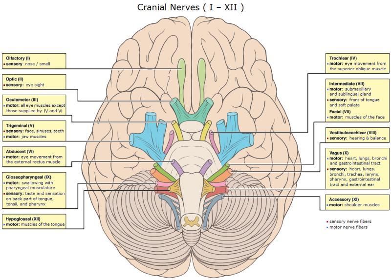
2

                                                             Table of Contents
1.1 Evidence-Based Practice ........................................................................................................................ 3

1.2 What is an ABI? ...................................................................................................................................... 3

1.3 Grading ABIs ........................................................................................................................................... 6

1.4 Neuroanatomy Review .......................................................................................................................... 8

1.5 Mechanism of Injury ............................................................................................................................ 11

1.6 Types of ABI .......................................................................................................................................... 12

   1.6.1 Diffuse Axonal Injury ..................................................................................................................... 12

   1.6.2 Focal Injury .................................................................................................................................... 14

1.7 Disorders of Consciousness.................................................................................................................. 18

1.8 Post Traumatic Amnesia ...................................................................................................................... 21

1.9 References ............................................................................................................................................ 24
3

    Introduction to Moderate to Severe Acquired Brain Injury
1.1 Evidence-Based Practice
The ERABI Clinical Guidebook has been developed through extensive collaboration with clinical experts,
researchers, the Ontario Neurotrauma Foundation, St. Joseph’s Healthcare London, the Lawson Health
Research Institute, Western University, and our partners in Toronto and Ottawa.

The Evidence-Based Review of Acquired Brain Injury (ERABI) Clinical Guidebook is designed to include
clinical information relevant to the identification of ABI sequelae, diagnosis of ABI deficits, outcome
measures appropriate for diagnosis of specific deficits, and valuable evidence-based interventions for
ABI rehabilitation. In addition to this framework, we have also included supplementary materials to
support education in ABI rehabilitation such as clinical algorithms, key studies from the literature, quiz
questions, and case studies to consolidate your knowledge. There are 14 Chapters contained in the
ERABI Clinical Guidebook, focused around ABI specific deficits. Although we recognize research from
other but similar disease states, such as stroke rehabilitation, may be relevant, we have restricted our
content to ABI specific populations in order to maximize applications for ABI rehabilitation.

The ERABI Clinical Guidebook has been developed to provide a summary document of best evidence,
and hopefully serve to help minimize the gaps that exist in evidence-based ABI rehabilitation clinical
care. Some articles cite that the time lag from research to practice in healthcare may be as long as 17
years (Morris, Wooding & Grant, 2011; Hanney et al., 2015; Munro & Savel, 2016). Canada has
recognized the need for high impact tools to support evidence-based care as stated in the Federal
Mandate to the Minister of Health in 2017. This project also directly supports access to care, increased
efficiency, and patient outcomes through technology, knowledge translation, and open access material.

 Click HERE to access the INESSS-ONF Clinical Practice Guideline for the Rehabilitation of adults with
                                      Moderate to Severe TBI

1.2 What is an ABI?
For the purposes of this evidence-based review, we used the definition of ABI employed by the Toronto
Acquired Brain Injury Network (2005). ABI is defined as damage to the brain that occurs after birth and is
not related to congenital disorders, developmental disabilities, or processes that progressively damage
the brain. ABI is an umbrella term that encompasses traumatic and non-traumatic etiologies. ABI
typically involves a wide range of impairments affecting physical, neurocognitive and/or psychological
functioning. A person with an ‘ABI’ might therefore refer to an individual with a traumatic brain injury
(TBI) of any severity, or a non-traumatic brain injury such as a person with Herpes encephalitis, viral
meningitis or acute hypertensive encephalopathy. As opposed to an insidious developmental process,
an ‘ABI’ infers that a person, previously intact from a neurological perspective, subsequently ‘acquired’
some form of brain pathology during their lifespan. Common traumatic causes include motor vehicle
accidents, falls, assaults, gunshot wounds, and sport injuries (Greenwald, Burnett, & Miller, 2003). Non-
traumatic causes of ABI include focal brain lesions, anoxia, tumours, aneurysm, vascular malformations,
and infections of the brain (Toronto Acquired Brain Injury Network, 2005).
4

For the purposes of this Clinical Guidebook vascular causes of focal ABIs, such as stroke, will be excluded
from our evidence synthesis. Included and excluded aetiologies of ABI for the purposes of this
Guidebook can be found in Table 1.1.

                        Click HERE to access the complete methodology for ERABI

Table 1.1 Definitions of ABI used in the ERABI Clinical Guidebook.
             Included in ABI definition                            Excluded from ABI definition
 Traumatic Causes                                     Vascular and Pathological Incidents
  • Motor vehicle accidents                              • Intracerebral hemorrhage (focal)
  • Falls                                                • Cerebrovascular accident (i.e., stroke)
  • Assaults                                             • Vascular accidents
  • Gunshot wounds                                       • Malignant/metastatic tumours
  • Sport Injuries                                    Congenital and Developmental Problems
 Non-traumatic Causes                                    • Cerebral Palsy
  • Tumours (benign/meningioma only)                     • Autism
  • Anoxia                                               • Developmental delay
  • Subarachnoid hemorrhage (non-focal)                  • Down’s syndrome
  • Meningitis                                           • Spina bifida with hydrocephalus
  • Encephalitis/encephalopathy (viral, bacterial,       • Muscular dystrophy
      drug, hepatic)                                  Progressive Processes
  • Subdural Hematoma                                    • Alzheimer’s disease
                                                         • Pick’s disease
                                                         • Dementia
                                                         • Amytrophic Lateral Sclerosis
                                                         • Multiple Sclerosis
                                                         • Parkinson’s disease
                                                         • Huntington’s disease

The vast majority of ABIs result from trauma and the two are essentially synonymous. ABIs are one of
the leading causes of lifelong disability in North America (Greenwald et al., 2003; Pickett, Ardern, &
Brison, 2001; Thurman & Guerrero, 1999). In the United States between 1.4 and 1.7 million people
sustain a TBI every year (Faul, Xu, Wald, & Coronado, 2010; Zaloshnja, Miller, Langlois, & Selassie, 2008),
with more than 120,000 people expected to develop long-term disabilities (Zaloshnja et al., 2008). In the
province of Ontario, more than 80,000 individuals sustained a TBI between 2002 and 2006 (Colantonio
et al., 2010). Although the majority of individuals who sustain a TBI have mild impairment, the
frequency of moderate severity injury is increasing; moderate TBIs accounted for 19% of injuries in 1992
compared with 37% of injuries in 2002 (Colantonio, McVittie, Lewko, & Yin, 2009).

It is important to know which factors are significantly related to outcomes post ABI. Prognostic
indicators can include such variables as injury severity, etiology of injury, age, rehabilitation length of
stay, duration of post-traumatic amnesia, etc. Table 1.2 summarizes the most common ABI prognostic
indicators identified in the literature.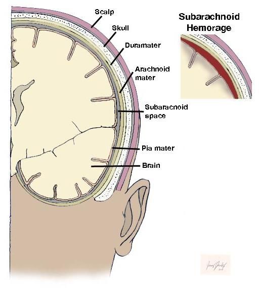
5

Table 1.2 Common Prognostic Indicators for ABI
 • Age                                               •    Rehabilitation length of stay
 • Sex                                               •    Duration of post-traumatic amnesia
 • Presence of prior brain injury                    •    Timing of rehabilitation
 • Injury severity                                   •    Intensity of rehabilitation
 • Length of coma                                    •    Nature of injury (TBI versus nTBI)
 • Initial Glasgow Coma Scale (GCS) score            •    Presence of comorbidities
 • Injury etiology

The majority of TBIs are related to motor vehicle or transportation accidents and falls, together
accounting for approximately 75% of TBIs (Colantonio et al., 2009). Based on literature, falls account for
approximately 35% to 42% of TBIs whereas MVAs are responsible for 12% to 17% (Colantonio et al.,
2010; Faul et al., 2010). These trends have been consistent for approximately ten years (Roozenbeek,
Maas, & Menon, 2013).

The rates of TBIs are typically higher among males, with males in the United States being 1.4 times more
likely to sustain a TBI than a female (Faul et al., 2010). Data from Ontario, Canada is consistent with this
finding and also shows greater rates of TBI among males (Chan, Zagorski, Parsons, & Colantonio, 2013a).
This may be due to greater participation in risk-taking behaviors by males compared to females. A
cohort study has shown that females have higher rates of TBIs due to falls, while TBIs in males are more
likely to be caused by being struck by or against an object (Colantonio et al., 2010).

Another significant demographic factor when evaluating ABI incidence and outcomes is age. Evidence
suggests that etiology of TBIs and outcome varies with age. For children aged 0-14, 50.2% of TBIs in the
United States were caused by falls (Faul et al., 2010), however rates of abusive head trauma can be as
high as 66% (Greenwald et al., 2003). For older adults, aged 65-85, falls make up approximately 60.7% of
TBIs, with falls being the cause of 90% of TBIs for those over 85 (Chan, Zagorski, Parsons, & Colantonio,
2013b, 2013c).

As might be expected, the prognosis of good outcomes long term is also associated with age. Evidence
suggests that age influences the trajectory of one’s recovery following injury. Individuals in the older age
bracket generally had poorer outcomes when compared to younger individuals (Marquez de la Plata et
al., 2008). Older age at the time of injury has also been associated with poorer performance in various
cognitive domains (Senathi-Raja, Ponsford, & Schönberger, 2010). A study by Ashman and Mascialino
(2008) noted that deficits in encoding and retention of verbal information as well as inattention were
more common and more serious post TBI in those over the age of 65 years. It has been postulated, for
those who are older at the time of injury, that less neuronal plasticity may negatively affect the brain’s
ability to compensate or adapt in the same way a younger brain does post injury (Senathi-Raja et al.,
2010). Mosenthal et al. (2002) found older subjects (>64 years of age) had a significantly higher
mortality rate than their younger peers at all levels of TBI severity (p

6

1.3 Grading ABIs

Q1. List three widely used indicators of severity in acute TBI

1. The Glasgow Coma Scale (GCS) (best score within 24 hours of injury).
2. Duration of unconsciousness.
3. Duration of posttraumatic amnesia (PTA).

TBI severity is usually classified according to the level of altered consciousness experienced by the
patient following injury (Table 1.3). The use of level of consciousness as a measurement arose because
the primary outcome to understand the severity of an injury has been the GCS. Consciousness levels
following ABI can range from transient disorientation to deep coma. Patients are classified as having a
mild, moderate or severe ABI according to their level of consciousness at the time of initial assessment.
Various measures of altered consciousness are used in practice to determine injury severity. Common
measures include the GCS, the duration of loss of consciousness (LOC), and the duration of PTA.

Table 1.3 Definitions of Traumatic Brain Injury Severity

 Mild:                      Moderate:                 Severe:                    Very Severe:
  • PTA 7 days
  • GCS 13-15               • GCS 9–12                 • GCS between 3-8         • LOC >48 hours
  • LOC
7

PTA is the time period post trauma for which the conscious patient has no recall for events. PTA is
formally defined as the period following emergence from coma in which the patient may appear
confused, disoriented, or agitated (Campbell, 2000). Research indicates a dose-response relationship,
with the length of PTA frequently being proportional to the severity of injury. Injury severity is defined
as mild if the duration of PTA is less than 1 hour, moderate if between 1–24 hours, and severe if PTA is
between 1–7 days. PTA exceeding 7 days is considered to represent a very severe injury (Campbell,
2000; Russell, 1932).

Q2. Describe the GCS including its strengths and limitations

Description
• The GCS is a quick, simple and objective tool used during the initial examination to estimate TBI
   severity.
• The GCS consists of 15 items in three basic categories: (1) eye opening – 4 items, (2) verbal response
   – 5 items, and (3) motor response – 6 items.
• Minimum score of 3
• Maximum score of 15

Strengths
1. Simple, straightforward and brief bedside assessment.
2. Most familiar and most widely used instrument in the assessment of level of consciousness.
3. Established categories related to depth of coma and severity of injury.
4. Significant predictor of outcome following head injury.
5. Can be used by various groups of healthcare professionals regardless of level of education or ICU
    experience

Weaknesses
1. The application of painful stimulus is controversial.
2. Assessment can be compromised by early interventions such as intubation and sedation.
3. Use of a global score may result in loss of information that adversely affects the predictive accuracy
   of the GCS.
4. The motor response sub score has the greatest influence on the total GCS score.
5. Individuals with the same GCS scores in varying permutations can have significantly different
   survival outcomes.
6. Lack of experience may result in inaccurate assessment.
7. It is an ordinal scale whereby, for example, the difference between scores of 3 and 4 Is not the same
   as the difference between scores of 13 and 14

The GCS score has been shown to have a significant correlation with outcome following severe TBI,
both as the summary score (Choi et al., 1994; Choi, Narayan,
Anderson, & Ward, 1988) and as the motor sub score (Beca
et al., 1995; Bhatty & Kapoor, 1993; Choi et al., 1988;
Michaud, Rivara, Grady, & Reay, 1992). In a prospective
study, the positive predictive value for a poor outcome (dead,
vegetative, or severely disabled) was calculated to be 77% for
patients with a GCS score of 3-5 and 26% for a GCS score of 6-
8

8 (Table 1.5) (Narayan et al., 1981). As is commonly done, this study grouped GCS measurements versus
outcome. In a larger study each GCS level would have its own predictive value. For example, in a series
of 315 patients with TBI from Australia, a significant inverse correlation was demonstrated between the
initial GCS score (obtained 6-48 hours after injury) and mortality (Fearnside, Cook, McDougall, & McNeil,
1993).

1.4 Neuroanatomy Review
As might be expected, the location of the ABI can play a significant role in the deficits observed
afterwards, recognizing that many of these injuries are diffuse in nature or a combination of focal and
diffuse injuries. Figure 1.1 provides an overview of the types of deficits that can be seen given the
location of the insult. Although this list is not exhaustive, it provides the basic functions and concepts
(singular or multiple) that can be negatively impacted by a brain injury.

Figure 1.1 A sagittal view of the human brain which identifies the major regions of function as well as
the common observed deficits which can arise from insult to each specific region of the brain
(Adapted from Excel Care Nurse Case Management, 2019).

Overall, there are 6 regions of the brain which can be impacted by a brain injury. These are the frontal
lobe, the parietal lobe, the occipital lobe, the temporal lobe, cerebellum, and the brain stem. Each of
these regions is characterized by the functions that they are responsible for. The frontal lobe (red) of the
brain is responsible for higher level cognition, motor skills, expressive language, and other functions. It
terminates at the central sulcus and includes the motor cortex. The parietal lobe (yellow) includes the
somatosensory cortex and is primarily responsible for processing tactile and sensory information such as

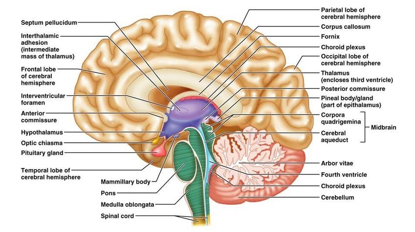
9

pain, pressure, and touch. The temporal lobe (blue), located at the bottom of each hemisphere, controls
the comprehension of language, auditory functions and memory. The occipital lobe (purple) located at
the posterior aspect of the brain, is associated with interpreting visual stimuli and information. The
cerebellum (green), which is part of the hindbrain, is responsible for coordinating gross and fine
movements, walking, while damage to this region can result in tremors, dizziness, and slurred speech.
Although these are not complete descriptions of what each area of the brain is responsible for, it is
sufficient background knowledge to put the existing ABI literature and relevant rehabilitation
interventions into context.

In addition to these regions of the brain, there are internal structures consisting of cranial nerves,
ventricles, and specialized structures which can also be damaged with specific consequences. Damage to
one of the 12 cranial nerves can result in pain, tingling, or loss of sensation. Damage to the pituitary can
result in neuroendocrine dysfunction, while damage to the hippocampus can result in memory deficits.
For more details if a deficit is linked to the dysfunction or damage of a specific region of the brain it will
be discussed in the chapter related to that specific deficit (ex. The ERABI module on Neuroendocrine
Dysfunction discusses damage to both the anterior and posterior pituitary). Figures 1.2 and 1.3 show the
neuroanatomy of the 12 cranial nerves as well as the internal specialized structures of the brain for
reference.
10

                             (Original image attribution goes to Patrick J. Lynch; and was taken from https://my-
                                                                               ms.org/anatomy_brain_part4.htm)

Figure 1.2 Ventral view of the brain with each of the 12 cranial nerves identified along with their
primary functions.
11
(Copywrite © 2009 Pearson Education, Inc, publishing as Pearson Benjamin Cummings)
Figure 1.3 Sagittal view of the brain showing the internal structures of the brain including the
ventricle, hypothalamus, and pituitary gland.
1.5 Mechanism of Injury
The two important factors when considering an ABI are the mechanism of injury (contusion or rotational
forces) and being aware of, and managing, both primary and secondary injuries. The mechanism of the
injury refers to the circumstance of the acquired damage. The two primary categories of injury
mechanisms are presented in Table 1.6. As you can see the majority of mechanisms of injury are derived
from traumatic instances, such as motor vehicle accidents or by making contact with an object.
Table 1.6 Mechanisms of Injury for ABI
Mechanistic Category Description
Contact/Contusion - Injury to scalp
- Fracture of skull with or without associated
extradural hematoma
- Surface contusions, lacerations, and
associated intracerebral hematomas
Acceleration/Deceleration/Rotational Forces - Tearing of bridging veins with the formation
of subdural hematoma
- Diffuse axonal injury, tissue tears and
associated intracerebral hematomas
- Diffuse vascular injury12 When discussing primary and secondary injuries, the primary injury refers to the damage caused as a direct result of the ABI; this can be the forces at impact, vascular injury (Subarachnoid hemorrhage; SAH), benign tumors, cortical disruption or other aetiologies. Secondary injuries encompass the evolution of the primary injury to include additional damage. Four categories of secondary injuries have been described by Kochanek et al. (2013). The first three categories are independent and include 1) “ischemia, excitotoxicity, energy failure”, 2) secondary cerebral swelling, and 3) axonal injury, with the fourth category of inflammation and regeneration, contributing to the evolution of other secondary injury categories. The most common resultants from secondary injury are increased intracranial pressure (discussed in Module 16 of ERABI) and excitotoxicity leading to eventual apoptosis cascades (Kochanek, 2013). Regardless of the severity of the ABI both primary and secondary injuries can play a significant role in determining the outcome of an individual with an ABI. 1.6 Types of ABI 1.6.1 Diffuse Axonal Injury Q3. Describe diffuse axonal injuries 1. Diffuse axonal injury (DAI) is seen exclusively following TBI and results from acceleration- deceleration and rotational forces associated with a high-velocity impact (ie. motor vehicle accidents). 2. Physical shearing of axons results in hemorrhage, tissue tears, axonal swelling and the formation of axonal bulbs acutely. Subacute clusters of microglia and macrophages are seen. Chronically Wallerian degeneration occurs. 3. DAI can be responsible for the initial loss of consciousness seen in acute TBI. 4. Damage is most often seen within midline structures and at interfaces between gray and white matter. In particular, the corpus callosum, the parasagittal white matter, the interventricular septum, the walls of the third ventricle and the brainstem, in particular the midbrain and the pons. Figure 1.4 Two scans showing diffuse axonal injuries in the subcortical regions.
13

DAI is the distinguishing feature of traumatic brain injury due to high-speed accidents. The
predominant causes of DAI include:
    1. High-speed motor vehicle collisions above 15 mph or 24 km/h.
    2. Shaken baby syndrome.
    3. High-speed collisions in sports (i.e. football, hockey, soccer, rugby) (see figure 1.5).

Rotational forces can cause diffuse tearing of neural processes and blood vessels throughout the white
matter resulting in diffuse axonal injury (Figure 1.5). Hemorrhagic changes involving the midline
structures are often associated with rotational acceleration and tend to involve the parasagittal white
matter, corpus callosum, structures in the walls of the 3rd ventricle and striatum (basal ganglia)
(Goldberg, 2001). Strain tends to be concentrated at the interfaces between gray and white matter, at
the midbrain juncture between the brainstem and diencephalon, and at the juncture between the
corpus callosum and the cerebral hemispheres (Goldberg, 2001).

Figure 1.5 Image showing the axonal tearing and shearing that can occur as a result of rotational
forces on the cranium during a traumatic brain injury.
14 Q4. Describe some of the clinical features seen following diffuse axonal injuries. 1. Rostral brain stem involvement results in initial loss of consciousness, poor attention and concentration. 2. Corticospinal tract involvement results in hemiparesis. 3. Shearing of the grey-white matter junction results in slowed mental processing and fatigue. 4. Cerebellar peduncle involvement results in ataxia. 5. Brainstem injury involvement results in dysarthria and dysphagia. Given the breadth of symptoms that can result as a consequence of DAI, specific considerations must be made in order to compensate for deficits in an appropriate manner during early recovery. Q5. How does diffuse axonal injury impact recovery and rehabilitation? 1. Disrupted connections between nerves results in slowed mental processing, fatigue, poor attention and concentration. 2. Rehabilitation must be organized in a manner that compensates for these difficulties such as by providing a structured environment. 3. Physical and cognitive stamina may be reduced and proper pacing will need to be implemented. 4. Poor attention combined with memory difficulties and behavioural concerns may require attendant care. 1.6.2 Focal Injury Cortical Contusions Q6. What are cerebral contusions? Where do they tend to occur most often? 1. Cerebral contusions are cortical “bruises” 2. They occur at the crests of gyri and extend to variable depths. 3. They occur commonly at the inferior frontal, anterior temporal, and inferior occipital lobes.
15

Figure 1.6 Example of a coup-contrecoup injury that might occur where an individual strikes their
head twice on opposite poles of the cranium.

Cortical contusions are quite common following traumatic brain injury and in some cases can be quite
involved, extending through the cortex and into the subcortical white matter (Burke & Ordia, 2000).
Cortical contusions tend to occur in characteristic areas, in part because of the movement of the brain in
the skull with acceleration-deceleration and rotational forces and because of the location of bony
protrusions where the brain can strike inside the skull (Burke & Ordia, 2000). One common combination
of contusions is the coup-contrecoup injury (Figure 1.6). In this instance the patient strikes the front of
their head and the brain accelerates forward in the direction of the impact striking the skull. The brain
then rebounds in the opposite direction and strikes the back of the skull, creating the countercoup
injury. Contusions usually occur bilaterally in the frontal poles, the anterior tips of the temporal lobes
and in some cases the lateral parts of the temporal lobes and the occipital regions (Burke & Ordia,
2000).

Intracranial Hemorrhages

Intracranial hemorrhage, regardless of whether it is epidural, subdural, intracerebral, intraventricular,
or subarachnoid in location, is a major concern post TBI (Burke & Ordia, 2000). A hematoma can
exasperate damage to the brain by: 1) placing direct pressure on the underlying brain structures, or 2)
causing a portion of the brain to herniate leading to secondary compression of the brainstem (Burke &
Ordia, 2000).
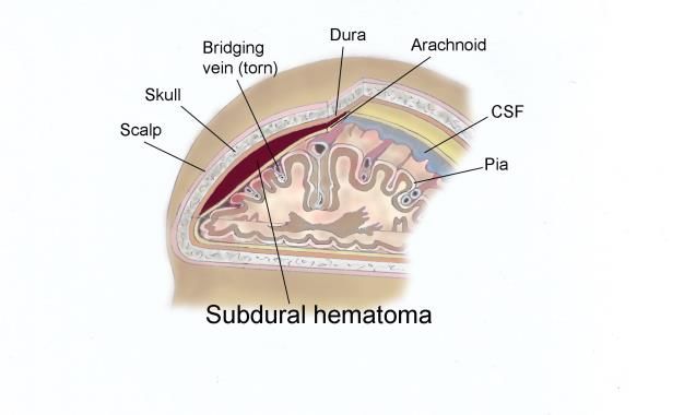
16

Intracranial Hemorrhages: Epidural Hematomas

Q7. What is the general prognosis of epidural hematomas?

1. The underlying brain injury is often not severe if treated promptly.

Figure 1.7 An illustration and two scans which exemplify both the nature of epidural hematomas as
well as their observed pathology.

Epidural hematomas (EDH) are often the result of an impact to the head that causes disruption of the
middle meningeal artery and/or its branches (Burke & Ordia, 2000). Less commonly an EDH can occur
due to a dural venous sinus injury. Blood collects between the dura and the skull. On imaging, an EDH
has a characteristic “lens-shaped” appearance that crosses the midline but does not cross the skull’s
suture lines. Symptom presentation can be delayed and may only become apparent 5 days post injury
(Zasler, Katz, & Zafonte, 2007). However, if the EDH is related to an arterial laceration, it may evolve
quickly resulting in rapid deterioration and even death (Burke & Ordia, 2000). If treated promptly the
underlying brain injury may not be that severe (Burke & Ordia, 2000). Signs of an EDH include a
decreased level of consciousness or “clouding”. If lesions occur in the frontal lobe, onset of symptoms
may be slow and vague (Zasler et al., 2007). Patients must be monitored carefully.

Intracranial Hemorrhages: Subdural Hematomas

Q8. What is the general prognosis of subdural hematomas (SDH)?

1. Prognosis is poor with a high mortality because of the severity of the underlying brain injury

SDH are common in severe brain trauma and often occur in individuals who are on blood thinners or
who are older. SDH occur due to injuries of the cortical bridging veins or rarely the pial artery (Burke &
Ordia, 2000). On imaging, a SDH has a crescent shape that does not cross the midline (it is confined by
dural reflections) but can cross skull suture lines. Due to the severity of injury to the underlying brain
tissue, the prognosis is poor and mortality rates are high.
17

Figure 1.8 An illustration and a scan which exemplifies both the nature and the observed pathology of
subdural hematomas.

Intracranial Hemorrhages: Intracerebral Hemorrhage

Figure 1.9 An illustration and scan showing the nature and pathology of intracerebral hemorrhages.

Intracerebral haemorrhages may result from ruptured blood vessels in association with a penetrating or
non-penetrating head injury. Although they often occur in the frontal or temporal lobes, they may also
be found in the cerebellum and brain stem.
18

Intracranial Hemorrhages: Subarachnoid Hemorrhage

The subarachnoid space is located between the arachnoid membrane and the pia mater that covers the
surface of the brain. This space contains CSF, originating from the choroid plexus. The CSF flows from
the ventricular system to the basal cisterns, and to the surface of the brain. Small arachnoid granulations
within the dural venous sinuses absorb the CSF.

SAH can be atraumatic or traumatic in etiology. SAH
most often occurs secondary to a ruptured
aneurysm of the Circle of Willis causing blood to
accumulate near the site of aneurysmal rupture. In
contrast, SAH related to traumatic cerebral
contusions is variable in terms of its location and is
the result of microvessel shearing in the
subarachnoid space. SAH can be associated with
complications such as intraventricular haemorrhage,
vasospasm, arachnoiditis, seizures, and
communicating hydrocephalus because blood
products obstruct the arachnoid granulations.
Although rare, communicating hydrocephalus can
progress to non-communicating hydrocephalus.

SAH presents on CT scan as bright hyperdense blood
within the sulci and/or basal cisterns. CT is very
sensitive for the detection of acute SAH. MRI is less      Figure 1.10 Illustration of a subarachnoid
sensitive for the detection of acute SAH, however it is    hemorrhage.
very good for identifying chronic blood products or
hemosiderin deposits.

Intracranial Hemorrhages: Intraventricular Hemorrhage

Intraventricular hemorrhage (IVH) can be classified as primary or secondary. Primary IVH occurs due to
injury of the supependymal veins surrounding the ventricles. Secondary IVH occurs due to extension of
adjacent SAH or Intracerebral haemorrhage. On CT, IVH appears bright and is often layered within the
dependent portions of the ventricles. IVH can obstruct cerebrospinal fluid flow resulting in
hydrocephalus.

1.7 Disorders of Consciousness

Q9. Which part of the brain determines consciousness?

1. Consciousness is a function of the ascending reticular activating system (RAS) and the cerebral
   cortex
2. The cell bodies of the RAS are located in the central reticular core of the upper brainstem (primarily
   midbrain) and connect to the cerebral cortex via thalamic and extra-thalamic projections.
19

Consciousness can be defined as an awareness of self, and one’s environment (Plum & Posner, 1982).
Both arousal and awareness are required to be in a conscious state. ABI can significantly impact the
brainstem and the cortex which are responsible for these processes. Severe injuries result in disorders of
consciousness (DOC) defined as a coma, a vegetative state (VS) (Ashwal et al., 1994), or a minimally
conscious state (MCS) (Giacino et al., 2002). The American Academy of Neurology establishes a ≥28 day
loss of consciousness in order to diagnose a DOC. To confirm a diagnoses of either a VS or a MCS the
American Academy of Neurology Guidelines on Disorders of Consciousness (2018) state that
electromyography, evoked potentials, and functional MRIs are able to discriminate between a MCS and
VS and assist in the diagnosis of DOC. According to the American Academy of Neurology only 35% of
individuals with a DOC recover consciousness within the first three months, however this increases to
67% of individuals recovering consciousness six months post-injury (Giaicino et al., 2018).

              Click here to see the AAN Practice Guidelines for Disorders of Consciousness

Q10. Describe the clinical features of coma.

1.   There is no evidence of self- or environmental-awareness.
2.   Eyes remain continuously closed.
3.   No sleep-wake cycles on EEG
4.   No spontaneous purposeful movement
5.   Inability to discretely localize noxious stimuli
6.   No evidence of language comprehension or expression

The criteria for diagnosing a coma has remained relatively unchanged since first described by Plum and
Posner in 1982. The key features of a coma include a) the complete loss of spontaneous or stimulus
induced arousal, b) no detectible sleep/wake cycles on electroencephalography (EEG), c) no evidence of
intentional motor activity such as those in response to a command, d) no language ability, e) eyes
remain closed, even in the presence of adverse stimuli.

Q11. Define what is meant by the term vegetative state.

1. Loss of capacity to interact with the environment despite the preserved potential for spontaneous
   or stimulus-induced arousal

Q12. Describe the clinical features of the vegetative state.

1.   Patient opens eyes (either spontaneously or with noxious stimuli)
2.   Intermittent wakefulness with sleep-wake cycles
3.   No evidence of purposeful behaviour; may startle to verbal/auditory stimuli but will not localize
4.   No evidence of intelligible verbal or gestural communication
5.   Visual tracking is considered a sign of patient transitioning out of the vegetative state
20
VS occurs when the capacity for stimulus-induced or spontaneous arousal is maintained while
awareness surrounding oneself or one’s environment is lost (Giacino, 2013). A diagnosis of VS is made
with the re-emergence of spontaneous eye opening, and the loss of intentional behavioral responses, as
well as the ability to engage in language expression or comprehension. A VS lasting more than one
month can be described as a persistent vegetative state.
Q13. Define MCS.
1. Severely altered consciousness with minimal but definite behavioural evidence of self- or
environmental awareness (Giacino, 2013)
Q14. Describe the clinical features of the MCS.
1. Sleep-wake cycles
2. The patient demonstrates minimal evidence of self- and/or environmental-awareness
3. Purposeful movements are reproducible but remain inconsistent: Simple command following, object
manipulation, and gestural or verbal yes/no responses
4. Eyes will open spontaneously. The patient may also show visual fixation, smooth pursuit tracking
and emotional or motor behaviours contingent upon specific eliciting stimuli (ie. patient may
respond best to voices of family members)
MCS typically reflect a period of recovery from comas or vegetative state. In a post-mortem analysis of
those with TBI and confirmed MCS, typical lesions were found to be grade 2 or 3 axonal injuries (Jennett,
Adams, Murray, & Graham, 2001). To diagnosis a MCS one or more of the following behaviors must be
present and reproducible (Zasler et al., 2007):
1. Simple command following
2. Intelligible verbalization
3. Recognizable verbal or gestural “yes/no” responses (without regard to accuracy)
4. Movements or emotional responses that are triggered by relevant environmental stimuli and
cannot be attributed to reflexive activity. Examples of the fourth criterion include (a) smiling or
crying following exposure to emotional (e.g., family photographs) but not neutral stimuli (e.g.,
photographs of objects), (b) vocalizations or gestures that occur in direct response to specific
linguistic prompts, (c) accurate reaching toward objects laced within the immediate visual field,
(d) manipulation of objects placed in the hand, and (e) sustained visual fixation or pursuit eye
movements.
Emergence from a MCS is typically identified by functional object use as well as increased interactive
communication. These represent the criteria to recover from a MCS as both features require widely
distributed cortical connectivity. Socially, these behaviors also represent a significant increase in
personal autonomy, and therefore are used to determine if an individual has left a MCS.21
1.8 Post Traumatic Amnesia
Q15. What is Post Traumatic Amnesia (PTA)?
1. PTA is defined as the “immediate and dramatic loss of memory that TBI can cause”
2. PTA is the period during which individuals with a TBI “are unable to effectively encode and retain
new information and experiences”
3. PTA varies in duration from a few moments to a few months, either retroactively, proactively, or
both.
4. Resolution of PTA clinically correlates with the period when incorporation of ongoing daily events
occurs in the working memory.
If an individual has post-traumatic amnesia they may appear confused, lack self-awareness of their
injuries, and even be agitated or combative. Individuals may even seem alert during the acute phases of
their injury and engage normally, however, later they may report that they were unconscious for this
period of time and have no memory of it (Eslinger, 2013).
Table 1.7 The relationship of PTA to likely outcomes in individuals with ABI.
Duration of PTA Likely Outcome
1 day or less Expect quick and full recovery with appropriate management (a few patients
may show persisting disability)
More than 1 day and Recovery period is more prolonged lasting weeks or months. However, for
less than 1 week most patients full recovery is possible with good management.
1-2 weeks Recovery occurs over many months. Many patients will be left with residual
problems even after the recovery process has ended, but one can be
reasonably optimistic about functional recovery with good management.
2-4 weeks Process of recovery is very prolonged – 1 year or longer is not unusual.
Permanent deficits are likely. There must be increasing pessimism about
functional recovery when PTA reaches these lengths.
More than 4 weeks Permanent deficits, indeed significant disability, are now certain. It is not just
a matter of recovery but of long-term retraining and management.
(Adapted from Brooks DN and McKinlay WW, Evidence and Quantification in Head Injury: Seminar
notes. Unpublished material, 1989).
PTA gradually improves over time in the majority of cases. The Galveston Orientation and Amnesia Test
(GOAT) is typically used to diagnose and track PTA (Eslinger, 2013). Overall, researchers conclude that
recovery can be predicted from early daily screening and the GOAT can be a moderately strong
predictor of length of stay, functional independence, and short-term memory (Eslinger, 2013).22
Q16. Describe the GOAT including advantages and disadvantages.
Description
The GOAT consists of 10 items regarding orientation to:
1. person: name, address and birth date
2. place: city/town and building they are in
3. time: current time, date, month year and date of hospital admission
4. memory of events both after and prior to the injury (Bode, Heinemann, & Semik, 2000).
Advantages
1. The GOAT provides an objective rating of early cognitive recovery eliminating the need for
sometimes ambiguous terminology used to describe mental status, such as “confused” (Levin,
O'Donnell, & Grossman, 1979).
2. Due to its design, the scale has been shown to be useful for assessing patients with a wide range of
cognitive impairments (Salter, Jutai, & Teasell, 2008).
3. Can be used to guide timing of neuropsychological testing which should not be attempted until
GOAT score consistently >70.
Limitations
1. The standard GOAT response format makes administration difficult with nonverbal patients
(Novack, Dowler, Bush, Glen, & Schneider, 2000). It is important to note that A-GOAT has been
developed for use in aphasic patients but requires further evaluation.
2. The requirement for oral or written expression may result in penalizing patients who are
experiencing deficits of expression but not in orientation or in the retrieval or consolidation of
memory (Jain, Layton, & Murray, 2000).
3. While the GOAT does contain items intended to provide an assessment of memory, it is primarily a
measure of orientation. Eight of the 10 GOAT items evaluate orientation while only two examine
memory (Forrester, Encel, & Geffen, 1994).
The GOAT provides an objective assessment with a standardized cut-off for the presence of PTA.
However, in its original form, the GOAT is not well suited to assessment of patients with aphasia. It may
be too lengthy for a simple, repeated bedside assessment of mental status. However, it is freely
available and can be used by any healthcare professional (Salter et al., 2008).
Assessment consists of 10 items regarding orientation to person (name, address & birthdate), place
(city/town and building they are in) and time (current time, date, month, year & date of hospital
admission) as well as memory of events both after and prior to the injury (Bode et al., 2000). Oral
questions are posed directly to the patient who may respond either orally or in writing (Jain et al., 2000;
Levin et al., 1979). Error points are awarded for each incorrect response and are summed and deducted
from 100 to arrive at the total score. Both the scale and instructions for assigning error points are
available in Levin et al. (1979).
The GOAT was intended to evaluate orientation to time, place and person and to provide an estimation
of the intervals prior to and following the injury for which there is no recall (Levin et al., 1979). The
duration of PTA is defined as the period following coma in which the GOAT score is 75 or less (Levin et al.,23 1979). PTA is considered to have ended if a score greater than 75 is achieved on 2 consecutive administrations (Novack et al., 2000; Wade, 1992; Zafonte et al., 1997). In the initial standardization study of Levin et al. (1979) using patients with mild head injury as a reference group, it was determined that a score of 75 represented a level achieved by 92% of the standardization group. No patients with mild head injury scored less than 65 on the GOAT. Scores between 66 and 75 are considered borderline- abnormal while scores above 75 fall into the range considered normal within the reference group (Levin et al., 1979; van Baalen et al., 2003).
24

1.9 References
Ashman, T. A., Cantor, J. B., Gordon, W. A., Sacks, A., Spielman, L., Egan, M., & Hibbard, M. R. (2008). A
      comparison of cognitive functioning in older adults with and without traumatic brain injury. J
      Head Trauma Rehabil, 23(3), 139-148. doi:10.1097/01.htr.0000319930.69343.64

Ashwal, S., Cranford, R., Bernat, J. L., Celesia, G., The Multi-Society Task Force on, P. V. S., & Multi-
       Society Task Force on, P. V. S. (1994). Medical Aspects of the Persistent Vegetative State. The
       New England Journal of Medicine, 330(21), 1499-1508. doi:10.1056/NEJM199405263302107

Beca, J., Cox, P. N., Taylor, M. J., Bohn, D., Butt, W., Logan, W. J., . . . BarkerG. (1995). Somatosensory
         evoked potentials for prediction of outcome in acute severe brain injury. J Pediatr, 126(1), 44-
         49.

Bhatty, G. B., & Kapoor, N. (1993). The Glasgow Coma Scale: a mathematical critique. Acta Neurochir
        (Wien), 120(3-4), 132-135.

Bode, R. K., Heinemann, A. W., & Semik, P. (2000). Measurement properties of the Galveston
        Orientation and Amnesia Test (GOAT) and improvement patterns during inpatient rehabilitation.
        J Head Trauma Rehabil, 15(1), 637-655.

Burke, D., & Ordia, J. I. (2000). Pathophysiology of Traumatic Brain Injury. In N. S. Woo BH (Ed.), The
        Rehabilitation of People with Traumatic Brain Injury (1st ed., pp. 19-34). Boston MA: Boston
        Medical Center.

Campbell, M. (2000). Rehabilitation for traumatic brain injury: physical therapy practice in context:
      Churchill Livingstone.

Chan, V., Zagorski, B., Parsons, D., & Colantonio, A. (2013a). Older adults with acquired brain injury: a
        population based study. BMC Geriatr, 13, 97. doi:10.1186/1471-2318-13-97

Chan, V., Zagorski, B., Parsons, D., & Colantonio, A. (2013b). Older adults with acquired brain injury:
        Functional independence measures after inpatient rehabilitation. Archives of physical medicine
        and rehabilitation, 94 (10), e9.

Chan, V., Zagorski, B., Parsons, D., & Colantonio, A. (2013c). Older adults with acquired brain injury:
        outcomes after inpatient rehabilitation. Canadian Journal on Aging, 32(3), 278-286.

Choi, S. C., Barnes, T. Y., Bullock, R., Germanson, T. A., Marmarou, A., & Young, H. F. (1994). Temporal
         profile of outcomes in severe head injury. J Neurosurg, 81(2), 169-173.
         doi:10.3171/jns.1994.81.2.0169

Choi, S. C., Narayan, R. K., Anderson, R. L., & Ward, J. D. (1988). Enhanced specificity of prognosis in
         severe head injury. J Neurosurg, 69(3), 381-385. doi:10.3171/jns.1988.69.3.0381

Colantonio, A., McVittie, D., Lewko, J., & Yin, J. (2009). Traumatic brain injuries in the construction
       industry. Brain Inj, 23(11), 873-878. doi:10.1080/02699050903036033
25
Colantonio, A., Saverino, C., Zagorski, B., Swaine, B., Lewko, J., Jaglal, S., & Vernich, L. (2010).
Hospitalizations and emergency department visits for TBI in Ontario. Can J Neurol Sci, 37(6),
783-790.
Eslinger, P. J., Zappala, G., Chakara, F., Barrett, A.M. . (2013). Cognitive Impairments. In K. D. Zasler ND,
Zafonte RD. (Ed.), Brain Injury Medicine: Principles and Practice (2nd ed., pp. 990-1001). New
York: Demos Medical Publishing.
Faul, M., Xu, L., Wald, M., & Coronado, V. (2010). Traumatic Brain Injury in the United States. Centers for
Disease Control and Prevention, National Center for Injury Prevention and Control. Atlanta, GA.
Fearnside, M. R., Cook, R. J., McDougall, P., & McNeil, R. J. (1993). The Westmead Head Injury Project
outcome in severe head injury. A comparative analysis of pre-hospital, clinical and CT variables.
Br J Neurosurg, 7(3), 267-279.
Forrester, G., Encel, J., & Geffen, G. (1994). Measuring post-traumatic amnesia (PTA): an historical
review. Brain Inj, 8(2), 175-184.
Giacino, J. T., Ashwal, S., Childs, N., Cranford, R., Jennett, B., Katz, D. I., . . . Zasler, N. D. (2002). The
minimally conscious state: definition and diagnostic criteria. Neurology, 58(3), 349-353.
doi:10.1212/WNL.58.3.349
Giacino, J. T., Katz, Douglas I., Garber, K., Schiff, Nicolas. (2013). Assessment and Rehabilitative
Management of Indiviuals with Disorders of Consciousness In K. D. Zasler ND, Zafonte RD. (Ed.),
Brain Injury Medicine: Principles and Practice (2nd ed., pp. 517-535). New York: Demos Medical
Publishing, LLC
Giaicino, J. T., Katz, D. I., Schiff, N. D., Whyte, J., Ashman, E. J., Ashwal, S., . . . Armstrong, M. J. (2018).
Report of the Guideline Development, Dissemination, and Implementation Subcommittee of the
American Academy of Neurology; the American Congress of Rehabilitation Medicine; and the
National Institute on Disability, Independent Living, and Rehabilitation Research. Retrieved from
Goldberg, G. (2001). Mild Traumatic Brain Injury and Concussion. Physical Medicine and Rehabilitation:
State of the Art Reviews, 15, 363-398.
Greenwald, B. D., Burnett, D. M., & Miller, M. A. (2003). Congenital and acquired brain injury. 1. Brain
injury: epidemiology and pathophysiology. Archives of physical medicine and rehabilitation, 84(3
Suppl 1), S3-7.
Jain, N. S., Layton, B. S., & Murray, P. K. (2000). Are aphasic patients who fail the GOAT in PTA? A
modified Galveston Orientation and Amnesia Test for persons with aphasia. Clin Neuropsychol,
14(1), 13-17. doi:10.1076/1385-4046(200002)14:1;1-8;ft013
Jennett, B., Adams, J. H., Murray, L. S., & Graham, D. I. (2001). Neuropathology in vegetative and
severely disabled patients after head injury. Neurology, 56(4), 486-490.
doi:10.1212/WNL.56.4.48626
Kochanek, P. M., Clark, R.S.B., Jenkins, L.W. . (2013). Pathobiology of Secondary Brain Injury. In K. D.
Zasler ND, Zafonte RD. (Ed.), Brain Injury Medicine: Princples and Practice (2nd ed., pp. 148-161).
New York: Demos Medical Publishing.
Levin, H. S., O'Donnell, V. M., & Grossman, R. G. (1979). The Galveston Orientation and Amnesia Test. A
practical scale to assess cognition after head injury. J Nerv Ment Dis, 167(11), 675-684.
Marquez de la Plata, C. D., Hart, T., Hammond, F. M., Frol, A. B., Hudak, A., Harper, C. R., . . . Diaz-
Arrastia, R. (2008). Impact of age on long-term recovery from traumatic brain injury. Archives of
physical medicine and rehabilitation, 89(5), 896-903.
Michaud, L. J., Rivara, F. P., Grady, M. S., & Reay, D. T. (1992). Predictors of survival and severity of
disability after severe brain injury in children. Neurosurgery, 31(2), 254-264.
Mosenthal, A. C., Lavery, R. F., Addis, M., Kaul, S., Ross, S., Marburger, R., . . . Livingston, D. H. (2002).
Isolated traumatic brain injury: age is an independent predictor of mortality and early outcome.
Journal of Trauma-Injury, Infection, and Critical Care, 52(5), 907-911.
Murdoch, B., & Theodoros, D. (2001). Introduction: Epidemiology, neuropathophysiology and medical
aspects of traumatic brain injury. San Diego, CA: Singular/Thomson Learning.
Narayan, R. K., Greenberg, R. P., Miller, J. D., Enas, G. G., Choi, S. C., Kishore, P. R., . . . Becker, D. P.
(1981). Improved confidence of outcome prediction in severe head injury. A comparative
analysis of the clinical examination, multimodality evoked potentials, CT scanning, and
intracranial pressure. J Neurosurg, 54(6), 751-762. doi:10.3171/jns.1981.54.6.0751
Novack, T. A., Dowler, R. N., Bush, B. A., Glen, T., & Schneider, J. J. (2000). Validity of the Orientation Log,
relative to the Galveston Orientation and Amnesia Test. J Head Trauma Rehabil, 15(3), 957-961.
Pickett, W., Ardern, C., & Brison, R. J. (2001). A population-based study of potential brain injuries
requiring emergency care. Cmaj, 165(3), 288-292.
Plum, F., & Posner, J. B. (1982). The Diagnosis of Stupor and Coma. Philadelphia: F.A. Davis Co. .
Roozenbeek, B., Maas, A. I., & Menon, D. K. (2013). Changing patterns in the epidemiology of traumatic
brain injury. Nat Rev Neurol, 9(4), 231-236. doi:10.1038/nrneurol.2013.22
Russell, W. R. (1932). Cerebral Involvement in Head Injury a Study Based on the Examination of Two-
hundred Cases. Brain, 55(4), 549-603.
Salter, K., Jutai, J., & Teasell, R. (2008). Assessment of Outcomes Following Acquired/Traumatic Brain
Injury. In M. S. Teasell R, Cullen N., Bayley M (Ed.), Evidence-Based Review of Moderate to
Severe Acquired Brain Injury (4th ed., pp. 1-73). London, ON: Ontario Neurotrauma Foundation.
Senathi-Raja, D., Ponsford, J., & Schönberger, M. (2010). Impact of age on long-term cognitive function
after traumatic brain injury. Neuropsychology, 24(3), 336.
Teasdale, G., & Jennett, B. (1974). Assessment of coma and impaired consciousness: a practical scale.
The Lancet, 304(7872), 81-84.27
Teasdale, G., & Jennett, B. (1976). Assessment and prognosis of coma after head injury. Acta
neurochirurgica, 34(1-4), 45-55.
Thurman, D., & Guerrero, J. (1999). Trends in hospitalization associated with traumatic brain injury.
Jama, 282(10), 954-957.
Toronto Acquired Brain Injury Network. (2005). Definition of acquired brain injury. Retrieved from
http://www.abinetwork.ca/downloads/binder-b3.pdf
van Baalen, B., Odding, E., Maas, A. I., Ribbers, G. M., Bergen, M. P., & Stam, H. J. (2003). Traumatic
brain injury: classification of initial severity and determination of functional outcome. Disabil
Rehabil, 25(1), 9-18.
Wade, D. T. (1992). Measurement in neurological rehabilitation. Curr Opin Neurol Neurosurg, 5(5), 682-
686.
Zafonte, R. D., Mann, N. R., Millis, S. R., Black, K. L., Wood, D. L., & Hammond, F. (1997). Posttraumatic
amnesia: its relation to functional outcome. Arch Phys Med Rehabil, 78(10), 1103-1106.
Zaloshnja, E., Miller, T., Langlois, J. A., & Selassie, A. W. (2008). Prevalence of long-term disability from
traumatic brain injury in the civilian population of the United States, 2005. The Journal of head
trauma rehabilitation, 23(6), 394-400.
Zasler, N. D., Katz, D. I., & Zafonte, R. D. (2007). Brain Injury Medicine. New York: Demos Medical
Publishing.You can also read