Traumatic Injuries to the Spinal Cord and Peripheral Nervous System - BINASSS
←
→
Page content transcription
If your browser does not render page correctly, please read the page content below
Tra u m a t i c I n j u r i e s t o t h e
   S p i n a l C o rd a n d P e r i p h e r a l
   Nervous System
                                  a                            b,
   Lucas Sjeklocha,          MD       , J. David Gatz,    MD        *

     KEYWORDS
      Peripheral nerve injury  Spinal cord injury  Trauma  Neurogenic shock
      Secondary injury

     KEY POINTS
      Injuries to the peripheral nervous system and spinal cord regularly occur during blunt and
       penetrating trauma.
      Other life-threatening injuries should be prioritized in the setting of polytrauma before pur-
       sing definitive management of injuries to peripheral nerves or the spinal cord.
      Sharply transected peripheral nerve injuries should prompt consultation for immediate
       repair.
      Providers should have a low threshold for intubation during the acute management of spi-
       nal cord injuries because lower cervical and even thoracic injuries can result in insufficient
       airway protection or breathing.
      First-line management of neurogenic shock should be intravenous fluids followed by, if
       necessary, norepinephrine to maintain a mean arterial pressure of at least 85 mm Hg.

   INTRODUCTION

   All trauma, whether blunt or penetrating, has the potential to cause injury to the ner-
   vous system. This includes the brain and spinal cord of the central nervous system
   and the somatic and autonomic components of the peripheral nervous system
   (PNS). Traumatic injuries to the PNS are a significant source of morbidity. Peripheral
   nerve injury (PNI) can result in permanent disability and entail significant health care
   and patient costs. Acute costs associated with these injuries average nearly $6000
   in the emergency department (ED) and $20,000 to $60,000 in inpatient expenses.1–3
   These costs do not consider the burden of decreased quality of life and long-term
   health care costs.

     a
       R Adams Cowley Shock Trauma Center, 22 South Greene Street, Room S4D03, Baltimore, MD
     21201, USA; b Department of Emergency Medicine, University of Maryland School of Medicine,
     110 South Paca Street, 6th Floor, Suite 200, Baltimore, MD 21201, USA
     * Corresponding author.
     E-mail address: jgatz@som.umaryland.edu
     Twitter: @DrDavidGatz (J.D.G.)

     Emerg Med Clin N Am 39 (2021) 1–28
     https://doi.org/10.1016/j.emc.2020.09.001                                                   emed.theclinics.com
     0733-8627/21/ª 2020 Elsevier Inc. All rights reserved.

Descargado para BINASSS Circulaci (binas@ns.binasss.sa.cr) en National Library of Health and Social Security de ClinicalKey.es por
Elsevier en febrero 15, 2021. Para uso personal exclusivamente. No se permiten otros usos sin autorización. Copyright ©2021. Elsevier
                                                  Inc. Todos los derechos reservados.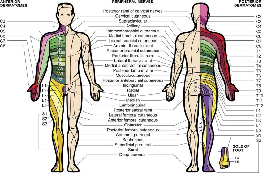
2 Sjeklocha & Gatz
Spinal cord injuries (SCIs) similarly can cause significant permanent disability and
even death. The financial cost to patients and society from SCIs is significant, accu-
mulating average expenses in the United States between $375,000 and $1,150,000
in the first year alone, depending on severity of injury. Subsequent annual expenses
average between $45,000 to $200,000.4
Emergency medicine providers play an essential role in the recognition and subse-
quent management of these patients. Presentations can be subtle and sometimes are
missed, risking morbidity and even mortality for patients, and subjecting providers to
high medicolegal liability. Prior malpractice suits involving missed cervical injuries in
blunt trauma patients, for example, have resulted in multimillion-dollar awards.5
This article covers the pathophysiology, clinical assessment, and management of
traumatic PNIs and SCIs. The primary population of this review is adult patients.
Please see the article “Neurologic Emergencies at the Extremes of Age,” by Khoujah
and Cobb for further discussion of pediatric and geriatric populations.
PERIPHERAL NERVE INJURY
Epidemiology
Traumatic PNI represents a significant burden of disease. PNIs are thought to have an
incidence of more than 350,000 per year in the United States.6 Previous estimates us-
ing the National Inpatient Sample and National Emergency Department Sample show
widely varying estimates of injuries based on diagnosis codes and likely underesti-
mate the true burden of injury. The overall trend in injuries has been relatively stable,
with upper extremity PNI more common than lower extremity PNI.1,2 Among trauma
patients evaluated at a level 1 trauma center, 2.8% were identified as having a
PNI.7 European trauma registry data showed PNIs associated with 3.3% of severe up-
per extremity trauma and 1.8% of severe lower extremity trauma.8,9 PNIs may remain
occult in severely injured patients given priorities of resuscitation and concomitant in-
juries that limit the examination. A previous case series of traumatic brain injury pa-
tients identified a 34% incidence of PNIs not recognized on initial evaluation.10
Males account for 80% of traumatic PNIs with a mean age of approximately 40
years in registry data.8,9 Blunt mechanisms, including motor vehicle accidents and
falls, account for the majority of injuries but disproportionately higher rates of PNIs
are observed in penetrating trauma then compared to the overall trauma population.9
In upper extremity trauma, the ulnar nerve is injured most commonly, followed by the
radial and median nerves.2 The peroneal nerve is the lower extremity nerve most
commonly injured followed by the sciatic and tibial nerves.1 Sports-related acute
nerve injury represents less than 1% of all injuries but has been increasing.11
Key Anatomy and Pathophysiology
The terms, peripheral nerve and PNS, encompass nervous tissue and supporting
structures between the central nervous system and target tissues or sensory areas.
These include cranial nerves (except for the optic nerves) and the autonomic nervous
system, in addition to motor and sensory branches originating in the spinal cord. Pe-
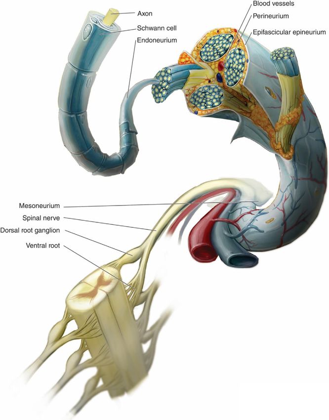
ripheral nerves have several specialized connective tissues: the endoneurium, the
perineurium, and the epineurium (Fig. 1). These define the structure of the nerve
and are critical for regeneration.12
Most PNIs can be attributed to a combination of mechanisms including traction or
stretch, contusion, transection, and compression. Other mechanisms of traumatic
injury include ischemia, burns, and electrical injuries. Nerves are vulnerable to different
mechanisms along their length due to the changing composition of the nerves and
Descargado para BINASSS Circulaci (binas@ns.binasss.sa.cr) en National Library of Health and Social Security de ClinicalKey.es por
Elsevier en febrero 15, 2021. Para uso personal exclusivamente. No se permiten otros usos sin autorización. Copyright ©2021. Elsevier
Inc. Todos los derechos reservados.Traumatic Injuries of the Spinal Cord and Peripheral Nerves 3
Fig. 1. Major anatomic components of a peripheral nerve. (From Smith BW, Sakamuri S,
Spain DA, Joseph JR, Yang LJ, Wilson TJ. An update on the management of adult traumatic
nerve injuries-replacing old paradigms: A review. J Trauma Acute Care Surg. 2019;86(2):299-
306; with permission.)
regional anatomy. Nerve roots, for instance, lack both epineurium and perineurium
and are relatively tethered to the spinal cord, making them vulnerable to traction
and compression.6 Proximity to bone makes nerves vulnerable to injury from fractures,
whereas superficial nerves may be more easily contused or lacerated.
Nerve stretching can be part of normal function with changes in length as nerves
cross over joints and at extremes of physiologic movement. Extremes of stretching
overwhelm the ability of the connective tissue to compensate and result in injuries
Descargado para BINASSS Circulaci (binas@ns.binasss.sa.cr) en National Library of Health and Social Security de ClinicalKey.es por
Elsevier en febrero 15, 2021. Para uso personal exclusivamente. No se permiten otros usos sin autorización. Copyright ©2021. Elsevier
Inc. Todos los derechos reservados.4 Sjeklocha & Gatz
with associated hematomas and scarring.13 Avulsion is an extreme stretch or traction
injury causing mechanical failure and disruption of the nerve, often occurring at nerve
roots and is associated with significant morbidity.
Compression can cause ischemic injury from direct or indirect pressure (eg, asso-
ciated compartment syndrome). A classic example is compression of the radial nerve
against the humerus as it travels in the radial groove, producing a Saturday night
palsy. Hydrostatic forces from penetrating injury also can cause nerve injury or disrup-
tion. Crush injuries can occur directly or via entrapment from dislocation-relocation or
associated fractures. Laceration or transection mechanisms can be divided into sharp
and blunt.
Classification of Peripheral Nerve Injuries
In 1942, Seddon14 proposed a classification scheme that is still in primary use today
for grading nerve injuries based on severity of disruption to the nerve and supporting
structures. Seddon divided injuries as neurapraxia, axonotmesis, and neurotmesis
(Fig. 2). Sunderland15 later expanded this to 5 degrees of injury (Table 1).
Presentation and Examination of Peripheral Nerve Injuries
Traumatic PNI initially is a clinical diagnosis and, because PNIs by themselves
typically are not life threatening, other more dangerous and time-sensitive causes
and associated injuries must be considered. In the trauma patient, prompt global
assessment and resuscitation should be undertaken prior to detailed investigation
for nerve injury. All sensory or motor abnormalities should be evaluated for alter-
native causes, especially central causes, such as intracranial hemorrhage and
SCIs.16 An evolving deficit should prompt evaluation for a dynamic process like
progressive edema, hematoma formation, pseudoaneurysm formation, or shifting
of fractures.
Evidence of nerve injury should prompt consideration of associated fractures, he-
matomas, compartment syndrome, and arterial injuries. Because nerves typically
travel along the neurovascular bundles, and blood vessels are vulnerable to the
same forces, approximately 13% of upper extremity PNIs from civilian trauma have
an associated vascular injury.9,17 Injuries associated with warfare and penetrating
injury have an even higher association between PNIs and vascular injuries, with arterial
injuries present in 48 of 119 patients in a case series of PNIs from the Balkan conflict.18
Traumatic injuries typically present with maximal deficits.
It is critical to determine open versus closed injuries because this significantly alters
management.19–23 Exploration of an open wound and assessment of the wound
mechanism can help identify an associated clinical nerve injury. A clean, sharp tran-
section versus a blunt, ragged transection can affect the urgency of repair.20,21,23 Pro-
viders should use motor grading and sensory testing to determine the severity and
likely anatomic location(s) of injury (Fig. 3 for sensory distribution of major peripheral
nerves). Two-point discrimination is the preferred mode of testing for sensory injury
with a recent case series of hand injuries, demonstrating 98.6% sensitivity for detect-
ing nerve injury with a 2-point discrimination tool compared to 82.5% for dry
gauze.16,24 Tinel sign also may be present acutely at the area of injury with advancing
location and increased pain present in regenerating injuries and developing neuromas,
respectively.25
Providers should be prepared to recognize several classic PNIs of the upper
(Table 2) and lower (Table 3) extremities.
Descargado para BINASSS Circulaci (binas@ns.binasss.sa.cr) en National Library of Health and Social Security de ClinicalKey.es por
Elsevier en febrero 15, 2021. Para uso personal exclusivamente. No se permiten otros usos sin autorización. Copyright ©2021. Elsevier
Inc. Todos los derechos reservados.Traumatic Injuries of the Spinal Cord and Peripheral Nerves 5
Fig. 2. (A) Intact peripheral nerve anatomy. (B) Anatomic schematic of PNI grades.
Diagnostic Evaluation of Peripheral Nerve Injuries
Clinical examination combined with potential surgical exploration, electromyog-
raphy, and nerve conduction studies is important for overall assessment of PNIs.
Additional diagnostics during acute presentations in the ED are largely supplements
to the clinical examination and evaluate primarily for associated injuries and alterna-
tive causes.
Descargado para BINASSS Circulaci (binas@ns.binasss.sa.cr) en National Library of Health and Social Security de ClinicalKey.es por
Elsevier en febrero 15, 2021. Para uso personal exclusivamente. No se permiten otros usos sin autorización. Copyright ©2021. Elsevier
Inc. Todos los derechos reservados.6 Sjeklocha & Gatz
Table 1
Seddon and Sunderland classification schemes of peripheral nerve injuries
Prognosis for
Clinical Pathologic Spontaneous Surgical
Seddon Sunderland Correlate Correlate Recovery Intervention
Neurapraxia 1 Compression, Demyelination Good Unnecessary
ischemia
Axonotmesis 2 Ischemia, Axon Good to fair Usually
crush, degeneration unnecessary
percussion
3 Endoneural injury Intermediate May be required
4 Perineural injury Poor May be required
Neurotmesis 5 Avulsion, Epineural Injury Poor Required
transection
From National Spinal Cord Injury Statistical Center. 2019 Annual Statistical Report for the Spinal
Cord Injury. Model Systems. University of Alabama at Birmingham: Birmingham, Alabama; With
permission.
X-ray and computed tomography evaluation
Peripheral nerves are not imaged by plain radiographs and are poorly imaged with
computed tomography (CT). These images can evaluate for associated injuries. Indi-
vidual nerve injuries may prompt specific radiographs to identify commonly associ-
ated fractures or dislocations (eg, hook of hamate fracture in distal ulnar nerve
injury or evaluation for a Bankart or Hill-Sachs lesion suggestive of previous disloca-
tion in axillary nerve dysfunction). Although CT is inadequate for direct evaluation of
Fig. 3. Comparison of peripheral nerve fields and dermatomes. (From Smith BW, Sakamuri S,
Spain DA, Joseph JR, Yang LJ, Wilson TJ. An update on the management of adult traumatic
nerve injuries-replacing old paradigms: A review. J Trauma Acute Care Surg. 2019;86(2):299–
306; with permission.)
Descargado para BINASSS Circulaci (binas@ns.binasss.sa.cr) en National Library of Health and Social Security de ClinicalKey.es por
Elsevier en febrero 15, 2021. Para uso personal exclusivamente. No se permiten otros usos sin autorización. Copyright ©2021. Elsevier
Inc. Todos los derechos reservados.Traumatic Injuries of the Spinal Cord and Peripheral Nerves 7
Table 2
Classic peripheral nerve injuries of the upper extremities
Nerves Example Mechanism(s) Major Deficits Pathologic Correlate
Brachial Stinger/burner Variable; typically, Neuropraxia, nerve
plexus Seatbelt injury C5-C6 or avulsion in
C8-T1, depending on severe trauma
direction of forces
Autonomic deficits
(eg, Horner
syndrome)
in C8-T1 lesions
Axillary Shoulder dislocation Sensory: deltoid area Neuropraxia
Surgical neck fracture Motor: shoulder flexion
of humerus and abduction
Radial Midshaft humerus Sensory: dorsal medial Neuropraxia
fracture hand
Saturday night palsy Motor: wrist extension,
finger extension
Ulnar Elbow dislocation Sensory: ulnar hand Axonotmesis
(proximal) Medial epicondyle Motor: grip strength,
fracture fourth and fifth digits
cubital compression flexion (proximal)
Median Supracondylar fracture Sensory: palmar radial Variable
of humerus hand
Laceration (typically Motor: thumb
distal) opposition,
second and third
digits flexion
Long thoracic Penetrating chest, Motor: scapular Neurotmesis
nerve axilla, protraction
or supraclavicular (may significantly
trauma impair upper
extremity function)
Table 3
Classic peripheral nerve injuries of the lower extremities
Pathologic
Nerves Example Mechanism(s) Major Deficits Correlate
Sciatic Posterior hip dislocation Sensory: posterior and lateral leg, Variable
Penetrating trauma dorsal and plantar foot
Motor: knee flexion,
ankle dorsiflexion
and plantarflexion
Peroneal Knee dislocation Sensory: dorsal foot Variable
Fibular fracture Motor: ankle dorsiflexion
Contusion and eversion
Inferior gluteal Posterior hip dislocation Sensory: none Variable
Motor: hip extension and
extension of the flexed thigh
Tibial Tibial fracture Sensory: plantar foot Variable
Knee dislocation Motor: ankle plantarflexion
Descargado para BINASSS Circulaci (binas@ns.binasss.sa.cr) en National Library of Health and Social Security de ClinicalKey.es por
Elsevier en febrero 15, 2021. Para uso personal exclusivamente. No se permiten otros usos sin autorización. Copyright ©2021. Elsevier
Inc. Todos los derechos reservados.8 Sjeklocha & Gatz
nerve injury, it has added utility for evaluating soft tissue lesions and vascular struc-
tures25 CT myelography is sensitive and specific for brachial plexus injury and nerve
root avulsion in later phases of injury.26
Magnetic resonance imaging
Magnetic resonance imaging (MRI), also called magnetic resonance neurography, is
superior to CT for PNIs due to significantly improved contrast resolution, ability to
assess nerve edema, and evolving use of sequences to assess nerve integrity.25,27,28
The utility of MRI in the immediate or early evaluation of suspected injury is unclear,
because there are no established guidelines and because of typically conservative
overall management strategy of closed nerve injuries. Given the limitations of electro-
diagnostic testing in the acute phase, there may be select cases of MRI that allow for
earlier intervention.29
Ultrasonography
Ultrasonography, along with MRI, is the other preferred imaging technique for nerve
injury. Ultrasound offers high spatial resolution, the ability to perform dynamic maneu-
vers, and comparatively low cost but with limited contrast resolution, limited ability to
image deeper structures, and significant operator dependence.28,30–32 Nerves are
imaged best with a high-frequency linear array and have a characteristic echotexture
due to bundles of nerve fibers or fascicles.
Superficial nerves are well visualized and can be traced along their course to eval-
uate for swelling, size, or echotexture changes (eg, loss of internal architecture) that
may indicate neuropraxia, axonotmesis/neurotmesis, and disruption of nerve continu-
ity.30–32 These can be accentuated by dynamic maneuvers. Ultrasound is able to char-
acterize much of the course of the most commonly injured nerves and can identify
areas of nerve entrapment.27,33,34
The role of ultrasound in the ED is evolving but has been shown in some cases to be
superior to MRI in evaluation of nerve lesions.35 Ultrasound evaluation changes man-
agement in as many as 58% of cases, including decisions on immediate versus
delayed surgery, identification of complete nerve disruption, detection of foreign
bodies, and detection of multiple areas of injury.36,37
Electrodiagnostic testing
Electrodiagnostic testing is the interrogation of nerve function using electrical im-
pulses and is widely used for evaluation of nerve function, including in traumatic in-
juries. This technique does not have a role in the acute setting because
electromyography and nerve conduction studies cannot differentiate between neuro-
praxia, axonotmesis, and neurotmesis immediately after injury.38,39 Neuropraxia and
higher-grade injuries can be differentiated by 1 week postinjury. PNI features are var-
iable between injury areas and type, and serial evaluations over time are used to help
gauge recovery and plan interventions.
Management of Peripheral Nerve Injuries
Disposition and follow-up
Treatment can vary widely after initial evaluation of possible nerve injury because,
depending on the type of injury, it may be supportive or surgical. Smith and colleagues
outlined a proposed approach to management of nerve injury building on the
approach of Grant and colleagues (Fig. 4).20,23
The rule of 3s can be helpful in considering the appropriate timing of follow-up and
intervention in subspecialty care. Sharp nerve transections are best explored and
repaired within 3 days. Open injuries that are ragged or contused may be best
Descargado para BINASSS Circulaci (binas@ns.binasss.sa.cr) en National Library of Health and Social Security de ClinicalKey.es por
Elsevier en febrero 15, 2021. Para uso personal exclusivamente. No se permiten otros usos sin autorización. Copyright ©2021. Elsevier
Inc. Todos los derechos reservados.Traumatic Injuries of the Spinal Cord and Peripheral Nerves 9
Acute Neurological Deficits
Associated with Trauma
Medical or Trauma Stabilizaon
Evaluaon for Central Causes
Evaluaon for Associated Injuries
Assess Deficits
Acute Peripheral Nerve Injury
Open Closed
Sharp Transecon Ragged Transecon Referral to
Specialty
Follow-up
ED consult (if ED consult (if
available) or available)
Transfer for Consider Referral
Repair Evaluaon Center Consultaon
and/or Transfer
Consider Tagging Nerves and Wound Repair Typically
Closure in Consult with Specialist Considered in
3–6 Mo
Repair within 2–3 Repair in 2–3
D Wk
Fig. 4. Proposed treatment algorithm for PNIs. (Adapted from Smith BW, Sakamuri S, Spain
DA, Joseph JR, Yang LJ, Wilson TJ. An update on the management of adult traumatic nerve
injuries-replacing old paradigms: A review. J Trauma Acute Care Surg. 2019;86(2):299–306;
with permission.)
explored for repair after 3 weeks to allow demarcation and healing of associated in-
juries as healthy nerve ends are needed for repair. Closed injuries typically are consid-
ered for surgery after 3 months postinjury.16
After stabilization and assessment, sharply transected PNIs should prompt consul-
tation for immediate repair or transfer. Transections without cleanly incised ends for
anastomosis should still prompt discussion with specialty care and should have urgent
follow-up. Closed injuries also should have urgent referral to specialty care not only for
Descargado para BINASSS Circulaci (binas@ns.binasss.sa.cr) en National Library of Health and Social Security de ClinicalKey.es por
Elsevier en febrero 15, 2021. Para uso personal exclusivamente. No se permiten otros usos sin autorización. Copyright ©2021. Elsevier
Inc. Todos los derechos reservados.10 Sjeklocha & Gatz
possible surgery but also because such patients benefit from comprehensive rehabil-
itation services.
Wound Management
Wound management in the ED in part is driven by need for specialty care or transfer.
Wounds should be decontaminated, explored, and assessed for foreign bodies,
tetanus status updated, and pain addressed. Closure should be done in consultation
with a specialist if the patient is not a candidate for immediate evaluation or transfer
and if within the scope of the emergency provider’s practice. Nerve ends can be
tagged with suture to local structures to maintain nerve length, which facilitates better
identification of nerves and preserves nerve length on re-exploration.
SPINAL CORD INJURY
Epidemiology
SCI affects approximately 300,000 individuals in the United States, with approximately
17,810 new cases occurring per year.4 Like most trauma patients, these individuals
tend to be younger and male. Overall, there is an almost 4:1 male predominance
among new SCIs in the United States. Paralleling the aging population of the United
States, the mean age of patients with acute traumatic SCI has risen gradually from
29 years to 43 years.4 The most common age at the time of injury for the past several
years is 19 years, and more than a quarter (25.61%) of all cases occur between the
ages of 16 years and 22 years.40 Non-Hispanic blacks make up approximately 24%
of new cases despite representing approximately only 13% of the US population.4
The most common cause of SCIs varies with age and other factors, such as gender
and race. Notably, the top 3 causes for both genders are the same: auto accidents,
followed by falls and gunshot wounds (Table 4). Over the age of 45 years, falls become
the leading cause of SCIs in the United States. The proportion of SCIs from vehicular
accidents, acts of violence, and sports-related injuries have been declining from their
peaks, while the proportion of SCIs from falls and medical/surgical complications have
been increasing.40
Globally, approximately 750,000 traumatic SCIs occur each year.41 Etiologies and
consequences of SCIs vary in other countries. Higher-income countries tend to
Table 4
Ten most common causes of spinal cord injury by gender (all ages)
Cause of Spinal Cord Injuries Cause of Spinal Cord Injuries
Rank Among Men (% of Total Cases) Among Women (% of Total Cases)
1 Auto accident (28.6) Auto accident (46.6)
2 Fall (22.8) Fall (23.1)
3 Gunshot wound (16.6) Gunshot wound (9.3)
4 Motorcycle accident (7.1) Medical/surgical complication (5.4)
5 Diving (6.5) Diving (2.4)
6 Hit by falling/flying object (3.2) Motorcycle accident (2.2)
7 Medical/surgical complication (2.3) Pedestrian (2.0)
8 Bicycle (1.9) Horseback riding (1.2)
9 Pedestrian (1.4) Person-to-person contact (1.1)
10 Person-to-person contact (1.0) Bicycle (1.0)
Data from National Spinal Cord Injury Statistical Center. 2019 Annual Statistical Report for the Spi-
nal Cord Injury. Model Systems. University of Alabama at Birmingham: Birmingham, Alabama.
Descargado para BINASSS Circulaci (binas@ns.binasss.sa.cr) en National Library of Health and Social Security de ClinicalKey.es por
Elsevier en febrero 15, 2021. Para uso personal exclusivamente. No se permiten otros usos sin autorización. Copyright ©2021. Elsevier
Inc. Todos los derechos reservados.Traumatic Injuries of the Spinal Cord and Peripheral Nerves 11
have older populations and see a bimodal distribution of traumatic SCIs, with peaks
between the ages of 18 years and 32 years and at ages greater than 65 years. These
older populations also see higher rates of tetraplegia with falls. Work-related falls in
younger patients are more common in low-income countries.42
Although acute SCIs may involve any part of the spine, certain regions are more
common. The needed flexibility of the cervical spine for flexion, extension, and rotation
makes this region highly vulnerable to injury. The cervical spine is the most common
site of injury in motor vehicle accidents and falls. Complete and incomplete tetraplegia
consequently has made up approximately 60% of acute traumatic SCI cases since
2015.4
Key Anatomy and Pathophysiology
The spinal cord exits the foramen magnum and travels the length of the spine to the
conus medullaris. The anterior-posterior diameter remains relatively constant, with
transverse enlargements occurring in cervical and lumbar spine, around C5 and
L3, respectively.43 The bony boundaries of the spinal canal are relatively wide in
the upper cervical spine, which can help protect the spinal cord from potentially
devasting injuries in this area. The relative area of the cervical canal compared to
the cord gets progressively smaller, increasing the chance of SCI in the lower cer-
vical spine.44
The spinal cord contains several important paired nerve tracts:
Corticospinal tracts—located both anteriorly/medially and posteriorly/laterally.
These are the major descending motor pathways.
Spinothalamic tracts—located anteriorly/laterally. These ascending pathways
communicate light touch, temperature, and pain to the brain.
Dorsal columns—located posteriorly/medially. These ascending pathways
communicate deep touch, proprioception, and vibration to the brain.
The spinal cord branches into 31 pairs of spinal nerves, named for the anatomic
location of their origin. This includes 8 cervical nerves (C1–C8) that exit the spinal col-
umn above their associated vertebra except for the C8 spinal nerve, which exits be-
tween the seventh cervical and first thoracic vertebra. The thoracic, lumbar, and
sacral spinal nerves all exit below their associated vertebra.
Although SCI can occur in isolation, it frequently is associated with injuries of the
vertebral column. Any underlying spinal disease can significantly increase the risk of
injury to the bony spine and consequently the spinal cord. Many examples are asso-
ciated with aging (like cervical spondylosis and osteoporosis). Spinal arthropathies like
ankylosing spondylitis or rheumatoid arthritis may affect younger patients as well.45,46
Additionally, congenital conditions like the atlantoaxial instability seen in Down syn-
drome and medication side effects like corticosteroid-induced osteoporosis may
place patients at increased risk.47
The mechanism of any traumatic neurologic injury may be classified broadly as blunt
versus penetrating. Blunt mechanisms are the leading cause of trauma in general and
can cause SCI through excessive flexion/extension, rotational movements, shearing,
or compressive forces. Penetrating injuries may be due to bullets, knives, or other mis-
siles (like shrapnel) related to the traumatic event. This mechanism classically pro-
duces a transection injury of the spinal cord or vertebral fractures with associated
SCIs. In rare cases, indirect damage to the spinal cord may occur. High-velocity mis-
siles may cause contusion of the spinal cord as their kinetic energy dissipates despite
never physically violating the spinal axis.48 Case reports describe this phenomenon
also occurring with low-velocity bullets.49
Descargado para BINASSS Circulaci (binas@ns.binasss.sa.cr) en National Library of Health and Social Security de ClinicalKey.es por
Elsevier en febrero 15, 2021. Para uso personal exclusivamente. No se permiten otros usos sin autorización. Copyright ©2021. Elsevier
Inc. Todos los derechos reservados.12 Sjeklocha & Gatz
Classification of Spinal Cord Injuries
The source of SCIs may be primary or secondary. Primary injury encompasses all the
initial mechanical insults (eg, compression, shearing, and laceration) affecting nerves
at the time of injury. Secondary injury occurs over the following minutes to hours and
causes further damage to the spinal cord through edema and additional cellular death.
Secondary injury is a complex and poorly understood collection of processes like hyp-
oxia, inflammation, and ischemia but represents an important therapeutic target for
emergency physicians and spinal cord specialists.
The degree of injury is classified broadly as complete or incomplete. A complete
injury causes total loss of sensation and motor function below the level of injury.
Incomplete injuries are highly variable with symptoms that may range from relatively
minor to near-complete paralysis. The most widely accepted scale for classifying
SCI severity is the American Spinal Injury Association (ASIA) Scale. Grade A is
assigned to patients with a complete cord injury, whereas grades B, C, and D identify
progressively less severe degrees of incomplete injury. ED providers should be
familiar with the ASIA International Standards for Neurological Classification of Spinal
Cord Injury (ISNCSCI) worksheet (Fig. 5). Its use allows for a rapid and accurate
assessment of a patient’s deficits, clear communication with specialists, and longitu-
dinal assessment of the patient.
Fig. 5. (A) Page 1 of the ISNCSCI worksheet, including dermatomal map and key motor as-
sessments. (B) Page 2 of the ISNCSCI worksheet, including motor and sensory grading scales
and overall ASIA impairment scale. (From ª2020 American Spinal Injury Association; reprin-
ted with permission.)
Descargado para BINASSS Circulaci (binas@ns.binasss.sa.cr) en National Library of Health and Social Security de ClinicalKey.es por
Elsevier en febrero 15, 2021. Para uso personal exclusivamente. No se permiten otros usos sin autorización. Copyright ©2021. Elsevier
Inc. Todos los derechos reservados.Traumatic Injuries of the Spinal Cord and Peripheral Nerves 13
Fig. 5. (continued)
Presentation and Examination of Spinal Cord Injuries
Initial evaluation can be challenging, and providers must have a low suspicion for sus-
pecting SCI in trauma patients. Mechanism and associated injuries can be important
clues (Table 5). High-energy blunt trauma should raise concern, because most SCIs in
Table 5
Classic mechanisms of traumatic spinal cord injuries and commonly associated spinal and
systemic injuries
Mechanism Common Spinal Injuries Common Associated Injuries
Car accident Variable Variable
Motorcycle accident Thoracic injuries Head injury
(especially if unhelmeted)
Pedestrian struck Variable Lower limb fractures
Fall from a height Thoracolumbar injuries if feet first Pelvic and lower limb fractures
Diving C1 burst fracture, C5–C6 fractures Head injury
Winter sports Thoracolumbar injuries Variable
Football/rugby Cervical injuries
Gunshot wounds Variable Variable
Data from Aito S, D’Andrea M. Clinical Assessment in Spinal Cord Injury. In: Chhabra HS, ed. ISCoS
Textbook on Comprehensive Management of Spinal Cord Injuries. Wolters Kluwer; 2015.
Descargado para BINASSS Circulaci (binas@ns.binasss.sa.cr) en National Library of Health and Social Security de ClinicalKey.es por
Elsevier en febrero 15, 2021. Para uso personal exclusivamente. No se permiten otros usos sin autorización. Copyright ©2021. Elsevier
Inc. Todos los derechos reservados.14 Sjeklocha & Gatz
the United States occur during blunt trauma and as many as 80% of SCI patients have
associated polytrauma.4,50 The emergency provider must remember that SCIs also
can result from a low-energy mechanism, especially within vulnerable populations
like the elderly.
Associated injuries and contributing factors may limit the ability to get an accurate
history. SCIs may occur in the context of substance use and up to 35% of cases may
be associated with moderate or severe traumatic brain injury.51 Thus, providers should
assume any confused or unconscious trauma patient to have a possible SCI until
proved otherwise.
Patients who can provide a history may describe symptoms concerning for SCIs.
Any report of spinal pain, sensory loss, weakness, or other potentially neurologic
symptom (eg, urinary retention) should raise concern.
Initial physical examination of a patient with suspected SCI should not differ from
that of any other trauma patient. It should follow a protocolized trauma algorithm,
such as the primary and secondary surveys of advance trauma life support (ATLS),
with additional emphasis on spinal precautions.
The primary survey in the ED may reveal several key complications in severe SCI
patients (Table 6).
The secondary survey better characterizes the injury or even recognize subtle SCI
not identified during the primary survey. Providers should assess major myotomes
and dermatomes bilaterally (Table 7). Approximately 20% of patients present with a
recognizable spinal cord syndrome (Table 8).52
A rectal examination is required in all cases of potential SCI because decreased
tone may be the only presenting abnormality. This examination also is essential for dis-
tinguishing incomplete versus complete injuries, which greatly affects prognosis and
potentially the timing of interventions. The anal mucocutaneous junction is the lowest
dermatome (S4/S5) and should be assessed by light touch and/or deep anal palpation
(DAP). DAP is performed by inserting the provider’s index finger and applying gentle
pressure against the anorectal wall or by squeezing the anus between the examiner’s
inserted index finger and external thumb. Although DAP is not required for the sensory
evaluation, a digital rectal examination is required to assess voluntary contraction of
Table 6
Key complications of acute spinal cord injury to identify during primary survey
Primary
Survey
Component Key Assessments
Airway Exclude associated face/neck injuries that may directly compromise the
airway (eg, swelling, bleeding, deformity).
Identify if paralysis prevents patient from protecting the airway (eg,
insufficient cough to clear secretions).
Breathing Use continuous pulse oximetry and capnography.
Insufficient oxygenation and/or ventilation may be due to SCI (eg,
paradoxic abdominal breathing) or an alternative injury (eg,
pneumothorax, hemothorax, or flail chest).
Circulation Assess for systemic hypotension.
Hypotension, with or without associated bradycardia, may be seen in
neurogenic shock.
Exclude other potential sources of shock first (eg, hemorrhage).
Disability Estimate level and severity of injury as quickly as possible.
Descargado para BINASSS Circulaci (binas@ns.binasss.sa.cr) en National Library of Health and Social Security de ClinicalKey.es por
Elsevier en febrero 15, 2021. Para uso personal exclusivamente. No se permiten otros usos sin autorización. Copyright ©2021. Elsevier
Inc. Todos los derechos reservados.Traumatic Injuries of the Spinal Cord and Peripheral Nerves 15
Table 7
Major myotomes and dermatomes
Spinal
Nerve Associated Myotome Associated Dermatome
C5 Elbow flexion (biceps)
C6 Wrist extension
C7 Elbow extension (triceps)
C8 Finger flexion
T1 Finger abduction
T4 Nipple
T10 Umbilicus
L2 Hip flexion
L3 Knee extension
L4 Ankle dorsiflexion
L5 Great toe extension
S1 Plantar flexion
S2–S4 Voluntary anal contraction
S4/S5 Anal mucocutaneous junction
the external anal sphincter. The presence of priapism in male patients suggests, but is
not required for, diagnosis of complete cord injury.
Diagnostic Evaluation of Spinal Cord Injuries
Laboratory evaluation
There are no laboratory tests specific to the diagnosis of SCI within the ED. Patients
with significant blunt or penetrating injuries should empirically receive typical trauma
laboratory studies, including any required prior to operative intervention or reversal
of coagulopathy. Some laboratory abnormalities may identify potential contributors
to secondary injury. Providers should identify and potentially treat
Anemia
Significant electrolyte abnormalities
Hypoxia or hyperoxia
Ongoing research outside the ED uses the presence of inflammatory cytokines
within a patient’s cerebrospinal fluid to predict the degree of injury and likelihood of
neurologic recovery but does not currently have a role in ED diagnosis.55
Imaging
Initial imaging should be used to identify unstable bony injuries, especially within the
cervical spine. Historically, it was felt plain radiographs were sufficient to detect
most bony injuries.56 These images involve multiple views and can require manipula-
tion of the patient. A significant portion of patients, despite this optimization, does not
have sufficient visualization of the entire cervical spine on plain radiography and sub-
sequently require CT.57 Head-to-head comparisons of plain radiographs and CT are
limited, but CT has been promoted consistently in obtunded patients and has a higher
sensitivity.58 National guidelines, such as those from the Eastern Association for the
Surgery of Trauma (EAST), and more recent studies support the regular use of cervical
CT in appropriate patients.59–61 Patients with SCI demonstrate neurologic deficits and,
Descargado para BINASSS Circulaci (binas@ns.binasss.sa.cr) en National Library of Health and Social Security de ClinicalKey.es por
Elsevier en febrero 15, 2021. Para uso personal exclusivamente. No se permiten otros usos sin autorización. Copyright ©2021. Elsevier
Inc. Todos los derechos reservados.16
Elsevier en febrero 15, 2021. Para uso personal exclusivamente. No se permiten otros usos sin autorización. Copyright ©2021. Elsevier
Descargado para BINASSS Circulaci (binas@ns.binasss.sa.cr) en National Library of Health and Social Security de ClinicalKey.es por
Table 8
Presentation of classic of spinal cord injuries
Sjeklocha & Gatz
Name Mechanism/Pathology Presentation Pearls
Complete Complete cord injury Variable mechanisms Areflexic flaccid paralysis distal to The immediately adjacent
All tracts damaged level of injury dermatome and myotome may
Complete less of sensation distal have partial function.
to level of injury Males may have transiently high-
flow priapism at the time of
injury, which rarely requires
intervention.54
Incomplete Anterior cord syndrome Flexion, retropulsion of fracture Motor function and sensations of Poor likelihood of recovery52
fragments, or occlusion of the pain/temperature are lost
anterior spinal artery (below level of injury).
Inc. Todos los derechos reservados.
Damages anterior 2/3 of the spinal Deep touch, pressure, vibration,
cord (corticospinal and and proprioception are
spinothalamic tracts) preserved.
Brown-Sequard syndrome Classically from a penetrating True hemisection causes loss of Incomplete hemisections are
injury ipsilateral motor function, common and cause symptoms
Lateral hemisection of spinal cord ipsilateral light touch and related to which tracts are
proprioception, and involved.52
contralateral pain and Best prognosis for ambulation52
temperature sensation.
CCS Classically from a hyperextension Bilateral weakness, greatest in the One of the most common SCIs in
injury upper extremities, and greatest adults (approximately 10% of
Buckling of the ligamentum in the distal muscle groups cases)
flavum causes localized injury to Variable sensory loss Has a favorable prognosis
the center of the spinal cord compared with other SCI
syndromes53
Conus medullaris syndrome Traumatic injuries at T12 or L1 Urinary retention and stool Will have both upper and lower
causing damage to the sacral incontinence with possible motor neuron signs
cord (usually mild) lower extremity
involvement (mix of upper and
lower motor neuron findings)
Saddle anesthesia
Posterior cord syndrome Mechanisms include Bilateral loss of vibration and Traumatic causes are extremely
hyperextension and occlusion of proprioception rare.
the posterior spinal artery.Elsevier en febrero 15, 2021. Para uso personal exclusivamente. No se permiten otros usos sin autorización. Copyright ©2021. Elsevier
Descargado para BINASSS Circulaci (binas@ns.binasss.sa.cr) en National Library of Health and Social Security de ClinicalKey.es por
Temporary Spinal shock Temporary physiologic injury to Flaccid paralysis, hypotonia, Usually lasts hours to days, but
the spinal cord (nerve pathways areflexia weeks also are possible
remain anatomically intact) Loss of sensory function Hypotension is not a defining
Loss of autonomic function feature, but it may cause low
blood pressure via neurogenic
shock.
Non-SCI Cauda equina syndrome Injury to lumbosacral nerve roots Bladder/bowel dysfunction and Often has asymmetric lower
and therefore not a true SCI possible lower extremity extremity weakness52
involvement (lower motor signs Better prognosis for recovery
only) given regenerative properties of
Saddle anesthesia nerve roots52
Traumatic Injuries of the Spinal Cord and Peripheral Nerves
Inc. Todos los derechos reservados.
1718 Sjeklocha & Gatz
therefore, likely already require a head CT in the setting of trauma. It is time efficient
and appropriate to pair this head imaging with a cervical CT.
Not all trauma patients require cervical imaging and several well-known clinical
decision rules may be used to limit imaging in low-risk patients. The National Emer-
gency X-Radiography Utilization Study (NEXUS) criteria were developed in the late
1990s and validated shortly afterward and demonstrate high sensitivity but relatively
poor specificity.62 The Canadian C-Spine Rule (CCR) was developed shortly after-
ward and appears to have better sensitivity, better specificity, and an overall lower
rate of imaging utilization than the NEXUS criteria.63 The CCR notably excludes pa-
tients 65 years of age or older, which is reasonable given the possibility of significant
injury despite a relatively minor mechanism in this demographic.64 Both of these
clinical decision rules exclude patients with neurologic findings, such those ex-
pected in SCIs.
Unfortunately, no validated clinical decision rules exist for imaging other areas of the
spine and providers should defer to clinical judgment. The American Association for
the Surgery of Trauma TL-Spine Multicenter Study Group proposed the following
criteria for imaging of the thoracolumbar spine in the setting of trauma65:
Physical examination findings (pain, tenderness, and deformity)
High-risk mechanism (eg, crush injury, motor vehicle collision with roll-over or
ejection, or pedestrian struck)
Neurologic deficit
Glasgow coma scale less than 15
Distracting injury
Intoxication
Age greater than 60 years
The American College of Radiology Appropriateness Criteria for suspected spine
trauma rate noncontrast CT of the thoracic and lumbar spine as “usually appropriate”
in the setting of blunt trauma meeting these criteria.66 It is important to recognize there
is a significant occurrence of noncontiguous vertebral fractures.67,68 Consequently,
the entire spine should be imaged if 1 vertebral column fracture is identified.
CT imaging can suggest SCI but does not routinely allow direct visualization of the
spinal cord. MRI is an important adjunct in SCI assessment and captures the spinal
cord and associated soft tissue in exquisite detail. SCIs can occur in the absence of
vertebral fracture from processes better seen on MRI, such as hemorrhage or edema.
Consequently, MRI still should be considered in patients even after a negative CT if
concerning features still exist (eg, neurologic deficit).69 The 2015 EAST guidelines,
acknowledging very low-quality evidence, provided a conditional recommendation
that cervical collars can be removed from obtunded adult blunt trauma patients after
high-quality cervical spine CT.70
Decisions about MRI typically should be made in conjunction with a surgical consul-
tant. There are numerous potential indications for MRI in the setting of spinal trauma,
such as71
Distinguishing hemorrhagic versus nonhemorrhagic SCIs (important for
prognosis)
Distinguishing acute versus chronic vertebral fractures
Identifying ligamentous injuries (may be missed on CT)
Identifying disc herniations (important before some closed reduction attempts)
Identifying hematomas causing cord compression (important for operative plan-
ning and before some closed reduction attempts)
Descargado para BINASSS Circulaci (binas@ns.binasss.sa.cr) en National Library of Health and Social Security de ClinicalKey.es por
Elsevier en febrero 15, 2021. Para uso personal exclusivamente. No se permiten otros usos sin autorización. Copyright ©2021. Elsevier
Inc. Todos los derechos reservados.Traumatic Injuries of the Spinal Cord and Peripheral Nerves 19
Identifying vascular injuries that can cause spinal cord infarctions
The term, SCI without radiographic abnormality (SCIWORA), was developed prior to
the widespread availability of MRI. It usually is described in pediatric populations. The
term, adult SCIWORA, is more controversial. Central cord syndrome (CCS), for
example, is relatively common and frequently occurs without any vertebral fracture.
MRI is essential to the diagnosis and prognosis of such cases.
Management of Spinal Cord Injuries
The initial management of a patient with suspected SCI should not differ from that of
any other trauma patient, other than emphasizing stabilization of the spine. Although
SCIs can be lethal, such deaths often occur in the prehospital environment second-
ary to respiratory arrest. The identification and treatment of more common life-
threatening injuries (hemorrhage, pneumothorax, splenic laceration, and so forth)
take precedent in patients with SCIs who survive to the ED. Providers should follow
a standardized trauma algorithm, such as ATLS, to identify and address such
injuries.
That said, patients with SCIs are a vulnerable population at risk of unique complica-
tions. Providers must promptly take several key steps (Table 9) and consider the
following therapeutic actions and medical complications:
Spinal motion restriction
Respiratory compromise
Hemodynamic compromise
Minimizing secondary injury
Surgical intervention
Table 9
Key management items for patients with acute traumatic spinal cord injury in the emergency
department
Key Step Key Details
Spinal motion restriction Apply a cervical collar
Miami J or Philadelphia collar preferred
Medical resuscitation Exclude other life-threatening injuries
Obtain definitive airway if indicated
Maintain oxygen saturation >92%
Maintain MAP >85–90 mm Hg
Neurologic examination Ideally per the ISNCSCI worksheet
At minimum assess for
major motor/sensory deficits,
rectal tone/sensation, and an
estimated spinal level of deficit
Radiographic evaluation CT is appropriate initial imaging
Whole-spine imaging if any
vertebral fracture is identified
Obtain MRI if indicated
Early consultation with Assess need for closed reduction
a surgical specialist Assess need for early surgical intervention
Avoid steroid administration Unless specifically requested after discussion
with surgeon or mandated by institutional policy
Transfer (if needed) Ideally to a definitive SCI care facility
Consider need for intubation prior to transport
Descargado para BINASSS Circulaci (binas@ns.binasss.sa.cr) en National Library of Health and Social Security de ClinicalKey.es por
Elsevier en febrero 15, 2021. Para uso personal exclusivamente. No se permiten otros usos sin autorización. Copyright ©2021. Elsevier
Inc. Todos los derechos reservados.20 Sjeklocha & Gatz
Spinal motion restriction
Most patients with potential SCI arrive with a cervical collar and often on an EMS back-
board. Patients should be removed from the backboard as soon as is safely allowed.
Cervical collars should remain only for suspected or confirmed cervical fractures and
ideally be exchanged from EMS collars to a Miami J collar or equivalent.72 As empha-
sized within the 2018 joint policy statement from the American College of Emergency
Physicians and the American College of Surgeons Committee on Trauma, the term,
spinal motion restriction, is preferred to the term, spinal immobilization.73
Efforts should be made throughout the physical examination and subsequent
management to minimize movements of the spine. These includes keeping the pa-
tient flat, using log-roll techniques, and maintaining cervical spine support during
movement or procedures like intubation. Head of bed elevation still can be accom-
plished if there is concern of concomitant head injury with reverse Trendelenburg
positioning.
Respiratory compromise
A significant proportion of patients with SCI require intubation, especially those with
cervical injury.74 Patients with complete cord injuries above C5 should be intubated
prophylactically.72 Intubation should be performed with manual in-line stabilization
in which a second individual maintains the patient’s cervical spine in a neutral position
after careful removal of the anterior component of the cervical collar.75 Video-assisted
laryngoscopy can be used to maintain a neutral position. This is distinctly different
from the sniffing or bed-up–head-elevated positions commonly used during ED intu-
bations. The second provider may approach from either the head or side of the bed.
Rapid sequence intubation is appropriate, although fiberoptic intubation also may be
considered if time permits.
High cervical spine injuries above C3 typically result in respiratory and subsequently
cardiac arrest unless rapidly intubated. Traumatic arrest in the field without signs of
alternative causes may provide a clue to a high cervical injury.
Patients with incomplete and/or lower SCIs also may require intubation. Although
the phrenic nerve to the diaphragm originates from the C3–C5 spinal nerves, the in-
nervations of additional respiratory and accessory muscles originate lower in the
spine. The intercostal muscles, for example, are controlled by the thoracic spinal
nerves T1–T11 via intercostal nerves. Loss of the internal intercostals for inhalation
can reduce vital capacity and lead to atelectasis and hypoxia. The intercostals are
critical to stabilizing the chest wall. Paralysis of these muscles consequently allows
the chest wall to contract with activation of the diaphragm, leading to paradoxical
abdominal breathing (ie, quad breathing) and a corresponding severe drop in venti-
latory ability.
The external intercostals and abdominal wall muscles assist with active exhalation
and coughing. Abdominal wall muscles are innervated by a combination of branches
from the lower intercostal nerves (T6–T12) and the ilioinguinal/iliohypogastric nerves
(L1).76 Compromise of these muscles can prevent a patient from coughing and
clearing secretions adequately. This issue may be exacerbated in acute tetraplegia
because these patients may develop increased secretions and bronchial constriction
(theorized to be secondary to unopposed vagal activity).77
Given the many respiratory complications of acute SCIs, providers should use
continuous pulse oximetry and continuous capnography to monitor the oxygenation
and ventilatory effort. There are numerous potential indications for intubation in
SCIs, and emergency providers should have a low threshold to preemptively establish
a definitive airway under controlled conditions (Table 10).
Descargado para BINASSS Circulaci (binas@ns.binasss.sa.cr) en National Library of Health and Social Security de ClinicalKey.es por
Elsevier en febrero 15, 2021. Para uso personal exclusivamente. No se permiten otros usos sin autorización. Copyright ©2021. Elsevier
Inc. Todos los derechos reservados.Traumatic Injuries of the Spinal Cord and Peripheral Nerves 21
Table 10
Potential indications for intubation in spinal cord injury
Indication Details
Severe injury Complete SCI of C4 or above
Airway compromise Physical compromise
Inability to clear secretions
Work of breathing Persistent or increasing tachypnea
Persistent or progressive hypoxemia
Elevated or progressive end-tidal carbon dioxide
Consider in cases of subjective shortness
of breath and/or development of
paradoxic abdominal breathing
Travel Consider preemptively for transfers to
other facilities (especially cervical)
Consider ahead of prolonged studies (such as MRI)
Hemodynamic compromise
Cord injuries above T6 can cause hemodynamic compromise. Loss of sympathetic
outflow to the peripheral vasculature and heart causes a distributive shock picture
with decreased vascular resistance and, sometimes, bradycardia. This process is
known as neurogenic shock. The exact incidence is unknown, but 1 review estimated
it to occur in 20% of cervical injuries and 7% of thoracic injuries.78 Bradycardia is not
required for diagnosis but tends to occur with more rostral injuries. There are no stan-
dardized cutoffs, but several studies have used systolic blood pressure of less than
100 mm Hg and heart rate less than 50 beats per minute to identify neurogenic shock.
Hypothermia also may be seen.
Neurogenic shock should be a diagnosis of exclusion. Emergency providers should
thoroughly evaluate for other causes of hypotension or bradycardia (eg, hemorrhagic
shock, cardiac tamponade, tension pneumothorax, medications, and age). Hypovole-
mic/hemorrhagic shock (compared to neurogenic) classically demonstrates tachy-
cardia instead of bradycardia, cool skin instead of warm, and reduced urine output
instead of normal.
If a diagnosis of neurogenic shock still is suspected, then patients should initially
receive intravenous fluids (or transfusion, if indicated) to mitigate the underlying vaso-
dilatory effects.79 Intractable hypotension should be treated further with vasopressors.
Historically, patients with neurogenic shock received phenylephrine or dopamine.
More recent guidelines recommend norepinephrine as the first-line agent because
of its more favorable side-effect profile.72 Providers should target a mean arterial pres-
sure (MAP) of 85 to 90 mm Hg. Atropine may be used for bradycardia.
Minimizing secondary injury
Damage to the spinal cord continues after the initial injury. Emergency providers
should take steps to minimize this ongoing harm. Key principles include avoiding
any physiologic extremes, such as hypoxia/hyperoxia or hyperthermia. Hypotension
should be corrected. The role of therapeutic hypothermia in SCIs is unclear and
without evidence to recommend routine use at this time, but it remains an active
area of research.80,81 Regardless of this research, fever should be avoided.
Current guidelines recommend a MAP goal greater than 85 mm Hg for all patients
with acute traumatic SCIs.82 Several early uncontrolled case series arbitrarily used
this value for 7 days.83,84 Subsequent study has continued to suggest neurologic
Descargado para BINASSS Circulaci (binas@ns.binasss.sa.cr) en National Library of Health and Social Security de ClinicalKey.es por
Elsevier en febrero 15, 2021. Para uso personal exclusivamente. No se permiten otros usos sin autorización. Copyright ©2021. Elsevier
Inc. Todos los derechos reservados.You can also read