Sus1, Sac3, and Thp1 mediate post-transcriptional tethering of active genes to the nuclear rim as well as to non-nascent mRNP
←
→
Page content transcription
If your browser does not render page correctly, please read the page content below
Downloaded from www.rnajournal.org on February 14, 2008 - Published by Cold Spring Harbor Laboratory Press

Sus1, Sac3, and Thp1 mediate post-transcriptional
tethering of active genes to the nuclear rim as well as
to non-nascent mRNP

JULIA A. CHEKANOVA,1,2 KATHARINE C. ABRUZZI,2 MICHAEL ROSBASH,2 and DMITRY A. BELOSTOTSKY1
1
 School of Biological Sciences, University of Missouri–Kansas City, Kansas City, Missouri 64110, USA
2
 Howard Hughes Medical Institute and Department of Biology, Brandeis University, Waltham, Massachusetts 02454, USA

ABSTRACT
Errors in the mRNP biogenesis pathway can lead to retention of mRNA in discrete, transcription-site-proximal foci. This RNA
remains tethered adjacent to the transcription site long after transcriptional shutoff. Here we identify Sus1, Thp1, and Sac3 as
factors required for the persistent tethering of such foci (dots) to their cognate genes. We also show that the prolonged
association of previously activated GAL genes with the nuclear periphery after transcriptional shutoff is similarly dependent on
the Sac3-Thp1-Sus1-Cdc31 complex. We suggest that the complex associates with nuclear mRNP and that mRNP properties
influence the association of dot-confined mRNA with its gene of origin as well as the post-transcriptional retention of the
cognate gene at the nuclear periphery. These findings indicate a coupling between the mRNA-to-gene and gene-to-nuclear
periphery tethering. Taken together with other recent findings, these observations also highlight the importance of nuclear
mRNP to the mobilization of active genes to the nuclear rim.
Keywords: mRNA export; mRNP; Saccharomyces cerevisiae

INTRODUCTION                                                                   conditions, these strains accumulate mRNA in discrete,
                                                                               transcription-site-proximal foci (hereafter referred to as
The export of eukaryotic mRNA to the cytoplasm is
                                                                               ‘‘dots’’) that can be visualized by fluorescent in situ hy-
preceded by nuclear RNA processing. This includes cova-
                                                                               bridization (FISH) with gene-specific probes.
lent events (capping, splicing, and polyadenylation) as well
                                                                                  Nuclear mRNA dots can also be observed in wild-type
as the noncovalent addition of hnRNP proteins, which
                                                                               cells under physiological conditions. For example, robust,
facilitate binding and translocation of mRNA through nu-
                                                                               intense dots arise from a GAL-driven GFP-encoding re-
clear pores. There are also elaborate quality-control mech-
                                                                               porter construct, which bypasses normal 39-end processing
anisms, which monitor the biogenesis of nuclear mRNP
                                                                               because it terminates in a hammerhead ribozyme (GAL-
(Jensen et al. 2003; Fasken and Corbett 2005). Indeed,
                                                                               GFP-RZ). Dots also arise from a reporter with a wild-type
Saccharomyces cerevisiae strains bearing temperature-
                                                                               GAL 39-UTR (containing GAL1 39-end formation signals;
sensitive mutations in genes encoding nuclear processing
                                                                               GAL-GFP-pA) and even from the endogenous GAL1 gene
factors manifest a pronounced nuclear mRNA retention.
                                                                               (Dower et al. 2004; Abruzzi et al. 2006). Therefore, dot
These genes include poly(A) polymerase PAP1 (Hilleren
                                                                               formation likely reflects a regular feature of gene expres-
et al. 2001), the mRNA export receptor MEX67 (Jensen et al.
                                                                               sion, which is quantitatively increased when nuclear mRNA
2001b), and 39-end processing factors RNA14 and RNA15
                                                                               processing is suboptimal or perturbed. Importantly, both
(Libri et al. 2002), as well as the components of the nuclear
                                                                               GAL-GFP-RZ and GAL-GFP-pA reporters give rise to dots
THO/TREX complex SUB2, HPR1, and MFT1 (Jensen et al.
                                                                               containing RNA that is largely post-transcriptional (i.e.,
2001a; Libri et al. 2002). Upon a shift to nonpermissive
                                                                               non-nascent). This is because the dots are spatially distinct
                                                                               from their transcription sites and because they persist long
                                                                               after the transcriptional shutoff (Abruzzi et al. 2006).
   Reprint requests to: Dmitry A. Belostotsky, School of Biological            Moreover, dots remain adjacent to their transcription sites
Sciences, University of Missouri–Kansas City, Kansas City, MO 64110,           during the shutoff.
USA; e-mail: belostotskyd@umkc.edu; fax: (816) 235-5595.
   Article published online ahead of print. Article and publication date are      Intriguingly, dot formation correlates with the tendency
at http://www.rnajournal.org/cgi/doi/10.1261/rna.764108.                       of active genes to associate with the nuclear periphery

66                                        RNA (2008), 14:66–77. Published by Cold Spring Harbor Laboratory Press. Copyright Ó 2008 RNA Society.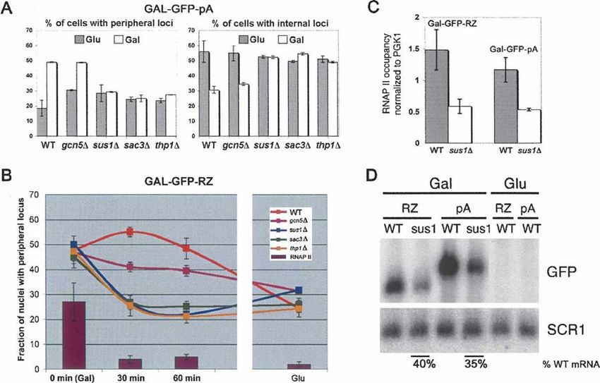
Downloaded from www.rnajournal.org on February 14, 2008 - Published by Cold Spring Harbor Laboratory Press

                                                                             Post-transcriptional tether of mRNP to active genes

(Abruzzi et al. 2006), which suggests mechanistic links             via an extensive manual survey of genes implicated in
between these two processes. Several mechanisms have been           mRNA biogenesis, processing, and export (Table 1).
proposed to contribute to the recruitment, capture, and/or             Deletion of SUS1 resulted in a striking enlargement and/
subperipheral retention of genes at the nuclear envelope.           or fragmentation of the dot (Fig. 1A,B), reproducibly
These include direct interactions between transcriptional           observed in $80% of cells. Importantly, these effects were
activators and nucleoporins (Menon et al. 2005; Schmid              not dependent on the specific construct or on its integra-
et al. 2006), the act of transcription itself (Cabal et al. 2006;   tion site, because substituting the conditional GAL1 pro-
Taddei et al. 2006), transcription-associated chromatin             moter for the TDH3 promoter as well as placing the
remodeling (Brickner et al. 2007), as well as unspecified           reporter at a different genomic location led to an identical
mRNA- and/or 39-UTR-dependent interactions (Casolari                dot enlargement and fragmentation in an sus1D back-
et al. 2005; Taddei et al. 2006). Our own experiments have          ground (Fig. 1C). Moreover, simultaneous visualization
also highlighted the importance of 39-end formation signals         of the GAL-GFP-RZ locus using TetR-GFP bound to a
on gene-periphery associations. Moreover, these have a strong       tandem array of 448 Tet operators integrated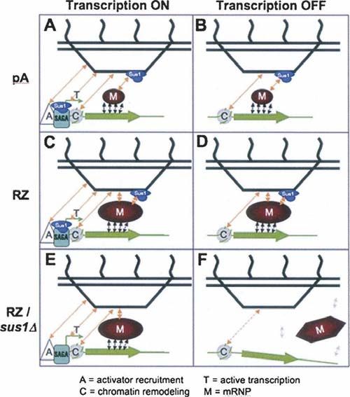
Downloaded from www.rnajournal.org on February 14, 2008 - Published by Cold Spring Harbor Laboratory Press

Chekanova et al.

 TABLE 1. A sublibrary of viable mRNA export-relevant deletion mutants used in the FISH-based screen

 Gene ID              Name                 Biological process (GO terms)                                Description

 YMR044W           IOC4               Chromatin remodeling                           Component of a complex (Isw1b) with Isw1p
                                                                                        and Ioc2p that exhibits nucleosome-stimulated
                                                                                        ATPase activity
 YGL133W           ITC1               Chromatin remodeling                           Component of the ATP-dependent Isw2–Itc1
                                                                                        chromatin-remodeling complex
 YIL079C           AIR1               RNA export from nucleus                        TRAMP complex, also interacts with the
                                                                                        arginine methyltransferase Hmt1 to regulate
                                                                                        Npl3, which modulates Npl3 function in
                                                                                        mRNA processing and export
 YDL175C           AIR2               RNA export from nucleus                        TRAMP complex, also interacts with the
                                                                                        arginine methyltransferase Hmt1 to regulate
                                                                                        Npl3, which modulates Npl3 function in
                                                                                        mRNA processing and export
 YFL007W           BLM10              Proteasome assembly                            Found in association with proteasome core
                                                                                        particles, with and without the 19S regulatory
                                                                                        particle
 YPR057W           BRR1               Spliceosome assembly                           In null mutant, newly synthesized snRNAs are
                                                                                        destabilized, and 39-end processing is slowed
 YLR226W           BUR2               Transcription                                  Cyclin for Bur1 kinase; transcriptional regulation
                                                                                        through its phosphorylation of RNAPII CTD
 YAL021C           CCR4               mRNA catabolism, regulation of                 CCR4-NOT transcriptional complex, also
                                        transcription from RNA polymerase II            component of the major cytoplasmic
                                        promoter                                        deadenylase
 YLR418C           CDC73              RNA elongation from RNA polymerase             Constituent of Paf1 complex with RNA polymerase
                                        II promoter                                     II, Paf1, Hpr1, Ctr9, Leo1, Rtf1, and Ccr4
 YER64W            CHD1               RNA elongation from RNA polymerase             Transcription elongation, nucleosome
                                        II promoter                                     remodeling, part of SAGA
 YOR061w           CKA2               Regulation of transcription from RNA           Catalytic subunit of casein kinase 2;
                                        polymerase II promoter                          phosphorylates Pta1 and RNAPII
 YKL139W           CTK1               Regulation of transcription from RNA           Phosphorylates Ser2 of CTD, affects transcription
                                        polymerase II promoter                          and pre-mRNA 39-end processing
 YDR069C           DOA4               Protein deubiquitination                       Required for recycling ubiquitin from
                                                                                        proteasome-bound ubiquitinated intermediates
 YGL043W           DST1               RNA elongation from RNA polymerase             Elongation factor TFIIS, stimulates cleavage of
                                       II promoter                                      nascent transcripts stalled at transcription
                                                                                        arrest sites
 YHL030W           ECM29              Protein catabolism                             Tethers proteasome core particle to the
                                                                                        regulatory particle
 YGR200C           ELP2               Regulation of transcription from RNA           Part of RNAPII Elongator histone
                                        polymerase II promoter                          acetyltransferase
 YPL086C           ELP3               Regulation of transcription from RNA           Part of RNAPII Elongator histone
                                        polymerase II promoter                          acetyltransferase
 YER032W           FIR1               mRNA polyadenylylation                         Involved in 39-mRNA processing, interacts
                                                                                        with Ref2
 YCL011C           GBP2               RNA export from nucleus                        Poly(A)+ RNA-binding protein
 YGR252W           GCN5               Chromatin modification                         Catalytic subunit of SAGA histone
                                                                                        acetyltransferase complex
 YNL199C           GCR2               Positive regulation of transcription from      Transcriptional activator, interacts with
                                        RNA polymerase II promoter                      DNA-binding protein Gcr1
 YMR255W           GFD1               RNA export from nucleus                        High-copy suppressor of dbp5 mutation
 YBR034C           HMT1               RNA export from nucleus                        Arginine methylation of hnRNPs, including
                                                                                        Npl3 and Hrp1
 YNL004W           HRB1               RNA export from nucleus                        Poly(A)+ RNA-binding protein; similar to
                                                                                        Gbp2 and Npl3
 YLR384C           IKI3               Regulation of transcription from RNA           Part of RNAPII Elongator histone
                                        polymerase II promoter                          acetyltransferase
 YGL016W           KAP122             Nucleocytoplasmic transport                    Karyopherin b, binds to nucleoporins Nup1 and
                                                                                        Nup2, synthetic lethal with gcr1 mutation
 YER110C           KAP123             Nucleocytoplasmic transport                    Karyopherin b; conditional loss of Pse1 in
                                                                                        kap123 mutant cells blocks mRNA export

                                                                                                                            (continued )

68     RNA, Vol. 14, No. 1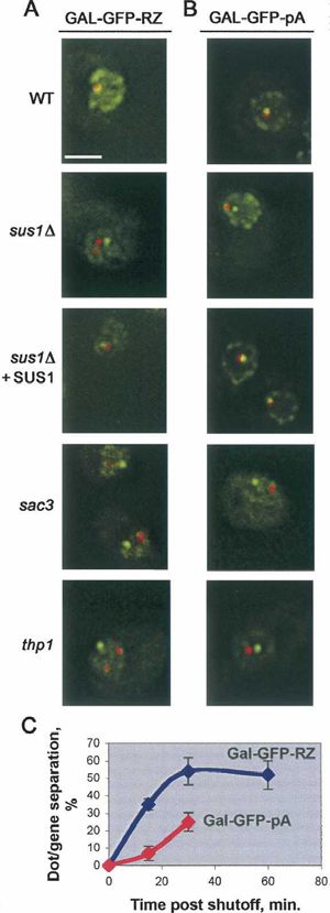
Downloaded from www.rnajournal.org on February 14, 2008 - Published by Cold Spring Harbor Laboratory Press

                                                                               Post-transcriptional tether of mRNP to active genes

TABLE 1. Continued

Gene ID              Name               Biological process (GO terms)                                   Description

YOR123C          LEO1              RNA elongation from RNA polymerase              Component of the Paf1 complex, histone
                                    II promoter                                       methylation; loss affects CTD
                                                                                      phosphorylation at Ser2
YDL051W          LHP1              tRNA processing                                 La homolog
YHR081W          LRP1              mRNA catabolism                                 Nuclear exosome
YHR121W          LSM12             RNA metabolism                                  Interacts with Pbp1 and Pbp4 with likely role
                                                                                      in RNA processing
YML062C          MFT1              RNA export from nucleus                         THO comlex subunit
YHR015W          MIP6              RNA export from nucleus                         Putative RNA-binding protein, interacts with
                                                                                      Mex67
YKP095W          MLP1              mRNA export from nucleus                        Involved with Pml1 and Pml39 in nuclear
                                                                                      retention of unspliced mRNAs
YIL149C          MLP2              RNA export from nucleus                         Nuclear retention of unspliced mRNA
YKL074C          MUD2              U2-type nuclear mRNA branch site                Similar to metazoan splicing factor U2AF65,
                                     recognition                                      synthetic rescue of sub2 mutant
YGR232W          NAS6              Proteolysis                                     Non-ATPase subunit of the 26S proteasome
YKL068W          NUP100            RNA export from nucleus                         GLFG motif-type nucleoporin, interacts
                                                                                      with Mex67
YBL079W          NUP170            RNA export from nucleus                         Nucleoporin, synthetic lethal with gcr1/2
YML103C          NUP188            RNA export from nucleus                         Nucleoporin, synthetic lethal with gcr1/2
YMR153W          NUP53             RNA export from nucleus                         Nucleoporin, synthetic lethal with gcr1/2
YGL094C          PAN2              mRNA 39-end processing                          Poly(A) nuclease subunit
YKL025C          PAN3              mRNA 39-end processing                          Poly(A) nuclease subunit
YGR178C          PBP1              mRNA polyadenylylation                          Poly(A)-binding protein binding protein
YMR129W          POM152            RNA export from nucleus                         Nuclear pore membrane glycoprotein,
                                                                                      synthetic lethal with gcr1/2
YLR018C          POM34             Nucleocytoplasmic transport                     Integral membrane protein of the nuclear pore,
                                                                                      synthetic lethal with gcr1/2
YGR135W          PRE9              Ubiquitin-dependent protein catabolism          The only nonessential 20S proteasome subunit
YEL037C          RAD23             Negative regulation of protein catabolism       Protein with ubiquitin-like N terminus, binds
                                                                                      damaged DNA
YDR195W          REF2              mRNA processing                                 Subunit of the APT subcomplex, involved
                                                                                      in the pre-mRNA 39-end cleavage
YGL244W          RFT1              Oligosaccharide transport                       Loss affects CTD phosphorylation at Ser2
YHR200W          RPN10             Ubiquitin-dependent protein catabolism          Non-ATPase base subunit of the 19S regulatory
                                                                                      particle (RP) of the 26S proteasome
YLR421C          RPN13             Ubiquitin-dependent protein catabolism          Subunit of the 19S regulatory particle of 26S
                                                                                      proteasome lid
YOR001W          RRP6              mRNA catabolism                                 Nuclear exosome
YDR159W          SAC3              RNA export from nucleus                         NPC-associated protein, part of
                                                                                      Sac3-Thp1-Sus1-Cdc31 complex
YML013W          SEL1              Proteasomal ubiquitin-dependent protein         UBX domain-containing protein, interacts
                                     catabolism                                       with Cdc48
YBL058W          SHP1              Proteasomal ubiquitin-dependent protein         UBX domain-containing protein that
                                     catabolism                                       regulates Glc7 phosphatase activity
                                                                                      and interacts with Cdc48
YMR216C          SKY1              Nucleocytoplasmic transport                     NPL3 kinase
YBR172C          SMY2              Cytoskeleton organization and biogenesis        Homolog of pombe Mpd2 (multicopy
                                                                                      suppressor of Ptr1/Tom1)
YOL148C          SPT20             Chromatin modification                          SAGA subunit, required for its integrity
YIL030C          SSM4              ER-associated protein catabolism                Ub ligase; ssm4 mutation suppresses mRNA
                                                                                      instability caused by an rna14 mutation
YBR11W-A         SUS1              mRNA export from nucleus                        Part of Sac3-Thp1-Sus1-Cdc31 complex; component
                                                                                      of the SAGA histone acetylase complex
YOR179C          SYC1              Transcription termination from                  Subunit of APT subcomplex of cleavage and
                                     Pol II promoter                                  polyadenylation factor; role in 39-end formation of
                                                                                      polyadenylated and non-polyadenylated RNAs
YNL253W          TEX1              RNA export from nucleus                         Component of THO/TREX complex
YNL139C          THO2/RLR1         mRNA export from nucleus                        Component of THO/TREX complex

                                                                                                                             (continued )

                                                                                                           www.rnajournal.org          69
Downloaded from www.rnajournal.org on February 14, 2008 - Published by Cold Spring Harbor Laboratory Press
Chekanova et al.
TABLE 1. Continued
Gene ID Name Biological process (GO terms) Description
YOL072W THP1 RNA export from nucleus NPC-associated protein, part of
Sac3-Thp1-Sus1-Cdc31 complex
YBR082C UBC4 Protein monoubiquitination, Ubiquitin-conjugating enzyme, interacts with
protein polyubiquitination many SCF ubiquitin protein ligases
YDR059C UBC5 Protein polyubiquitination Ubiquitin-conjugating enzyme
YFR010W UBP6 Protein deubiquitination Ubiquitin-specific protease in the base
subcomplex of 26S proteasome
YGR184C UBR1 Protein monoubiquitination, protein Ubiquitin-protein ligase (E3)
polyubiquitination
YBR173C UMP1 Proteasome assembly Chaperone required for correct maturation
of the 20S proteasome
YGL227W VID30 Proteasomal ubiquitin-dependent Involved in proteasome-dependent
protein catabolism catabolite degradation of FBPase
YKL214C YRA2 RNA export from nucleus mRNA export adaptor, redundant with Yra1
YMR273C ZDS1 RNA export from nucleus ceg1-ts suppressor, implicated in mRNA
nuclear export, Y2H interaction with Dbp5
YNL016W PUB1 mRNA catabolism Poly(A)+ RNA-binding protein, required
for stability of many mRNAs
YOR304W ISW2 Chromatin remodeling Member of the imitation-switch (ISWI)
class of ATP-dependent chromatin
remodeling complexes
YLR095C IOC2 Chromatin remodeling Member of an Isw1b complex with
nucleosome-stimulated ATPase activity
YFR013W IOC3 Chromatin remodeling Member of an Isw1a complex with
nucleosome-stimulated ATPase activity
the transcription-site-proximal GFP-RZ mRNP pool, ever, it similarly detaches from its gene in the three deletion
which is visualized by FISH as an exaggerated and/or strains after transcriptional shutoff (Fig. 2). These data
fragmented dot. Importantly, this effect is specific, since indicate that the Sac3-Thp1-Sus1-Cdc31 complex contrib-
other mutants that affect poly(A)+ mRNA export to a utes to retention of a post-transcriptional dot near its gene
comparable or greater extent (e.g., lrp1D) (Hieronymus of origin. Moreover, the lack of an effect before transcrip-
et al. 2004; data not shown) had no effect on dot morphology. tional shutoff suggests that there are additional, Sac3-,
Thp1-, and Sus1-independent mechanisms contributing to
the dot-gene tether during active transcription.
The Sac3-Thp1-Sus1-Cdc31 complex impacts
the dot-gene tether
The Sac3-Thp1-Sus1-Cdc31 complex also impacts
To further explore the role of the Sac3-Thp1-Sus1-Cdc31
the gene–nuclear periphery tether
complex in the tethering of the mRNA dot to its cognate
gene during transcription as well as after the transcriptional The difference in dot phenotype between GAL-GFP-pA and
shutoff, we simultaneously monitored the locations of the GAL-GFP-RZ in the mutant strains (Fig. 1) parallels a
dots with FISH and the reporter locus with the TetR-GFP/ difference in dot-gene dissociation kinetics, as the GAL-
TetO448 system as described above. As previously reported, GFP-RZ mRNA dot separates more quickly from the gene
the dot as well as its tether to the reporter gene persist for after transcriptional shutoff (Fig. 2C). Moreover, differ-
at least 60 min after transcriptional shutoff in WT cells ences in dot phenotypes were previously observed in wild-
(Abruzzi et al. 2006). In the sus1D, sac3D, and thp1D mutant type cells: the GAL-GFP-pA dots disappear more quickly
strains, however, we observed that the GAL-GFP-RZ dots than the GAL-GFP-RZ dots after transcriptional shutoff,
progressively detach from their loci of origin after transcrip- which further correlates with differences in the dissociation
tional shutoff (Fig. 2). To extend this observation, we also of the two genes from the nuclear rim after transcriptional
tested the GAL-GFP-pA reporter construct integrated at the shutoff in WT cells: GAL-GFP-pA dot dissociates more
same genomic location. GAL-GFP-pA construct possesses a quickly than the GAL-GFP-RZ dot (Abruzzi et al. 2006).
normal GAL1 39-UTR and polyadenylation signal (while its These differences presumably reflect the different RNP com-
GFP chromophore has been inactivated to enable visualiza- positions of the two transcripts (see Discussion).
tion of TetR-GFP/TetO448), and the GAL-GFP-pA dot Motivated by these observations, we examined the
appears normal during active transcription (Fig. 1I). How- relationship of the reporter loci to the nuclear periphery
70 RNA, Vol. 14, No. 1Downloaded from www.rnajournal.org on February 14, 2008 - Published by Cold Spring Harbor Laboratory Press
Post-transcriptional tether of mRNP to active genes
(Cabal et al. 2006; Drubin et al. 2006).
In contrast, the GAL-GFP-RZ gene still
localized to the nuclear periphery in the
deletion strains. However, its association
was rapidly lost upon transcriptional
shutoff, with kinetics that paralleled that
of RNAP II runoff (Fig. 3B). This con-
trasts with WT cells, in which the GAL-
GFP-RZ locus dissociates from the
nuclear rim only very slowly after glu-
cose addition (>60 min) (Abruzzi et al.
2006). The results suggest the existence
of additional transcription-dependent
tether(s) that maintain an actively tran-
scribing GAL-GFP-RZ locus at the nu-
clear periphery. They also suggest that
the post-transcriptional retention of the
GAL-GFP-RZ locus at the nuclear pe-
riphery requires the Sac3-Thp1-Sus1-
Cdc31 complex.
Because Sus1 directly participates in
transcription from the GAL1 promoter
(Rodriguez-Navarro et al. 2004; Kohler
et al. 2006), we addressed the possibility
that the differences in intranuclear posi-
tioning between the active GAL-GFP-pA
and GAL-GFP-RZ genes in the sus1D
mutant is a consequence of differential
transcriptional effects on these two genes.
To this end, we compared the RNAP
II occupancy on these two reporter
constructs, using chromatin immuno-
precipitation (ChIP). The magnitude of
reduction in RNAP II occupancy of
GAL-GFP-RZ and GAL-GFP-pA in
sus1D relative to WT cells was identical
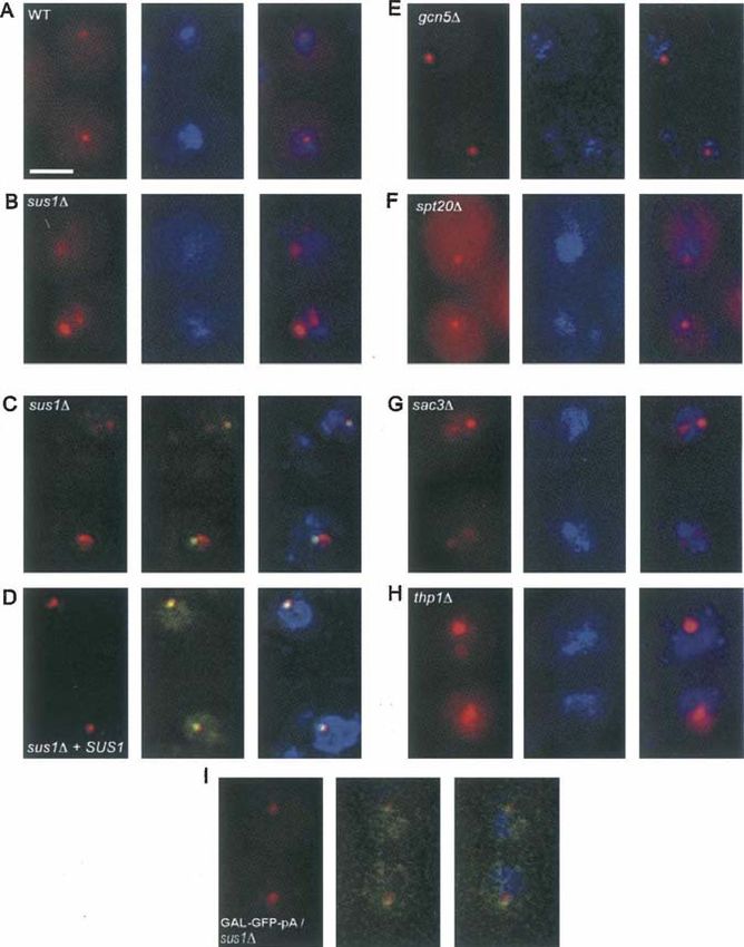
FIGURE 1. Altered nuclear-retained (A,B,E,F) TDH-GFP-RZ and (C,D) GAL-GFP-RZ
mRNA dot morphology in sus1D, sac3D, and thp1D cells. (A,B,E–H), fluorescent in situ (Fig. 3C), and it was also identical to that
hybridization (FISH) for the TDH-GFP-RZ mRNA in (A) wild-type (WT), (B) sus1D, (E) of the endogenous GAL1 gene in sus1D
gcn5D, (F) spt20D, (G) sac3D, and (H) thp1D cells. (Left panels) FISH signal (red); (middle cells (data not shown). Moreover, the
panels) DAPI staining for total DNA (blue); (right panels) overlay. Scale bar, 2 mm. (C,D)
FISH for the GAL-GFP-RZ mRNA with simultaneous visualization of the TetO448 array
decrease was quantitatively mirrored by
integrated at the BMH1 locus on Chromosome V,Downloaded from www.rnajournal.org on February 14, 2008 - Published by Cold Spring Harbor Laboratory Press
Chekanova et al.
dot morphology. They also affect the persistent tethering of
dots to their cognate genes as well as to the nuclear rim
after transcriptional shutoff. These findings reveal a novel,
post-transcriptional function of the Sac3-Thp1-Sus1-Cdc31
complex in addition to its previously described roles in
transcription itself, mRNA export, and the tethering of
genes to the nuclear periphery during active transcription.
Indeed, it is striking that the disruption of this complex
affects the tethering of post-transcriptional mRNPs to their
respective genes as well as the capture and retention of
transcriptionally activated loci at the nuclear rim. Although
we cannot rule out indirect effects, a parsimonious inter-
pretation of this unexpected dual role is that the dot-to-
gene and gene-to-nuclear periphery interactions are both
mediated by post-transcriptional mRNP decorated with the
Sac3-Thp1-Sus1-Cdc31 complex. Although the steady-state
distribution of these proteins is predominantly at the
nuclear rim, this does not preclude an association with
mRNP. Indeed, Sus1 and its partners interact with mRNA
and chromatin, in addition to their interactions with
the nuclear pores (Fischer et al. 2002; Gallardo et al.
2003; Lei et al. 2003; Rodriguez-Navarro et al. 2004; Kohler
et al. 2006).
Characterization of the sus1D, thp1D, and sac3D strains
indicates that fragmentation of the GAL-GFP-RZ mRNP
dot is intimately related to weakening of the gene-dot
tethering. This might indicate that dot integrity is de-
pendent on contacts between dot mRNP, Sac3-Thp1-
Sus1-Cdc31 complex, and chromatin. When these are
diminished (e.g., in sus1D), dot fragmentation as well as se-
paration of the dot and the gene occur (Figs. 1, 4). Notably,
despite their fragmentation in the mutant backgrounds,
the GAL-GFP-RZ dots do not completely disperse and
disappear, indicating that there must be Sus1-, Sac3-, and
FIGURE 2. Deletions of SUS1, SAC3, and THP1 lead to a loss of Thp1-independent associations between the individual
association of GAL-GFP-RZ and GAL-GFP-pA mRNA dots with their mRNP particles within the dot that contribute to its
respective genes. (A) GAL-GFP-RZ mRNA or (B) GAL-GFP-pA integrity.
mRNA is visualized by FISH (red); respective reporter loci are
visualized using the TetR-GFP/TetO448 system (green); and nuclear
We find that deletions of Sus1, Sac3, and Thp1 affect the
periphery is visualized using Nup49-GFP (green). Images are taken at dot–gene interactions not only in the case of the 39-end-
the 30-min time point after the transcriptional shutoff. Scale bar, impaired GFP-RZ construct, but also in the case of the
2 mm. (C) relative kinetics of the GAL-GFP-RZ and GAL-GFP-pA GAL-GFP-pA mRNP, suggesting that Sus1, Sac3, and Thp1
mRNA dots’ separation from respective genes after transcriptional
shutoff in sus1D cells. (Very few GAL-GFP-pA mRNA dots remain at are also components of the GAL-GFP-pA-containing dots.
60 min in sus1D cells; dot/locus separation in WT never exceeded Moreover, sus1D, sac3D, and thp1D mutations strongly
5%.) Error bars represent standard deviation. impact the association of the GAL-GFP-pA gene with the
nuclear rim. Yet their effects on this reporter differ in two
respects from their effects on GAL-GFP-RZ: the GAL-GFP-
rim association of these genes is not due to its SAGA-related pA dots are neither enlarged nor fragmented in the
functions but to the Sac3-Thp1-Sus1-Cdc31 complex. We mutants, and the association of the GAL-GFP-pA gene
also speculate that post-transcriptional mRNP may more gen- with the nuclear periphery is abolished completely, i.e.,
erally facilitate gene repositioning to the nuclear periphery. even during active transcription. As only the GAL-GFP-RZ
gene remains associated with the nuclear rim during active
transcription in the mutant strains, there must be addi-
DISCUSSION
tional transcription-dependent contacts that are stronger or
Using a FISH-based genetic screen, we identified Sus1, more numerous between the GAL-GFP-RZ gene and the
Thp1, and Sac3 as factors that have an impact on mRNA nuclear periphery.
72 RNA, Vol. 14, No. 1Downloaded from www.rnajournal.org on February 14, 2008 - Published by Cold Spring Harbor Laboratory Press
Post-transcriptional tether of mRNP to active genes
FIGURE 3. Deleting SUS1, SAC3, and THP1 has distinct transcription-independent effects on retention of the activated GAL-GFP-pA and GAL-
GFP-RZ genes at the nuclear periphery. (A) Intranuclear positioning of the GAL-GFP-pA gene is indistinguishable in sus1D, sac3D, and thp1D
cells under activating and repressing conditions. The proportion of cells with (left panel) peripherally and (right panel) internally positioned loci is
plotted for galactose-grown (Gal) and glucose-grown (Glu) cells. The intranuclear position of the locus was revealed using the TetR-GFP/TetO448
system. The nuclear periphery was visualized using Nup49-GFP fusion coexpressed in the same strain. Subperipheral fraction remains unchanged
at z20% (data not shown). Error bars represent standard deviation. (B) Dissociation of the GAL-GFP-RZ gene locus from the nuclear periphery
in sus1D, sac3D, and thp1D cells is dramatically accelerated and parallels the kinetics of the RNAP II runoff from the gene. The proportion of cells
with a peripherally positioned GAL-GFP-RZ gene locus after 0, 30, or 60 min of transcriptional shutoff or in steady-state glucose conditions is
plotted. For comparison, RNAP II occupancy after transcriptional shutoff as well as in steady-state glucose conditions, measured by ChIP, is
shown as vertical bars. Error bars represent standard deviation. (C) RNAP II occupancy of the GAL-GFP-pA and GAL-GFP-RZ reporter
constructs in sus1D cells is affected to the same extent. The RNAP II levels are normalized to the RNAP II occupancy on the endogenous PGK1
gene, which is unaffected by the loss of SUS1. Error bars represent standard deviation. (D) Northern blot quantification of the steady-state GAL-
GFP-pA and GAL-GFP-RZ mRNA levels using a GFP-specific probe in WT and sus1D cells grown on galactose and glucose. (SCR1) Loading
control used for normalization.
Although we cannot rule out that such differential This view implies that there are multiple contacts
contacts may be mediated by chromatin per se, there are between a tethered gene and the NPC. Indeed, it has
no known chromatin effects associated with replacing the been shown that transcriptional activator binding (Schmid
GAL1 39-UTR with the ribozyme. An alternative (or ad- et al. 2006), transcription-associated chromatin remodeling
ditional) possibility would be due to a difference between (Brickner et al. 2007), and perhaps the act of transcription
GAL-GFP-pA and GAL-GFP-RZ mRNP structure or com- itself (Casolari et al. 2005; Cabal et al. 2006; Drubin et al.
position, which can be qualitative, quantitative, or both. 2006) all contribute to the recruitment of the activated GAL
Indeed, we reported previously that the GAL-GFP-RZ genes to the nuclear periphery. Moreover, diminished
gene shows an altered cotranscriptional recruitment profile perinuclear positioning during active transcription was
of Yra1 (Abruzzi et al. 2006). Moreover, a quantitative observed for endogenous GAL genes in sus1D and sac3D
difference is indicated by the consistently larger GAL-GFP- cells (e.g., Cabal et al. 2006; Drubin et al. 2006). Our
RZ dots than the GAL-GFP-pA dots (data not shown; D. findings extend these conclusions by suggesting that this
Zenklusen and R. Singer, pers. comm.). Larger dots would effect is mediated by the post-transcriptional mRNP and
provide more contact area between RZ mRNP and the becomes relevant only after initial contact of the activated
nuclear periphery than between pA RNP and the periphery, gene with the nuclear rim (modeled in Fig. 4). Whereas the
which might contribute to RZ gene retention at the nuclear initial encounter of the locus with the nuclear periphery is
rim during active transcription even in a Sus1-deleted transcriptional activator-dependent, independent of Sus1
strain (Fig. 4). (Schmid et al. 2006), and precedes the onset of transcription
www.rnajournal.org 73Downloaded from www.rnajournal.org on February 14, 2008 - Published by Cold Spring Harbor Laboratory Press
Chekanova et al.
tate rapid mRNA export, as originally proposed by Blobel
in the gene gating hypothesis (Blobel 1985).
MATERIALS AND METHODS
Strain design and growth conditions
Construction of strains bearing the TDH-GFP-RZ reporter loci
marked with nourseothricin resistance and KanMX-marked dele-
tions of selected nonessential genes was done using the approach
originally developed for genome-wide scoring of synthetic lethal
interactions (Tong et al. 2001). Reporter strains designed for
mating with deletion array strains were in a Y7092 background
(MATa can1DTSTE2pr-SpDhis5 his3D1 leu2D0 ura3D0 met15D0
lyp1D trp1DTGAL1-IpgB1-URA3) (Alto et al. 2006; gift from
Charlie Boone).
Yeast strains other than the ones used in the FISH-based screen
for altered dot morphology as well as their derivation are
described in detail in Table 2. To delete the SUS1, SAC3, and
THP1, the kanamycin (KanMX) cassette plus respective flanking
regions were PCR-amplified from the respective KanMX-
marked deletion strains in the BY4741 background (MATa
his3D1 leu2D0 met15D0 ura3D0), obtained from Open Biosys-
tems. The fragment encompassing NUP49-GFP fusion and HIS3
(Huh et al. 2003) was amplified from the Invitrogen strain col-
FIGURE 4. A model that integrates the multiple interactions between lection. Integrations were verified by PCR and/or Southern blotting.
the activated GAL genes (green), non-nascent mRNP pools (red), and Yeast cell growth conditions for FISH, visualization of GFP
nuclear pores (black), emphasizing the dual role of the Sac3-Thp1- fusion proteins, as well as for galactose-to-glucose shifts were as
Sus1-Cdc31 complex (blue) at the site of transcription as well as at
the nuclear periphery. (A) In WT cells, perinuclear repositioning of described (Abruzzi et al. 2006).
the GAL-GFP-pA locus occurs via a combined action of multiple
mechanisms including transcriptional activator (A)-dependent re-
cruitment as well as transcription (T)-, chromatin (C)-, and mRNP Plasmid constructs and oligonucleotide primers
(M)-mediated capture and retention. (B) Upon shutoff of transcrip-
tion, persistent chromatin- and mRNP-mediated retention prevents
The primers used in this study are listed in Table 3. Plasmid
the immediate departure of the GAL-GFP-pA locus from the nuclear pDB700 was designed for integrating the TDH3-GFP-RZ reporter
rim. (C) The abnormal and large GAL-GFP-RZ mRNP pool pro- marked by nureseothricin resistance marker gene NatMX (Gold-
duced when mRNA 39-end processing is bypassed by RZ makes stein and McCusker 1999) into the genomic trp1 locus. To this
additional contacts at the nuclear pore, which are not Sac3-Thp1- end, NatMX was amplified with oDB1082/1083 and cloned into
Sus1-Cdc31 complex-dependent, and hence is strongly retained at the AatII site of the pRS304/2m bearing TDH3-GFP-RZ (Dower
the nuclear rim. (D) Upon transcriptional shutoff, the synergy of
et al. 2004), thus replacing its 2m origin of replication. Integration
these additional contacts with the NPC, acting together with the
Sac3-Thp1-Sus1-Cdc31 complex-dependent and chromatin-depen- was conducted after linearization at the Bsu36I site within the
dent contacts, allows the GAL-GFP-RZ mRNP to persist at the TRP1 gene sequence.
nuclear periphery longer than GAL-GFP-pA. (E) Even in the absence pDB716 and pDB719 (described in Abruzzi et al. 2006) were
of the Sac3-Thp1-Sus1-Cdc31 complex at the NPC, the synergy of designed for inserting the GAL-GFP-RZ and GAL-GFP-pA,
the activator-mediated contacts, ongoing transcription, chromatin- respectively, by the gamma-integration method (Sikorski and
mediated and the Sac3-Thp1-Sus1-Cdc31 complex-independent Hieter 1989) into the intergenic region between ECM32 and
interactions of the exaggerated GAL-GFP-RZ mRNP with the NPC
support a modest degree of peripheral tethering. (F) Upon tran- BMH1, after linearization with NotI and selecting for Trp+
scriptional shutoff, this modest tethering fails immediately, while the transformants.
aberrant mRNP remodeling (illustrated by the change in shape of the To generate pDB729, WT genomic SUS1 fragment was ampli-
mRNP pool) and/or compromised export of the GAL-GFP-RZ fied by PCR with oDB1152 and oDB1153, cloned into the Not site
mRNP in the absence of Sac3-Thp1-Sus1-Cdc31 complex causes of pDB700, followed by releasing the TDH-GFP-RZ reporter from
its separation from the gene. the polylinker after digesting with SpeI + ApaI, repairing the ends
with T4 DNA polymerase, and religating. Integration was con-
(Brickner et al. 2007), stable retention becomes independent ducted into TRP1 locus after linearization with Bsu36I, with
of active transcription and is facilitated by the Sac3-Thp1- selection for nurseothricin resistance.
Sus1-Cdc31 complex; it is more generally aided by intrinsic
Microscopy
properties of post-transcriptional mRNP. We suggest that
multiple tethering mechanisms serve to strengthen the asso- GFP fusion proteins were observed in cells grown to OD z 0.5
ciation between the active gene and NPC and hence facili- and fixed for 15 min in 4% paraformaldehyde (without acetic
74 RNA, Vol. 14, No. 1Downloaded from www.rnajournal.org on February 14, 2008 - Published by Cold Spring Harbor Laboratory Press
Post-transcriptional tether of mRNP to active genes
TABLE 2. Yeast strains other than the deletion sublibrary used in FISH-based screen
Y7092 MATa can1DTSTE2pr-SpDhis5 his3D1 leu2D0 ura3D0 met15D0 lyp1D trp1DTGAL1-IpgB1-URA3 Tong and
Boone (2007)
YDB526 TDH-GFP-RZ reporter (pDB700) integrated into Y7092 This study
YDB527 TDH-GFP-RZ (pDB700)TNatMX, sus1DTKanMX segregant of a cross with YDB526 This study
YDB528 TDH-GFP-RZ (pDB700)TNatMX, sac3DTKanMX segregant of a cross with YDB526 This study
YDB529 TDH-GFP-RZ (pDB700)TNatMX, thp1DTKanMX segregant of a cross with YDB526 This study
YDB530 TDH-GFP-RZ (pDB700)TNatMX, gcn5DTKanMX segregant of a cross with YDB526 This study
YDB531 TDH-GFP-RZ (pDB700)TNatMX, spt20DTKanMX segregant of a cross with YDB526 This study
YDB532 MATa, ura3, trp1, his3, leu2TLEU2 tetR-GFP, 2x224tet0 URA3 integrated between AE3 in Abruzzi
BMH1 and PDA1 et al. (2006)
YDB533 GAL-GFP-RZ (pDB716) + NUP49-GFP, HIS3 in YDB532 This study
YDB534 GAL-GFP-pA (pDB719) + NUP49-GFP, HIS3 in YDB532 This study
YDB535 sus1DTKanMX in YDB533 This study
YDB536 sus1DTKanMX in YDB534 This study
YDB537 sac3DTKanMX in YDB533 This study
YDB538 sac3DTKanMX in YDB534 This study
YDB539 thp1DTKanMX in YDB533 This study
YDB540 thp1DTKanMX in YDB534 This study
YDB541 WT SUS1 gene (pDB729) integrated into trp1 in YDB535 This study
YDB542 WT SUS1 gene (pDB729) integrated into trp1 in YDB536 This study
acid) using an Olympus IX70-based DeltaVision workstation Walter 2004) into intranuclear, peripheral, and subperipheral (i.e.,
(Applied Precision). Z-stacks were taken at 0.2 mm step size and locus touching the nuclear envelope but not coplanar with it). The
subjected to constrained iterative deconvolution. Positions of the subperipheral fraction varied little in all conditions and therefore
TetR-GFP marked locus were scored in the z-section that cuts is not reported in Figure 3. Each experiment was done in replicate,
through the middle of the nucleus as described (Brickner and and between 100 and 150 cells were scored per sample per time
TABLE 3. Oligonucleotide primers
Name Sequence (59 to 39) Purpose/notes
oDB1082 ACATGGAGGCCCAGAATACCC Amplifying NatMX gene
oDB1083 CAGTATAGCGACCAGCATTCAC Amplifying NatMX gene
oDB1128 CGTACTTGCCATCCCTTACG Upstream of SUS1 for disruption
oDB1129 AAGGTGGGTAACGTGAATTAGG Downstream from SUS1 for disruption
oDB1130 TGAAGGCTGTCTGACGTTGT Upstream of oDB1128 for checking
disruption junction
oDB1146 GGAGAGAGGAAAGCAGCAGA Upstream of SAC3 for disruption
oDB1147 GTATTTGCAACCCTGGCTTC Downstream from SAC3 for disruption
oDB1148 ATTCTCTCGCTGACCCAAGA Upstream of oDB1146 for checking
disruption junction
oDB1149 TGATCATGGCTAGCTTGGTG Upstream of THP1 for disruption
oDB1150 TTGTAGCCTTGCAACGACAG Upstream of THP1 for disruption
oDB1151 GGCAACTAAGGGAACCACAA Upstream of oDB1149 for checking
disruption junction
oDB1152 TTGAATTCAAGGAAAATGCCGAAAGAAT Amplifying WT SUS1 for complementation
oDB1153 TTGAGCTCCAATTCATTCATTATGTTGTGGA Amplifying WT SUS1 for complementation
KA56 GCCTTATTTCTGGGGTAATTAATCAGCGAAGCGATG ChIP, 59-UTR of GAL1 (forward)
KA100 AACTCCAGTGAAGAGTTCTTCTCCTTT ChIP, within ORF of GFP (reverse)
KD209 GT*GCCCATTAACAT*CACCATCTAAT T*CAACA AGAAT*TGGGACAACT*CCAGT FISH probe for GFP
KD210 GTACAT*AACCTTCGGGCAT*GGCACTCTT*GAAAAAGTCAT*GCCGTTTCAT*AT FISH probe for GFP
KD211 GATTCCAT*TCTTTTGTT*TGTCTGCCAT*GATGTATACAT*TGTGTGAGTT*ATA FISH probe for GFP
KD212 CCCAGCAGCT*GTTACAAACT*CAAGAAGGACCAT*GTGGTCT*CTCTTTTCGT*T FISH probe for GFP
KA480 ACGGAAGAGCTGCTGAAAAA Amplification of NUP49-GFP,
HIS3 fragment
KA481 TTGAATTGGGGTAGGCTCAG Amplification of NUP49-GFP,
HIS3 fragment
(T*) Amino-modified dT.
www.rnajournal.org 75Downloaded from www.rnajournal.org on February 14, 2008 - Published by Cold Spring Harbor Laboratory Press
Chekanova et al.
point in each replicate in all experiments shown. FISH with Cy3- Casolari, J.M., Brown, C.R., Drubin, D.A., Rando, O.J., and
labeled oligonucleotide probes was carried out according to Silver, P.A. 2005. Developmentally induced changes in transcrip-
Dower et al. (2004). Red/green channel signal offset due to chro- tional program alter spatial organization across chromosomes.
Genes & Dev. 19: 1188–1198.
matic aberration alone, as estimated by imaging TetraSpec beads
Dhasarathy, A. and Kladde, M.P. 2005. Promoter occupancy is a
(100 nm diameter, Invitrogen/Molecular Probes) under identical major determinant of chromatin remodeling enzyme require-
conditions, was negligible compared to the separation of FISH ments. Mol. Cell. Biol. 25: 2698–2707.
(Cy3) and GFP signals. Dower, K., Kuperwasser, N., Merrikh, H., and Rosbash, M. 2004. A
synthetic A tail rescues yeast nuclear accumulation of a ribozyme-
Chromatin immunoprecipitation and RNA analyses terminated transcript. RNA 10: 1888–1899.
Drubin, D.A., Garakani, A.M., and Silver, P.A. 2006. Motion as a
RNAP II ChIP was performed using monoclonal antibody 8WG16 phenotype: The use of live-cell imaging and machine visual
(Covance). Target DNA levels in input and IP samples were deter- screening to characterize transcription-dependent chromosome
dynamics. BMC Cell Biol. 7: 19.
mined by real-time PCR using RotorGene (Corbett Research),
Dudley, A.M., Rougeulle, C., and Winston, F. 1999. The Spt
and results were normalized as described (Abruzzi et al. 2004). The components of SAGA facilitate TBP binding to a promoter at a
Northern hybridization signals in Figure 3 obtained with a GFP- post-activator-binding step in vivo. Genes & Dev. 13: 2940–2945.
specific probe were normalized to respective signals for SCR1, a Fasken, M.B. and Corbett, A.H. 2005. Process or perish:
RNAP III transcript, using ImageQuant software. Signal ratios Quality control in mRNA biogenesis. Nat. Struct. Mol. Biol. 12:
were identical to the ChIP signal ratios in the respective strains. 482–488.
Fischer, T., Strasser, K., Racz, A., Rodriguez-Navarro, S., Oppizzi, M.,
Ihrig, P., Lechner, J., and Hurt, E. 2002. The mRNA export
machinery requires the novel Sac3p–Thp1p complex to dock at
ACKNOWLEDGMENTS the nucleoplasmic entrance of the nuclear pores. EMBO J. 21: 5843–
5852.
We appreciate the technical assistance of Prabhat Mallik and
Fischer, T., Rodriguez-Navarro, S., Pereira, G., Racz, A., Schiebel, E.,
Andrey Belostotsky. We are grateful to the Boone, Nasmyth, and and Hurt, E. 2004. Yeast centrin Cdc31 is linked to the nuclear
Wente laboratories for strains, as well as to Kristine O’Brien, other mRNA export machinery. Nat. Cell Biol. 6: 840–848.
members of the Melissa Moore laboratory, and Alexey Khodjakov Gallardo, M., Luna, R., Erdjument-Bromage, H., Tempst, P., and
for help with deconvolution microscopy. This work was sup- Aguilera, A. 2003. Nab2p and the Thp1p–Sac3p complex func-
ported in part by grants from NIH to D.A.B. and J.A.C. tionally interact at the interface between transcription and mRNA
(#GM073872) and to M.R. (#GM23549) and from the NSF to metabolism. J. Biol. Chem. 278: 24225–24232.
Goldstein, A.L. and McCusker, J.H. 1999. Three new dominant drug
D.A.B. (Grant MCB0424651). resistance cassettes for gene disruption in Saccharomyces cerevisiae.
Yeast 15: 1541–1553.
Received August 9, 2007; accepted September 21, 2007. Grant, P.A., Duggan, L., Cote, J., Roberts, S.M., Brownell, J.E.,
Candau, R., Ohba, R., Owen-Hughes, T., Allis, C.D., Winston, F.,
et al. 1997. Yeast Gcn5 functions in two multisubunit complexes
to acetylate nucleosomal histones: Characterization of an Ada
REFERENCES
complex and the SAGA (Spt/Ada) complex. Genes & Dev. 11:
Abruzzi, K.C., Lacadie, S., and Rosbash, M. 2004. Biochemical analysis 1640–1650.
of TREX complex recruitment to intronless and intron-containing Hieronymus, H., Yu, M.C., and Silver, P.A. 2004. Genome-wide
yeast genes. EMBO J. 23: 2620–2631. mRNA surveillance is coupled to mRNA export. Genes & Dev. 18:
Abruzzi, K.C., Belostotsky, D.A., Chekanova, J.A., Dower, K., and 2652–2662.
Rosbash, M. 2006. 39-End formation signals modulate the associ- Hilleren, P., McCarthy, T., Rosbash, M., Parker, R., and Jensen, T.H.
ation of genes with the nuclear periphery as well as mRNP dot 2001. Quality control of mRNA 39-end processing is linked to the
formation. EMBO J. 25: 4253–4262. nuclear exosome. Nature 413: 538–542.
Alto, N.M., Shao, F., Lazar, C.S., Brost, R.L., Chua, G., Mattoo, S., Huh, W.K., Falvo, J.V., Gerke, L.C., Carroll, A.S., Howson, R.W.,
McMahon, S.A., Ghosh, P., Hughes, T.R., Boone, C., et al. 2006. Weissman, J.S., and O’Shea, E.K. 2003. Global analysis of protein
Identification of a bacterial type III effector family with G protein localization in budding yeast. Nature 425: 686–691.
mimicry functions. Cell 124: 133–145. Jensen, T.H., Boulay, J., Rosbash, M., and Libri, D. 2001a. The DECD
Basehoar, A.D., Zanton, S.J., and Pugh, B.F. 2004. Identification and box putative ATPase Sub2p is an early mRNA export factor. Curr.
distinct regulation of yeast TATA box-containing genes. Cell 116: Biol. 11: 1711–1715.
699–709. Jensen, T.H., Patricio, K., McCarthy, T., and Rosbash, M. 2001b. A
Blobel, G. 1985. Gene gating: A hypothesis. Proc. Natl. Acad. Sci. 82: block to mRNA nuclear export in S. cerevisiae leads to hyper-
8527–8529. adenylation of transcripts that accumulate at the site of transcrip-
Brickner, J.H. and Walter, P. 2004. Gene recruitment of the activated tion. Mol. Cell 7: 887–898.
INO1 locus to the nuclear membrane. PLoS Biol. 2: e342. doi: Jensen, T.H., Dower, K., Libri, D., and Rosbash, M. 2003. Early
10.1371/journal.pbio.0020342. formation of mRNP: License for export or quality control? Mol.
Brickner, D.G., Cajigas, I., Fondufe-Mittendorf, Y., Ahmed, S., Cell 11: 1129–1138.
Lee, P.C., Widom, J., and Brickner, J.H. 2007. H2A.Z-mediated Kohler, A., Pascual-Garcia, P., Llopis, A., Zapater, M., Posas, F.,
localization of genes at the nuclear periphery confers epigenetic Hurt, E., and Rodriguez-Navarro, S. 2006. The mRNA export
memory of previous transcriptional state. PLoS Biol. 5: e81. doi: factor Sus1 is involved in Spt/Ada/Gcn5 acetyltransferase-medi-
10.1371/journal.pbio.0050081. ated H2B deubiquitinylation through its interaction with Ubp8
Cabal, G.G., Genovesio, A., Rodriguez-Navarro, S., Zimmer, C., and Sgf11. Mol. Biol. Cell 17: 4228–4236.
Gadal, O., Lesne, A., Buc, H., Feuerbach-Fournier, F., Olivo- Lei, E.P., Stern, C.A., Fahrenkrog, B., Krebber, H., Moy, T.I., Aebi, U.,
Marin, J.C., Hurt, E.C., et al. 2006. SAGA interacting factors and Silver, P.A. 2003. Sac3 is an mRNA export factor that localizes
confine subdiffusion of transcribed genes to the nuclear envelope. to cytoplasmic fibrils of nuclear pore complex. Mol. Biol. Cell 14:
Nature 441: 770–773. 836–847.
76 RNA, Vol. 14, No. 1Downloaded from www.rnajournal.org on February 14, 2008 - Published by Cold Spring Harbor Laboratory Press
Post-transcriptional tether of mRNP to active genes
Libri, D., Dower, K., Boulay, J., Thomsen, R., Rosbash, M., and Sterner, D.E., Grant, P.A., Roberts, S.M., Duggan, L.J.,
Jensen, T.H. 2002. Interactions between mRNA export commit- Belotserkovskaya, R., Pacella, L.A., Winston, F., Workman, J.L.,
ment, 39-end quality control, and nuclear degradation. Mol. Cell. and Berger, S.L. 1999. Functional organization of the yeast SAGA
Biol. 22: 8254–8266. complex: Distinct components involved in structural integrity,
Menon, B.B., Sarma, N.J., Pasula, S., Deminoff, S.J., Willis, K.A., nucleosome acetylation, and TATA-binding protein interaction.
Barbara, K.E., Andrews, B., and Santangelo, G.M. 2005. Reverse recruit- Mol. Cell. Biol. 19: 86–98.
ment: the Nup84 nuclear pore subcomplex mediates Rap1/Gcr1/Gcr2 Taddei, A., Van Houwe, G., Hediger, F., Kalck, V., Cubizolles, F.,
transcriptional activation. Proc. Natl. Acad. Sci. 102: 5749–5754. Schober, H., and Gasser, S.M. 2006. Nuclear pore association
Rodriguez-Navarro, S., Fischer, T., Luo, M.J., Antunez, O., confers optimal expression levels for an inducible yeast gene.
Brettschneider, S., Lechner, J., Perez-Ortin, J.E., Reed, R., and Nature 441: 774–778.
Hurt, E. 2004. Sus1, a functional component of the SAGA histone Tong, A. and Boone, C. 2007. High-throughput strain construction and
acetylase complex and the nuclear pore-associated mRNA export systematic synthetic lethal screening in Saccharomyces cerevisiae.
machinery. Cell 116: 75–86. In Methods in microbiology: Yeast gene analysis, 2nd ed. (eds., I.
Schmid, M., Arib, G., Laemmli, C., Nishikawa, J., Durussel, T., and Stansfield and M.J.R. Stark), Vol. 36, pp. 369–386, 706–707.
Laemmli, U.K. 2006. Nup-PI: The nucleopore–promoter interac- Elsevier, Amsterdam.
tion of genes in yeast. Mol. Cell 21: 379–391. Tong, A.H., Evangelista, M., Parsons, A.B., Xu, H., Bader, G.D.,
Sikorski, R.S. and Hieter, P. 1989. A system of shuttle vectors and Page, N., Robinson, M., Raghibizadeh, S., Hogue, C.W., Bussey, H.,
yeast host strains designed for efficient manipulation of DNA in et al. 2001. Systematic genetic analysis with ordered arrays of yeast
Saccharomyces cerevisiae. Genetics 122: 19–27. deletion mutants. Science 294: 2364–2368.
www.rnajournal.org 77You can also read