Protein Adductomics: Analytical Developments and Applications in Human Biomonitoring - MDPI
←
→
Page content transcription
If your browser does not render page correctly, please read the page content below
toxics
Review
Protein Adductomics: Analytical Developments and
Applications in Human Biomonitoring
George W. Preston           and David H. Phillips *
 Environmental Research Group, Department of Analytical, Environmental and Forensic Science,
 School of Population Health and Environmental Sciences, King’s College London, Franklin-Wilkins Building,
 150 Stamford Street, London SE1 9NH, UK; george.preston@kcl.ac.uk
 * Correspondence: david.phillips@kcl.ac.uk

 Received: 22 March 2019; Accepted: 20 May 2019; Published: 25 May 2019

 Abstract: Proteins contain many sites that are subject to modification by electrophiles. Detection and
 characterisation of these modifications can give insights into environmental agents and endogenous
 processes that may be contributing factors to chronic human diseases. An untargeted approach,
 utilising mass spectrometry to detect modified amino acids or peptides, has been applied to blood
 proteins haemoglobin and albumin, focusing in particular on the N-terminal valine residue of
 haemoglobin and the cysteine-34 residue in albumin. Technical developments to firstly detect
 simultaneously multiple adducts at these sites and then subsequently to identify them are reviewed
 here. Recent studies in which the methods have been applied to biomonitoring human exposure to
 environmental toxicants are described. With advances in sensitivity, high-throughput handling of
 samples and robust quality control, these methods have considerable potential for identifying causes
 of human chronic disease and of identifying individuals at risk.

 Keywords: haemoglobin; albumin; mass spectrometry; biomarkers; protein adducts

1. The Exposome and Adductomics
      Many decades of epidemiological observations have indicated that incidences of chronic human
diseases are likely to result from a combination of environmental exposures to chemical and physical
stressors, and predispositions inherent in human genetics. The wide geographical variation of many
such diseases implies that it is environmental factors that play the dominant role, and not inherited
predisposition, in disease causation [1], but knowledge of what the environmental factors are is often
far from complete. As a consequence, estimations of overall risks associated with these factors are
inaccurate and important associations may go undetected. These limitations have recently been
framed within the context of the exposome, which can be thought of as the environmental counterpart
of the genome. Conceptually, the exposome aims to reflect the totality of environmental exposures
throughout the human lifespan, and to take into account both external components (e.g., exogenous
environmental agents) and internal ones (e.g., endogenous cellular processes that give rise to altered
stasis or function) [2–5]. For strategies to improve human health to be effective, it is essential to
unravel the causes of chronic human diseases and to assess accurately their risks. The goal of
studying the exposome (i.e., of exposomics) is disease prevention through the acquisition of a broad
scientific perspective that encompasses health, environmental, educational, socioeconomic and political
factors [6–9].
      Two recent collaborative projects have applied the exposome concept to investigating
environmental impacts on human health by assessing environmental exposure at personal and
population levels within existing short- and long-term population studies. In the EXPOsOMICS
project the emphasis has been on the measurement and impact of air and water pollution, studied

Toxics 2019, 7, 29; doi:10.3390/toxics7020029                                    www.mdpi.com/journal/toxics
Toxics 2019, 7, 29 2 of 17
in a number of adult and child study populations [10]. In the HELIX project the focus has been on
early-life events, examining exposure to a range of chemicals and physical agents in existing birth
cohorts [11]. Both these projects utilised a combination of exposure monitoring, using mobile and static
monitors, smartphone and satellite data, and omics techniques to investigate biomarkers associated
with exposures. The multi-omic approach has included metabolome, proteome, transcriptome,
epigenome and adductome profiles. While many of the results of these interrelated analyses have
yet to emerge, it is anticipated that new insights into the importance of environmental factors in the
aetiology of human diseases will ensue and that the studies will point the way to improved strategies
for monitoring human exposures and their health consequences.
While these projects have focused on human exposures and health outcomes, broader ecological
issues may also be addressed by the exposome concept. The adverse outcome pathway (AOP) concept
seeks to define the initial molecular events that culminate in adverse (toxicological) endpoints [12].
There is currently much discussion of how to assess the properties of complex mixtures of chemicals,
taking into consideration possible positive and negative interactions between their components,
in order to refine hazard identification and risk assessment. It has been proposed that considering the
relative contributions of components of the exposome in relation to complex mixtures combined with a
mechanistic understanding of the induced adverse effects, may improve the integrated risk assessment
for both human and environmental health [13].
Electrophiles have long been suspected in the causality of cancer and other chronic diseases.
Because they are reactive, they can be measured indirectly through the adducts they form with
protein and DNA. Indeed, damage to, or modification of, DNA by reactive intermediates of chemical
carcinogens or by ionising and non-ionising radiation is a key early event in the carcinogenic process.
The exposome concept encompasses a “top-down” approach to identifying environmental factors
that determine susceptibility to disease throughout the entire lifespan. In parallel, a “bottom-up”
approach can investigate biomarkers specific for certain environmental exposures, based on knowledge
of environmental carcinogens and their pathways of metabolic activation. As part of this approach,
protein adductomics constitute the untargeted investigation of modification of proteins by endogenous
or exogenous agents.
2. Approaches to Protein Adductomics
The concept of an adductome (that is, a collection of additional products) implicates two types
of reactant: Those that add, and those to which are added. In the context of the present discussion,
these are nucleophilic protein sites (amino acid residues) and electrophilic toxicants, respectively1 .
Reactants of either type are potentially diverse, meaning that the adductome could be vast. From an
analytical standpoint, this potential vastness (i.e., structural diversity) is problematic because of the
lack of a common ‘handle’ or ‘signature’ by which to purify and identify the adducts. Accordingly,
investigators have focused on adducts of either specific nucleophiles or specific electrophiles. If the
investigator’s aim is to discover biomarkers of exposure, a nucleophile is selected and the electrophiles
to which it adds are captured; if the aim is instead to discover targets, an electrophile is selected and
the nucleophiles that add to it are captured. Given that the focus of this review is on biomarkers
of environmental exposure, we will concentrate on the former approach. The latter approach is
also important, however, because it is a route by which novel adducts could be accessed, either
directly [14,15] or indirectly [16].
1 A note regarding language. For the reaction of a nucleophile with an electrophile, the view of the chemist is that the
nucleophile is the active participant, providing electrons for the chemical bond (‘nucleophilic addition’, ‘nucleophilic attack’,
and so on). Toxicologists, on the other hand, tend to speak of the toxicant as active (toxicant ‘binds’ to target), and since the
toxicant is usually an electrophile the roles would seem to switch. This second interpretation is equally logical because the
nucleophilic targets are often endogenous and less mobile (e.g., DNA or protein) and therefore seem to be passive entities.Toxics 2019, 7, 29 3 of 17
Of the methods that capture electrophiles, the most advanced methods are based on haemoglobin
(Hb) and human serum albumin (HSA). There have been a number of important methodological
developments since Rappaport et al. reviewed the subject in 2012 [17]. Another, related review [18]
was published during the preparation of the present review.
2.1. Hb as A Target of Electrophiles
Hb is found in the erythrocytes, where it functions as an oxygen carrier. Its high concentration
and reactivity (see below) make it a likely target of electrophiles, and its long lifetime in vivo (126 days,
the lifetime of an erythrocyte [19]) presumably gives the resulting adducts an opportunity to accumulate.
Human Hb A, the major form of Hb in adults, is a tetramer composed of two α-chains and two
β-chains. The four chains, each of which binds one molecule of haem, all adopt similar folds in the
tetramer. The α- and β-chains have several amino acid residues in common, including the N-terminal
valine residues [20]. The α-amino groups of these terminal residues are nucleophilic, and have been
observed to react with toxicologically-relevant electrophiles [21]. The N-terminal α-amino groups of
the α- and β-chains have similar pKa values and similar reactivity towards certain electrophiles (e.g.,
the acetylating agent acetic anhydride), but not necessarily towards all electrophiles [22]. For example,
another acetylating agent, methyl acetyl phosphate, has been observed to modify the N-terminus of
only the β-chain [23].
The β-chain of Hb possesses a cysteine residue (Cys-β93) for which there is no equivalent in the
α-chain [20]. Adducts of Hb Cys-β93 have been the subject of both targeted and, to a lesser extent,
untargeted adductomic analyses (see below). A targeted adductomic method (i.e., a method involving
simultaneous monitoring of multiple known/hypothesised adducts) was used to monitor Hb adducts
of 15 different aromatic amines (e.g., 4-aminobiphenyl) in tobacco smokers’ blood [24]. These, it should
be pointed out, are not adducts of the amines themselves, but rather of the corresponding arylnitroso
compounds [25]. Arylnitroso compounds form via oxidation of the amines’ N-hydroxy metabolites,
in a reaction for which, in the erythrocyte at least, the oxidant is the oxy form of Hb itself. The Cys-β93
adducts of arylnitroso compounds are N-arylsulfinamides, which hydrolyse under acidic conditions to
regenerate their corresponding aromatic amines [26]. On this basis, detection of the aromatic amines
liberated by acid hydrolysis of N-arylsulfinamides has been used as an indirect way of detecting the
adducts [24].
2.2. The N-alkyl Edman Method
The analytical tractability of Hb N-terminal adducts is due to a general property of N-terminal
amino acid residues, namely their ability to be detached from the rest of the protein via Edman
degradation. This is a procedure that was originally developed for protein sequencing, but which was
modified in the 1980s by Ehrenberg and co-workers for the analysis of Hb N-terminal adducts [27].
Ehrenberg and co-workers’ procedure has been referred to as the ‘N-alkyl Edman method’ because of
its ability to detect, for example, Nα -methyl and Nα -ethyl substituents [28,29]. In fact, the observed
Nα -substituents have not been limited to simple alkyl groups, but for convenience the modified
N-terminal amino acid is referred to as N-alkylvaline. Edman’s original procedure involved
reacting the α-amino group of a peptide with phenyl isothiocyanate, which rendered an acid-labile
product [30]. Treatment of this product with anhydrous acid liberates the terminal amino acid as an
anilinothiazolinone, which is then isomerised in aqueous acid to a phenylthiohydantoin (PTH) [31].
Ehrenberg and co-workers found that Hb with N-terminal N-alkylvaline (i.e., a secondary amine)
reacted with isothiocyanate reagents in the same way as unmodified Hb, but that the resulting
derivatives were labile even under neutral conditions [27]. The final product, a substituted PTH,
could therefore be isolated using conditions under which unmodified Hb remained intact.
In subsequent iterations of the N-alkyl Edman method, the isothiocyanate reagent was varied so as
to generate analytes appropriate for particular analytical methods. The most recent iteration, the ‘FIRE
procedure’, uses fluorescein isothiocyanate (‘FIRE’ being a contraction of ‘fluorescein isothiocyanate’,Toxics 2019, 7, 29 4 of 17
‘R-group’ and ‘Edman degradation’) [32]. The FIRE procedure was initially developed with targeted
analysis in mind, but was later adapted for untargeted analyses (‘FIRE screening procedure’ [28]).
2.3. The Role of Tandem Mass Spectrometry in Protein Adductomics
Like most other adductomic methodologies, the FIRE screening procedure utilises tandem mass
spectrometry (MS/MS) for the detection of adducts. MS/MS, as its name suggests, involves two stages
of mass analysis. The first stage is for intact precursor ions (e.g., protonated molecules) and the second
stage is for product ions (i.e., fragments of precursor ions). A process of fragmentation takes place
in between the two stages. Mass analysis can be performed in either a static mode, whereby ions of
specified mass-to-charge ratio (m/z) are isolated, or a dynamic mode, whereby a continuous range of
m/z values is scanned. Either stage can be performed in either mode, meaning that a number of different
types of experiment are possible. In selected reaction monitoring (SRM), a technique commonly used
for targeted analyses, ions of pre-specified m/z are isolated at both stages. Isolation is achieved by
defining a narrow window of permissible m/z values and is often done using a quadrupole mass filter.
An apparatus commonly used for SRM is the triple quadrupole mass spectrometer, which consists
of two quadrupole mass filters, with a collision cell between them, connected in series. The first and
second stages of mass analysis take place in the first and second filters, respectively, with fragmentation
taking place in the collision cell. Other MS/MS techniques of relevance to this review are precursor ion
scanning, data-dependent acquisition (DDA) and data-independent acquisition (DIA). These will be
covered in more detail in the sections concerning HSA adductomics.
2.4. Stepped MS/MS Methods
Several adductomic studies have employed stepped methods, which can be thought of as hybrids
of SRM and scanning. A stepped method consists of a sequence of SRM experiments that collectively
resemble a scan. In considering how the methods work, it is instructive to think of adducts’ structures in
terms of two distinct parts: A constant part that derives from the nucleophile (common to all precursor
ions) and a variable part that derives from the electrophile (variable among precursor ions). It follows,
therefore, that a given product ion (or neutral fragment) will be either constant or variable depending on
how the precursor ion becomes broken up into fragments. Given that the variable parts of the precursor
ions are unlikely to be known a priori, the constituent SRM experiments of a stepped method must be
necessarily arbitrary. For this reason, it is common to see lists of equally-spaced integer or half-integer
m/z values [28]. We have referred to these arbitrary values as sampling points [33]. The idea of an
arbitrary SRM experiment might strike the reader as odd, since SRM is traditionally used for targeted
analyses, but for untargeted analyses it does not matter where the sampling points fall. The important
thing is that, collectively, they are able to capture all relevant adducts. The limitation of stepped
methods is their low resolution, which means that they are unable to identify adducts unambiguously
purely on the basis of mass. Their value, therefore, tends to be in providing a quantitative description
of the distribution of adducts.
2.5. The FIRE Screening Procedure
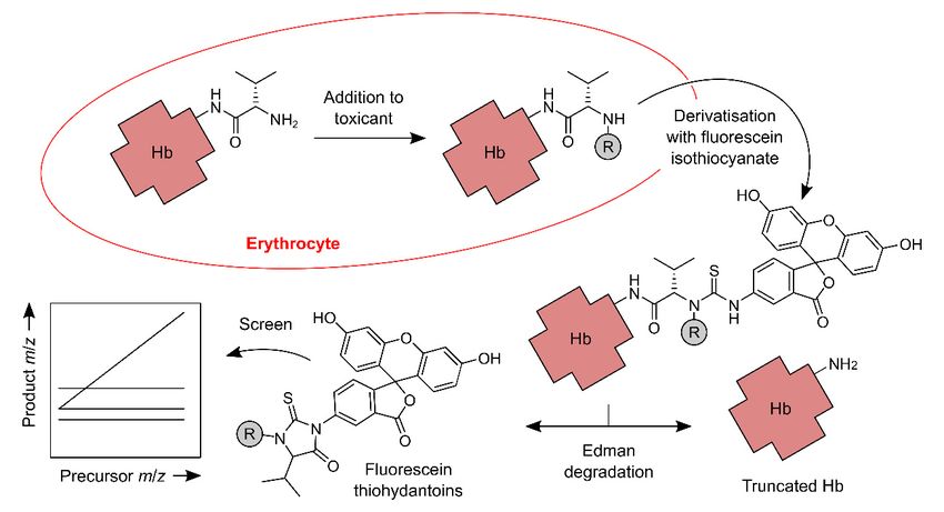
The FIRE screening procedure [28] is a method for untargeted detection of Hb adducts (Figure 1).
It is a stepped method akin to the ‘adductome approach to detect DNA damage’ developed by
Kanaly et al. [34]. In the FIRE screening procedure, different precursor ions (protonated fluorescein
thiohydantoins, FTHs) are captured at the first stage of mass analysis via one of 136 different windows.
Each window is approximately 0.7 m/z units wide, and the m/z values on which the windows are
centred are 1 Da apart. Thus, by cycling through all 136 windows, the method can capture a wide range
of precursor ions and can, therefore, detect the corresponding range of mass shifts (between +14 and
+149 Da). Once captured, a precursor ion is fragmented, and its products are passed to the second stage
of mass analysis. Here, a set of fixed windows permit only constant product ions to pass to the detector
(implicates loss of variable neutral fragments), and a variable window permits only variable productToxics 2019, 7, 29 5 of 17
ions to pass (implicates loss of constant neutral fragments). If the right combination of constant and
variable product ions is detected, then the presence of a corresponding FTH, and therefore Hb adduct,
can be inferred. This MS/MS is done ‘online’ following the chromatographic separation of the FTHs,
Toxics 2019, 7, x FOR PEER REVIEW 5 of 17
and the data thus generated are, like those reported by Kanaly et al., visualised as an ‘adductome map’,
usually a plot of m/z against retention time [28,34,35].
Figure 1. Main steps of the FIRE (‘fluorescein isothiocyanate’, ‘R-group’ and ‘Edman degradation’)
Figure 1. Main
screening steps for
procedure of the
Hb FIRE (‘fluorescein
N-terminal isothiocyanate’,
adductomics. ‘R-group’
The procedure and ‘R’
detects ‘Edman degradation’)
groups, which are
screening procedure for Hb N-terminal adductomics. The procedure detects ‘R’ groups, which are
generated when an N-terminus of Hb reacts with an electrophile in vivo. The N-termini are derivatised
generated when an N-terminus of Hb reacts with an electrophile in vivo. The N-termini
with fluorescein isothiocyanate, and derivatives with ‘R’ groups are selectively decomposed to the are
derivatised with fluorescein isothiocyanate, and derivatives with ‘R’ groups
corresponding fluorescein thiohydantoins. The thiohydantoins are analysed using LC and online are selectively
decomposed
‘stepped’ toquadrupole
triple the corresponding fluorescein thiohydantoins. The thiohydantoins are analysed using
mass spectrometry.
LC and online ‘stepped’ triple quadrupole mass spectrometry.
Carlsson et al. used their procedure to screen the blood of smokers and non-smokers and detected
Carlsson
26 features et al. used
of interest; their procedure
this study is describedtobelow
screen
in the blood
Section 3. of smokers and non-smokers and
detected 26 features of interest; this study is described below in Section 3.
2.6. HSA as A Target of Electrophiles
2.6. HSA
HSAas is A
theTarget
majorofprotein
Electrophiles
in human plasma. Its lifetime in vivo, whilst shorter than that of Hb, is
presumably still long enough
HSA is the major protein in forhuman
adducts to accumulate.
plasma. Its lifetime Ininvivo,
vivo,HSA
whilstbinds fattythan
shorter acids,
that scavenges
of Hb, is
metal ions, and
presumably stillcontributes
long enough to the
foroncotic
adducts pressure of blood In
to accumulate. [22]. Extensive
vivo, HSA binds use offatty
HSAacids,
has been made
scavenges
for the biological monitoring of toxicants, and a detailed account of this
metal ions, and contributes to the oncotic pressure of blood [22]. Extensive use of HSA has been madecan be found in the recent
review
for the by Sabbionimonitoring
biological and Turesky of [36]. For the
toxicants, purposes
and a detailedof the present
account ofreview,
this canwe befocus
foundoninproviding
the recenta
background to the untargeted
review by Sabbioni and Turesky HSA adductomics
[36]. studies.
For the purposes of the present review, we focus on providing
a background to the untargeted HSA adductomics studies. including (but not limited to) histidine
Thus far, HSA contains a number of nucleophilic sites,
residues,
Thuslysine residues
far, HSA and aasingle
contains number reduced cysteine residue
of nucleophilic (Cys-34). Notably,
sites, including (but not histidine
limited to) residues in
histidine
HSA, as inlysine
residues, Hb, are targets and
residues of epoxides
a single[37,38].
reducedLysine
cysteineresidues
residuein serum
(Cys-34).albumins
Notably, arehistidine
notable targets of
residues
aflatoxin B dialdehyde [39,40].
in HSA, as1 in Hb, are targets of epoxides [37,38]. Lysine residues in serum albumins are notable
Cys-34
targets is the only
of aflatoxin site in HSA[39,40].
B1 dialdehyde for which untargeted adductomic methods have been developed.
The motivation
Cys-34 is the only site in HSA forsite
to look at this particular is related
which to the unique
untargeted chemistry
adductomic of thiol
methods havegroups,
beenand the fact
developed.
that HSA Cys-34 accounts for the majority of such groups in human plasma [41].
The motivation to look at this particular site is related to the unique chemistry of thiol groups, and Given that the reacting
species
the factisthat
a thiolate
HSA Cys-34anion rather
accountsthan fora the
thiolmajority
group proper
of such[41], adduct
groups formation
in human should
plasma beGiven
[41]. promoted
that
by alkaline conditions and/or basic groups within the local protein environment.
the reacting species is a thiolate anion rather than a thiol group proper [41], adduct formation should The pK a of the HSA
Cys-34 thiol group
be promoted is controversial,
by alkaline conditionsbut is generally
and/or regarded
basic groups to be
within thelower
local than thatenvironment.
protein of a typical thiol
The
group
pKa of[41].
the HSAIn theCys-34
three-dimensional
thiol group isstructure of HSA,
controversial, butas is
determined by X-ray crystallography,
generally regarded to be lower thanthe side
that of
a typical thiol group [41]. In the three-dimensional structure of HSA, as determined by X-ray
crystallography, the side chain of Cys-34 is partially buried [42]. On this basis, it has been inferred
that there might be a limit to the size of the electrophiles that HSA Cys-34 can add to. It has also been
recognised, however, that the tertiary structure of HSA is dynamic and that Cys-34 may become less
buried upon deprotonation of the thiol group [17,43]. HSA Cys-34 is reactive towards a variety of[44,45], and can also undergo oxidative transformations [46,47]. It appears that, in vivo, a substantial
proportion of the HSA Cys-34 thiol groups is S-thiolated (S-[cystein-S-yl], S-[glutathion-S-yl] and so
on), and a smaller, but appreciable proportion is found as the corresponding sulfenic, sulfinic or
sulfonic acids [41,47].
Toxics 2019, 7, 29 6 of 17
2.7. HSA Cys-34 Adductomics
chainTo of date,
Cys-34methods for HSA
is partially buried Cys-34
[42]. adductomics
On this basis,have it hasbeen
beenbased exclusively
inferred that thereon peptide
might be aanalytes
limit to
(Figure
the size 2). When
of the HSA is digested
electrophiles that HSA with trypsin,
Cys-34 can and
add no to. cleavages
It has alsoare beenmissed, Cys-34
recognised, and its adducts
however, that the
are found
tertiary in a 21-amino-acid
structure peptide
of HSA is dynamic and[48,49]. This peptide,
that Cys-34 may become which lessRappaport’s
buried upongroup has referred
deprotonation to
of the
as ‘T3’
thiol (i.e., the
group third-heaviest
[17,43]. HSA Cys-34 tryptic peptide
is reactive [49]), has
towards been used
a variety as an analyte in a number
of toxicologically-relevant of studies
electrophiles,
[49–51]. When
including sulphur a combination
mustard andofmetabolites
trypsin and ofchymotrypsin
aromatic amines is used,
[44,45], the Cys-34-containing
and can also undergopeptide
oxidative is
transformations [46,47]. It appears that, in vivo, a substantial proportion of the HSA Cys-34 thiola
instead the LQQCPF hexapeptide [43]. The use of Pronase, suggested by Sabbioni and Turesky as
means of
groups is generating
S-thiolated lower-molecular-weight analytes, hasand
(S-[cystein-S-yl], S-[glutathion-S-yl] notso to on),
our knowledge
and a smaller,beenbutimplemented
appreciable
for untargeted HSA Cys-34 adductomics [36]. When Noort et al.
proportion is found as the corresponding sulfenic, sulfinic or sulfonic acids [41,47]. [44,52] used Pronase to digest HSA
adducts of either sulphur mustard or acrylamide, the respective modifications were found in the CPF
2.7. HSA Cys-34 Adductomics
tripeptide.
Some
To date,of methods
the first untargeted
for HSA Cys-34 HSAadductomics
adductomic have analyses
beenwere
based performed
exclusively byonAldini
peptideet al. using
analytes
the technique
(Figure 2). When of precursor ion scanning
HSA is digested [43] (seeand
with trypsin, alsonothe ‘chemical
cleavages aremodificomics’
missed, Cys-34 method
and itsproposed
adducts
are found in a 21-amino-acid peptide [48,49]. This peptide, which Rappaport’s groupthe
by Goto et al. [53]). Precursor ion scanning is an MS/MS technique involving a scan at hasfirst stage
referred
to as ‘T3’ (i.e., the third-heaviest tryptic peptide [49]), has been used as an analyte in a numberisofa
of mass analysis and the isolation of a constant product ion at the second stage. The result
spectrum
studies of the different
[49–51]. precursor ions
When a combination of that
trypsingiveandrisechymotrypsin
to a given product. is used,Aldini et al. [43] reacted
the Cys-34-containing
purified HSA with a mixture of α,β-unsaturated aldehydes (4-hydroxy-2-nonenal,
peptide is instead the LQQCPF hexapeptide [43]. The use of Pronase, suggested by Sabbioni 4-hydroxy-2- and
hexenal and acrolein), and digested the products with trypsin and
Turesky as a means of generating lower-molecular-weight analytes, has not to our knowledge been chymotrypsin. Analysis of the
digestion products, using liquid chromatography (LC) and online precursor
implemented for untargeted HSA Cys-34 adductomics [36]. When Noort et al. [44,52] used Pronase to ion scanning, revealed
peaks corresponding
digest HSA adducts oftoeither substituted
sulphurLQQCPF
mustard peptides. These,
or acrylamide, theinrespective
turn, corresponded
modificationsto HSA
wereCys-34
found
Michael adducts
in the CPF tripeptide. of the α,β-unsaturated aldehyde reactants.
Figure
Figure 2. Main steps
2. Main steps of
of published
published HSAHSA Cys-34
Cys-34 adductomic
adductomic workflows. The reaction
workflows. The reaction of
of HSA
HSA with
with an
an
electrophile
electrophile in the blood plasma installs an ‘R’ group at the Cys-34 site. The HSA is isolated from
in the blood plasma installs an ‘R’ group at the Cys-34 site. The HSA is isolated from
plasma
plasma or or serum
serum and
anddigested
digested—usually
– usually withwithtrypsin
trypsin—to produce
– to produce a mixture
a mixture of of peptides.
peptides. SomeSome of
of the
the
peptides contain ‘R’ groups and others do not (the introduction of an enrichment step prior to
peptides contain ‘R’ groups and others do not (the introduction of an enrichment step prior to
digestion can limit the number of those that do not). Peptides are then separated chromatographically
digestion can limit the number of those that do not). Peptides are then separated chromatographically
and
and analysed
analysed using
using MS/MS.
MS/MS. One
One ofof the MS/MS methods,
the MS/MS methods, aa stepped
stepped triple-quadrupole
triple-quadrupole method
method termed
termed
FS-SRM, is depicted. This method monitors three variable product ions of the tryptic ‘T3’ peptide
FS-SRM, is depicted. This method monitors three variable product ions of the tryptic ‘T3’ peptide (y15,
(y15 , y16 and y17 ).
y16 and y17).
Some of the first untargeted HSA adductomic analyses were performed by Aldini et al. using the
2.8. Fixed-step SRM of HSA Adducts
technique of precursor ion scanning [43] (see also the ‘chemical modificomics’ method proposed by
Goto et al. [53]). Precursor ion scanning is an MS/MS technique involving a scan at the first stage of
mass analysis and the isolation of a constant product ion at the second stage. The result is a spectrum
of the different precursor ions that give rise to a given product. Aldini et al. [43] reacted purified HSA
with a mixture of α,β-unsaturated aldehydes (4-hydroxy-2-nonenal, 4-hydroxy-2-hexenal and acrolein),
and digested the products with trypsin and chymotrypsin. Analysis of the digestion products, using
liquid chromatography (LC) and online precursor ion scanning, revealed peaks corresponding toToxics 2019, 7, 29 7 of 17
substituted LQQCPF peptides. These, in turn, corresponded to HSA Cys-34 Michael adducts of the
α,β-unsaturated aldehyde reactants.
2.8. Fixed-Step SRM of HSA Adducts
An important development, reported by Li et al. in 2011, was the demonstration of a stepped
method called fixed-step SRM (FS-SRM [49]). FS-SRM consists of a sequence of SRM experiments that
collectively resemble a linked scan [54]. In developing the method, Li et al. drew on elements of the
‘adductome approach to detect DNA damage’ described by Kanaly et al. [34,55], and also a method
of analysing mercapturic acids described by Wagner et al. [56]. Being a stepped method, FS-SRM
is broadly analogous to the FIRE screening procedure (which, in fact, it pre-dates). The analytes in
FS-SRM are substituted T3 peptides, and the precursor ions captured in the first stage of mass analysis
are triply-protonated peptides. The product ions isolated in the second stage are doubly-charged
variable y-ions and a singly-charged constant b-ion. Together, these precursor and product ions
constitute what is effectively a peptide sequence tag [57]. The sampling points used for FS-SRM are
4.5 Da apart and, in the Li and co-workers’ study, there were 77 of them. FS-SRM differs from the other
stepped methods in that, for FS-SRM, the sample is infused into the mass spectrometer as a mixture of
adducts rather than as a series of eluted components. There is still an LC step but it is disconnected
from the mass spectrometry, and it serves to capture the entire population of adducts rather than to
separate them. The method is therefore freed from a major constraint imposed by LC, namely the need
for a full set of SRM experiments to be done within the width of a chromatographic peak.
Our personal experience with protein adductomics has been in the implementation of FS-SRM for
epidemiological studies [10,33]. Such studies, which typically involve tens or hundreds of samples,
pose challenges that are not necessarily encountered in smaller pilot studies. In implementing the
method of Li et al., the main challenge that we faced was the need for higher throughput. This was
addressed by evaluating the various stages of sample preparation (HSA purification, adduct enrichment,
digestion and peptide clean-up) and optimising these where possible. Notably, we deleted the adduct
enrichment step, and we changed the method of sample clean-up from HPLC (serial) to solid-phase
extraction (SPE; effectively parallel). A model adduct, prepared by treating HSA with N-ethylmaleimide,
proved useful for evaluating the performance of the methods.
In parallel with our work on FS-SRM, Grigoryan et al. [50] developed a new analytical workflow
based on LC with on-line DDA mass spectrometry. In DDA, the data on which the acquisition is
dependent are precursor ions’ m/z values, and they are obtained via a high-resolution scan—using,
for example, an Orbitrap mass analyser. The data are used to direct the isolation of precursor ions,
and so only these precursor ions are fragmented. The acquisition is the scan via which the resulting
product ions are detected. In addition to their analytical method, Grigoryan et al. [50] also developed
methods for sample preparation and data analysis (the ‘adductomics pipeline’). The method of sample
preparation is essentially a streamlined version of the one developed by Li et al. [49]. One major
difference with respect to the earlier method, however, was the omission of a reducing agent, which
had previously been used to reduce protein disulphide bonds prior to tryptic digestion. The effect
of omitting the reducing agent was to preserve S-thiolated forms of Cys-34. The method of data
analysis begins with the detection of a tag (a combination of constant and variable product ions)
in the product-ion scan data. The corresponding precursor ion is then identified, and an ion count
chromatogram for this precursor ion is extracted. A particularly innovative part of the pipeline is
the method by which the peptide analytes are quantified. Each analyte is quantified relative to a
‘housekeeping peptide’, which is another tryptic peptide of HSA. In this way, the method is able to
control for variation in the quantity of digested HSA. Grigoryan et al. [50] used their pipeline to analyse
samples of plasma from smokers and non-smokers, and found a total of 43 putative adducts (see
Section 3 below).Toxics 2019, 7, 29 8 of 17
2.9. Multiplex Adduct Peptide Profiling
Another promising method for HSA Cys-34 adductomics (and potentially also Hb Cys-β93
adductomics) is ‘multiplex adduct peptide profiling’ (MAPP [51]). MAPP utilises DIA mass
spectrometry, which is perhaps the least prescriptive of all MS/MS techniques. Similar to a stepped
SRM-based method, DIA captures precursor ions via a series of contiguous windows. The windows
are, however, rather wider than those used for SRM, and it is therefore likely that a given window will
capture multiple precursor ions (in MAPP, for example, the width of each window is 10 m/z units).
As in DDA mass spectrometry, the second stage of mass analysis is a scan, and a high-resolution scan
is done as an alternative first stage.
The MAPP method, like the ‘adductomics pipeline’, requires prior knowledge of the peptide
analyte’s sequence and the site of modification. Series of constant product ions (e.g., b-ions from
backbone scission near the N-terminus) are recognised and are linked back to their respective precursor
ions via common chromatographic retention times. The substituted peptide’s mass shift is then
confirmed by the presence of corresponding variable product ions. Although the authors were only
able to identify oxidised and S-thiolated forms of HSA Cys-34, their method has the potential to detect
toxicologically-relevant adducts (e.g., if the samples could be further enriched for these adducts prior
to analysis).
2.10. Hb and HSA Compared
Given that Hb and HSA contain some of the same nucleophilic functional groups, these proteins
might be expected to have overlapping reactivity towards electrophiles. The observation that cysteine
residues in HSA and Hb can add to comparable amounts of benzene oxide in vivo, for example,
is evidence of such overlap [58]. On the other hand, Dingley et al. [59] found that dietary exposure
to 2-amino-1-methyl-6-phenylimidazo[4,5-b]pyridine (PhIP; see Section 3.3) caused the formation of
substantially larger amounts of HSA adducts than Hb adducts. A similar fate has been observed
for aflatoxin B1 in rats: of a given dose of this toxicant, a substantially higher proportion is found
bound to serum albumin than to Hb [60,61]. This might also be expected to be the case in humans,
and indeed assays for HSA adducts of aflatoxin B1 dialdehyde have been developed [62]. Possible
reasons for differences in the amount or type of adducts include (i) the fact that Hb and HSA are
synthesised at different sites in the body (in different cell types), and as a result could be exposed
to different electrophiles [36]; (ii) the fact that Hb resides inside the erythrocyte, whereas HSA is
secreted [18]; (iii) the influence of neighbouring amino acid side chains and cofactors on the reactivity
of the nucleophilic groups (see Sections 2.1 and 2.6); and (iv) the possibility that the erythrocyte
membrane could shield Hb from electrophiles, or even sequester electrophiles [63]. It is also worth
considering that apparent differences in the extent of adduct formation could reflect differences in
chemical and biological stability of the proteins and/or modifications.
2.11. Other Target Proteins
Few proteins other than Hb and HSA have been discussed as candidates for untargeted adductomic
analyses, and fewer still have been investigated experimentally. Hb and HSA adducts are probably
two of the richest and most accessible sources of potential biomarkers, but this is not to say that other
proteins could not provide additional and unique information. Three other proteins of relevance to the
present review have been discussed: Collagen, histones and apolipoproteins. Collagen is mentioned
by Scheepers in his workshop report [19], presumably because of its abundance in the body and its
extremely long lifespan in certain tissues [64]. However, there have been few attempts to use collagen
adducts for biological monitoring, probably because of the heterogeneity, physical properties and
limited accessibility of collagen [65–67]. Histones, which are also mentioned by Scheepers, represent
a more promising source of biomarkers. Work on histone adducts has not been extensive, but some
interesting results have been obtained. N-Terminal segments of histones are of particular interestToxics 2019, 7, 29 9 of 17
because they protrude from nucleosomal core particles, and, on this basis, it is plausible that they could
be accessible to electrophiles. Consistent with this idea, SooHoo et al. [68] observed modifications
near the N-termini of histones isolated from cultured human lymphoblasts that had been exposed
to anti-benzo[a]pyrene 7,8-dihydrodiol-9,10-oxide (BPDE). Fabrizi et al. [69] used a model peptide to
infer the reactivity of an N-terminal segment of histone H2B towards phosgene, and observed the
incorporation of carbonyl groups into the peptide.
Apolipoproteins have been investigated as targets of endogenous electrophiles, such as the lipid
oxidation product 4-hydroxy-2-nonenal. By definition, endogenous adducts cannot be biomarkers
of exposure in the strict sense, but they could potentially be biomarkers of effect. We mention them
here because they have been the subject of a recent untargeted adductomics study. This study focused
on adducts of histidine and lysine residues in human low density lipoprotein [35]. Unlike the FIRE
screening procedure or FS-SRM, the method is not site-specific; rather, it detects modifications to any
and all residues of particular amino acid. The analytes are ‘free’ amino acids, which are prepared from
lipoprotein by acid hydrolysis. Consequently, they may represent a mixture of sites, and perhaps a
mixture of proteins. The analytical method, like others described elsewhere in this article, involves
ultraperformance LC and triple quadrupole mass spectrometry. Apparently it is a stepped method,
in which a constant product ion is isolated at the second stage of mass analysis. For adducts of histidine
residues, the constant product is the immonium ion of histidine, and for adducts of lysine residues,
it is a deaminated immonium ion of lysine. Shibata et al. [35] used their method to analyse low density
lipoprotein that had been first purified from human plasma, and then oxidised in vitro. The oxidised
lipoprotein was treated with sodium borohydride to reduce imine linkages (as in, for example, a lysine
residue adducts of 9-oxononanoic acid), before being hydrolysed and the resulting amino acids
analysed. The authors produced adductome maps for lipoprotein with and without oxidation, and by
comparing these maps they were able to attribute the formation of the aforementioned 9-oxononanoic
acid adduct to the oxidising condition.
2.12. Adduct Enrichment
Enrichment, in the context of untargeted adductomics, entails depletion of the unmodified
nucleophile and possibly also other substances that might interfere with the detection of the adducts.
In the FIRE screening procedure for Hb adducts, enrichment is facilitated by the detachment of the
N-alkylvaline residues. This exaggerates the relatively minor difference in structure between Hb
and its adducts, thereby allowing the unmodified Hb to be removed readily [28]. For HSA Cys-34
adductomics, methods of enrichment have mainly exploited the reactivity of the Cys-34 thiol group,
which is present in the unmodified HSA but not in the adducts. Funk et al. [70] demonstrated the
use of a disulfide-functionalised resin for scavenging unmodified HSA, and this method was later
used in adductomic workflows [49,51,71]. The main limitation of the thiol scavenging method is that
it does not remove S-thiolated HSA: If a reducing agent is later added to reduce the other disulfide
bonds in HSA (i.e., those of the cystine residues) then the S-thiolation is reversed and the Cys-34 thiol
would seem to reappear. Funk et al. [70] sought to limit this effect by removing the S-thiolation prior to
the scavenging step. In our hands, the thiol scavenging method proved difficult to implement in a
high-throughput setting, and so we deleted it from our workflow [33]. Chung et al. [71] used thiol
scavenging as the first of two stages of enrichment, the second stage being an antibody-mediated
purification of the substituted T3 peptides using a polyclonal antibody raised against the T3 peptide
but having cross-reactivity with adducts.
3. Human Biomonitoring
3.1. Methodological Considerations
Human biomonitoring refers to the quantification of xenobiotics or their derivatives (and sometimes
their early effects) in human biospecimens [72]. As well as confirming the nature of the exposure,Toxics 2019, 7, 29 10 of 17
biomonitoring aims to measure the internal dose of the xenobiotic(s). The biomonitoring of protein
adducts is usually done as part of the ‘bottom up’ (targeted) approach (see Section 1). A typical targeted
method might involve isotope dilution (i.e., the addition of a known amount of an isotopically-labelled
standard) followed by LC-MS/MS. This would require prior characterisation of the adduct and synthesis
of a suitable standard.
In principle, data collected via the untargeted approach (e.g., peak areas from LC-MS/MS) could
be used in the same way as those collected in targeted studies. However, this would depend on the
untargeted method achieving an acceptable accuracy, precision and dynamic range for each relevant
adduct. At some stage, a synthetic reference compound would be needed to confirm a particular
adduct’s identity, and to implicate the corresponding electrophile [18]. For hitherto unknown adducts,
possible identities must first be proposed. Methods that have assisted in this endeavour have included
database searching, the use of calculator software, and the comparison of measured and predicted
physicochemical properties [50,73]. The characterisation of novel adducts—a challenging aspect of the
research—has been reviewed in detail by Carlsson et al. [18].
Accuracy, in practice, may suffer as a consequence of the need to capture a range of adducts.
It is likely that the use of generic standards (e.g., the S-carbamidomethylated T3 peptide for FS-SRM)
affects accuracy, and therefore precludes absolute quantification [33]. Dynamic ranges are dependent
on the analytical method, and presumably also on the ability to enrich adducts. As judged from
lowest reported adduct concentrations, the detection limits of Grigoryan and co-workers’ LC-MS-based
method, and of the FIRE screening procedure, are good (Toxics 2019, 7, 29 11 of 17
Applying their untargeted Hb adductomic approach to a larger study population, Carlsson et
al. [76] analysed blood samples from healthy children about 12 years old (n = 51). In this cohort,
a total of 24 adducts (12 of them previously identified; see above) were observed and their levels
quantified. Relatively large interindividual variations in adduct levels were observed. The frequencies
of micronuclei in erythrocytes were also determined. Analysis using a partial least-squares regression
model showed that as much as 60% of the micronucleus variation could be explained by the adduct
levels. This indicates the ability of such studies to align measurements of internal dose (protein
adducts) with endpoints of genotoxicity (micronucleus formation).
3.3. Human Biomonitoring of HSA Adducts
An early study that demonstrated the utility of monitoring HSA for alkylated cysteine involved
exposure of human blood to 14 C-labelled sulfur mustard (the chemical warfare agent mustard gas) [44].
Isolation and tryptic digestion of albumin produced the 21-amino acid fragment containing a sulfur
mustard-cysteine adduct, detected by micro-LC-MS/MS. An alternative method, which employed
Pronase for the digestion, yielded a modified tripeptide (Cys-Pro-Phe), which was detected with
greater sensitivity than the 21-amino acid fragment. The method was used to analyse samples of
blood from nine Iranians exposed to sulfur mustard during the Iran-Iraq war of 1986. In all nine cases,
the sulfur mustard-adducted tripeptide was detected.
Application of the FS-SRM method to analyses of archived plasma protein that had been pooled
according to subjects’ ethnicities and tobacco smoking habits demonstrated differences between
pools [49] and suggested that FS-SRM might be able to detect statistically significant differences
between groups of individual samples that had not been pooled.
A pilot study of 20 smokers and 20 never-smokers provided evidence of the effect of smoking
on levels of putative HSA adducts. Differences between smokers and never-smokers were most
apparent in putative adducts with net gains in mass between 105 Da and 114 Da (relative to unmodified
HSA) [33].
Further investigations of the effects of tobacco smoking have revealed around 43 adduct features,
some of which are positively associated with smoking and but also some that are negatively associated.
The former result from genotoxic constituents of tobacco smoke, such as ethylene oxide and acrylonitrile,
while the latter, which include Cys-34 oxidation products and disulfides, may reflect alterations in the
serum redox state of smokers, resulting in lower adduct levels [50].
Grigoryan et al. used LC and high-resolution mass spectrometry to investigate interactions
between the Cys-34 and reactive oxygen species (ROS) [47]. Chronic exposure to ROS is linked to
many chronic diseases and, in this study, a number of adducts originating from ROS were detected in
human serum: Sulfinic acid, sulfonic acid and a proposed sulfinamide structure (a mono-oxygenated
moiety also with the loss of two hydrogen atoms).
Antibody enrichment may pave the way to a more sensitive assay. Using a polyclonal antibody,
raised against the T3 peptide, but with cross-reactivity to the peptide containing adducts (see
Section 2.12), ten modified T3 peptides were detected in human plasma samples; eight of them were
characterised and they included Cys-34 oxidation products, modification involving loss of water or
lysine, cysteinylation, and transpeptidation of arginine [68].
In a study of women from the Xuanwei and Fuyuan counties in China, where extensive use of
smoky coal for heating and cooking has resulted in very high rates of lung cancer among non-smokers,
HSA Cys-34 adducts were compared in 29 females who used smoky coal and 10 controls using other
energy sources [77]. Fifty different modified T3 peptides were identified, including oxidation products,
mixed disulfides, rearrangements and truncations. Two peptides that were detected at significantly
lower levels in the smoky coal group were adducts of glutathione and γ-glutamylcysteine. The results
are interpreted as evidence that exposure to the indoor combustion products results in depletion of
glutathione, an essential antioxidant, as well as its precursor γ-glutamylcysteine [77].Toxics 2019, 7, 29 12 of 17
A recent study on the health effects of urban air pollution, the Oxford Street II study [78], involved a
randomised crossover design whereby three groups of volunteers (healthy subjects, chronic obstructive
pulmonary disease (COPD) sufferers and patients with ischaemic heart disease (IHD)) walked for two
hours along a busy street in London where traffic is restricted to diesel buses and taxis. The volunteers
also spent two hours walking in a London park on a separate occasion. They were monitored for
respiratory and cardiovascular function in both environments and, in addition, two studies have
analysed their HSA samples for adducts. In the first report, Liu et al. [79] analysed 50 HSA samples
by high-resolution mass spectrometry to determine whether protein modifications differ between
COPD or IHD patients and healthy subjects. The untargeted analysis of adducts at the Cys-34 locus
of HSA detected 39 adducts with sufficient data, and these adducts were examined for associations
with estimated exposures to air pollution and health status. Multivariate linear regression revealed
21 significant associations, mainly with the underlying diseases, but also with air-pollution exposures.
Interestingly, most of the associations indicated that adduct levels decreased with the presence of
disease or increased pollutant concentrations. Negative associations of COPD and IHD with the Cys-34
disulfide of glutathione and two Cys-34 sulfoxidations were consistent with results from smokers and
non-smokers [50] and from non-smoking women exposed to indoor combustion of coal and wood [74].
In the second study, Preston et al. [80] examined a larger number of Oxford Street II samples by
the FS-SRM method. Associations between amounts of putative adducts and two types of measure
were tested: Pollution (e.g., ambient concentrations of nitrogen dioxide and particulate matter) and
health outcome (e.g., measures of lung health and arterial stiffness). There were 11 instances of a
response variable being associated with a pollution measurement and eight instances of a response
variable being associated with a health outcome measure. However, no two measures of different
types were associated with the same adduct amount, suggesting that the internal changes responsible
for health outcomes may differ from those that effect changes in adduct amounts.
In a more targeted study, Bellamri et al. [81] investigated the formation in human subjects of HSA
adducts at Cys-34 by PhIP, which is formed in cooked meats and may be associated with colorectal,
prostate and mammary cancer. Volunteers abstained from eating cooked well-done meat or fish for
three weeks, then ate a semi-controlled diet that included cooked beef containing known quantities of
PhIP for four weeks. The volunteers then returned to their regular diets, but with the exclusion of
cooked well-done meat and fish for a further four weeks. The authors found that an adduct of oxidised
PhIP, which was below the limit of detection (LOD) (10 femtograms PhIP/mg HSA) in most subjects
before the meat feeding, increased by up to 560-fold at week 4 in subjects who ate meat containing 8.0
to 11.7 µg of PhIP per 150–200 g serving. In contrast, the adduct remained below the LOD in subjects
who ingested 1.2 or 3.0 µg PhIP per serving, and PhIP-HSA adduct levels did not correlate with PhIP
intake levels across four exposure groups (p = 0.76). There were also indications that the PhIP adduct
was unstable, having a half-life of fewer than two weeks. Nevertheless, the study demonstrates that
the Cys-34 site in HSA is accessible by a relatively large molecule like PhIP, despite concerns about
possible steric hindrance (see Section 2.6.).
4. Prospects
A key advantage of monitoring proteins for adducts is the abundance of material that can be
obtained from tissue banks; for example, red blood cells are an abundant source of Hb and blood
plasma or serum is an abundant source of HSA. The proteins’ lifespans in blood mean that there
is a substantial “capture period” for monitoring exposure to genotoxicants; and protein adducts,
unlike DNA adducts, are not subject to loss through repair processes. Full implementation of the
exposome concept requires monitoring individuals or populations at several points in time over the
course of their lives [3,4]. This is achievable if biobanks collect material from individuals not just once
but multiple times, and such biobanks already exist.
Dried blood spots can also be a suitable source of protein for investigation [82]. If obtained
from neonatal blood spots (i.e., Guthrie spots) then the material provides a valuable opportunity forToxics 2019, 7, 29 13 of 17
investigating exposures in utero. A single blood spot of about 50 µL is estimated to contain about
9.6 mg of protein, of which about 7.7 mg will be Hb and 1.2 mg HSA [79]. In a proof-of-principle study,
Yano et al. [83] identified 26 Cys-34 adducts (oxidation and S-thiolation products) in HSA isolated from
dried blood spots of 49 newborn babies and were able to distinguish between newborns of smoking
and non-smoking mothers on the basis of the levels of a putative cyano modification to Cys-34.
There is also the potential to broaden the scope of adductomics by investigating novel modifiable
loci in blood proteins. Cys-34 of HSA and the N-terminus of Hb are undoubtedly major targets for
electrophiles, but the literature hints at wider reactivity within the blood proteome. Consequently,
a hitherto-untapped source of analytes for protein adductomics can be envisaged. Mapping the loci at
which adducts can form will be beneficial and, for this purpose, new chemical tools will be required.
Identifying adducts detected by the top-down approaches will be a challenge, necessitating chemical
synthesis of candidate structures for unequivocal characterisation.
Protein adductomics is a component of the exposome concept that is still relatively novel, but it
is one that has already demonstrated the ability to capture electrophiles of both endogenous and
exogenous origin; this suggests the potential to contribute meaningfully to the aims of the exposome
concept—to describe the totality of all biologically relevant exposures. Rapid advances in mass
spectrometry instrumentation, with significant increases in sensitivity and resolution, will drive further
advances in protein adductomics methodology. When coupled with other omics approaches, such as
proteomics, transcriptomics and metabolomics, all of which have the potential for high-throughput
screening of populations, a future can be envisaged in which it will be possible to capture snapshots
of human exposure to genotoxicants and the resultant biological consequences at multiple stages
throughout life. Building this comprehensive picture should shed significant light on the causes and
courses of chronic diseases in humans. Such knowledge will provide new opportunities for early
intervention to reduce potentially harmful human exposure, to monitor the effectiveness of intervention
strategies and, ultimately, to prevent diseases before they occur.
Author Contributions: Both authors critically reviewed the literature and contributed equally to writing
the manuscript.
Funding: The authors’ research was funded by the European Community’s Seventh Framework Programme
(FP7/2007-2013) under grant agreement number 308610 (the EXPOsOMICS project). Additional funding was from
Cancer Research UK (Programme Grant CRUK/A14329) and the MRC-PHE Centre for Environment and Health
(MRC grant number G0801056/1).
Conflicts of Interest: The authors declare no conflict of interest.
References
1. Rappaport, S.M. Genetic Factors Are Not the Major Causes of Chronic Diseases. PLoS ONE 2016, 11, e0154387.
[CrossRef] [PubMed]
2. Rappaport, S.M.; Smith, M.T. Epidemiology. Environment and disease risks. Science 2010, 330, 460–461.
[CrossRef] [PubMed]
3. Wild, C.P. Complementing the genome with an “exposome”: The outstanding challenge of environmental
exposure measurement in molecular epidemiology. Cancer Epidemiol. Biomark. Prev. 2005, 14, 1847–1850.
[CrossRef] [PubMed]
4. Wild, C.P. The exposome: From concept to utility. Int. J. Epidemiol. 2012, 41, 24–32. [CrossRef] [PubMed]
5. Rappaport, S.M.; Barupal, D.K.; Wishart, D.; Vineis, P.; Scalbert, A. The blood exposome and its role in
discovering causes of disease. Environ. Health Perspect. 2014, 122, 769–774. [CrossRef]
6. Rappaport, S.M. Biomarkers intersect with the exposome. Biomarkers 2012, 17, 483–489. [CrossRef]
7. Dennis, K.K.; Marder, E.; Balshaw, D.M.; Cui, Y.; Lynes, M.A.; Patti, G.J.; Rappaport, S.M.; Shaughnessy, D.T.;
Vrijheid, M.; Barr, D.B. Biomonitoring in the Era of the Exposome. Environ. Health Perspect. 2017, 125, 502–510.
[CrossRef]
8. Stewart, B.W.; Bray, F.; Forman, D.; Ohgaki, H.; Straif, K.; Ullrich, A.; Wild, C.P. Cancer prevention as part of
precision medicine: ‘Plenty to be done’. Carcinogenesis 2016, 37, 2–9. [CrossRef] [PubMed]You can also read