Complementary Food Ingredients Alter Infant Gut Microbiome Composition and Metabolism In Vitro
←
→
Page content transcription
If your browser does not render page correctly, please read the page content below
microorganisms

Article
Complementary Food Ingredients Alter Infant Gut Microbiome
Composition and Metabolism In Vitro
Shanthi G. Parkar * , Doug I. Rosendale, Halina M. Stoklosinski, Carel M. H. Jobsis                              , Duncan I. Hedderley
and Pramod Gopal *

                                          The New Zealand Institute for Plant and Food Research Limited, Private Bag 11600,
                                          Palmerston North 4442, New Zealand; doug.rosendale@anagenix.com (D.I.R.);
                                          halina.stoklosinski@plantandfood.co.nz (H.M.S.); carel.jobsis@plantandfood.co.nz (C.M.H.J.);
                                          duncan.hedderley@plantandfood.co.nz (D.I.H.)
                                          * Correspondence: shanthi.parkar@gmail.com (S.G.P.); pramod.gopal@plantandfood.co.nz (P.G.)

                                          Abstract: We examined the prebiotic potential of 32 food ingredients on the developing infant
                                          microbiome using an in vitro gastroileal digestion and colonic fermentation model. There were sig-
                                          nificant changes in the concentrations of short-chain fatty-acid metabolites, confirming the potential
                                          of the tested ingredients to stimulate bacterial metabolism. The 16S rRNA gene sequencing for a
                                          subset of the ingredients revealed significant increases in the relative abundances of the lactate-
                                          and acetate-producing Bifidobacteriaceae, Enterococcaceae, and Lactobacillaceae, and lactate- and
                                          acetate-utilizing Prevotellaceae, Lachnospiraceae, and Veillonellaceae. Selective changes in specific
                                          bacterial groups were observed. Infant whole-milk powder and an oat flour enhanced Bifidobacte-

                                   riaceae and lactic acid bacteria. A New Zealand-origin spinach powder enhanced Prevotellaceae
                                          and Lachnospiraceae, while fruit and vegetable powders increased a mixed consortium of beneficial
Citation: Parkar, S.G.; Rosendale,
                                          gut microbiota. All food ingredients demonstrated a consistent decrease in Clostridium perfringens,
D.I.; Stoklosinski, H.M.; Jobsis,
C.M.H.; Hedderley, D.I.; Gopal, P.
                                          with this organism being increased in the carbohydrate-free water control. While further studies
Complementary Food Ingredients            are required, this study demonstrates that the selected food ingredients can modulate the infant gut
Alter Infant Gut Microbiome               microbiome composition and metabolism in vitro. This approach provides an opportunity to design
Composition and Metabolism In             nutrient-rich complementary foods that fulfil infants’ growth needs and support the maturation of
Vitro. Microorganisms 2021, 9, 2089.      the infant gut microbiome.
https://doi.org/10.3390/
microorganisms9102089                     Keywords: infant complementary foods; baby foods; gut microbiome; infant complementary feeding;
                                          infant solid foods; short-chain fatty acids; SCFAs
Academic Editor: Julio Villena

Received: 15 August 2021
Accepted: 30 September 2021
                                          1. Introduction
Published: 3 October 2021
                                               The trajectory of microbiome development in the first 1000 days of the child’s life [1,2]
Publisher’s Note: MDPI stays neutral
                                          has long-term implications in terms of the individual’s immune and metabolic health [3].
with regard to jurisdictional claims in
                                          A dysbiosis in the early-life gut and the interlinked alterations in the immune signaling
published maps and institutional affil-   have been associated with childhood immune-mediated disorders such as type 1 diabetes,
iations.                                  juvenile asthma, and allergies [4,5].
                                               Early feeding patterns, such as breast milk and/or formula and the duration of
                                          breastfeeding are some of the key factors in regulating gut bacterial colonization and
                                          composition and the associated immunological maturation of the growing infant [1,4].
Copyright: © 2021 by the authors.
                                          Bifidobacteriaceae are the most abundant group in the gut of both breast-fed and formula-
Licensee MDPI, Basel, Switzerland.
                                          fed infants [2,6,7]. Seen in lesser abundance are bacterial families such as Bacteroidaceae,
This article is an open access article
                                          Lachnospiraceae, Veillenollaceae, Clostridiaceae, Lactobacillaceae, Enterococcaceae, and
distributed under the terms and           Streptococcaceae [2,6,7]. Formula-fed infants show a greater diversity in their gut bacteria
conditions of the Creative Commons        with a higher abundance of families from the Firmicutes phylum [7,8]. Lactate produc-
Attribution (CC BY) license (https://     ers such as Bifidobacteriaceae, Lactobacillaceae, Enterococcaceae, and Streptococcaceae
creativecommons.org/licenses/by/          have been shown to protect infants against antibiotic- and enteropathogen-induced diar-
4.0/).                                    rhea [9], inflammatory responses [10], and atopic dermatitis [11]. Bifidobacteria possess the

Microorganisms 2021, 9, 2089. https://doi.org/10.3390/microorganisms9102089                       https://www.mdpi.com/journal/microorganisms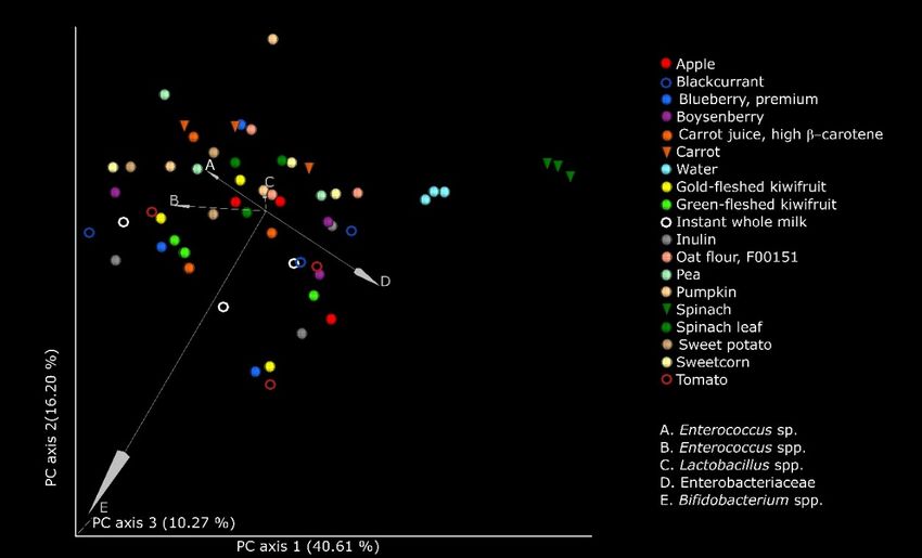
Microorganisms 2021, 9, 2089                                                                                            2 of 17

                               metabolic capacity to degrade and utilize gut epithelial mucin and the structurally analo-
                               gous human milk oligosaccharides [12,13]. Consequently, Bifidobacteriaceae have evolved
                               to occupy prime niches in the infant gut [12,13] and may persist into adulthood [14,15].
                               A founder–colonizer species of the infant gut, B. bifidum lays the foundation for the sub-
                               sequent colonization by diverse microbiota. This is enabled by sharing carbohydrate
                               resources with other bifidobacteria, as well as intermediary metabolites such as lactate and
                               acetate with butyrate- and propionate-producing bacteria such as Eubacterium hallii [12,16].
                               B. bifidum strains have also been associated with beneficial effects on gut functionality
                               such as intestinal homeostasis, modulation of gut immunity, and anti-inflammatory pro-
                               tection [17,18]. Similarly, other bifidobacteria and lactic acid bacteria (LAB) have also
                               been found to be autochthonous and remain gut-associated throughout the host’s lifespan,
                               conferring beneficial effects on the host [14,19].
                                     The transition from the milk-based infant diet to a mixed diet, where milk is com-
                               plemented with plant-based foods, meat, and dairy, is recommended to start at around
                               6 months of age [1]. The period, up until 24 months of age, is a critical window of oppor-
                               tunity for shaping the structure of the developing infant’s gut microbiota [1]. Exposure
                               to new foods triggers developmental maturation of the gut, and it presents novel, nondi-
                               gestible carbohydrates to the microbes in the colon of the growing infant. Nondigestible
                               carbohydrates have a major impact on the proliferation of gut bacteria such as Bacteroides
                               and Faecalibacterium (the most abundant genera in Bacteroidetes and Firmicutes phyla,
                               respectively, in the adult gut), and the concomitant reduction in enterobacteria, bifidobacte-
                               ria, and clostridia that predominate in the infant gut [3,5,20]. The cessation from milk as the
                               primary food and the transition to solid foods play an important role in the acquisition and
                               proliferation of new bacteria and lay the foundation for a stable and adult-type microbial
                               population. Foods that influence this early colonization may, thus, permanently shape gut
                               microbiota composition, maturation of the gut and immune system, and even long-term
                               health effects [1,5,21].
                                     A variety of foods are being gradually introduced to growing infants as they adapt
                               to the family’s dietary patterns. These early foods generally include cheese, meat, eggs,
                               fruits, vegetables, and cereals, which are customized to local availability and family pref-
                               erences [1,22,23]. As the food transits the growing child’s gastrointestinal tract, the fibers
                               (and other phytochemicals such as polyphenols) that are resistant to host digestion reach
                               the colon partially or fully undigested. The founding bacteria of the child, including
                               bifidobacteria and LAB, adapt to the newly introduced dietary components, including
                               fiber, which is broken down to generate lactate, an organic acid, and acetate, a short-chain
                               fatty acid (SCFA) [4,5,14,24]. These microbial acid metabolites play an important role in the
                               maturation of gut and gut ecology, including the development of syntrophic microbiota, by
                               providing substrates to enhance the growth of secondary consortium that can deconstruct
                               more complex carbohydrates to generate SCFAs such as propionate and butyrate. Lactate
                               and the SCFAs also regulate microbial homeostasis by maintaining an acidic milieu that
                               inhibits colonization by pathogens [25,26]. The SCFAs, mainly acetate, propionate, and
                               butyrate, are also important for host health, with butyrate being particularly important
                               as an energy source for colonocytes and for strengthening of gut barrier function [26,27].
                               Butyrate is generated mainly by Lachnospiraceae and Ruminococcaceae, either by breaking
                               down polysaccharides or by utilizing the lactate produced by lactic acid bacteria and the
                               acetate, an early metabolite of microbial metabolism of glycans [26]. The gut microbiome,
                               especially when highly diverse and abundant in beneficial members of Bifidobacterium, Bac-
                               teroides, Lachnospiraceae, and Ruminococcaceae, can aid in the prevention of gut disorders,
                               as well as autoimmune and allergic disease, during childhood and later in life [4,5,28,29].
                                     Prebiotics are recognized as some of the most promising dietary supplements, with
                               numerous health benefits in both children and adults. One of the most studied prebiotics
                               in pediatric nutrition (infant formulae) is a mixture of short-chain galactooligosaccharide
                               and long-chain fructooligosaccharide in a ratio of 9:1 [30]. Solid foods supplemented
                               with galacto- and fructooligosaccharides were found to enhance gut bifidobacteria when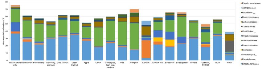
Microorganisms 2021, 9, 2089 3 of 17
Microorganisms 2021, 9, 2089 3 of 17
galacto- and fructooligosaccharides were found to enhance gut bifidobacteria when fed in
a 6 week intervention study involving 4–6 month old infants who were about to start con-
suming
fed in asolid
6 week foods [31]. Inulin
intervention is another
study involving well-characterized
4–6 month old infants prebioticwhothatwere
has about
been
shown
to start consuming solid foods [31]. Inulin is another well-characterized prebiotic as
to beneficially affect the gut microbiome of children [32,33] and is often used thata
supplement
has been shown in many food products
to beneficially affect formulated for children.
the gut microbiome Looking [32,33]
of children beyondand galactooli-
is often
gosaccharides and fructans,
used as a supplement in many exposure of the early-life
food products formulatedgut microbiome
for children.toLooking
a wide array
beyondof
nondigestible carbohydrates
galactooligosaccharides with varied
and fructans, physicochemical
exposure complexities
of the early-life helps totofurther
gut microbiome a wide
enhance
array of the plasticity ofcarbohydrates
nondigestible the microbiome with[1,21].
variedBefore progression tocomplexities
physicochemical family foods,helps
and toto
complement breast and/or formula feeding, infants are given suitable
further enhance the plasticity of the microbiome [1,21]. Before progression to family foods, homemade or com-
mercial ready-to-serve
and to complement foods.
breast Infant
and/or complementary
formula foods are
feeding, infants comprise plant-based,
given suitable dairy,
homemade
and meat products
or commercial and aim to meet
ready-to-serve foods.the nutritional
Infant needs of infants
complementary [34–37]. plant-based,
foods comprise
Plant-based
dairy, foods areand
and meat products a richaim source
to meet of structurally
the nutritional varied
needscarbohydrates, with varying
of infants [34–37].
capacities to alterfoods
Plant-based gut microbial
are a richcomposition [1,38–40].varied
source of structurally While carbohydrates,
different fractionswithofvarying
plant-
derived carbohydrates
capacities are recognized
to alter gut microbial to play [1,38–40].
composition an important Whilerole in shaping
different the infant
fractions gut
of plant-
derived carbohydrates
microbiome, few studiesare recognized
have examined tothe
play an important role in shaping
microbiome-modulating role ofthe infant
whole gut
foods
microbiome,
or ingredientsfew thatstudies
may behaveusedexamined the microbiome-modulating
in the formulation of infant foods. Werole of whole foods
hypothesized that
or ingredients
food ingredients that may be used
(predigested in anin the
upper formulation
gut model) of will
infant foods. We hypothesized
differentially modulate colonic that
food ingredients
microbiota (predigested
from infants in an upper
in a simulated hindgutgutmodel)
model.will differentially
In this study, wemodulate
selected acolonic
range
microbiota
of from available
commercially infants in plant-based
a simulated hind food gut model. Inalong
ingredients, this study, we selected powders,
with whole-milk a range of
commercially available plant-based food ingredients, along with
and evaluated their ability to modulate infant gut microbiota using a batch fermentation whole-milk powders, and
evaluated their ability to modulate infant gut microbiota using
model of the colon. We examined these ingredients for their fermentative capacity by fol- a batch fermentation model
of the colon.
lowing We examined
the changes these ingredients
in the metabolites generated forbytheir fermentative
microbial breakdowncapacity bysubstrates
of the following
the changes
over 24 h, and in the
we metabolites
further studied generated
a subset byof microbial
the selectedbreakdown of the substrates
food ingredients in terms over
of
24 h, andinwe
changes further studiedmicrobiome
substrate-driven a subset of the selected food ingredients in terms of changes
structure.
in substrate-driven microbiome structure.
2. Materials and Methods
2. Materials and Methods
A schematic workflow of the experimental protocol used in this study is shown in
A schematic workflow of the experimental protocol used in this study is shown in
Figure 1.
Figure 1.
Figure1.
Figure Anoutline
1.An outlineof
ofthe
theexperimental
experimentalprotocol
protocol and
and analyses.
analyses.
2.1. Food Ingredients
2.1. Food Ingredients
Thirty-two different food ingredients were selected from commercial sources and
Thirty-two different food ingredients were selected from commercial sources and
tested for their ability to modulate infant gut microbiota. They were classified as per the
tested
Unitedfor theirDepartment
States ability to modulate infant gut
of Agriculture [41]microbiota. They
into milk, fruit were classified
(blackcurrant, as per the
Boysenberry,
blueberry, kiwifruit, apple, feijoa, and passionfruit), vegetables (carrot, pea, pumpkin,
spinach, sweetcorn, sweet potato, and tomato), flavor agents (honey and kaffir lime leaf),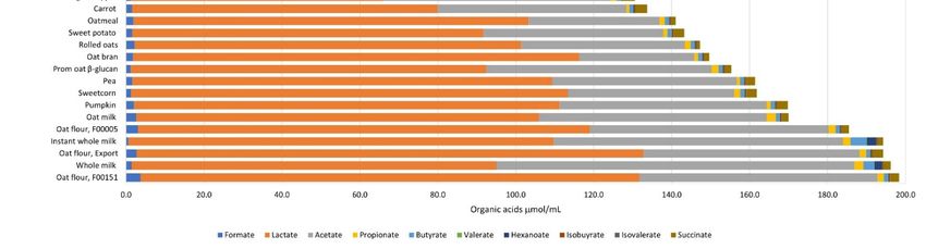
Microorganisms 2021, 9, 2089                                                                                                     4 of 17

                                   and different forms of cereal grain (oats) (Table 1). Orafti® Synergy1 (ORAFTI Active Food
                                   Ingredients, Tienen, Belgium, hereafter referred to as inulin) was included as a positive
                                   control. This inulin is a 1:1 mixture of short-chain oligofructans and long-chain fructans that
                                   vary in their degrees of polymerization, i.e.,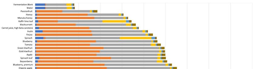
Microorganisms 2021, 9, 2089                                                                                         5 of 17

                               2.2. In Vitro Simulated Gastroileal Digestion
                                    As outlined in Figure 1, food ingredients were digested in vitro using previously
                               published protocols [42]. Each food ingredient (n = 32) was weighed in triplicate (25 g) and
                               hydrated in 46 mL of sterile de-ionized water. Three replicates of sterile deionized water
                               (25 mL) were included as the vehicle control. The hydrated food ingredients and the water
                               control were incubated with acidified 10% pepsin (P7000, >250 units/mL, Sigma-Aldrich,
                               St. Louis, MO, USA) with slow constant stirring (130 rpm) at 37 ◦ C for 30 min. The reaction
                               mixture was buffered to pH 6.0 with 0.1 M maleate buffer and incubated with 0.05 mL of
                               amyloglucosidase (E-AMGDF, Megazyme, Bray, Ireland) and 1.25 mL of 2.5% pancreatin
                               (P7545; 8 × USP specifications, Sigma, St. Louis, MO, USA) at 37 ◦ C for 120 min in a final
                               volume of 27.5 mL. In a simulation of intestinal passive absorption of small molecules such
                               as glucose, the reaction mixture (representing the digesta) was dialyzed using a 1000 Da
                               molecular weight cutoff Spectra/Por® CE membrane (Thermo Fisher Scientific, Auckland,
                               New Zealand) with at least six changes of cold deionized water over 24 h. Additionally,
                               three extra replicates of water were similarly prepared, and inulin (2.5 g) was added to
                               the predigested water in a final volume of 27.5 mL. Inulin is resistant to host digestive
                               enzymes [32] and is, therefore, not expected to change after digestion. Hence, inulin was
                               added after the digestion and dialysis steps to avoid the loss of small-molecular-weight
                               fructooligosaccharides during dialysis.

                               2.3. Preparation of Fecal Inoculum
                                    Fresh feces were obtained from 14 healthy infants (aged between 5 and 12 months)
                               with informed consent from their primary care givers. Approval #16/NTA/154, dated
                               7 October 2016, was obtained from the Health and Disability Ethics Committees, Ministry
                               of Health, New Zealand. Relevant details of the infant donors who donated feces as a
                               source of gut microbiota are provided as a supplementary table (Table S1).
                                    The nappy liners containing fresh feces were transferred to gastight bags containing
                               one Anaeropouch™ (Mitsubishi Gas Chemical Company Inc., Tokyo, Japan) and trans-
                               ported in an insulated lunch bag containing an icepack to the laboratory. The feces were
                               processed within 1 h of defecation into a 25% v/v fecal slurry by homogenizing with
                               chilled sterile pre-reduced glycerol in phosphate-buffered saline with 0.05% w/v cysteine,
                               and aliquots were stored at −80 ◦ C. All processing of feces was carried out anaerobically
                               under an atmosphere of CO2 :H2 :N2 at 5:5:90 in a Coy anaerobic chamber (Coy Laboratory
                               Products Inc., Grass Lake, MI, USA). Two hours before the fermentation, one aliquot of
                               each of 14 fecal slurries was removed from −80 ◦ C, thawed in the anaerobic chamber, and
                               pooled in equal proportions for immediate use as the inoculum.

                               2.4. Simulated Colonic Fermentation
                                     First, 3 mL of a 10× sterile pre-reduced carbohydrate-free basal medium, prepared as
                               described previously [42], was added to each digesta (resulting from gastroileal digestion,
                               as described in Section 2.3). The pooled fecal slurry was then added to the reaction mixture
                               at a final concentration of 1% v/v. The final concentration of the food ingredients and
                               inulin was 2.5 g in a final fermentation volume of 30 mL.
                                     In addition to the water control, a fermentation blank, containing sterile deionized
                               water with no digesta but incubated with fecal slurry, was included in triplicate. Thus,
                               one set of 32 food ingredients, inulin, water, and the fermentation blank were fermented
                               with freshly prepared pooled fecal inoculum at 37 ◦ C for up to 24 h on each of three
                               separate days (Figure 1). The fermentation blank was included to examine changes in
                               microbiota-generated acid metabolites, as an indication of fermentative changes with
                               “undigested” water. Two 1 mL aliquots were collected from the fermentation mixture at
                               0, 5, 10, 16, and 24 h, immediately centrifuged at 13,000× g for 5 min at 4 ◦ C, and the
                               pellets and supernatants were separated and stored at −80 ◦ C until further processing.
                               The supernatants were used for analysis of organic acids, while the pellets were used for
                               extraction of DNA and subsequent microbiome compositional analysis.
Microorganisms 2021, 9, 2089 6 of 17
2.5. Analysis of Organic Acid Metabolites
The concentrations of the organic acids, formate, acetate, propionate, isobutyrate,
butyrate, isovalerate, valerate, hexanoate, heptanoate, lactate, and succinate, were quanti-
fied using gas chromatography as described previously [43], and data were measured as
µmol/mL fermenta.
2.6. Characterization of Microbial DNA
DNA was extracted from 0, 5, and 10 h fermenta of the substrates, i.e., water, inulin,
and a subset of the food ingredients, employing the Qiagen DNeasy PowerLyzer PowerSoil
Kit (Bio-Strategy Ltd., Auckland, New Zealand), with some modification. Briefly, the
bacterial pellets were shaken in the PowerBead tubes at 55 m/s for 60 s using a FastPrep-
24™ 5G (MP Biomedicals, Solon, OH, USA) and cooled on ice for 5 min before further
processing according to the manufacturer’s instructions. The extracted DNA was stored
at −80 ◦ C until use, and its quantity and quality were measured using Qiagen QIAxpert
(Bio-Strategy Ltd., Auckland, New Zealand).
The V3–V4 region of the 16S rRNA gene was sequenced using an Illumina MiSeq
2× 250 base paired-end run [44]. The sequence data were analyzed using Quantitative
Insights Into Microbial Ecology 2 (QIIME 2, v 2018.8) [45] using the DADA2 method for
denoising and inferring the amplicon sequence variants (ASVs) [46]. The taxonomical
identity of the ASVs was assigned using Greengenes database (v.13.8, with 99% sequence
similarity) trained on the naïve Bayes classifier [47]. Diversity analysis was performed with
unfiltered ASVs, with rarefaction at 21,000 reads using the QIIME2 pipeline. Microbial α-
diversity examining variety and abundance of species within a sample was measured using
Shannon index, richness, evenness, and phylogenetic diversity. Microbial β-diversity to
examine similarities/differences in microbial communities between samples was measured
in terms of Bray–Curtis index (dissimilarities in microbial abundance), unweighted uniFrac
(measures community membership, as it records absence and presence of different ASVs),
and weighted uniFrac (measures community structure, by accounting for the relative
abundance of each ASV). Microbiota data ordination was done using a principal coordinates
analysis (PCoA) plot based on the weighted uniFrac metric within the QIIME2 workflow
and visualized as EMPeror plot [45].
2.7. Statistical Analyses
For the organic acid data, analysis of variance was used, with substrate (i.e., food
ingredients, inulin or water control, and fermentation blank) and time as factors, and the
replicate as a block (random). The data were log-transformed to stabilize the variance.
Multiple comparisons between substrate means were performed using Tukey’s honestly
significant difference (HSD).
The ASV dataset was filtered to include reads which were present at >0.05% relative
abundance in at least one sample prior to differential abundance analysis using DESeq2 [48].
Combining DESeq2 with likelihood ratio tests and a nested factorial structure allowed
testing for differences between the treatments at each timepoint, and then between the
averages for each timepoint. The p-values were adjusted for false discovery rate.
The significant changes in terms of microbial α-diversity measures were calculated
using the Kruskal–Wallis test. The significances in the β-diversity measures were calculated
using the one-way permutational multivariate analysis of variance (PERMANOVA) pseudo-
F method [45]. Differences were considered significant at p < 0.05.
Spearman’s rank correlation test was used to analyze correlations between organic
acid concentrations and microbiome composition. Spearman’s correlations (r) significant at
a false discovery rate-adjusted p < 0.05 are quoted.
3. Results
The organic acid profile was generated after 0, 5, 10, 16, and 24 h of fermentation of
the food ingredients, inulin, water control, and the fermentation blank. Formate, acetate,Microorganisms 2021, 9, 2089 7 of 17
Microorganisms 2021, 9, 2089
3. Results 7 of 17
The organic acid profile was generated after 0, 5, 10, 16, and 24 h of fermentation of
the food ingredients, inulin, water control, and the fermentation blank. Formate, acetate,
propionate, lactate, and succinate were present in all samples (Table S2). They showed
propionate, lactate, and
significant substrate, succinate
time, were present
and substrate in all samples
× time interactions, with (Table S2). They
the biggest showed
effects being
significant substrate, time, and substrate × time interactions, with the
changes over time, along with differences between the mean values of the various samples biggest effects being
changes
(Table S3).over
Manytime,ofalong with differences
the substrates showed between the mean
significant changes values of organic
in the the various
acidssamples
at 0 h.
(Table S3). Many of the substrates showed significant changes
During the course of the fermentation, formate concentrations were low and steadyin the organic acids at 0for
h.
During the course of the fermentation, formate concentrations were low and steady for
most substrates, while the values started high and declined for oat flour (F00151). Early
most substrates, while the values started high and declined for oat flour (F00151). Early
formate peaks were seen for kaffir lime leaf (5 h) and spinach leaf (10 h). Acetate increased
formate peaks were seen for kaffir lime leaf (5 h) and spinach leaf (10 h). Acetate increased
over time with all the substrates, with the increases being higher for the two milk powders
over time with all the substrates, with the increases being higher for the two milk powders
and spinach. Lactate, like acetate, increased over time with all substrates, with increases
and spinach. Lactate, like acetate, increased over time with all substrates, with increases
being high for three oat flours (F00005, F00151, and Export) and low for spinach, water,
being high for three oat flours (F00005, F00151, and Export) and low for spinach, water,
and the fermentation blank. Propionate also increased for all the substrates, but more so
and the fermentation blank. Propionate also increased for all the substrates, but more so
with spinach and kaffir lime leaf. Butyrate increased only in milk powders, spinach, kaffir
with spinach and kaffir lime leaf. Butyrate increased only in milk powders, spinach, kaffir
lime leaf powder, water, and the fermentation blank. Valerate increases were seen only
lime leaf powder, water, and the fermentation blank. Valerate increases were seen only
with spinach, kaffir lime leaf, water, and the fermentation blank. Hexanoate increased
with spinach, kaffir lime leaf, water, and the fermentation blank. Hexanoate increased with
withmilk
the the powders.
milk powders. Heptanoate
Heptanoate concentrations
concentrations were below
were below theof
the limit limit of detection
detection (Table(Ta-
S4).
ble S4). Isobutyrate and isovalerate were detected only in the water
Isobutyrate and isovalerate were detected only in the water control and the fermentation control and the fer-
mentation
blank fromblank from 10 24
10 h through h through
h. 24 h.
At the end of 10 h (Figure 2 and Table
At the end of 10 h (Figure 2 and S2), there
Table S2), there were
were no no significant
significant changes
changes (p (p <
< 0.05)
0.05)
in valerate, heptanoate or the branched-chain fatty acids, isobutyrate
in valerate, heptanoate or the branched-chain fatty acids, isobutyrate and isovalerate. In and isovalerate. In
case of
case of the
the ingredients,
ingredients, formate
formate concentrations
concentrations were were the
the lowest
lowest with
with instant
instant whole
whole milk
milk
and the highest with spinach powder. Lactate concentrations were
and the highest with spinach powder. Lactate concentrations were lowest with spinach lowest with spinach
and highest
and highest with
with the
the export
export type
type of
of oat
oat flour.
flour. Acetate
Acetate concentrations
concentrations at at 10
10 hh were
were lowest
lowest
in passionfruit powder and highest in whole-milk powder. Propionate
in passionfruit powder and highest in whole-milk powder. Propionate was lowest was lowest in car-
in
carrot and highest in spinach. Butyrate remained at 1 µmol/mL fermenta for most in-
rot and highest in spinach. Butyrate remained at 1 µmol/mL fermenta for most of the of
gredients
the and was
ingredients andincreased ~3-fold
was increased with kaffir
~3-fold lime leaf,
with kaffir limespinach, and whole
leaf, spinach, milk pow-
and whole milk
der, andand
powder, ~4-fold withwith
~4-fold instant whole
instant wholemilk
milkpowder.
powder. The
The1010hhinulin
inulinfermenta
fermenta were
were rich in
rich in
lactate and
lactate and acetate,
acetate,while
whilewater
waterandandthethe fermentation
fermentation blank
blank acid
acid profiles
profiles werewere similar—
similar—low
low
in in lactate
lactate and acetate,
and acetate, and high
and high in formate
in formate and propionate.
and propionate.
Figure 2. Average organic acid profiles of the 32 food ingredients, inulin (positive control), water, and the fermentation
blank at 10 h fermentation with the pooled infant fecal slurry (n = 3). The statistical differences are given in Supplementary
Tables S2 and S3.Microorganisms 2021, 9, 2089 8 of 17
The microbiome characterized for the 0, 5, and 10 h fermenta from a subset of ingre-
dients, inulin, and water revealed that a total of 7,036,016 reads were obtained, with the
minimum number of reads per sample being 21,766. In the case of the 0 h sample, which is
representative of the pooled fecal inoculum, the four major phyla present were Firmicutes,
Proteobacteria, Actinobacteria, and Bacteroidetes, with their relative abundances (RAs)
being 40%, 24%, 20%, and 16% respectively (Table S5a). The most abundant families that
were over 10% RA were Enterobacteriaceae, Bifidobacteriaceae, Bacteroidaceae, and Veil-
lonellaceae (Table S5b). Differential abundance analysis of the changes in the microbiome
at 5 and 10 h fermentation revealed substrate-related effects on the microbiome at the phy-
lum, family, and species levels or the closest classifiable taxonomical level (Tables S6–S8,
respectively). At 10 h, significant substrate-mediated effects (p < 0.005) were seen in Acti-
nobacteria, Bacteroidetes and Cyanobacteria (Table S6). Spinach powder showed the least
Actinobacteria (3% RA compared to 37% for instant milk powder) and highest Bacteroidetes
(33% RA compared to 4% with tomato powder). Presence of plant-derived Cyanobacteria
(resolved to an unclassified family of Streptophyta) [49] was increased to ≥1% RA with
blackcurrant and Boysenberry.
At 10 h, several significant differences (p < 0.005) were observed at the family level
(Table S7 and Figure 3). The relative abundance of Bifidobacteriaceae was the highest at 37%
with instant milk powder and was between 15–35% for all other ingredients, except spinach
powder (3%). Comparatively, spinach leaf powder increased the RA of Bifidobacteriaceae
to 22%. Prevotellaceae was increased to 24% RA with spinach powder, while it was 4% RA
with spinach leaf powder, with the values being between 0% and 3% RA for the remaining
substrates. Bacillaceae was at 8% RA with sweetcorn powder, 5% RA with spinach leaf
powder, and between 0% and 1% RA for the remaining substrates. Enterococcaceae relative
abundance was highest with instant milk powder (13% RA) and lowest with pumpkin
powder (0.25% RA), and it was generally low with fruit, carrot, pea, tomato, and oat
powders, and high with spinach leaf, sweetcorn, and sweet potato powders. The RA of
Lactobacillaceae (Lactobacillales) was highest at 35% with pumpkin powder, with the
values being over 20% RA with some vegetable powders (i.e., pea, pumpkin, carrot, carrot
juice, sweet potato) and fruit powders (blackcurrant, Boysenberry, gold-fleshed kiwifruit),
with spinach powder far lower than the rest (0.6% RA). Streptococcaceae (also from the
order Lactobacillales) was increased to 7% RA with sweetcorn powder compared with the
smaller changes seen with the other ingredients. Lachnospiraceae relative abundance was
high with spinach powder (12%), and to a smaller extent with pumpkin, carrot, and pea
powders, at 8%, 7%, and 5% RA, respectively. Small but significant changes in relative
abundance (Microorganisms 2021, 9, 2089 9 of 17
While acknowledging a loss of resolution in ascribing genus- and species-level data
with 16S rRNA gene sequencing [50], we studied the microbiome changes at the species
level at 10 h and observed some significant changes, as shown in Table S8. In the case
of Bifidobacteriaceae, significant changes were seen in Bifidobacterium spp. (p < 0.05), B.
bifidum (p < 0.005), B. adolescentis (p < 0.005), and B. longum (p < 0.05). B. bifidum was
increased highest by green-fleshed kiwifruit powder (12% RA) followed by tomato powder
(10% RA). B. longum was at similar relative abundances for most ingredients, the greatest
for instant milk and green-fleshed kiwifruit powders (14% RA), and the lowest for spinach
powder (2% RA). An unclassified Bifidobacterium species was significantly higher (p = 0.01)
for green-fleshed kiwifruit (13% RA) and spinach leaf powders (14% RA, in contrast to
spinach powder, which was the lowest at 1% RA), while all the other ingredients were
between 4% and 12% RA. Of the changes in the Bacteroidetes phylum, the most remarkable
effect was the 30% RA in Prevotella copri with spinach powder (p < 0.005). Other changes
where the RA of the species was at least 5% with at least one substrate were Bacteroides
spp. (p < 0.05) and B. ovatus (p < 0.04). These species were at lower RA with instant whole
milk, blueberry, carrot juice, sweet potato, and tomato powders. Carrot (in contrast to
carrot juice, high β-carotene at 3% RA), apple, and oat flour, F00151 powder, among others,
were stimulatory of Bacteroides spp. (9%, 7%, and 7% RA, respectively). The changes
in Firmicutes were driven mainly by alteration in the classes Bacilli (Bacillus spp. And
LAB) and Clostridia (families Clostridiaceae, Lachnospiraceae, Ruminococcaceae, and
Veillonellaceae). Bacillus spp. Were less than 1% RA for all substrates, except sweetcorn and
spinach leaf powders at 12% and 7% RA, respectively. Of the LAB, all the substrates, except
spinach powder and water, increased at least one of the following genera by over 5% RA:
Enterococcus spp., Enterococcus sp., Lactobacillus spp., Lactobacillus zeae, and Weissella spp.
(all at p < 0.005) and Lactobacillus reuteri (p < 0.05). Some significant differences included
the 9% RA in Enterococcus spp. And 5% RA in Lactobacillus reuteri with instant milk powder.
The fruit powders (blackcurrant, Boysenberry, gold-fleshed and green-fleshed kiwifruit,
and apple), vegetable powders (carrot juice, pea, and tomato) and inulin increased only L.
reuteri as compared to the other LAB. The increase was greatest with carrot juice powder
(19% RA) and lowest with inulin (7% RA). Carrot juice and pea powders caused a similar
trend, except that L. reuteri values were 16% and 19%, respectively. Two different groups of
LAB were increased to >5% RA with instant milk and sweet potato powders (Enterococcus
spp. and L. reuteri), blueberry powder, and oat flour, F00151 (L. reuteri and Weissella spp.),
carrot and pumpkin powders (Lactobacillus spp. and L. reuteri), and sweetcorn powder
(Enterococcus sp. and L. reuteri). Spinach leaf powder modulated four different LAB groups
to >5% RA, namely, Enterococcus spp., one unclassified Enterococcus sp., L. reuteri, and
Weissella spp. Notable ingredient-driven effects on Clostridia with significant changes ≥5%
RA in any one putative species were observed. The major changes in Lachnospiraceae were
with spinach powder, which caused a bloom in at least two unidentified Lachnospiraceae
species, and pumpkin powder, which increased Blautia spp. (5% RA). Compared with the
other substrates, water increased Clostridium perfringens to 16% RA, and this was suppressed
to 5% RA being seen with the fruit powders, carrot juice powder, spinach
powder, tomato powder, and water. Other small but significant (p < 0.005) increases were
with two Proteobacteria, an unclassified group from Rhizobiales, and Sutterella spp. (7%
RA by spinach powder).
The Spearman’s correlation coefficient for the 10 h organic acid and microbiome
abundances revealed that Bifidobacterium longum correlated with formate (r = −0.731),
Veillonella spp. with lactate (r = −0.774) and propionate (r = 0.819), and unclassified
Acetobacteraceae species with propionate (r = −0.766). In the cases where both the acid
and the organism were found in only one substrate, i.e., milk powder, a Spearman’s rwith the fruit powders, carrot juice powder, spinach powder, tomato powder, and water.
Other small but significant (p < 0.005) increases were with two Proteobacteria, an unclas-
sified group from Rhizobiales, and Sutterella spp. (7% RA by spinach powder).
The Spearman’s correlation coefficient for the 10 h organic acid and microbiome
abundances revealed that Bifidobacterium longum correlated with formate (r = −0.731), Veil-
Microorganisms 2021, 9, 2089 10 of 17
lonella spp. with lactate (r = −0.774) and propionate (r = 0.819), and unclassified Acetobac-
teraceae species with propionate (r = −0.766). In the cases where both the acid and the
organism were found in only one substrate, i.e., milk powder, a Spearman’s r of 1 was
calculated. These wereThese
of 1 was calculated. for Pediococcus
were for with hexanoate
Pediococcus withand Pseudomonas
hexanoate and fragi with hexano-
Pseudomonas fragi
ate.
with hexanoate.
The changes in the microbiome after after fermentation
fermentation were
wereexamined
examinedat at55and
and1010hh(sepa-
(sep-
arately)
rately) ininterms
termsofofmicrobial
microbialdiversity.
diversity.There
There were
were no
no significant
significant changes
changes in terms of the
within-community α-diversity
α-diversity atat55oror1010h h (data
(data notnot presented).
presented). There
There were,
were, however,
however, sig-
significant changes (p < 0.005) in the β-diversity at both 5 and 10 h, with both
nificant changes (p < 0.005) in the β-diversity at both 5 and 10 h, with both nonphylogenetic nonphylo-
genetic (Bray–Curtis
(Bray–Curtis dissimilarity
dissimilarity index) andindex) and phylogenetic
phylogenetic (weighted (weighted and unweighted
and unweighted UniFrac)
UniFrac) metrics.
metrics. The The EMPeror
EMPeror plot demonstrating
plot demonstrating the separation
the separation of the communities
of the communities using theus-
weighted
ing uniFrac distances,
the weighted which is awhich
uniFrac distances, measure
is aof the structure
measure of theofstructure
the microbial
of thecommunity
microbial
at 10 h of fermentation,
community is depicted in
at 10 h of fermentation, Figure 4. in
is depicted The taxa that
Figure 4. Themost
taxainfluenced
that most the separa-
influenced
tionseparation
the of the microbial
of the communities between the
microbial communities different
between thesubstrates
different were Enterobacteriaceae,
substrates were Enter-
Lactobacillus spp.,
obacteriaceae, Enterococcus
Lactobacillus spp.,spp., and Bifidobacterium
Enterococcus spp., andspp. Communityspp.
Bifidobacterium structure clusters
Community
(weightedclusters
structure UniFrac) were most
(weighted clearly visualized
UniFrac) were mostfor spinach
clearly and spinach
visualized leaf powders
for spinach and
and spin-
water
ach control.
leaf powders and water control.
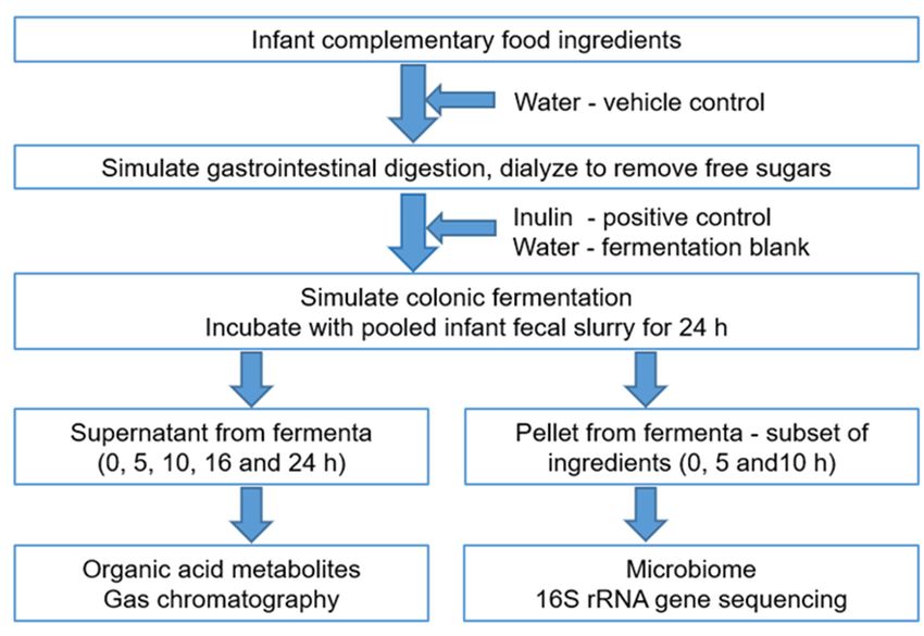
Figure
Figure 4.4. Principal
Principalcoordinates
coordinatesanalysis
analysis(PCoA)
(PCoA) plots
plots based
based on weighted
on weighted UniFrac
UniFrac distances
distances demonstrating
demonstrating significant
significant shifts
shifts in the microbiome structure at 10 h of fermentation of the food ingredients, inulin (positive control), and water. Each
in the microbiome structure at 10 h of fermentation of the food ingredients, inulin (positive control), and water. Each sample
sample was anaerobically fermented in triplicate, in the presence of pooled infant fecal slurry. The axes represent the
was anaerobically fermented in triplicate, in the presence of pooled infant fecal slurry. The axes represent the dimensions
dimensions explaining the greatest proportion of variances in the communities. The statistical significance (p < 0.005) was
explainingusing
computed the greatest proportion
permutational of variances
multivariate in theof
analysis communities. The statistical significance (p < 0.005) was computed
variance (PERMANOVA).
using permutational multivariate analysis of variance (PERMANOVA).
4. Discussion
Thirty-two food ingredients were taken in equal quantities and investigated for their
ability to modulate the gut microbiome of the growing infant using simulated in vitro
models of gastroileal digestion, intestinal absorption, and colonic fermentation. Examining
the changes in microbial organic acid metabolites and microbial composition, we inferred
the fermentative potential of these ingredients with the infant gut microbiome.
Similar in vitro models have previously been used to study the fermentative capacity
of foods and ingredients with infant gut microbiome and validated using clinically proven
prebiotics, including fructo- and galactooligosaccharides as positive controls [32,51]. In
our study, water was included as a control to confirm the potential effect of the digestive
enzymes that may escape digestion and dialysis, thereby reaching the colonic fermentation
stage. Infants up to the age of 9 months harbor very low numbers of groups such as theMicroorganisms 2021, 9, 2089 11 of 17
butyrate-producing Lachnospiraceae and Ruminococcaceae that are more indicative of
microbial diversity and adaptation to different foods [2,4,52]. Therefore, to have a baseline
that includes different microbiota that may potentially be modulated in this study, fecal
bacteria obtained from infants 5–12 months of age were pooled to generate an inoculum
with a diverse representation of bacteria that might be encountered by the infant. Thus,
limitations of an in vitro system (vs. a clinical trial) and a fixed and sparse microbial
baseline (vs. acquisition of new microbes) were mitigated by using appropriate standards
and a pooled microbial inoculum, respectively.
Food ingredients selected were powders of different fruits, vegetables, varieties of
oats, and milk, which are all often components of infant foods [1,34]. Powdered food
ingredients have a stable shelf-life compared to fresh foods, are more consistent in terms
of composition, are easier to transport, and are often used in baby food formulation.
Commercially available sources were chosen to allow future use in the formulation of
new infant complementary food with reproducibility of results. A standardized amount
(2.5 g/30 mL) was chosen rather than a normalization with respect to the fiber content of
such powders, as bioactives other than fiber are now being recognized as modulators of
the gut microbiome. These bioactives include polyphenols that are present within the plant
cell structures and potentially reach the colon [53,54].
The presence of organic acids in the baseline fermenta of many of the samples is
indicative of the organic acids that were present in the substrate or generated during diges-
tion, but not dialyzed out, despite their low molecular weight. While the fermenta were
collected within 0.5 h, we do not consider that the fecal microbial metabolism was suffi-
ciently activated to generate microbial acids. The increase in lactate (and acetate) to varying
degrees throughout the fermentation may be attributed to the fact that most plant-based
foods contain oligosaccharides that are rapidly metabolized. The exceptions, including
spinach powder and lime leaf powder, which both showed increases in propionate and
acetate, and to a smaller extent butyrate, indicated a syntrophic metabolism that comes
into play when the early acid metabolites are further utilized by specialist bacteria. In the
case of spinach powder, the early increase in succinate (at 5 h) depleted with the increase
in propionate at 10 h. The smaller early rise in butyrate that built up over the course of the
fermentation again indicates the increase in butyrogenic clostridia [26,55]. The accompany-
ing increase in formate is a recognized mechanism to generate more acetate via mutualistic
cross-feeding of hydrogen-utilizing acetogens, which then generate butyrate [56]. Most
other ingredients caused lactate and acetate production to varying degrees, which is an
indication of the microbial utilization of the carbohydrates that are still available in this
closed system. The comparatively low propionate and butyrate production with infant
fecal microbiota as compared to adult fecal microbiota indicates the immaturity of the mi-
crobiome and its inability to utilize carbohydrates efficiently [4,5,39,42]. In addition to the
development stage of the infant microbiome, the fermentation rates and metabolic profiles
of the different foods are influenced by the food components, especially the amounts and
the physicochemical nature of the carbohydrates [21,57]. Indeed, fermentation is higher
for linear polysaccharides (inulin, β-glucan, resistant starch) than for carbohydrates with
sidechains such as arabinoxylans. This is consistent with the high lactate and acetate for
the milk powders and certain vegetable powders (pea, pumpkin, sweet corn, and sweet
potato, but not carrot, spinach, and tomato), as well as the oats.
For the first-pass step of fermentation followed by SCFA analysis, the variety of
ingredients included different products from the same starting material, to determine the
extent of variability that similar foods show in terms of their microbial stimulatory capacity.
The variation in the SCFA profile indicates that the nature and the processing of the food
are important factors. While honey is not usually recommended in foods for infants until
the age of 1 year [58], we included it in this in vitro study to gather information on its
prebiotic properties [43,59]. Most of the honey sugars were anticipated to be absorbed; the
increase in microbial SCFAs indicates that the prebiotic mono- and oligosaccharides known
to be present in honey may have been available to the fecal bacteria.Microorganisms 2021, 9, 2089 12 of 17
A subset of the ingredients that were anticipated to be more commonly used in infant
foods was further analyzed for changes in microbiome composition. The timepoints of
5 and 10 h are close to the mean gastrointestinal transit time of 8.5 h (for 1–3 months old)
to 10 h (for 1–2 years old) [60]. The inoculum obtained from infants may be considered
representative of an in vivo system, while the batch fermentation employed in this study
is a closed system precluding the replenishment of substrates or removal of bacterial
metabolites and wastes. Furthermore, since pH was controlled only by buffering capacity
of the bacterial fermentation medium, later timepoints may have confounding influences
due to altered pH affecting microbial growth [61]. For these reasons, changes in the
microbiome and its metabolites were compared within each timepoint. Acquisition was
not simulated in this study, and this study, therefore, examines the ability of the ingredient–
inoculum interactions to stimulate bacteria that are already present, but in very low relative
abundances. The zero-hour composition analysis ascertained the presence of a diverse
species of bacteria much like the adult gut microbiome. This implies an opportunity
for the diverse bacteria to utilize the carbohydrate resources within ingredients to result
in populational shifts in the microbiota. Indeed, changes in the community structure
(weighted uniFrac) were driven by the different substrates.
Instant milk powder, which is often a component of infant complementary foods,
is a source of lactose and milk oligosaccharides, both of which stimulate bifidobacteria,
particularly B. longum subsp. longum in vitro [21,62]. Consistent with these in vitro results,
when 7–90 day old infants were fed exclusively a cow’s milk-based formula or breast
milk, an increase in bifidobacteria was observed at 3 weeks of feeding in both cases,
although there was no difference in the concentration of SCFAs [63]. Interestingly only
one, but not all the products tested behaved in a similar manner with respect to the
effect on abundance of Bifidobacterium spp. This may have been due to differences in the
composition of different products. Bifidobacterium spp. and B. longum were increased with
the infant milk powder in this study. This suggests potential benefits due to its ability to
persist in the gut beyond infancy and to metabolize a variety of dietary carbohydrates
that are introduced during the complementary feeding [15,64]. Thus, higher bifidobacteria
and LAB, and the associated lactate and acetate generation indicate microbial ecology
driven by prebiotic components in the instant milk powder [62]. The greater lactate and
acetate seen with foods such as sweet potato, sweetcorn, pea, and pumpkin powders,
as well as oat flour, may be a consequence of the higher bifidobacteria, enterococci, and
lactobacilli. This indicates a presence of easily metabolizable carbohydrates with fewer
complexities or sidechains [65,66]. Carrots and fruits such as kiwifruit, apple, blackcurrant,
and other berries are known to contain pectin-rich cell-wall polysaccharides [67,68]. Pectins
enable the growth of a more diverse microbial consortium, led by the early blooms of
bifidobacteria, lactobacilli, and streptococci [21,39,40,69]. The resultant lactate and/or
acetate may have facilitated the growth of a second line of bacteria, e.g., Lachnospiraceae.
While the increases in Firmicutes were not sufficient to cause large increases in butyrate,
a prebiotic environment may be generated that supports beneficial propionigenic and
butyrogenic commensals [6,70]. Spinach and tomato have a cell wall mainly composed of
cellulose, hemicellulose, and lignin, and these components are known to be comparatively
slowly fermentable by human gut bacteria [71,72]. This explains the lower lactate and
acetate, and higher propionate and butyrate for these substrates, as some members utilize
the easily accessible sugars, while other members further metabolize the primary end-
products. Spinach cell-wall components also show phenolic cross-linkages, which makes
it particularly recalcitrant to microbial breakdown [71]. This has a beneficial role, as the
undigested fiber is moved further to the normally carbohydrate-poor distal colon for further
microbial utilization. The high P. copri seen here with spinach powder is known to drive
an increase mostly in succinate [26,73]. The further conversion of succinate to propionate
and butyrate by cross-feeders such as Faecalibacterium prausnitzii and Phascolarctobacterium
spp. [74,75] was not clearly evident in this study owing to the microbial composition ofMicroorganisms 2021, 9, 2089 13 of 17
the immature inoculum, with its lower Firmicutes relative abundance, compensated for by
Proteobacteria and Actinobacteria.
The differential effects on the microbiome changes caused by powders sourced from
similar cultivars, such as carrots and spinach, may be explained by the different processing
conditions. Thus, the type of food processing and consequent impact on food structure
and composition (fiber, polyphenol content) may potentially influence microbiome compo-
sition [54,57,76,77]. This may explain the increase in Bacteroides spp. and Lachnospiraceae
with the fiber-rich carrot powder made from whole carrots, but not the fiber-free carrot
juice powder which was high in β-carotene. Similarly, spinach powder sourced from New
Zealand Spinacia oleracea enhanced propionate (and butyrate), with the largest increase in P.
copri. This propionigenic effect was not observed with spinach leaf powder, which had a
similar macronutrient composition but “was sourced from imported spinach” according to
the manufacturer’s product description.
Exposure of infants and toddlers to a variety of foods helps to build a more versatile
gut microbiome. This study, along with other studies, demonstrates that foods differentially
modulate infant gut microbiota [39,51]. Blending different food groups may help to
improve the food palatability [78], as well as the nutritive content and variety [79], and
generate combinations that provide age-specific support to the developing infant’s gut
microbiome [28,52].
The inulin (positive control) was a commercial mixture of low- and high-molecular-
weight oligofructans and was added to aliquots of dialyzed water digesta to avoid removal
by the 1000 Da molecular weight cutoff dialysis membrane. The strong bifidogenic effect of
inulin, as expected [32], validated our in vitro gut model. The water control was essentially
a mixture of undialyzed digestive enzymes fermented with infant fecal bacteria in a
peptone-rich broth. It served as a vehicle control in this study, but also mimicked a gut
environment that was devoid of carbohydrate, but rich in proteins. The increase in C.
perfringens, along with smaller increases in other Firmicutes families and Bacteroides spp.,
supports a metabolomic profile rich in propionate, butyrate, and branched-chain fatty
acids such as isobutyrate and isovalerate, characteristic of the degradation of glycoproteins
or proteins that reach the distal gut [26,80]. The ingredients were able to suppress C.
perfringens [81], a potential pathogen, and this may be attributed to the increase in other
beneficial bacteria. Spinach powder, which increased isobutyrate and isovalerate, still
suppressed C. perfringens, while favoring the butyrate-producing Lachnospiraceae.
5. Conclusions
Our results show that powdered food ingredients displayed varied abilities to stim-
ulate microbial metabolism, evidenced by the generation of beneficial SCFAs. The foods
also enhanced desirable bacteria such as Bifidobacterium, Bacteroides, Prevotella, LAB, and
Lachnospiraceae. Different foods were shown to selectively enhance specific groups; for
example, infant whole-milk powder and oat flour, F00151, enhanced Bifidobacteriaceae and
LAB, and spinach powder enhanced Prevotellaceae and Lachnospiraceae, while fruit and
vegetable powders modulated a mixed consortium of beneficial bacteria. In addition, all
the food ingredients were consistent in inhibiting the opportunistic pathobiont, C. perfrin-
gens, which was high only in the carbohydrate-free water control. More studies examining
these food ingredients and their appropriate dosages should be undertaken to understand
how different ingredients interact with the infant microbiome. This will help us to design
nutrient-rich foods suited to the developmental stage of the infant.
Supplementary Materials: The following are available online at https://www.mdpi.com/article/
10.3390/microorganisms9102089/s1: Table S1. Details of the infants donating the feces; Table S2.
Organic acid concentrations during the course of fermentation; Table S3. Analysis of variance in the
organic acid measurements; Table S4. Limits of detection of organic acid analysis; Table S5. Percent
relative abundance of microbial phyla at 0 h at phylum level (a), family level (b), and species level
(c); Table S6. Percent relative abundance of microbial phyla 5 and 10 h after fermentation; Table S7.Microorganisms 2021, 9, 2089 14 of 17
Percent relative abundance of microbial families 5 and 10 h after fermentation; Table S8. Percent
relative abundance of microbial species 5 and 10 h after fermentation.
Author Contributions: Conceptualization, P.G., S.G.P. and D.I.R.; methodology, S.G.P., D.I.R., H.M.S.
and C.M.H.J.; formal analysis, S.G.P. and D.I.H.; data curation, S.G.P., H.M.S. and C.M.H.J.; writing—
original draft preparation, S.G.P.; project administration, S.G.P.; funding acquisition, P.G. All authors
have read and agreed to the published version of the manuscript.
Funding: This research was funded by the New Zealand Ministry of Business, Innovation, and
Employment through the Foods for Health at Different Life Stages program, contract C11X1312.
Institutional Review Board Statement: The study was conducted according to the guidelines of
the Declaration of Helsinki, and approved by the Health and Disability Ethics Committees of the
Ministry of Health, New Zealand (protocol code #16/NTA/154, approved on 7 October 2016).
Informed Consent Statement: Written informed consent has been obtained from the primary care-
givers of all the subjects to publish this paper.
Data Availability Statement: The 16S rRNA gene sequence data and metadata were deposited into
the SRA database with links to the BioProject accession number PRJNA669972 (https://www.ncbi.
nlm.nih.gov/bioproject/).
Acknowledgments: We acknowledge Hannah Dinnan and Sheridan Martell for collection of fecal
samples from the donors, as well as Lee Huffman and Irene Ho for procuring and providing the food
ingredients. We acknowledge Simon Bulman for expert review of the manuscript.
Conflicts of Interest: The authors declare no conflict of interest.
References
1. Laursen, M.F.; Bahl, M.I.; Michaelsen, K.F.; Licht, T.R. First foods and gut microbes. Front. Microbiol. 2017, 8, 356. [CrossRef]
2. Koenig, J.E.; Spor, A.; Scalfone, N.; Fricker, A.D.; Stombaugh, J.; Knight, R.; Angenent, L.T.; Ley, R.E. Succession of microbial
consortia in the developing infant gut microbiome. Proc. Natl. Acad. Sci. USA 2011, 108, 4578–4585. [CrossRef] [PubMed]
3. Koleva, P.T.; Bridgman, S.L.; Kozyrskyj, A.L. The infant gut microbiome: Evidence for obesity risk and dietary intervention.
Nutrients 2015, 7, 2237–2260. [CrossRef]
4. Milani, C.; Duranti, S.; Bottacini, F.; Casey, E.; Turroni, F.; Mahony, J.; Belzer, C.; Palacio, S.D.; Montes, S.A.; Mancabelli, L.; et al.
The first microbial colonizers of the human gut: Composition, activities, and health implications of the infant gut microbiota.
Microbiol. Mol. Biol. Rev. 2017, 81, e00036-17. [CrossRef] [PubMed]
5. Tamburini, S.; Shen, N.; Wu, H.C.; Clemente, J.C. The microbiome in early life: Implications for health outcomes. Nat. Med. 2016,
22, 713–722. [CrossRef]
6. Solís, G.; Reyes-Gavilan, C.D.L.; Fernández, N.; Margolles, A.; Gueimonde, M. Establishment and development of lactic acid
bacteria and bifidobacteria microbiota in breast-milk and the infant gut. Anaerobe 2010, 16, 307–310. [CrossRef]
7. Tannock, G.W.; Lawley, B.; Munro, K.; Pathmanathan, S.G.; Zhou, S.J.; Makrides, M.; Gibson, R.A.; Sullivan, T.; Prosser, C.G.;
Lowry, D.; et al. Comparison of the compositions of the stool microbiotas of infants fed goat milk formula, cow milk-based
formula, or breast milk. Appl. Environ. Microbiol. 2013, 79, 3040–3048. [CrossRef]
8. Harmsen, H.J.M.; Wildeboer-Veloo, A.C.M.; Raangs, G.C.; Wagendorp, A.A.; Klijn, N.; Bindels, J.G.; Welling, G.W. Analysis of
intestinal flora development in breast-fed and formula-fed infants by using molecular identification and detection methods. J.
Pediatric Gastroenterol. Nutr. 2000, 30, 61–67. [CrossRef] [PubMed]
9. Hove, H.; Nørgaard, H.; Mortensen, P.B. Lactic acid bacteria and the human gastrointestinal tract. Eur. J. Clin. Nutr. 1999, 53,
339–350. [CrossRef]
10. Wang, S.; Hibberd, M.L.; Pettersson, S.; Lee, Y.K. Enterococcus faecalis from healthy infants modulates inflammation through
MAPK signaling pathways. PLoS ONE 2014, 9, e97523. [CrossRef]
11. Murphy, R.; Morgan, X.; Wang, X.; Wickens, K.; Purdie, G.; Fitzharris, P.; Otal, A.; Lawley, B.; Stanley, T.; Barthow, C.; et al.
Eczema-protective probiotic alters infant gut microbiome functional capacity but not composition: Sub-sample analysis from a
RCT. Benef. Microbes 2019, 10, 5–17. [CrossRef]
12. Gotoh, A.; Katoh, T.; Sakanaka, M.; Ling, Y.; Yamada, C.; Asakuma, S.; Urashima, T.; Tomabechi, Y.; Katayama-Ikegami, A.;
Kurihara, S.; et al. Sharing of human milk oligosaccharides degradants within bifidobacterial communities in faecal cultures
supplemented with Bifidobacterium bifidum. Sci. Rep. 2018, 8, 1–14. [CrossRef]
13. Turroni, F.; Duranti, S.; Milani, C.; Lugli, G.A.; Van Sinderen, D.; Ventura, M. Bifidobacterium bifidum: A key member of the early
human gut microbiota. Microorganisms 2019, 7, 544. [CrossRef]
14. Reuter, G. The Lactobacillus and Bifidobacterium microflora of the human intestine: Composition and succession. Curr. Issues Intest.
Microbiol. 2001, 2, 43–53.You can also read