Metabolites from Microbes Isolated from the Skin of the Panamanian Rocket Frog Colostethus panamansis (Anura: Dendrobatidae) - MDPI
←
→
Page content transcription
If your browser does not render page correctly, please read the page content below
H

         OH
               OH
                    metabolites
Article
Metabolites from Microbes Isolated from the Skin of
the Panamanian Rocket Frog Colostethus panamansis
(Anura: Dendrobatidae)
Christian Martin H. 1,2 , Roberto Ibáñez 3 , Louis-Félix Nothias 4 ,
Andrés Mauricio Caraballo-Rodríguez 4 , Pieter C. Dorrestein 4 and Marcelino Gutiérrez 1, *
 1            Centro de Biodiversidad y Descubrimiento de Drogas, Instituto de Investigaciones Científicas y Servicios de
              Alta Tecnología (INDICASAT AIP), Clayton, Panama 0843-01103, Panama; cmartin@indicasat.org.pa
 2            Department of Biotechnology, Acharya Nagarjuna University, Nagarjuna Nagar, Guntur 522510, India
 3            Smithsonian Tropical Research Institute, Balboa, Ancon, Panama 0843-03092, Panama; ibanezr@si.edu
 4            Collaborative Mass Spectrometry Innovation Center, Skaggs School of Pharmacy and Pharmaceutical
              Sciences, University of California San Diego, La Jolla, CA 92093, USA; nothias@ucsd.edu (L.-F.N.);
              amcaraballor@ucsd.edu (A.M.C.-R.); pdorrestein@health.ucsd.edu (P.C.D.)
 *            Correspondence: mgutierrez@indicasat.org.pa; Tel.: +507-517-0732; Fax: +507-517-0701

 Received: 14 September 2020; Accepted: 9 October 2020; Published: 13 October 2020

 Abstract: The Panamanian rocket frog Colostethus panamansis (family Dendrobatidae) has been affected
 by chytridiomycosis, a deadly disease caused by the fungus Batrachochytrium dendrobatidis (Bd). While
 there are still uninfected frogs, we set out to isolate microbes from anatomically distinct regions
 in an effort to create a cultivable resource within Panama for potential drug/agricultural/ecological
 applications that perhaps could also be used as part of a strategy to protect frogs from infections. To
 understand if there are specific anatomies that should be explored in future applications of this resource,
 we mapped skin-associated bacteria of C. panamansis and their metabolite production potential by
 mass spectrometry on a 3D model. Our results indicate that five bacterial families (Enterobacteriaceae,
 Comamonadaceae, Aeromonadaceae, Staphylococcaceae and Pseudomonadaceae) dominate the
 cultivable microbes from the skin of C. panamansis. The combination of microbial classification and
 molecular analysis in relation to the anti-Bd inhibitory databases reveals the resource has future
 potential for amphibian conservation.

 Keywords: 3D molecular cartography; Colostethus panamansis; Dendrobatidae; skin-associated
 bacteria; 16S rRNA sequencing; feature-based molecular networking

1. Introduction
      Among the three existing orders of amphibians, Caudata (salamanders), Gymnophiona (caecilians)
and Anura (frogs and toads) [1], the last one is the most diverse [2] and also widely affected by the
lethal amphibian disease chytridiomycosis, caused by the fungus Batrachochytrium dendrobatidis (Bd).
Bd is responsible for the high rate of amphibian loss in the world, particularly in the Neotropics [3–8].
This pathogen mainly infects amphibians at the ventral coronal plane (abdomen, pelvic patch, pes
and toes) [9]. However, not all amphibians that have been exposed to Bd in nature have been affected
equally. Some of them have shown asymptomatic responses and, therefore, are considered to be
resistant or tolerant [9,10].
      Colostethus panamansis (family Dendrobatidae) is considered a least-concerned species regarding
its conservation status, despite a decreasing population trend [11]. In fact, population declines of
this species have not been noticed in the lowlands of Panama. In contrast, highland populations of
C. panamansis in Panama have experienced dramatic declines [12]. Infection experiments have shown

Metabolites 2020, 10, 406; doi:10.3390/metabo10100406                                   www.mdpi.com/journal/metabolites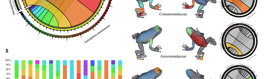
Metabolites 2020, 10, 406 2 of 19
that C. panamansis is a susceptible species to Bd, causing death in infected frogs [13]. It was hypothesized
that healthy frogs of this species could harbor bacterial symbionts on their skin as potential producers
of specialized metabolites that could protect them against microbial pathogens [14–19]. While a small
number of specialized metabolites produced by amphibian skin-associated bacteria has been reported,
including some endowed with antifungal activity against Bd in vitro [17,20–22], multi-omics methods
are needed to investigate the symbiotic metabolome of bacteria on the amphibian’s skin mucosome [23].
Recently, multi-omics approaches such as DNA sequencing and mass spectrometry-based
metabolomics have allowed researchers to deeply explore the microbial and chemical diversity
associated with a wide range of biological sources [24–26]. In the case of chemical diversity, the Global
Natural Products Social molecular networking web-platform (GNPS) facilitates the exploration of
the chemical space of metabolomes. GNPS is a tandem mass spectrometry (MS/MS) automated data
organizational tool that makes comparisons of MS/MS fragmentation patterns among samples in order
to cluster and visualize related molecules in a spectral network [27,28]. Feature-based molecular
networking (FBMN) is a recent analysis method in GNPS that enhances quantitative capability and
isomer resolution in molecular networks by coupling with mass spectrometry data processing tools,
such as MZmine [29,30]. In addition, this makes it possible to annotate the spectra with computational
tools, like SIRIUS [31] which can provide putative molecular formula and structural information.
Researchers can perform three dimensional (3D) cartographic maps to explore the spatial distribution
of host-derived molecules, along with bacterial specialized metabolites [32–38]. Characterization
of skin microorganisms and their related molecules is essential for understanding host–microbial
symbiont interactions. Although many studies have focused on the human-associated microbes, i.e.,
gut and skin, far less is known about the skin microbes of other mammals, amphibians, birds, fish,
and reptiles [39].
In this study, we performed 3D molecular cartography of the bacterial genomics and metabolomics
data obtained from bacterial isolates collected from different body locations of C. panamansis skin.
The mapping results allowed us to correlate the distribution of the most abundant bacterial isolates
with some of the specialized metabolites on multiple body parts of the frog, helping to understand
the function of skin-associated bacteria in C. panamansis. Additionally, we detected non-described
molecules in C. panamansis that could be produced by previously reported anti-Bd bacterial isolates.
That means that such molecules could serve as potential sources for future conservation treatments in
amphibian populations.
2. Results
2.1. 16S rRNA Amplicon Sequencing and Molecular Cartography
We obtained 170 isolates from the skin of four specimens of Bd-uninfected frogs of C. panamansis
collected in June 2016 and August 2017. Frogs collected in 2016 were tested for Bd infection through
q-PCR, displaying negative results. After performing Sanger amplicon sequencing, we found that
bacterial isolates belong to phylum Proteobacteria, Firmicutes, Bacteroidetes and Actinobacteria with a
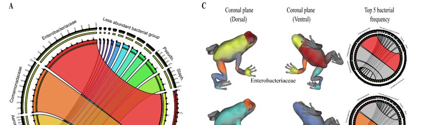
relative abundance of 85.3%, 10.6%, 3.5% and 0.6%, respectively. Within the phylum Proteobacteria, the
largest group, the most frequent families were Enterobacteriaceae (31.2%), Comamonadaceae (24.1%),
Aeromonadaceae (11.8%) and Pseudomonadaceae (7.7%). Within Bacteroidetes and Firmicutes, the
bacterial families were mainly composed of Staphylococcaceae (8.8%) and Flavobacteriaceae (3.5%),
respectively. Actinobacteria was composed by Streptomycetaceae (0.6%), being the less abundant
phylum (Figure 1A). We also found significant differences between the number of isolates, based on
bacterial families, and body parts sampled (dorsal and ventral: head, trunk, forelimbs, manus, thigh,
hind limbs, pes and toes) (Friedman test; p = 0.0445). Significant differences between the number of
bacterial isolates per family and the coronal plane (dorsal and ventral) of specimens collected (Pearson
Chi-Square; p = 0.0096) were found.
Flavobacteriaceae (3.5%), respectively. Actinobacteria was composed by Streptomycetaceae (0.6%),
being the less abundant phylum (Figure 1A). We also found significant differences between the
number of isolates, based on bacterial families, and body parts sampled (dorsal and ventral: head,
trunk, forelimbs, manus, thigh, hind limbs, pes and toes) (Friedman test; p = 0.0445). Significant
differences
Metabolites 2020, 10,between
406 the number of bacterial isolates per family and the coronal plane (dorsal 3and
of 19
ventral) of specimens collected (Pearson Chi-Square; p = 0.0096) were found.
Figure
Figure 1. Frequency
1. Frequency of isolated
of isolated bacteria
bacteria from thefrom
skinthe skinofsurface
surface of C. panamansis.
C. panamansis. (A) Relative(A) Relative
abundances
abundances
of bacterial of bacterial
families from thefamilies from
skin of C. the skinbased
panamansis of C.onpanamansis
16S rRNAbased on 16S
amplicon rRNA amplicon
sequencing. (B) The
sequencing.
proportional (B) The proportional
abundance (as a percentabundance (as a percent
of total sequences) of total sequences)
of bacterial of bacterial
families based on bodyfamilies
parts.
based
(C) 3D on body parts.
topographic map of(C) C. 3D topographic
panamansis basedmap of C.
on the panamansis
five based bacterial
most abundant on the five most abundant
families isolated at
eachbacterial familiesRed
body location. isolated at eachthe
represents body location.
highest Red represents
percentage of each the highest
family thatpercentage
was isolatedof and
eachblue
family
the
that was isolated
lowest percentage. and blue the lowest percentage.
In terms
In terms of topographical
of topographical distribution,
distribution, we weshowed
showedthetheresults
results of
of the
the five most
most frequent
frequentbacterial
bacterial
families
families of isolates
of isolates fromfrom the of
the skin skin of C. panamansis.
C. panamansis. We observed
We observed that: (1)that: (1) Enterobacteriaceae
Enterobacteriaceae was
was isolated
isolated
mainly on themainly
dorsalonhead,
the dorsal
thighs,head,
trunkthighs, trunk and
and manus, andmanus, and the
the ventral ventral
trunk, trunk, head,
forelimbs, forelimbs,
manushead,
and
thighs; (2) Comamonadaceae
manus was isolated on
and thighs; (2) Comamonadaceae wasthe dorsalon
isolated head/manus/hind limbs/toes and
the dorsal head/manus/hind the ventral
limbs/toes and
manus/forelimbs/thighs; (3) Aeromonadaceae
the ventral manus/forelimbs/thighs; (3) was isolated on the dorsal
Aeromonadaceae was head/pes/forelimbs
isolated on the and the
dorsal
ventral trunk/pes/toes/head;
head/pes/forelimbs (4) Staphylococcaceae
and the was isolated
ventral trunk/pes/toes/head; (4) on the dorsal head and
Staphylococcaceae wasventral forelimbs;
isolated on the
anddorsal
(5) Pseudomonadaceae was found
head and ventral forelimbs; and on the dorsal manus was
(5) Pseudomonadaceae and found
ventral onhead/forelimbs/manus
the dorsal manus and
(Figure 1B,C).
ventral head/forelimbs/manus (Figure 1B,C).
2.2. Molecular Networking and Molecular Cartography
MS/MS molecular networking of the 170 bacterial strains, 32 skin swabs and 2 pure compounds
(tetrodotoxin and viscosin) revealed 2498 nodes. Each node represents an LC-MS/MS molecular
feature that comprises detected m/z, MS/MS, and retention time. Thus, connected nodes generate
clusters based on their spectral similarity in the molecular network. We focused on sub-networks
containing GNPS and SIRIUS hits. The identified metabolites were detected from bacterial isolate
extracts only. Putative annotations of compound and molecular families based on GNPS and
Metabolites 2020, 10, 406                                                                            4 of 19

SIRIUS matches correspond to level 2, respectively, according to Sumner et al. 2007 [40]. Eighteen
selected bacterial metabolites, within m/z error of 10 ppm, were annotated as N-methyl tryptophan,
m/z 205.0969 [M + H − H2 O]+ (1), tryptophan, m/z 188.0696 [M + H − H2 O]+ (2), phenylalanine,
m/z 166.0871 [M + H]+ (3), 3-indole acetic acid, m/z 176.0706 [M + H]+ (4) and diketopiperazines,
m/z 261.1233 [M + H]+ (5), m/z 245.1286 [M + H − H2 O]+ (6), m/z 197.1281 [M + H]+ (7), m/z
211.1437 [M + H]+ (8), m/z 227.1386 [M + H]+ (9) and m/z 235.1189 [M + H − H2 O]+ (10) (Figure 2),
as well as the oligopeptides that included Pro-Leu-Ile, m/z 342.2383 [M + H]+ (11), Pro-Ile-Val, m/z
328.2226 [M + H]+ (12), Pro-Pro-Phe-Val, m/z 459.2600 [M + H]+ (13), Pro-Pro-Phe, m/z 360.1937
[M + H]+ (14), cyclodepsipeptide Leualacin, m/z 596.3386 [M + Na]+ (15), a poly-glutamic acid
analog, m/z 922.3167 [M + H]+ (16), Leu-Phe-Gly-Tyr-Pro-Val-Tyr-Val, m/z 957.5032 [M + H]+ (17),
and cyclo Ala-Val-3-hydroxy-4-methyloctanoyl-Gly-Val-Leu, m/z 596.3950 [M + H]+ (18) (Figure 3).
After feature-based molecular networking analysis, both tetrodotoxin (TTX) and viscosin were absent
in the bacterial crudes or swabs from the sampled specimens of C. panamansis.
       Through integration of MS/MS data to 0 ili web platform [33], we describe the molecular distribution
of annotated specialized metabolites. Compounds 1, 2 and 3 were found on the ventral trunk samples.
Compound 4 was found on the ventral head. Diketopiperazines-related metabolites were identified on
multiple body parts. MS/MS features of compound 5 were found on the dorsal trunk/pes and ventral
head and (6) on the dorsal pes and ventral head. Compounds 7, 8 and 9 were spotted at the dorsal toes,
dorsal pes, dorsal trunk, ventral head, ventral trunk, and ventral forelimbs. MS/MS feature 10 was
found in samples from the dorsal trunk (Figure 4).
       We also visualized; annotated oligopeptides produced by skin-associated bacterial isolates through
the 0 ili web platform. The tripeptide 11 was found in samples from the ventral thigh and hind limbs
while 12 was distributed among the dorsal pes, trunk, and toes. Compound 13 was observed in the
dorsal pes, toes, trunk, and ventral head and 14 was found in samples from the dorsal trunk and
pes. Compound 15 and 17 were both identified in dorsal trunk and toes samples. Compound 16 was
associated with samples from forelimbs only. Compound 18 was related to samples from dorsal hind
limbs and the ventral thigh (Figure 4).
       The specialized metabolites annotated through GNPS and SIRIUS were found in multiple samples
(crude extracts from bacterial isolates) obtained from different body parts on the skin of C. panamansis.
We found significant differences (Friedman test; p < 0.001) between bacterial families of isolates and the
number of small molecules annotated. These small molecules were produced by bacterial isolates that
belong to the families Enterobacteriaceae, Comamonadaceae, Pseudomonadaceae, Aeromonadaceae
and Staphylococcaceae (Supplementary Materials Table S1, Figure 5). We also found significant
differences (Friedman test; p < 0.001) when considering bacterial families and the number of peptides
annotated. Such peptides were detected in well-defined bacterial families, namely, Comamonadaceae,
Aeromonadaceae, Pseudomonadaceae and Staphylococcaceae (Table S2, Figure 5).

2.3. Comparison between 16S rRNA Sequences from C. panamansis and Bd-Inhibitory Public Datasets
     After comparing 170 bacterial sequences from isolates obtained from the skin of C. panamansis
along with 621 bacterial sequences that displayed inhibitory effects against Bd, deposited in a public
database [41], Pseudomonadaceae was the most abundant family (43%), followed by Enterobacteriaceae
(17%), Comamonadaceae (7%), Aeromonadaceae (4%) and Staphylococcaceae (3%) (Figure 6A). We
calculated the proportion of C. panamansis sequences within them as Enterobacteriaceae (39%),
Comamonadaceae (75%), Aeromonadaceae (63%), Staphylococcaceae (60%) and Pseudomonadaceae
(4%). Based on the coronal plane of isolates from C. panamansis, we found the major percentage samples
from dorsal body parts in Enterobacteriaceae and Comamonadaceae (53% and 66%, respectively),
while Aeromonadaceae, Staphylococcaceae and Pseudomonadaceae displayed on ventral body parts
(65%, 60% and 69%, respectively) (Figure 6B).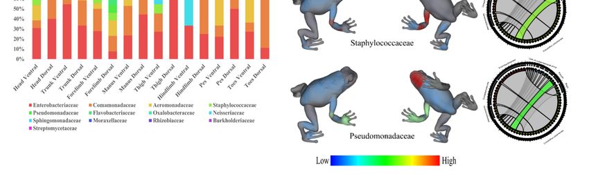
Metabolites 2020, 10, 406                                                                                              5 of 19
     Metabolites 2020, 10, x FOR PEER REVIEW                                                                      5 of 19

           Figure 2. Molecular network of specialized metabolites (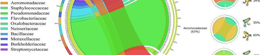
Metabolites 2020, 10, 406                                                                                             6 of 19
     Metabolites 2020, 10, x FOR PEER REVIEW                                                                     6 of 19

          Figure
      Figure      3. Molecular
              3. Molecular     network
                            network     of annotated
                                    of annotated     peptides
                                                  peptides    identified
                                                           identified  inin crude
                                                                          crude   extractsofof
                                                                                extracts       bacterialisolates
                                                                                             bacterial    isolatesfrom
          from   skin of C. panamansis by  LC-MS/MS   analysis. Highlighted   nodes correspond    to  annotated
      skin of C. panamansis by LC-MS/MS analysis. Highlighted nodes correspond to annotated metabolites
          metabolites by GNPS (yellow) and SIRIUS CSIFingerID (purple). Pie charts inside nodes denotes
      by GNPS (yellow) and SIRIUS CSIFingerID (purple). Pie charts inside nodes denotes abundance while
          abundance while colors correspond to the bacterial family where the feature was found. Edge width
      colors correspond to the bacterial family where the feature was found. Edge width between nodes
          between nodes displays cosine score values, which are related to the spectral similarities between
      displays cosine score values, which are related to the spectral similarities between nodes.
           nodes.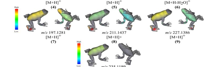
Metabolites 2020, 10, 406                                                                                              7 of 19
 Metabolites 2020, 10, x FOR PEER REVIEW                                                                                 7 of 19

     Figure
       Figure   Molecular
             4. 4. Molecularcartography
                                cartographyofofannotated
                                                  annotatedmetabolites
                                                              metabolitesidentified
                                                                           identifiedfrom
                                                                                       from bacterial
                                                                                             bacterialisolates sampled
                                                                                                        isolates sampled
     from  the skin surface   of C. panamansis.   These  metabolites were  annotated   as tryptophan   derivatives
       from the skin surface of C. panamansis. These metabolites were annotated as tryptophan derivatives           (1,2),
     phenylalanine    (3), 3-indoleacetic   acid (4), diketopiperazines  (5–10) and  peptides   (11–18).
       (1,2), phenylalanine (3), 3-indoleacetic acid (4), diketopiperazines (5–10) and peptides (11–18).  Intensities are
     related to the are
       Intensities  abundance
                         related of
                                  to the
                                      the specialized
                                           abundancemetabolites    in the samples
                                                        of the specialized         at eachinbody
                                                                            metabolites           part. Redatrepresents
                                                                                             the samples       each body
     the highest   abundance     while  blue  represents  the lowest  abundance.
       part. Red represents the highest abundance while blue represents the lowest abundance.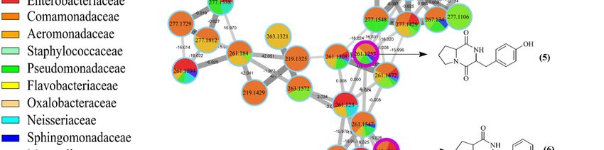
Metabolites 2020, 10, 406                                                                                     8 of 19

   Metabolites 2020, 10, x FOR PEER REVIEW                                                                  8 of 19

         Figure 5. Phylogenetic tree and structures of annotated metabolites from 170 cultivable bacteria
      Figure 5. Phylogenetic tree and structures of annotated metabolites from 170 cultivable bacteria isolated
         isolated from the skin of C. panamansis and a heatmap that displays the abundance of specialized
      from the skin of C. panamansis and a heatmap that displays the abundance of specialized metabolites
         metabolites annotated as molecular features by feature based molecular networking-analysis.
      annotated as molecular features by feature based molecular networking-analysis.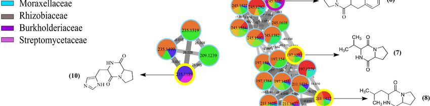
(3%) (Figure 6A). We calculated the proportion of C. panamansis sequences within them as
    Enterobacteriaceae (39%), Comamonadaceae (75%), Aeromonadaceae (63%), Staphylococcaceae
    (60%) and Pseudomonadaceae (4%). Based on the coronal plane of isolates from C. panamansis, we
    found the major percentage samples from dorsal body parts in Enterobacteriaceae and
Metabolites 2020, 10, 406
    Comamonadaceae        (53% and 66%, respectively), while Aeromonadaceae, Staphylococcaceae and9 of 19
    Pseudomonadaceae displayed on ventral body parts (65%, 60% and 69%, respectively) (Figure 6B).

          Figure
      Figure      6. Relative
              6. Relative     abundancesofof791
                           abundances         791bacterial
                                                  bacterial sequences
                                                            sequences from
                                                                       fromC.C.panamansis
                                                                                 panamansisand  anti-Bd
                                                                                              and       published
                                                                                                  anti-Bd  published
          sequences.  (A) Relative  abundance  of 16S rRNA   sequences from C. panamansis   and anti-Bd
      sequences. (A) Relative abundance of 16S rRNA sequences from C. panamansis and anti-Bd sequences  sequences
      fromfrom  published databases based on their bacterial family. (B) Comparison between the five most
            published    databases based on their bacterial family. (B) Comparison between the five most
          abundant bacterial families in C. panamansis and Bd-inhibitory sequences (light green) and the
      abundant bacterial families in C. panamansis and Bd-inhibitory sequences (light green) and the
          distribution of isolates based on the coronal plane in C. panamansis is also displayed.
      distribution of isolates based on the coronal plane in C. panamansis is also displayed.

    3. Discussion
3. Discussion
      TheThe    primary
            primary   rolerole of animal
                           of animal    skinskin
                                              is toisserve
                                                       to serve
                                                             as a as  a physical
                                                                  physical         barrier
                                                                              barrier       to protect
                                                                                       to protect      the body
                                                                                                  the body         against
                                                                                                             against   adverse
     adverse
effects  fromeffects   from the environment
                the environment      and harmful  andorganisms.
                                                         harmful organisms.
                                                                        Animal Animal      skin harbors
                                                                                 skin harbors    a widearange
                                                                                                           wide range   of
                                                                                                                  of microbes
     microbes    due  to  direct  contact  with    the   environment.      These   microbes
due to direct contact with the environment. These microbes may produce specialized metabolitesmay   produce   specialized
thatmetabolites
      could protectthattheir
                         couldhost
                               protect   theirpathogens
                                    against    host against     pathogens
                                                              [42].          [42]. of
                                                                     In the case   In amphibians,
                                                                                      the case of amphibians,
                                                                                                     microbes microbes
                                                                                                                 are acquired
from water, soil, plants and even other amphibians [18,43–47]. As C. panamansis is foundisinfound
     are acquired   from  water,  soil, plants  and   even   other  amphibians    [18,43–47].  As C. panamansis       riparian
     in riparian
habitats,   a few habitats, a fewthe
                  meters from       meters   from
                                       water’s      the and
                                                 edge     water’s  edge
                                                               under      and under
                                                                        rocks, this mayrocks, this may
                                                                                          explain  whyexplain    why the
                                                                                                         the skin-associated
     skin-associated
bacteria               bacteria
           in our samples        in our
                             mainly      samples
                                      belong        mainly
                                                to the        belong
                                                         phylum        to the phylum[46,48].
                                                                    Proteobacteria      Proteobacteria
                                                                                                The high[46,48]. The high
                                                                                                           prevalence   of this
     prevalence   of  this phylum     was   observed      in other   studies   where   culture-dependent
phylum was observed in other studies where culture-dependent and culture-independent approaches             and   culture-
     independent
for bacterial        approaches for
                 identification    werebacterial
                                           appliedidentification    were applied
                                                        [22,46,49–51].      In terms [22,46,49–51].
                                                                                        of bacterialIn terms  of bacterial
                                                                                                       families,   specimens
of C. panamansis from the General de División Omar Torrijos Herrera National Park, in central
Panama, mainly harbored cultivable bacteria of the families Enterobacteriaceae, Comamonadaceae,
Aeromonadaceae, Staphylococcaceae and Pseudomonadaceae. However, the proportion of these
bacterial families contrasted to previous reports in C. panamansis and other Panamanian frogs, where
Pseudomonadaceae (Pseudomonas sp.) and Aeromonadaceae (Aeromonas sp.) were the most abundant
bacterial families [22,46,49–51]. Although few studies have been carried out in tropical regions,
such differences could be attributed to distinct genomic approaches applied (culture-dependent vs.
culture independent), sampling sites, sampling size, host specimens and even host-susceptibility to
Bd [22,46,52].
      Through high-resolution mass spectrometry analyses, we obtained relevant information about
specialized metabolites through molecular networking. This allows for the arrangement of large sets
of tandem MS/MS data based on fragmentation pattern similarity, allowing the detection of molecules
with related structures [27,28,53]. At present, the chemistry of specialized microbial metabolites on
amphibian skin in tropical frogs and their possible protective and functional roles has not been studied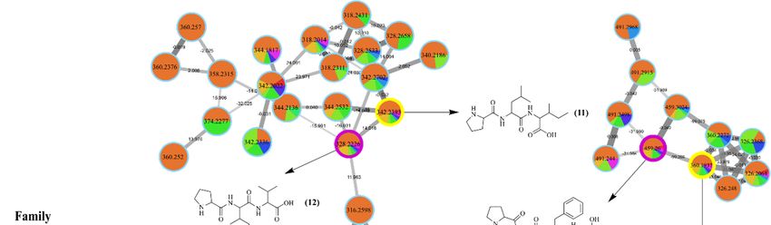
Metabolites 2020, 10, 406                                                                            10 of 19

in depth [22,49,50]. Considering that natural products and their derivatives constitute around half of
the pharmaceuticals on the market today and provide for many essential agricultural products [54],
eighteen specialized metabolites were annotated by means of GNPS and SIRIUS [27,31] and are
reported here for the first time as metabolites produced by frog-associated bacteria.
     Tryptophan derivatives such as 1 and 2, are well known for exhibiting antioxidant and
immunomodulatory properties [55,56]. In terms of immunoregulatory properties, these skin-associated
bacterial metabolites may influence the response from the host’s immune system to help fight off
infection [57]. However, it is important to mention that Bd also produces tryptophan derivative
molecules, which inhibit host immunity and even enhance the survival of this frog-killing fungus [58].
     Phenylalanine 3 is an essential amino acid, known to be directly taken up from water, which
promotes larval growth in the salamander Hynobius retardatus [59]. Considering that microbes can be
transmitted vertically through parental care to the embryos [60,61], if such vertical transmission occurs
in C. panamansis during egg attendance and tadpole transport, phenylalanine bacterial producers
might serve as growth promoters at the larval stages of this frog. The indole-3-acetic acid 4 is a
common biosynthetic product of tryptophan in bacteria through different biosynthetic pathways,
which is produced by a wide range of microbes [62]. It is very well known for contributing to
plant growth [62–64]; however, its presence and role on amphibian skin has not been reported. The
diketopiperazines (5–10) have been isolated from diverse microorganisms [65,66], and exhibit a wide
range of biological properties such as antimicrobials, immunosuppressant and anticancer [65,67–70].
This stresses the need for understanding how these skin-associated bacteria, which produce myriad
natural products, could be further used for amphibian conservation strategies.
     The antimicrobial peptides (AMPs) are small molecules with a broad spectrum of inhibitory
effects against bacteria, fungi, protozoa and viruses [71]. AMPs have been reported in amphibian skin
secretions for conferring host defenses against pathogens and predators [5,16]. In some cases, peptides
secreted by amphibian skin glands may not negatively affect bacterial growth, including bacteria with
anti-Bd properties [72]. Amphibian skin-associated bacteria are also known to produce peptides with
anti-Bd properties [22]. Nonetheless, peptides of bacterial origin had not been described frequently in
frogs even less in the family Dendrobatidae. In this study, we found bacterial oligopeptides composed
mainly by alpha amino acids. These oligopeptides were compounds 11, 12, 13 and 14. It is known
that oligopeptides containing proline, as part of amino acid residues, are able to target intracellular
membranes in microbes, leading to cell death by lysis [71]. Additionally, 15 is a cyclic depsipeptide,
known as leualacin, which does not exhibit antimicrobial activity; however, it has been reported to act
as a blood pressure regulator in mice [73], and could potentially have a similar role in frogs.
     Through the dereplicator workflow launched in GNPS, it was possible to extend the identification
of peptides and other natural products [74]. Thus, we identified the poly-glutamic acid analog
16. This compound is a polymer composed of D-glutamic acid residues that have skin protectant
properties, improving skin moisture and elasticity, even more than collagen and hyaluronic acid [75].
However, further studies are needed to determine whether this structural modification could provide
the same or higher skin protection in amphibians. Additionally, poly-glutamic acid is known to exhibit
high antibiotic activity against Gram positive and Gram negative bacteria, i.e., Listeria monocytogenes,
Stenotrophomonas typhimurium, Staphylococcus aureus, Klebsiella pneumoniae and Escherichia coli [76].
     The oligopeptide 17, was annotated with SIRIUS coupled to CSI:FingerID that is able to propose
molecular structure candidates [31]. However, in a few cases the biological roles of predicted metabolites
are unknown. Finally, compound 18 is a cyclic depsipeptide that has been reported as presenting
antifungal properties [77]. Since cyclic lipodepsipeptides have been recently reported by our group to
have biological in vitro activity against Bd [22], we infer a similar property for this bacterial metabolite
isolated from the skin of C. panamansis.
     3D molecular cartography studies have been implemented to integrate and interpret molecular
results in large studies (i.e., genomics and metabolomics) from human, host-parasites and plant-related
samples [32,35,36,78]; however, such studies have not been conducted in amphibians. By generating a
Metabolites 2020, 10, 406 11 of 19
3D molecular cartography model of an amphibian, i.e., the Panamanian rocket frog C. panamansis, it
was possible to visualize and understand the body distribution of skin-associated molecules and their
bacterial producers [32,35,79]. Although isolation of amphibian’s strains is common, understanding
their body distribution and their role in the environment can be helpful in the selection of isolates for
further novel secondary metabolite discovery. In this sense, the topographical distribution is particularly
relevant because of the amphibian-killing fungus, B. dendrobatidis, which infects vascularized body
parts such as the ventral abdomen, pelvic patch, pes and toes of frogs [9,80,81]. Therefore, bacterial
distribution and their specialized metabolites could represent key factors in studies related to amphibian
conservation since significant differences between the number of bacterial isolates per family on ventral
and dorsal body parts were found. Our results provide insight into how bacterial isolates on the
skin of C. panamansis could differ based on the coronal plane. Such differences could be attributed to
the environmental contact effect (ventral region) and the heterogeneity of skin gland (dorsal region)
density on the coronal planes [82].
Bacterial gene sequences from isolates obtained from different body parts in C. panamansis were
compared along with published sequences with anti-Bd properties [41]. We determined that a large
amount of anti-Bd bacteria predominantly belongs to the family Pseudomonadaceae [22] while the
families Enterobacteriaceae, Comamonadaceae, Aeromonadaceae and Staphylococcaceae comprised
around thirty percent. Within these five bacterial families, a genetic dispersion is shown, as has
also been shown in other amphibian-related studies [41]. It is important to mention that although
bacterial isolates from C. panamansis have displayed a level of relationship with bacteria with anti-Bd
properties published by Woodhams et al. 2015 [41], it needs to be determined experimentally that
bacteria found in C. panamansis exhibit antifungal activity against Bd and other pathogens. Given
the range of biological activities reported previously for compounds 1–18, including antimicrobial,
we could suggest that multiple isolates from C. panamansis very likely have the potential to fend off
pathogens in the natural environment through some of the specialized metabolites presented in this
study. Our results indicate that the bacterial strains reported here indeed have the capacity to produce
bioactive molecules. These molecules may have potential applications in medicine and agriculture and
are related to strains producing anti-Bd natural products and certainly represent a strain repository for
future studies on drug discovery and frog ecology.
4. Materials and Methods
4.1. Sampling Specimens
In June 2016 and August 2017, adult specimens of C. panamansis (n = 4, 2 males and 2 females)
were collected from the trail La Rana Dorada (N: 8.67143◦ , W: 80.59025◦ , elevation: 625 m) at the
General de División Omar Torrijos Herrera National Park, north of El Copé, Province of Coclé, Panama.
We did not seek to describe general differences between males and females, which would require a
larger sample size of each sex. Frogs were individually rinsed with sterile water to remove transient
bacteria from the skin [60]. After rinsing, 8 cotton swabs pre-moistened with distilled water, 50:50
ethanol/deionized water, were used to sample the cultivable skin-associated bacteria and specialized
metabolites, respectively. We also sampled two specimens collected in 2016 for testing infection
caused by Bd, based on a quantitative PCR technique [83]. The sampling of body parts was performed
10 times on dorsal and ventral regions, as presented in the supplementary information. To determine
Bd infection, only ventral regions at positions 2 and 5–8 were sampled (Figure S1) [84]. Swabs for
cultivable bacteria were streaked on Petri dishes containing fresh R2A agar [85] (Becton, Dickinson
and Company, Franklin Lakes, NJ, USA). Once bacterial colonies grew, they were characterized and
codified according to morphological characteristics (i.e., form, color, texture, and border type). Isolates
were streaked on R2A agar until pure colonies were obtained. The pure isolates were cryopreserved at
−80 ◦ C in R2A broth with 15% glycerol.Metabolites 2020, 10, 406 12 of 19
4.2. 16S rRNA Amplicon Sequencing of Cultivable Bacteria
Two universal primers, 27F (50 -AGAGTTTGATCCTGGCTCAG-30 ) and 1492R
(50 -GGTTACCTTGTTACGACTT-30 ), were used in PCR to amplify the 16S rRNA gene to
characterize the taxonomy of the cultured bacterial isolates. The amplification reactions were done in a
total volume of 50 µL (45 µL of Master Mix and 5 µL of bacterial DNA). The reaction mixtures were
amplified in a T3000 thermocycler (Biometra GmbH, Göttingen, Germany) at 95 ◦ C for 5 min, followed
by 30 cycles of 94 ◦ C for 1 min, 55 ◦ C for 1 min, 72 ◦ C for 90 s, and a final elongation for 10 min. PCR
products were verified by electrophoresis in 1% agarose gel. Amplicons were sent to Macrogen Inc.
(Seoul, Korea) for Sanger sequencing. Furthermore, DNA sequences were cleaned and assembled
using Geneious 8.1.9 (Biomatters, Auckland, New Zealand) [86]. The 16S taxonomic diversity was
obtained by BLASTn. These results were uploaded to the web-platform iTOL (https://itol.embl.de/)
and Circos (http://www.circos.ca/) for taxonomic visualization [87–89]. Gene sequences are available in
GenBank with the accession code MK533936-MK534105.
4.3. Fermentation and Crude Extracts Preparation
Fresh bacterial colonies at 0.5 McFarland (1.00 × 10⁶ CFU/mL) were aseptically inoculated into
15 mL centrifuge tubes (Celltreat Scientific Products, Pepperell, MA, USA) containing 5 mL of fresh
R2A broth. For fermentation, inoculated tubes were put in a MaxQ 3000 orbital shaker (Thermo Fisher
Scientific, Waltham, MA, USA) at 225 rpm at room temperature for 10 days. Afterwards, the samples
were centrifuged, and supernatants (3 mL) were collected for organic extraction with ethyl acetate
(3 mL). The organic phase was retrieved and dried under vacuum in a Speed-Vac SC210A (Thermo
Fisher Scientific, Waltham, MA, USA) for 24 h. Dry mass yield of the organic extracts is provided in
Table S3.
4.4. LC-MS/MS Analysis
Organic extracts (0.05 mg) were reconstituted in LC-MS grade 80% MeOH/Water containing 2 µM
sulfamethazine as internal standard. LC-MS/MS analysis was performed in an UltiMate 3000 UPLC
system (Thermo Scientific) using a Scherzo SM-C18 (Imtakt, Portland, OR, USA) column (250 × 2 mm,
3 µm) and Maxis Q-TOF mass spectrometer (Bruker Daltonics, Fremont, CA, USA) equipped with an
electrospray ionization source. Gradient elution was conducted with 100% solvent A (LC-MS grade
99.9% water, 0.1% formic acid) for 5 min, followed by a linear gradient from 100% A to 100% B (LC-MS
grade 99.9% acetonitrile, 0.1% formic acid) in 5 min, held at 100% B for 2 min. Then, the process was
repeated with 100% B to 100% A in 2.5 min and maintained at 100% A for 1 min, followed by a linear
gradient from 100% A to 100% B in 2 min, held at 100% B for 1 min, then 100% B to 100% A in 1 min and
held at 100% A for 1.5 min. A flow rate of 0.5 mL/min throughout the 21 min run was maintained. MS
spectra were acquired in positive ion mode in the range of 100–2000 m/z. A mixture of 10 mg/mL of each
sulfamethazine, sulfamethizole, sulfachloropyridazine, sulfadimethoxine, amitriptyline, and coumarin
was run after every 96 injections for quality control. An external calibration with ESI-Low concentration
tuning mix (Agilent technologies, Santa Clara, CA, USA) was performed prior to data collection and
internal calibrant Hexakis (1H, 1H, 2H-perfluoro-ethoxy) phosphazene (CAS 186817-57-2) was used
throughout the runs. The capillary voltage of 4500 V, nebulizer gas pressure (nitrogen) of 2 bar, ion
source temperature of 200 ◦ C, dry gas flow of 9 L/min source temperature, spectral rate of 3 Hz for
MS1 and 10 Hz for MS2 was used. For acquiring MS/MS fragmentation, the 5 most intense ions per
MS1 were selected [26]. The advanced stepping functions used to fragment ions and collision-induced
dissociation (CID) energies for MS/MS data acquisition are presented in the supplementary information
(Tables S4 and S5). The MS/MS active exclusion parameter was set to 2 and released after 30 s. The
mass of internal calibrant was excluded from the MS/MS list using a mass range of m/z 621.5–623.0.
The data was deposited in the MassIVE online repository (MSV000083487).Metabolites 2020, 10, 406 13 of 19
4.5. MS/MS Data Analysis and Molecular Networking
MS/MS data obtained from experiments were exported to 32-bit mzXML file, using Bruker
Compass Data analysis v4.1. Feature-based molecular networking (FBMN) was performed (https:
//ccms-ucsd.github.io/GNPSDocumentation/featurebasedmolecularnetworking/), and these files were
imported to MZmine 2.33 [29] for feature detection and then uploaded to the Global Natural Products
Social Molecular Networking online platform (GNPS) [27]. Feature extraction for MS1 and MS2
was performed for a centroid mass detector with a signal threshold of 1.0 × 10³ . Chromatogram
builder was run with a minimum height of 1.0 × 10³ and tolerance of 20 ppm. Chromatograms
were deconvoluted with a peak duration range of 0.0 to 1.00 min and a baseline cut-off algorithm
of 1.0 × 10³ . Isotopic peaks were grouped with a m/z tolerance of 0.02 Da and a retention time of
0.50 min. Detected peaks were aligned through the Join Aligner Module considering 0.02 Da and a
retention time tolerance of 0.2 min. The molecular formula was also computed from isotope pattern
analysis through in silico annotation using Sirius linked to the CSI:FingerID web service [31,90]. The
MGF file generated from MZmine 2.33 was uploaded to GNPS for generating molecular networks
of MS/MS spectra. Then, a molecular network was generated by filtering edges to have a cosine
score above 0.70 and more than 4 matched peaks. The spectra in the network were then searched
against GNPS public spectral libraries. The network and parameters can be accessed at the following
link (https://gnps.ucsd.edu/ProteoSAFe/status.jsp?task=8e9ce0e180464ef69dff302dde800d3e). The
dereplicator workflow and parameter can be accessed at https://gnps.ucsd.edu/ProteoSAFe/result.
jsp?task=569e5436c7e34b208653f75e54c28079&view=view_significant_unique. This network was
consequently imported to Cytoscape version 3.5.0 (www.cytoscape.org) for analysis.
4.6. 3D Modeling and Data Picturing
The 3D model of the frog was freely downloaded in STL format from https://www.cgtrader.
com/free-3d-models/animals/other/stylised-frog-model. Coordinates (x, y, z) were manually created
with MeshLab, labeled and exported into a CSV file [33]. Into this file, was inserted the bacterial
diversity on the skin of C. panamansis obtained through Geneious 8.1.7 (Biomatters, Auckland, New
Zealand) and the molecular features obtained from MZmine 2.33 GNPS. Data sets, genomics and
metabolomics obtained from samples were merged, and the 3D model was generated through the
0 ili online tool (https://ili.embl.de/) [33] by loading both files (.stl and .csv) described above. See
supplementary information.
4.7. Bacterial Correlation for Inhibitory Properties Based on 16S rRNA Data
A total of 170 sequences from C. panamansis were analyzed along with 621 sequences from
isolates that displayed in vitro inhibitory effects against B. dendrobatidis [41]. Inhibitory sequences (621)
were selected based on their length (Metabolites 2020, 10, 406 14 of 19
range of antimicrobial properties, which could lead future bioprospecting and amphibian conservation
biology studies. We also found that both annotated molecules, small molecules and peptides, were
widely distributed among bacterial families and are significantly different from anatomically coronal
planes (dorsal and ventral) on the skin of C. panamansis. Such differences will highlight an alternative
perspective for future isolation studies in amphibians and other host–microbial studies. Additionally,
we determined that bacterial sequences from isolates obtained from different body parts on the skin of
C. panamansis displayed a genetic dispersion among at least five bacterial families when compared with
sequences of bacterial strains with Bd-inhibitory activity from public databases. This might suggest
that multiple isolates from C. panamansis, potentially mediated by their specialized metabolites and
their wide body distribution, could provide additional evidence of the chemical role of skin-associated
bacteria in fending off fungal pathogens, such as Bd, in natural environments. Finally, viscosin,
an anti-Bd peptide of bacterial origin, was not detected in this study and, although C. panamansis has
been reported for presenting TTX, a water-soluble toxin on their skin; it was neither detected from
bacterial crude extracts nor frog skin swabs.
Supplementary Materials: The following are available online at http://www.mdpi.com/2218-1989/10/10/406/s1.
Table S1. Frequency of small molecules based on their corresponding bacterial family producer. Table
S2. Frequency of peptides based on their corresponding bacterial family producer. Table S3. Advanced
stepping function used for ions fragmentation. Table S4. Collision-induced dissociation (CID) energies
for MS/MS data acquisition. Table S5. Dry mass yield of the organic extracts. Figure S1. Colostethus
panamansis sampling sites for MS/MS, cultivable bacteria identification and Bd infection analysis. Dorsal
(L) and ventral (R). (1) Head, (2) trunk, (3) forelimb, (4) manus, (5) thigh, (6) hind limb, (7) pes and (8) toes. 3D
files are available at: https://github.com/cmartinhdz/3D-molecular-cartography-of-the-Panamanian-rocket-frog-
Colostethus-panamansis-Dendrobatidae-. Supporting information about annotated molecules is available
at: https://github.com/cmartinhdz/3D-molecular-cartography-of-the-Panamanian-rocket-frog-Colostethus-
panamansis-Dendrobatidae-/blob/master/Supporting%20information_annotated%20molecules.xlsx.
Author Contributions: Conceptualization, C.M.H., R.I., P.C.D. and M.G.; methodology, C.M.H., L.-F.N., A.M.C.-R.;
formal analysis, C.M.H., L.-F.N., R.I., M.G.; resources, P.C.D. and M.G.; data curation, C.M.H. and L.-F.N.;
writing—original draft preparation, C.M.H., R.I., L.-F.N. and A.M.C.-R.; writing—review and editing, C.M.H., R.I.,
L.-F.N., A.M.C.-R., P.C.D. and M.G.; supervision, M.G.; funding acquisition, C.M.H., R.I., P.C.D. and M.G. All
authors have read and agreed to the published version of the manuscript.
Funding: This research was funded by the National Secretariat for Science, Technology and Innovation of Panama
(SENACYT, for its initials in Spanish), grant number APY-GC-2015-032.
Acknowledgments: We are grateful to the Ministerio de Ambiente of Panama for granting permission for
collecting and exporting samples (permits SE/A-68-16 and SEX/A-66-16). We thank Cely T. González and Ricardo
Cossio for their support during the sampling of frogs. We thank Estefany Illueca for his laboratory assistance
in Bd diagnosis. CM was supported by a scholarship from SENACYT and the Instituto para la Formación y
Aprovechamiento de Recursos Humanos (IFARHU, for their initial in Spanish). CM, RI and MG are thankful for
the support of the Sistema Nacional de Investigación (SNI, for its initials in Spanish) of Panama, SENACYT. RI
was also supported by the Panama Amphibian Rescue and Conservation project and Minera Panamá.
Conflicts of Interest: The authors declare no conflict of interest.
References
1. Feller, A.E.; Hedges, S.B. Molecular evidence for the early history of living amphibians. Mol. Phylogenet.
Evol. 1998, 9, 509–516. [CrossRef]
2. Haddad, C.F.B.; Prado, C.P.A. Reproductive Modes in Frogs and Their Unexpected Diversity in the Atlantic
Forest of Brazil. Bioscience 2005, 55, 207. [CrossRef]
3. Berger, L.; Speare, R.; Daszak, P.; Green, D.E.; Cunningham, A.A.; Goggin, C.L.; Slocombe, R.; Ragan, M.A.;
Hyatt, A.D.; McDonald, K.R.; et al. Chytridiomycosis causes amphibian mortality associated with population
declines in the rain forests of Australia and Central America. Proc. Natl. Acad. Sci. USA 1998, 95, 9031–9036.
[CrossRef] [PubMed]
4. Lips, K.R.; Reeve, J.D.; Witters, L.R. Ecological Traits Predicting Amphibian Population Declines in Central
America. Conserv. Biol. 2003, 17, 1078–1088. [CrossRef]Metabolites 2020, 10, 406 15 of 19
5. Rollins-Smith, L.A.; Carey, C.; Longcore, J.; Doersam, J.K.; Boutte, A.; Bruzgal, J.E.; Conlon, J.M. Activity
of antimicrobial skin peptides from ranid frogs against Batrachochytrium dendrobatidis, the chytrid fungus
associated with global amphibian declines. Dev. Comp. Immunol. 2002, 26, 471–479. [CrossRef]
6. La Marca, E.; Lips, K.R.; Lotters, S.; Puschendorf, R.; Ibanez, R.; Rueda-Almonacid, J.V.; Schulte, R.; Marty, C.;
Castro, F.; Manzanilla-Puppo, J.; et al. Catastrophic Population Declines and Extinctions in Neotropical
Harlequin Frogs (Bufonidae: Atelopus)1. Biotropica 2005, 37, 190–201. [CrossRef]
7. Mendelson, J.R., 3rd; Lips, K.R.; Gagliardo, R.W.; Rabb, G.B.; Collins, J.P.; Diffendorfer, J.E.; Daszak, P.;
Ibáñez, D.R.; Zippel, K.C.; Lawson, D.P.; et al. Biodiversity. Confronting amphibian declines and extinctions.
Science 2006, 313, 48. [CrossRef]
8. Lips, K.R. Overview of chytrid emergence and impacts on amphibians. Philos. Trans. R. Soc. Lond. B Biol. Sci.
2016, 371. [CrossRef]
9. Van Rooij, P.; Martel, A.; Haesebrouck, F.; Pasmans, F. Amphibian chytridiomycosis: A review with focus on
fungus-host interactions. Vet. Res. 2015, 46, 137.
10. Voyles, J.; Woodhams, D.C.; Saenz, V.; Byrne, A.Q.; Perez, R.; Rios-Sotelo, G.; Ryan, M.J.; Bletz, M.C.;
Sobell, F.A.; McLetchie, S.; et al. Shifts in disease dynamics in a tropical amphibian assemblage are not due
to pathogen attenuation. Science 2018, 359, 1517–1519.
11. Grant, T.; Solís, F.; Ibáñez, R.; Jaramillo, C.; Fuenmayor, Q. IUCN Colostethus panamansis. In IUCN Red List of
Threatened Species 2004; IUCN: Gland, Switzerland, 2004.
12. Crawford, A.J.; Lips, K.R.; Bermingham, E. Epidemic disease decimates amphibian abundance, species
diversity, and evolutionary history in the highlands of central Panama. Proc. Natl. Acad. Sci. USA 2010, 107,
13777–13782. [PubMed]
13. Lips, K.R.; Brem, F.; Brenes, R.; Reeve, J.D.; Alford, R.A.; Voyles, J.; Carey, C.; Livo, L.; Pessier, A.P.; Collins, J.P.
Emerging infectious disease and the loss of biodiversity in a Neotropical amphibian community. Proc. Natl.
Acad. Sci. USA 2006, 103, 3165–3170. [CrossRef] [PubMed]
14. Daly, J.W.; Gusovsky, F.; Myers, C.W.; Yotsu-Yamashita, M.; Yasumoto, T. First occurrence of tetrodotoxin in a
dendrobatid frog (Colostethus inguinalis), with further reports for the bufonid genus Atelopus. Toxicon 1994,
32, 279–285. [CrossRef]
15. Wang, J.; Fan, Y.; Yao, Z. Isolation of a Lysinibacillus fusiformis strain with tetrodotoxin-producing ability from
puffer fish Fugu obscurus and the characterization of this strain. Toxicon 2010, 56, 640–643. [CrossRef]
16. Küng, D.; Bigler, L.; Davis, L.R.; Gratwicke, B.; Griffith, E.; Woodhams, D.C. Stability of microbiota facilitated
by host immune regulation: Informing probiotic strategies to manage amphibian disease. PLoS ONE 2014,
9, e87101.
17. Lauer, A.; Simon, M.A.; Banning, J.L.; Lam, B.A.; Harris, R.N. Diversity of cutaneous bacteria with antifungal
activity isolated from female four-toed salamanders. ISME J. 2008, 2, 145–157. [CrossRef]
18. Lauer, A.; Simon, M.A.; Banning, J.L.; André, E.; Duncan, K.; Harris, R.N. Common Cutaneous Bacteria from
the Eastern Red-Backed Salamander Can Inhibit Pathogenic Fungi. Copeia 2007, 2007, 630–640. [CrossRef]
19. Scheele, B.C.; Pasmans, F.; Skerratt, L.F.; Berger, L.; Martel, A.; Beukema, W.; Acevedo, A.A.; Burrowes, P.A.;
Carvalho, T.; Catenazzi, A.; et al. Amphibian fungal panzootic causes catastrophic and ongoing loss of
biodiversity. Science 2019, 363, 1459–1463.
20. Brucker, R.M.; Baylor, C.M.; Walters, R.L.; Lauer, A.; Harris, R.N.; Minbiole, K.P.C. The identification of
2,4-diacetylphloroglucinol as an antifungal metabolite produced by cutaneous bacteria of the salamander
Plethodon cinereus. J. Chem. Ecol. 2008, 34, 39–43. [CrossRef]
21. Woodhams, D.C.; LaBumbard, B.C.; Barnhart, K.L.; Becker, M.H.; Bletz, M.C.; Escobar, L.A.; Flechas, S.V.;
Forman, M.E.; Iannetta, A.A.; Joyce, M.D.; et al. Prodigiosin, Violacein, and Volatile Organic Compounds
Produced by Widespread Cutaneous Bacteria of Amphibians Can Inhibit Two Batrachochytrium Fungal
Pathogens. Microb. Ecol. 2018, 75, 1049–1062.
22. Martin, H.C.; Ibáñez, R.; Nothias, L.-F.; Boya, P.C.A.; Reinert, L.K.; Rollins-Smith, L.A.; Dorrestein, P.C.;
Gutiérrez, M. Viscosin-like lipopeptides from frog skin bacteria inhibit Aspergillus fumigatus and
Batrachochytrium dendrobatidis detected by imaging mass spectrometry and molecular networking. Sci. Rep.
2019, 9, 3019.
23. Woodhams, D.C.; Brandt, H.; Baumgartner, S.; Kielgast, J.; Küpfer, E.; Tobler, U.; Davis, L.R.; Schmidt, B.R.;
Bel, C.; Hodel, S.; et al. Interacting symbionts and immunity in the amphibian skin mucosome predict
disease risk and probiotic effectiveness. PLoS ONE 2014, 9, e96375.Metabolites 2020, 10, 406 16 of 19
24. Da Silva, R.R.; Lopes, N.P.; Silva, D.B. CHAPTER 3: Metabolomics. In Mass Spectrometry in Chemical Biology;
Royal Society of Chemistry: London, UK, 2017; pp. 57–81.
25. Brunetti, A.E.; Lyra, M.L.; Melo, W.G.P.; Andrade, L.E.; Palacios-Rodríguez, P.; Prado, B.M.; Haddad, C.F.B.;
Pupo, M.T.; Lopes, N.P. Symbiotic skin bacteria as a source for sex-specific scents in frogs. Proc. Natl. Acad.
Sci. USA 2019, 116, 2124–2129. [CrossRef] [PubMed]
26. Garg, N.; Kapono, C.; Lim, Y.W.; Koyama, N.; Vermeij, M.J.A.; Conrad, D.; Rohwer, F.; Dorrestein, P.C. Mass
spectral similarity for untargeted metabolomics data analysis of complex mixtures. Int. J. Mass Spectrom.
2015, 377, 719–727. [CrossRef] [PubMed]
27. Wang, M.; Carver, J.J.; Phelan, V.V.; Sanchez, L.M.; Garg, N.; Peng, Y.; Nguyen, D.D.; Watrous, J.; Kapono, C.A.;
Luzzatto-Knaan, T.; et al. Sharing and community curation of mass spectrometry data with Global Natural
Products Social Molecular Networking. Nat. Biotechnol. 2016, 34, 828–837.
28. Quinn, R.A.; Nothias, L.-F.; Vining, O.; Meehan, M.; Esquenazi, E.; Dorrestein, P.C. Molecular Networking
As a Drug Discovery, Drug Metabolism, and Precision Medicine Strategy. Trends Pharmacol. Sci. 2017, 38,
143–154. [CrossRef] [PubMed]
29. Nothias, L.F.; Petras, D.; Schmid, R.; Dührkop, K.; Rainer, J.; Sarvepalli, A.; Protsyuk, I.; Ernst, M.; Tsugawa, H.;
Fleischauer, M.; et al. Feature-based Molecular Networking in the GNPS Analysis Environment. bioRxiv
2019, 812404. [CrossRef]
30. Pluskal, T.; Castillo, S.; Villar-Briones, A.; Orešič, M. MZmine 2: Modular framework for processing,
visualizing, and analyzing mass spectrometry-based molecular profile data. BMC Bioinform. 2010, 11, 395.
31. Dührkop, K.; Fleischauer, M.; Ludwig, M.; Aksenov, A.A.; Melnik, A.V.; Meusel, M.; Dorrestein, P.C.;
Rousu, J.; Böcker, S. SIRIUS 4: A rapid tool for turning tandem mass spectra into metabolite structure
information. Nat. Methods 2019, 16, 299–302. [CrossRef]
32. Bouslimani, A.; Porto, C.; Rath, C.M.; Wang, M.; Guo, Y.; Gonzalez, A.; Berg-Lyon, D.; Ackermann, G.;
Moeller Christensen, G.J.; Nakatsuji, T.; et al. Molecular cartography of the human skin surface in 3D. Proc.
Natl. Acad. Sci. USA 2015, 112, E2120–E2129. [CrossRef]
33. Protsyuk, I.; Melnik, A.V.; Nothias, L.-F.; Rappez, L.; Phapale, P.; Aksenov, A.A.; Bouslimani, A.; Ryazanov, S.;
Dorrestein, P.C.; Alexandrov, T. 3D molecular cartography using LC-MS facilitated by Optimus and 0 ili
software. Nat. Protoc. 2018, 13, 134–154. [CrossRef] [PubMed]
34. Nothias, L.-F.; Nothias-Esposito, M.; da Silva, R.; Wang, M.; Protsyuk, I.; Zhang, Z.; Sarvepalli, A.; Leyssen, P.;
Touboul, D.; Costa, J.; et al. Bioactivity-Based Molecular Networking for the Discovery of Drug Leads in
Natural Product Bioassay-Guided Fractionation. J. Nat. Prod. 2018, 81, 758–767. [CrossRef]
35. Floros, D.J.; Petras, D.; Kapono, C.A.; Melnik, A.V.; Ling, T.-J.; Knight, R.; Dorrestein, P.C. Mass Spectrometry
Based Molecular 3D-Cartography of Plant Metabolites. Front. Plant Sci. 2017, 8, 429. [CrossRef]
36. Kapono, C.A.; Morton, J.T.; Bouslimani, A.; Melnik, A.V.; Orlinsky, K.; Knaan, T.L.; Garg, N.; Vázquez-Baeza, Y.;
Protsyuk, I.; Janssen, S.; et al. Creating a 3D microbial and chemical snapshot of a human habitat. Sci. Rep.
2018, 8, 3669. [CrossRef] [PubMed]
37. Garg, N.; Wang, M.; Hyde, E.; da Silva, R.R.; Melnik, A.V.; Protsyuk, I.; Bouslimani, A.; Lim, Y.W.; Wong, R.;
Humphrey, G.; et al. Three-Dimensional Microbiome and Metabolome Cartography of a Diseased Human
Lung. Cell Host Microbe 2017, 22, 705–716. [CrossRef] [PubMed]
38. Parrot, D.; Blümel, M.; Utermann, C.; Chianese, G.; Krause, S.; Kovalev, A.; Gorb, S.N.; Tasdemir, D. Mapping
the Surface Microbiome and Metabolome of Brown Seaweed Fucus vesiculosus by Amplicon Sequencing,
Integrated Metabolomics and Imaging Techniques. Sci. Rep. 2019, 9, 1061. [PubMed]
39. Ross, A.A.; Hoffmann, A.R.; Neufeld, J.D. The skin microbiome of vertebrates. Microbiome 2019, 7, 79.
40. Sumner, L.W.; Amberg, A.; Barrett, D.; Beale, M.H.; Beger, R.; Daykin, C.A.; Fan, T.W.M.; Fiehn, O.;
Goodacre, R.; Griffin, J.L.; et al. Proposed minimum reporting standards for chemical analysis. Metabolomics
2007, 3, 211–221.
41. Woodhams, D.C.; Alford, R.A.; Antwis, R.E.; Archer, H.; Becker, M.H.; Belden, L.K.; Bell, S.C.; Bletz, M.;
Daskin, J.H.; Davis, L.R.; et al. Antifungal isolates database of amphibian skin-associated bacteria and
function against emerging fungal pathogens. Ecology 2015, 96, 595. [CrossRef]
42. Elizabeth, A.; Grice, J.A.S. The skin microbiome. Nat. Rev. Microbiol. 2011, 9, 244.
43. Austin, R.M. Cutaneous Microbial Flora and Antibiosis in Plethodon Ventralis. In The Biology of Plethodontid
Salamanders; Springer: Boston, MA, USA, 2000; pp. 451–462.Metabolites 2020, 10, 406 17 of 19
44. Culp, C.E.; Falkinham, J.O.; Belden, L.K. Identification of the natural bacterial microflora on the skin of
eastern newts, bullfrog tadpoles and redback salamanders. Herpetologica 2007, 63, 66–71. [CrossRef]
45. Woodhams, D.C.; Vredenburg, V.T.; Simon, M.-A.; Billheimer, D.; Shakhtour, B.; Shyr, Y.; Briggs, C.J.;
Rollins-Smith, L.A.; Harris, R.N. Symbiotic bacteria contribute to innate immune defenses of the threatened
mountain yellow-legged frog, Rana muscosa. Biol. Conserv. 2007, 138, 390–398. [CrossRef]
46. Rebollar, E.A.; Hughey, M.C.; Medina, D.; Harris, R.N.; Ibáñez, R.; Belden, L.K. Skin bacterial diversity
of Panamanian frogs is associated with host susceptibility and presence of Batrachochytrium dendrobatidis.
ISME J. 2016, 10, 1682–1695. [PubMed]
47. Varela, B.J.; Lesbarrères, D.; Ibáñez, R.; Green, D.M. Environmental and Host Effects on Skin Bacterial
Community Composition in Panamanian Frogs. Front. Microbiol. 2018, 9, 298. [PubMed]
48. Grant, T. A new, toxic species of Colostethus (Anura: Dendrobatidae: Colostethinae) from the Cordillera
Central of Colombia. Zootaxa 2007, 1555, 39–51. [CrossRef]
49. Becker, M.H.; Walke, J.B.; Murrill, L.; Woodhams, D.C.; Reinert, L.K.; Rollins-Smith, L.A.; Burzynski, E.A.;
Umile, T.P.; Minbiole, K.P.C.; Belden, L.K. Phylogenetic distribution of symbiotic bacteria from Panamanian
amphibians that inhibit growth of the lethal fungal pathogen Batrachochytrium dendrobatidis. Mol. Ecol. 2015,
24, 1628–1641.
50. Belden, L.K.; Hughey, M.C.; Rebollar, E.A.; Umile, T.P.; Loftus, S.C.; Burzynski, E.A.; Minbiole, K.P.C.;
House, L.L.; Jensen, R.V.; Becker, M.H.; et al. Panamanian frog species host unique skin bacterial communities.
Front. Microbiol. 2015, 6, 1171. [CrossRef]
51. Becker, M.H.; Richards-Zawacki, C.L.; Gratwicke, B.; Belden, L.K. The effect of captivity on the cutaneous
bacterial community of the critically endangered Panamanian golden frog (Atelopus zeteki). Biol. Conserv.
2014, 176, 199–206.
52. Belden, L.K.; Harris, R.N. Infectious diseases in wildlife: The community ecology context. Front. Ecol. Environ.
2007, 5, 533–539.
53. Wolfender, J.L.; Allard, P.M. Integration of molecular networking and in-silico MS/MS fragmentation for
sensitive high throughput natural products dereplication. Planta Med. 2016, 81, PCS7. [CrossRef]
54. Demain, A.L.; Sanchez, S. Microbial drug discovery: 80 years of progress. J. Antibiot. 2009, 62, 5–16.
55. Demori, I.; El Rashed, Z.; Corradino, V.; Catalano, A.; Rovegno, L.; Queirolo, L.; Salvidio, S.; Biggi, E.;
Zanotti-Russo, M.; Canesi, L.; et al. Peptides for Skin Protection and Healing in Amphibians. Molecules 2019,
24, 347. [CrossRef] [PubMed]
56. Wirthgen, E.; Otten, W.; Tuchscherer, M.; Tuchscherer, A.; Domanska, G.; Brenmoehl, J.; Günther, J.; Ohde, D.;
Weitschies, W.; Seidlitz, A.; et al. Effects of 1-Methyltryptophan on Immune Responses and the Kynurenine
Pathway after Lipopolysaccharide Challenge in Pigs. Int. J. Mol. Sci. 2018, 19, 3009. [CrossRef] [PubMed]
57. Naik, S.; Bouladoux, N.; Wilhelm, C.; Molloy, M.J.; Salcedo, R.; Kastenmuller, W.; Deming, C.; Quinones, M.;
Koo, L.; Conlan, S.; et al. Compartmentalized control of skin immunity by resident commensals. Science
2012, 337, 1115–1119. [CrossRef]
58. Rollins-Smith, L.A.; Scott Fites, J.; Reinert, L.K.; Shiakolas, A.R.; Umile, T.P.; Minbiole, K.P.C.
Immunomodulatory Metabolites Released by the Frog-Killing Fungus Batrachochytrium dendrobatidis.
Infect. Immun. 2015, 83, 4565. [CrossRef]
59. Katayama, N.; Makoto, K.; Kishida, O. An aquatic vertebrate can use amino acids from environmental water.
Proc. R. Soc. B Biol. Sci. 2016, 283, 20160996. [CrossRef]
60. Walke, J.B.; Harris, R.N.; Reinert, L.K.; Rollins-Smith, L.A.; Woodhams, D.C. Social Immunity in Amphibians:
Evidence for Vertical Transmission of Innate Defenses. Biotropica 2011, 43, 396–400.
61. Bletz, M.C.; Loudon, A.H.; Becker, M.H.; Bell, S.C.; Woodhams, D.C.; Minbiole, K.P.C.; Harris, R.N. Mitigating
amphibian chytridiomycosis with bioaugmentation: Characteristics of effective probiotics and strategies for
their selection and use. Ecol. Lett. 2013, 16, 807–820. [CrossRef]
62. Spaepen, S.; Vanderleyden, J.; Remans, R. Indole-3-acetic acid in microbial and microorganism-plant signaling.
FEMS Microbiol. Rev. 2007, 31, 425–448.
63. Fu, S.-F.; Wei, J.-Y.; Chen, H.-W.; Liu, Y.-Y.; Lu, H.-Y.; Chou, J.-Y. Indole-3-acetic acid: A widespread
physiological code in interactions of fungi with other organisms. Plant Signal. Behav. 2015, 10, e1048052.
[CrossRef]You can also read