Alkaline phosphatase and cardiovascular disease - Journal of ...
←
→
Page content transcription
If your browser does not render page correctly, please read the page content below
Review Article
                                                                                                                                  Page 1 of 15

Alkaline phosphatase and cardiovascular disease
Gjin Ndrepepa

Department of Adult Cardiology, Deutsches Herzzentrum München, Technische Universität, Munich, Germany
Correspondence to: Gjin Ndrepepa, MD. Department of Adult Cardiology, Deutsches Herzzentrum München, Lazarettstrasse 36,
80636 München, Germany. Email: ndrepepa@dhm.mhn.de.

                Abstract: Alkaline phosphatase (AP) is a plasma membrane-anchored enzyme that is widely distributed
                in nature from bacteria to humans. AP has a broad specificity and catalyzes the hydrolysis of phosphate
                monoesters from a wide range of substrates. The best known physiological function of AP is participation in
                the bone mineralization by increasing the availability of phosphate that is used for hydroxyapatite synthesis.
                Measurement of AP activity in plasma is widely used for the diagnosis of bone and liver diseases. Population-
                based longitudinal studies strongly suggest an association between elevated AP and increased risk of incident
                cardiovascular disease (CVD) or all-cause mortality. Elevated AP in subjects with pre-existing coronary
                heart disease (CHD) is associated with the increased risk of mortality with no evidence for a stronger
                association between AP and mortality in these subjects compared to subjects free of CHD. AP is associated
                with cardiometabolic risk factors including conventional CVD risk factors, metabolic syndrome, systemic
                inflammation, vitamin D deficiency and various morbid conditions that may signify a poor CVD risk profile
                or impact unfavorably on prognosis. Experimental studies strongly suggest an involvement of AP in the
                vascular calcification and, through this mechanism, a direct role of the enzyme in the pathophysiology of CVD.
                However, criteria for a causal relationship between AP and CVD or mortality remain unfulfilled. Further
                explorations of the molecular mechanisms of the involvement of AP in the pathophysiology of CVD and the
                use of the enzyme as a therapeutic target to reduce CVD risk remain to be elucidated in future studies.

                Keywords: Alkaline phosphatase; cardiovascular disease (CVD); coronary heart disease (CHD); prognosis;
                vascular calcification

                Received: 18 September 2017; Accepted: 10 October 2017; Published: 26 October 2017.
                doi: 10.21037/jlpm.2017.10.02
                View this article at: http://dx.doi.org/10.21037/jlpm.2017.10.02

Historical perspective                                                     its physiological function. In 1923 Robison (3) found that
                                                                           bone extracts from rabbits and rats but not extracts from
Alkaline phosphatases [AP; orthophosphoric monoester
                                                                           non-ossifying cartilage hydrolyzed hexosephosphoric ester
phosphohydrolase (alkaline optimum); Enzyme Commission
                                                                           to liberate phosphate. Subsequently, the enzyme which was
number (EC) 3.1.3.1.] are plasma membrane-anchored                         present in bone was named “monophosphoric esterase”
enzymes that are widely distributed in nature from                         by Robison and Soames (4). In 1924 Kay and Robison (5)
bacteria to humans. The enzymes catalyze the hydrolysis                    measured the enzyme activity in human blood and called
of phosphate monoesters from a wide range of substrates                    it “bone enzyme”. Subsequently, terms “phosphatase” or
as well as a transphosphorylation reaction in the presence                 “bone phosphatase” were used by Martland and Robison (6).
of abundant concentrations of phosphate acceptors (1).                     Notably, Robison (3) hypothesized that AP may participate
AP was discovered by Suzuki et al. (2) who named the                       in bone physiology by increasing phosphate availability
enzyme “phytase” (because the enzyme released phosphate                    needed for bone mineralization—a hypothesis that has
by hydrolyzing phytic acid). A series of studies by Robison                stood the test of time. Historical aspects of AP discovery,
and colleagues in 1920s made a great contribution to                       characterization and evolution of laboratory assays used for
biochemical characterization of AP including postulation of                its measurement have recently been reviewed by Siller and

© Journal of Laboratory and Precision Medicine. All rights reserved.               jlpm.amegroups.com                 J Lab Precis Med 2017;2:83
Page 2 of 15 Journal of Laboratory and Precision Medicine, 2017
Whyte (7). AP has been considered the most frequently TNAP gene expression either by increasing transcriptional
assayed enzyme in medicine (7). activity or by increasing TNAP mRNA stability (19,20).
Mutations of TNAP gene cause hypophosphatasia, a rare
inherited systemic bone disease that is characterized by
Structure and function
bone hypomineralization (12,21). As of end of 2013, 275
Human AP consists of four different types: the tissue non- mutations in the TNAP gene have been reported to cause
specific AP (TNAP) and three tissue-specific AP (intestinal, hypophosphatasia (11).
placental and germ-cell types). TNAP is expressed in Physiological functions of AP in tissues other than
many tissues throughout the body but its highest activity is bone remain largely unknown. The participation of
found in bone, liver and kidney (also known as bone/liver/ bone isoenzyme of AP in the skeletal mineralization—
kidney AP type). The tissue specific APs are 90% to 98% a hypothesis first proposed by Robison (3)—is already
homologous whereas TNAP is 50% homologous to the confirmed and is the best known AP physiological action.
other three tissue specific APs (8). TNAP is a homodimeric The role of AP in bone mineralization has been verified,
glycoprotein with each monomer consisting of 524 amino particularly after recognition of an inborn error of
acid residues (57.2 kDa) (9,10). TNAP has several domains metabolism involving TNAP which was associated with
including active site, several mammalian specific domains defective bone mineralization (22). Although, TNAP has a
like long-terminal α-helix domain that is responsible for broad specificity and may cleave phosphate from multiple
catalysis, a non-catalytic Ca2+-binding domain and a crown substrates, natural substrates are pyrophosphate, pyridoxal
domain that is responsible for uncompetitive inhibition, 5’-phosphate (PLP) and perhaps, phosphoethanolamine
allosteric properties, heat stability and interaction with (23,24). Hydrolysis of pyrophosphate to produce two
extracellular matrix components including collagen (11). The inorganic phosphates by the enzyme is important for bone
active site has two binding sites for two Zn2+ ions, a binding mineralization for at least two reasons: first it increases the
site for a Mg 2+ ion and serine 102 to which phosphate availability of phosphate which afterwards is incorporated
binds (12). TNAP undergoes multiple post-translational in the structure of hydroxyapatite together with Ca2+; and
changes including extensive N-glycosylation in at least second it decreases the concentration of pyrophosphate, a
5 sites (asparagine residues 140, 230, 271, 303 and 430) potent inhibitor of mineralization process (22). Pyridoxal-
and at least 1 undetermined O-glycosylation (13,14). 5’-phosphate (activated vitamin B6)—a co-enzyme that
The glucan content is highly variable but it contributes is required for >150 enzymatic reactions (25)—is also
at least 20% of the dry weight of most preparations (15). a physiological substrate for TNAP in leukocytes (26).
All mammalian AP have five cysteine residues per unit Although, physiological consequences of TNAP action
that form two disulfide bonds that contribute to enzyme on pyridoxal-5’-phosphate remain largely unknown,
conformational stability (1). The enzyme is anchored to in adults, common variants of TNAP gene influence
the plasma membrane via its carboxyl terminal end by a plasma concentrations of this co-enzyme (27). Abnormal
phosphatidylinositol-glycan moiety (16). All four AP are metabolism of pyridoxal-5’-phosphate may also explain
coded by four different genes. The TNAP gene is located epileptic seizures that experience some patients with
at the tip of chromosome 1 (1p36.1-p34) whereas the hypophosphatasia (1). Other hypothetical but less known
genes for the other three tissue specific AP are located physiological AP actions have been reviewed (1,12,21,28).
at the end of the long arm of chromosome 2 (2q34-q37). Lipopolysaccharide—a constituent of the cell wall of gram-
The complete sequence of TNAP gene is known. TNAP negative bacteria—is also a substrate of intestinal AP. It is
gene is comprised of at least 50 kb of DNA and consists suggested that dephosphorilation of lipopolysaccharide by
of 12 exons, of which the first and part of the second are intestinal AP reduces its load and contributes to microbiome
noncoding (17,18). There are also two leader exons (exon homeostasis in the intestinal tract (29).
1B and 1L) with regulatory (promoter) function (10). Isoelectric focusing technique has detected more than
Regulation of TNAP gene expression is complex and 17 different AP isoforms in circulation (30). AP activity
far from being fully understood. However, transcription in serum originates mostly from liver and bone in similar
factors Sp1 and Sp3, retinoic acid (activated form of vitamin proportions. Intestinal AP contributes to approximately
A), progestin, granulocyte colony stimulating factor and 10% of overall AP activity (31). Circulating (soluble)
1.25-dihydroxyvitamin D have been reported to regulate AP is physiologically inactive (21). Placental type is
© Journal of Laboratory and Precision Medicine. All rights reserved. jlpm.amegroups.com J Lab Precis Med 2017;2:83Journal of Laboratory and Precision Medicine, 2017 Page 3 of 15
expressed only during the last semester of pregnancy (32). not differ according to presence of chronic kidney disease,
After fatty meal, AP of intestinal origin is elevated in some proteinuria, smoking status, body mass index (dichotomized
individuals but it makes a small contribution (less than 20%) (33). at 30 kg/m 2 ), diabetes or C-reactive protein (CRP;
Mechanism of AP release from tissues in circulation is unknown, dichotomized at 3 mg/L). The findings remained consistent
but C or D phosphatidases, detergents, cell turnover, proteolysis, in analyses restricted to participants without pre-existing
lipolysis or membrane fragmentation have been proposed (21). coronary heart disease (CHD) or when participants were
The AP activity in plasma ranges between 40 and 130 U/L categorized according to AP quintiles. Of note, cubic spline
in healthy men and between 35 and 105 U/L in healthy regression showed a linear relationship between higher AP
women (measured by a standardized, colorimetric enzyme- levels and the risk of all-cause mortality. The association
assay according to the International Federation of Clinical between AP and mortality was stronger in the presence of
Chemistry and Laboratory Medicine method) (31). elevated phosphate levels (35). Wannamethee et al. (36)
Measurement of circulating AP activity is extensively used assessed the association of AP with CVD outcomes and
for the diagnosis of bone and liver disease. Both elevation mortality in a sample of 3381 men, 60 to 79 years of age
(more common) and decrease of circulating AP activity have recruited in the British Regional Heart Study, all of whom
clinical utility (28,34). were without pre-existing diagnosis of myocardial infarction
Even though, measurement of AP is not used for the or stroke. During a mean follow-up of 11 years, there
diagnosis of cardiovascular disease (CVD), emerging were 317 CHD events [adjusted HR =1.15 (1.03–1.28)],
evidence suggests that elevated AP activity indicates an 230 stroke events [adjusted HR =1.09 (0.96–1.25)], 345
increased risk for CVD and total or CVD-related mortality. CVD deaths [adjusted HR =1.07 (0.96–1.18)] and 605
This review aimed to summarize the existing knowledge CVD events [fatal CHD, nonfatal myocardial infarction,
linking AP with CVD. stroke or CVD death; adjusted HR =1.09 (1.01–1.18)], with
all risk estimates calculated per standard deviation (SD)
increase in log-AP. Overall there were 984 deaths [adjusted
AP and CVD and mortality HR =1.09 (1.03–1.16) per each SD increase in log-AP].
Epidemiological evidence Notably, all associations remained consistent after exclusion
of subjects with chronic kidney disease. The study showed
Epidemiological evidence gathered from population- an association between AP and the risk of CHD and CVD
based, longitudinal studies suggests an association between events that was partially explainable by AP association with
elevated AP levels and CVD or mortality. Tonelli et al. (35) CVD risk factors and inflammation in elderly men free of
analyzed the association between AP and cardiovascular myocardial infarction or stroke (at entry). The association
outcomes in survivors of an acute myocardial infarction [the between AP and total mortality was independent of
Cholesterol And Recurrent Events (CARE) study; n=4,115] inflammation or chronic kidney disease.
and in a general population sample [the Third National Other studies offer additional evidence for an association
Health and Nutrition Examination Survey (NHANES III); between elevated AP and increased risk of CVD or related
n=14,716 adults collected between 1988–1994 and followed outcomes. Abramowitz et al. (37) tested the association of AP
up for a mean of 12 years]. The CARE study findings are (or phosphate) with the risk of mortality or hospitalization
discussed later in this review. In the NHANES III sample in a retrospective cohort study of 10,743 outpatients with
there was a graded association between AP and all-cause preserved renal function (estimated glomerular filtration
mortality [mortality rates, 4.7%, 8.2% and 13.0% in the 1st, rate of >60 mL/min/1.73 m 2) recruited between 2000
2nd and 3rd AP tertiles; adjusted hazard ratio (HR) =1.27, and 2002 and followed up to a median of 6.8 years. The
95% confidence interval (CI): 1.06 to 1.52, P=0.01 for 3rd mean age was 51 years and women comprised 64% of the
vs. 1st AP tertile]. With respect to cardiovascular mortality cohort. Overall 949 patients died during the follow-up.
(mortality rates, 1.9%, 3.5% and 5.9% in the 1st, 2nd and There was a progressive increase in the risk of mortality
3rd AP tertiles), the association was significant in partially from AP quartiles 1 (AP ≤66 U/L) to 4 (AP ≥104 U/L;
(age, sex and race-adjusted) adjusted analysis [HR =1.61 P for trendPage 4 of 15 Journal of Laboratory and Precision Medicine, 2017 associated with the risk of all-cause [adjusted HR =1.14 applicants (n=1,905,664). There were 50,174 deaths over (1.03–1.25)], CVD [adjusted HR =1.28 (1.10–1.48)], a median follow-up of 12 years. Using the risk of the infection [adjusted HR=1.53 (1.28–1.84)] or fracture-related middle 50% of the population (25th to 74th percentiles) [adjusted HR =1.63 (1.03–2.58)] hospitalization (with all risk as reference, the relative risk (RR) of mortality increased estimates calculated for 4th vs. 1st AP quartile). Filipowicz significantly and was linear from very low to relatively high et al. (38) assessed the association of skeletal and non-skeletal values of AP. Recently, Kunutsor et al. (41) assessed the AP with CRP or mortality in a sample of 10,707 adults from association of AP with CVD risk in 6,974 participants of the 1999–2004 NHANES participants. The total serum AP the Prevention of Renal and Vascular End-stage Disease showed a moderate correlation with skeletal AP (Pearson (PREVEND) study—an observational, general population- r=0.74, P95th percentile (124 U/L in higher risk of death in the subsequent 2.5 years. Three-year women and 119 U/L in men) vs. 55 years of age. Fulks et al. (40) disease (44). investigated the relationship between liver enzymes Three recent meta-analyses have summarized (including AP) and all-cause mortality in life insurance epidemiological evidence on the association between AP © Journal of Laboratory and Precision Medicine. All rights reserved. jlpm.amegroups.com J Lab Precis Med 2017;2:83
Journal of Laboratory and Precision Medicine, 2017                                                                       Page 5 of 15

and the risk of CVD or mortality (45-47). A meta-analysis              (118 ischemic; 89 hemorrhagic) in women.
by Kunutsor et al. (45) that included four prospective cohort             There was a U-shaped relationship between AP level and
studies with 33,727 participants and 2,097 cases reported              incidence of stroke in men and women which for the most
a 8% increase [RR =1.08 (1.03–1.14)] in the risk for CVD               part was confined to non-drinkers. The fully adjusted HR
for each SD increase in AP level. Another meta-analysis of             for association of AP with total strokes was 1.79 (1.20–2.68)
4 prospective studies reported a 38% increase in the risk for          in men and 1.29 (0.85–1.96) in women. According to
all-cause mortality [RR =1.38 (1.17–1.63)] in individuals in           drinking status the fully adjusted HRs in non-drinkers were
the top third vs. those in the bottom third of AP level (45).          5.90 (2.01–17.33) in men and 1.43 (0.92–2.22) in women; in
Finally, a recent meta-analysis by Li et al. (47) that included        drinkers, the fully adjusted HRs were 1.32 (0.83–2.11) and
only studies of subjects with preserved renal function                 0.31 (0.06–1.74), in men and women, respectively, with all
reported a 2% increase in the risk for CVD death [RR                   HR calculated for AP 5th quintile vs. 3rd quintile (used as
=1.02 (1.01–1.04)] and a risk of 57% [RR=1.57 (1.27–1.95)]             reference in the setting of U-shaped relationship). In non-
for all-cause mortality for high vs. low AP group. The AP-             drinkers, AP was associated with the risk of ischemic stroke
mortality relationship was non-linear.                                 in men and hemorrhagic stroke in women. Lower AP levels
   In conclusion, epidemiological evidence is strong for               were associated with higher risk of hemorrhagic stroke in
an association between elevated AP activity and the risk               both genders. Although, alcohol consumption is known
of all-cause mortality in general population. Although                 to be associated with the risk of stroke (both ischemic
several studies have reported an increased risk for incident           and hemorrhagic) (49), the CIRCS study showed an
CVD in subjects with elevated AP level, evidence is weaker             association between AP and stroke only in non-drinkers (48).
for an association between elevated AP and the risk for                In a cohort of 3,916 stroke-free participants ≥55 years of
incident CVD or CVD-related mortality. Evidence remains                age from the Rotterdam Study, AP was associated with the
inconclusive with respect to gender-related disparities in             risk of hemorrhagic stroke [55 hemorrhagic strokes over
the association between AP and the risk for mortality or               50,375 person years of follow-up: adjusted HR=1.30 (1.02–
CVD. The association between elevated AP and the risk                  1.68) per SD increment of AP level]. The association was
of atherosclerosis, CHD or acute coronary events such as               stronger in excessive alcohol drinkers than in non-excessive
acute myocardial infarction remains partially investigated.            alcohol drinkers (50).
An elevated AP level appears to be associated with reduced                Several studies have shown that increased AP is associated
survival in patients with chronic kidney disease.                      with poor outcomes after stroke (51,52). A prospective study
                                                                       of 2,029 stroke patients, followed up for a mean of 923 days
                                                                       showed a significant association between baseline AP
AP and stroke
                                                                       and all-cause death with adjusted HR for 3rd, 4th and
Epidemiological evidence suggests an association between               5th AP quintiles of 1.67 (1.12–2.49), 1.79 (1.20–2.67)
elevated AP and the risk of stroke. In the British Regional            and 2.83 (1.95–4.10) compared with 1st AP quintile. The
Heart Study, elevated AP was associated with the increased             associations were significant for ischemic and hemorrhagic
risk of stroke over a mean follow-up of 12 years in men                strokes. The risk for vascular death was also higher in
without myocardial infarction or stroke at baseline. In                patients with elevated AP level with HR for 4th and 5th
subjects with AP in the 1st, 2nd, 3rd and 4th AP quartiles,            AP quintiles (vs. the 1st quintile) of 1.81 (1.14–2.86) and
the stroke rates per 1,000 person-years were 5.5, 6.9, 6.5             2.78 (1.87–4.15), respectively (51). In another more recent
and 9.5, respectively. The association was significant in              study of 1,034 patients with first-ever acute cerebral
age-adjusted analysis [HR =1.15 (1.03–1.29)] but it was                infarction, AP levels were associated with a poor functional
attenuated after full adjustment [HR =1.09 (0.96–1.25)],               outcome [OR =1.25 (1.04–1.50) for each SD increment
with both HRs calculated per SD increase in log-AP (36).               of AP] but not with cerebral atherosclerosis (52). In other
Shimizu et al. (48) assessed the association of AP with                studies, AP has been associated with cerebral small vessel
the risk of stroke in a cohort of 10 754 Japanese subjects             disease—a marker of silent brain (lacunar) infarction (53),
(4,098 men and 6,656 women; aged 40–69 years) recruited                multi-cerebral microbleeds in patients with acute
in the Circulatory Risk in Communities Study (CIRCS)                   ischemic stroke (54) or transformation of ischemic to
study. During a 16-year follow-up, there were 264 strokes              hemorrhagic stroke in patients with atrial fibrillation (55).
(164 ischemic; 69 hemorrhagic) in men and 225 strokes                  In another recent study of 1,082 neurologically healthy

© Journal of Laboratory and Precision Medicine. All rights reserved.        jlpm.amegroups.com               J Lab Precis Med 2017;2:83
Page 6 of 15 Journal of Laboratory and Precision Medicine, 2017
subjects undergoing brain magnetic resonance imaging, mortality in the 1st, 2nd and 3rd AP tertiles were 3.1%,
elevated AP was independently associated with white 5.5% and 13.2% and 1.8%, 2.5% and 6.6% respectively:
matter hyperintensities and cerebral infarct but not with adjusted HR =4.21 (2.03–8.71) for all-cause mortality and
microbleeds (56). Although such conditions help to explain adjusted HR =3.92 (1.37–11.20) for cardiac mortality. AP
an elevated risk of stroke in subjects with elevated AP and also was associated with the risk of nonfatal myocardial
may impact unfavourably on prognosis of patients after infarction [adjusted HR =1.98 (0.91–4.29)], and stent
stroke, causality remains unproven. thrombosis [adjusted HR =2.73 (1.33–5.61)], with all HR
In aggregate, there is limited (particularly compared to calculated for highest vs. lowest AP tertile. Of note, AP was
evidence linking AP with the risk of all-cause mortality) significantly associated with the presence of angiographic
but strongly suggestive epidemiological evidence for coronary calcium (P=0.046). Our group assessed the
an association between AP and increased risk of stroke. association between AP and 3-year outcome in 5,540
Evidence available also suggests that subjects with elevated patients with angiography-proven coronary artery disease
AP may be at higher risk of a poor functional outcome treated with percutaneous coronary intervention (58). The
or mortality following a stroke event. Gender-related 3-year mortality rates according to AP tertiles were 7.2%,
disparities in the association between AP and the risk of 8.1% and 11.8% in the 1st, 2nd and 3rd AP tertiles with
stroke and confounding effect of drinking on the AP-stroke HR =1.33 (1.19–1.50) calculated per each tertile increment.
association require further study. After adjustment, the association between AP and all-
cause mortality remained significant [adjusted HR =1.33
(1.18–1.51), calculated per unit increment in log-AP].
AP and mortality in patients with CHD
Adding AP in multivariable models alongside cardiovascular
Patients with known CHD are at higher risk of mortality risk factors and CRP improved the risk prediction for all-
primarily due to CVD causes and biomarkers are valuable cause mortality as assessed by C-statistic changes [C-statistic
tools for risk stratification in these patients. A number of without AP, 0.820 (0.797–0.843) vs. C-statistic 0.825
studies have investigated the prognostic value of AP in (0.804–0.849) after AP inclusion; P98 and ≤98 U/L, respectively (adjusted HR
drug-eluting stent implantation in 1,636 patients without =1.32 (1.02–1.70), for each unit increment in log-AP]. The
known liver disease or cancer (at baseline) over a median C-statistic was improved by AP inclusion in multivariable
follow-up of 762 days. The rates of all-cause and cardiac models of all-cause mortality [0.836 (0.807–0.866)
© Journal of Laboratory and Precision Medicine. All rights reserved. jlpm.amegroups.com J Lab Precis Med 2017;2:83Journal of Laboratory and Precision Medicine, 2017 Page 7 of 15 before vs. 0.842 (0.814–0.874) after AP inclusion; P=0.045] but common in patients with congestive heart failure (63-65) not in the models of cardiac mortality [0.854 (0.823–0.884) and caused by liver congestion and injury in the setting of before and 0.857 (0.824–0.889) after AP inclusion; P=0.385]. this syndrome (66). Very high AP levels in patients with The study found a stronger association between AP and all- congestive heart failure have been described (67,68). In cause mortality in patients of younger age (
Page 8 of 15                                                                       Journal of Laboratory and Precision Medicine, 2017

pressure, low-density lipoprotein cholesterol, CRP, reduced            of CVD or mortality.
renal function, von Willebrand factor and reduced lung
function (36). In the CARE study that included subjects
                                                                       Putative mechanisms of the association
with definite diagnosis of previous infarction, AP was
                                                                       between AP and CVD risk and mortality
strongly associated with older age, sex (higher proportion of
women in top AP tertile), alcohol consumption, smoking,                Outside clinically overt bone or liver disease (both
proteinuria, systolic blood pressure, lower high-density               conditions may affect CVD risk and survival), mechanisms
lipoprotein, fasting glucose and diabetes (35). In the                 for the association of AP with CVD risk or mortality
NHANES III data set, AP was strongly associated with                   remain incompletely investigated. Nevertheless, a number
older age, male sex, smoking, alcohol consumption, body                of potential mechanisms may be mentioned.
mass index, diabetes, albuminuria, lower high-density                      First, as stated above, elevated AP level is associated with a
lipoprotein level, higher systolic blood pressure, lower               plethora of cardio-metabolic risk factors. This association has
25(OH)-vitamin D and higher CRP-level (35). In the                     at least two implications: firstly, the association implies that a
Rotterdam Study, higher AP levels were associated with                 part of the risk associated with elevated AP levels is mediated
older age, female sex, diabetes, current smoking and arterial          by cardio-metabolic risk factors; secondly, the association
hypertension as well as lower alcohol intake and elevated              raises concerns with respect to whether AP has a direct role
cholesterol (39). A particularly strong correlation between            in the pathophysiology of CVD and the risk of mortality or it
AP and elevated CRP has been reported (41,72-74). It has               is simply an epiphenomenon of CVD risk factors which tend
been suggested that AP represents an acute phase reactant              to cluster in subjects with elevated AP level.
and a marker of inflammation of hepatic origin (72) or                     Second, AP is strongly associated with markers of
that the association between AP and increased CVD risk                 systemic inflammation, in particular CRP. According to
may be mediated by inflammation (41). A retrospective                  current paradigm, inflammation plays a crucial role in all
analysis of 15,234 adult participants in the NHANES III                stages of atherosclerosis including atherosclerotic plaque
study showed a progressive increase in the frequency of                initiation, progression and transition from stable to unstable
metabolic syndrome with the increase in AP level (frequency            plaques with subsequent clinical events (77).
14% among subjects in lowest AP quartile and 41%                           Third, elevated AP has been reported to reflect low levels
among subjects in highest AP quartile). In subjects with               of vitamin D in population-based studies (35) and patients
AP in 2nd, 3rd and 4th AP quartiles, the adjusted ORs for              with chronic kidney disease on dialysis (78). A prospective
the risk of metabolic syndrome were 1.41, 1.66 and 1.75                study by Giovannucci et al. (79) showed that vitamin
compared with AP 1st quartile (reference). However, the                D deficiency was associated with the risk of myocardial
association of AP with mortality remained significant after            infarction in a graded manner and independent of CVD risk
adjusting for demographic factors [HR =1.52 (1.35–1.72)]               factors in men. Other studies have shown that up to 90% of
or demographic factors plus metabolic syndrome [HR =1.37               patients presenting with ST-segment elevation myocardial
(1.21–1.56)]. The association was stronger in a subgroup               infarction had low levels of vitamin D (80) or that vitamin
of subjects without any component of metabolic syndrome                D deficiency was associated with 1-year mortality in
[HR =1.83 (1.36–2.46)] (75). A large Chinese case-control              patients presenting with acute coronary syndromes (81).
study that included 6,268 subjects with metabolic syndrome             In fact, vitamin D deficiency is an emerging CVD risk
and 6,330 matched healthy controls showed that the risk                factor and potentially relevant mechanisms explaining the
of metabolic syndrome increased progressively with the                 association between vitamin D deficiency and cardiovascular
increase in AP activity with OR=1.43 (1.28–169); OR=1.87               risk and mortality may include the association of low
(1.68–2.08) and OR=2.80 (2.52–3.10) for the 2nd, 3rd and               vitamin D levels with smooth muscle cell proliferation,
4th AP quartiles vs. the 1st quartile. The risk of metabolic           endothelial dysfunction, vascular inflammation, vascular
syndrome was increased by 9% [adjusted OR=1.09 (1.08–                  calcification, increased activity of renin-angiotensin
1.10)] for each 5 units increase in the AP activity (76). Based        system and atherosclerosis (82-84). Despite this evidence
on these studies, the evidence is clear that elevated AP levels        a recent randomized, double-blinded, placebo-controlled
signify a worse cardio-metabolic risk profile and the close            study found no effect of vitamin D supplementation for
association between AP and CVD risk factors may account                8 weeks on bone turnover markers (including bone AP) in
at least in part for the association between AP and the risk           197 hypertensive patients (85).

© Journal of Laboratory and Precision Medicine. All rights reserved.        jlpm.amegroups.com                  J Lab Precis Med 2017;2:83
Journal of Laboratory and Precision Medicine, 2017 Page 9 of 15
Fourth, elevated AP may be associated with liver disease, artery calcification is associated with CHD and increased
primarily non-alcoholic fatty liver disease—a highly prevalent risk of mortality in patients with chronic kidney disease (101)
morbid condition and a common cause of elevated liver and diabetes (102). Coronary calcium also predicts
enzymes (86). Multiple lines of evidence suggest that non- mortality in community-living individuals (103). Whether,
alcoholic fatty liver disease is closely associated and shares atherosclerotic plaque calcification contributes to plaque
many features with metabolic syndrome and other factors instability remains controversial (92). Some authors have
predisposing for coronary atherosclerosis and CVD (87). suggested that presence of calcium destabilizes the plaque
CVD remains a common cause of death in patients with either because of a physical stress exerted by calcified
non-alcoholic fatty liver disease and patients with this nodules (104) or propensity of plaques to rupture at areas
condition may benefit from CVD risk assessment (88). of interface between high and low density tissue (105).
However, a recent analysis from the PREVEND study Spotty calcification has been suggested to predispose for
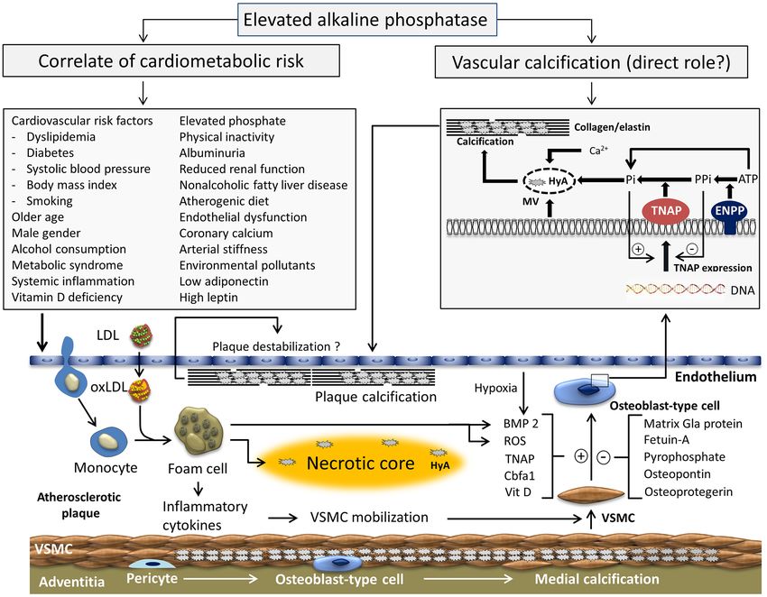
involving 6,340 participants aged 28–75 years without pre- atherosclerotic plaque rupture, as well (106). Putative
existing CVD found no association between non-alcoholic mechanisms of AP association with CVD are shown in
fatty liver disease and the risk of CVD, once the association Figure 1.
was adjusted for established CVD risk factors and other A number of studies have suggested an association of
potential confounders (89). AP with several conditions that may increase CVD risk or
Although important for understanding the association mortality. Thus AP has been shown to be associated with
between AP and the risk of CVD or mortality, none of the the extent of coronary artery disease (107), lack of collateral
above-cited mechanisms points out to a direct role of AP circulation (108), contrast-induced nephropathy in patients
in the pathophysiology of CVD or CHD. Demonstration with acute coronary syndromes (109), arterial stiffness after
of the involvement of AP in vascular calcification (90) renal transplantation (110), left ventricular hypertrophy
has led to the hypothesis that AP mediates elevated CVD in mice (111), endothelium-dependent vasodilation and
risk and a deleterious prognostic impact through vascular endothelial dysfunction in hypertensive patients (112),
calcification. Mechanisms of vascular calcification have adipogenesis (113), atherogenic diet (33), phosphate
been recently reviewed (91). Several lines of evidence metabolism (36), low adiponectin (114), high leptin (115),
seem to support such a role. AP activity is crucial for fibroblast growth factor-23 (116) or environmental
formation of hydroxyapatite—a crucial molecule in the pollutants (117). Although these conditions may increase the
endochondral calcification or vascular calcification when risk of CVD or have a negative impact on prognosis, causality
AP is expressed in osteoblast-type cells originating from relationship between them and AP remains unproven.
phenotypic transformation of smooth muscle cells (92). AP
stimulates vascular smooth muscle cell trans-differentiation
Conclusions and perspective
into chondrocyte-like cells (93), a pivotal step in vascular
calcification. Up-regulation of AP expression in vessels with Population-based longitudinal studies suggest an association
medial calcification (94) or advanced calcified atherosclerotic between AP and increased risk of incident CVD or all-
lesions (95), hydrolysis of pyrophosphate—an inhibitor cause mortality. Clinical studies involving patients with pre-
of calcification—by up-regulated AP (96) and inhibition existing CHD also suggest an association between AP and
of vascular calcification by AP inhibitors (97) appear to mortality in these patients. The association between AP
offer evidence in favor of AP involvement in vascular and increased risk of mortality seems to be stronger for all-
calcification. An experimental mouse model involving cause mortality than for cardiac mortality. Experimental
overexpression of human TNAP in vascular smooth muscle studies strongly suggest an involvement of AP in vascular
cells demonstrated extensive medial vascular calcification calcification and a direct role of the enzyme in the
early in life, in the absence of systemic changes in calcium, pathophysiology of CVD. There is no consistent evidence
phosphate, or renal function (98). Medial calcification leads with respect to association of AP with extent of CHD,
to vascular stiffness and consequently to increased pulse angiographic coronary calcification or acute coronary
pressure (99) and left ventricular hypertrophy (100)— events such as acute myocardial infarction. The association
factors associated with the risk of mortality in patients with between AP and other CVD such as arterial hypertension
CHD. Although, the association of vascular calcification or cardiac arrhythmias, primarily atrial fibrillation remains
with atherosclerosis remains poorly understood (92), medial unexplored. AP is associated with cardiometabolic risk
© Journal of Laboratory and Precision Medicine. All rights reserved. jlpm.amegroups.com J Lab Precis Med 2017;2:83Page 10 of 15 Journal of Laboratory and Precision Medicine, 2017
Figure 1 Putative mechanisms of the association between alkaline phosphatase and cardiovascular disease. ATP, adenosine triphosphate;
BMP, bone morphogenetic proteins; Ca2+, calcium ion; DNA, deoxyribonucleic acid; Cbfα1, transcription factor core binding factor-α1;
ENPP, ectonucleotide pyrophosphatase/phosphodiesterase-1; HyA, hydroxyapatite; LDL, low-density lipoprotein; oxLDL, oxidized low-
density lipoprotein; MV, microvesicles; Pi, phosphate; PPi, pyrophosphate; ROS, reactive oxygen species; TNAP, tissue-nonspecific alkaline
phosphatase; Vit D, vitamin D; VSMC, vascular smooth muscle cell. Plus sign shows a stimulatory effect; minus sign shows an inhibitory effect.
factors including conventional CVD risk factors, metabolic use of the enzyme as a therapeutic target to reduce CVD
syndrome, systemic inflammation, vitamin D deficiency risk remain to be elucidated in future studies.
and several morbid conditions that may signify a poor
CVD risk profile and increased risk for mortality. The
Acknowledgements
intricate relationship between AP and cardio-metabolic risk
factors and failure to improve risk prediction for incident None.
CHD (41) or cardiac mortality in patients with acute
coronary syndromes (59) by the enzyme seem to weaken
Footnote
arguments for a causal relationship between AP and CVD.
Further explorations of the molecular mechanisms of the Conflicts of Interest: The author has no conflicts of interest to
involvement of AP in the pathophysiology of CVD and the declare.
© Journal of Laboratory and Precision Medicine. All rights reserved. jlpm.amegroups.com J Lab Precis Med 2017;2:83Journal of Laboratory and Precision Medicine, 2017 Page 11 of 15
References A 1986;83:7182-6.
14. Nosjean O, Koyama I, Goseki M, et al. Human tissue
1. Millan JL. Alkaline Phosphatases : Structure, substrate
non-specific alkaline phosphatases: sugar-moiety-induced
specificity and functional relatedness to other members
enzymic and antigenic modulations and genetic aspects.
of a large superfamily of enzymes. Purinergic Signal
Biochem J 1997;321:297-303.
2006;2:335-41.
15. Butterworth PJ. Alkaline phosphatase. Biochemistry of
2. Suzuki U, Yoshimura K, Takaishi M. Uber ein enzyme
mammalian alkaline phosphatases. Cell Biochem Funct
“phytase” das anhydro-oxy-methylen-diphosphorsaure
1983;1:66-70.
spaltet. Bull Coll Agric Tokyo Imp Univ 1907;7:503-12.
16. Jemmerson R, Low MG. Phosphatidylinositol anchor
3. Robison R. The possible significance of hexosephosphoric
of HeLa cell alkaline phosphatase. Biochemistry
esters in ossification. Biochem J 1923;17:286-93.
1987;26:5703-9.
4. Robison R, Soames KM. The possible significance of
17. Kiledjian M, Kadesch T. Post-transcriptional regulation of
hexosephosphoric esters in ossification. part III: The
the human liver/bone/kidney alkaline phosphatase gene. J
action of the bone enzyme on the organic phosphorus
Biol Chem 1991;266:4207-13.
compounds in blood. Biochem J 1924;18:740-54.
18. Riancho-Zarrabeitia L, Garcia-Unzueta M, Tenorio JA,
5. Kay HD, Robison R. The possible significance of et al. Clinical, biochemical and genetic spectrum of low
hexosephosphoric esters in ossification. part III: The alkaline phosphatase levels in adults. Eur J Intern Med
action of the bone enzyme on the organic phosphorus 2016;29:40-5.
compounds in blood. Biochem J 1924;18:755-64. 19. Orimo H, Shimada T. Regulation of the human tissue-
6. Martland M, Robison R. Possible significance of nonspecific alkaline phosphatase gene expression by all-
hexosephosphoric esters in ossification: Part VI. trans-retinoic acid in SaOS-2 osteosarcoma cell line. Bone
phosphoric esters in blood-plasma. Biochem J 2005;36:866-76.
1926;20:847-55. 20. Orimo H, Shimada T. Posttranscriptional modulation
7. Siller AF, Whyte MP. Alkaline Phosphatase: Discovery and of the human tissue-nonspecific alkaline phosphatase
Naming of Our Favorite Enzyme. J Bone Miner Res 2017. gene expression by 1,25-dihydroxyvitamin D-3 in MG-
[Epub ahead of print]. 63 osteoblastic osteosarcoma cells. Nutrition Research
8. Mornet E, Stura E, Lia-Baldini AS, et al. Structural 2006;26:227-34.
evidence for a functional role of human tissue nonspecific 21. Whyte MP. Physiological role of alkaline phosphatase
alkaline phosphatase in bone mineralization. J Biol Chem explored in hypophosphatasia. Ann N Y Acad Sci
2001;276:31171-8. 2010;1192:190-200.
9. Weiss MJ, Cole DE, Ray K, et al. A missense mutation in 22. Millan JL, Whyte MP. Alkaline Phosphatase and
the human liver/bone/kidney alkaline phosphatase gene Hypophosphatasia. Calcif Tissue Int 2016;98:398-416.
causing a lethal form of hypophosphatasia. Proc Natl Acad 23. Fedde KN, Whyte MP. Alkaline phosphatase (tissue-
Sci U S A 1988;85:7666-9. nonspecific isoenzyme) is a phosphoethanolamine and
10. Matsuura S, Kishi F, Kajii T. Characterization of a pyridoxal-5'-phosphate ectophosphatase: normal and
5'-flanking region of the human liver/bone/kidney alkaline hypophosphatasia fibroblast study. Am J Hum Genet
phosphatase gene: two kinds of mRNA from a single gene. 1990;47:767-75.
Biochem Biophys Res Commun 1990;168:993-1000. 24. Fedde KN, Blair L, Silverstein J, et al. Alkaline
11. Silvent J, Gasse B, Mornet E, et al. Molecular evolution phosphatase knock-out mice recapitulate the metabolic
of the tissue-nonspecific alkaline phosphatase allows and skeletal defects of infantile hypophosphatasia. J Bone
prediction and validation of missense mutations responsible Miner Res 1999;14:2015-26.
for hypophosphatasia. J Biol Chem 2014;289:24168-79. 25. Percudani R, Peracchi A. A genomic overview of pyridoxal-
12. Orimo H. The mechanism of mineralization and the role phosphate-dependent enzymes. EMBO Rep 2003;4:850-4.
of alkaline phosphatase in health and disease. J Nippon 26. Wilson PD, Smith GP, Peters TJ. Pyridoxal 5'-phosphate:
Med Sch 2010;77:4-12. a possible physiological substrate for alkaline phosphatase
13. Weiss MJ, Henthorn PS, Lafferty MA, et al. Isolation and in human neutrophils. Histochem J 1983;15:257-64.
characterization of a cDNA encoding a human liver/bone/ 27. Carter TC, Pangilinan F, Molloy AM, et al. Common
kidney-type alkaline phosphatase. Proc Natl Acad Sci U S Variants at Putative Regulatory Sites of the Tissue
© Journal of Laboratory and Precision Medicine. All rights reserved. jlpm.amegroups.com J Lab Precis Med 2017;2:83Page 12 of 15 Journal of Laboratory and Precision Medicine, 2017
Nonspecific Alkaline Phosphatase Gene Influence 39. Koehler EM, Sanna D, Hansen BE, et al. Serum liver
Circulating Pyridoxal 5'-Phosphate Concentration in enzymes are associated with all-cause mortality in an
Healthy Adults. J Nutr 2015;145:1386-93. elderly population. Liver Int 2014;34:296-304.
28. Sharma U, Pal D, Prasad R. Alkaline phosphatase: an 40. Fulks M, Stout RL, Dolan VF. Using liver enzymes as
overview. Indian J Clin Biochem 2014;29:269-78. screening tests to predict mortality risk. J Insur Med
29. Fawley J, Gourlay DM. Intestinal alkaline phosphatase: 2008;40:191-203.
a summary of its role in clinical disease. J Surg Res 41. Kunutsor SK, Bakker SJ, Kootstra-Ros JE, et al. Serum
2016;202:225-34. Alkaline Phosphatase and Risk of Incident Cardiovascular
30. Wallace BH, Lott JA, Griffiths J, et al. Isoforms of alkaline Disease: Interrelationship with High Sensitivity C-Reactive
phosphatase determined by isoelectric focusing in patients Protein. PLoS One 2015;10:e0132822.
with chronic liver disorders. Eur J Clin Chem Clin 42. Bates CJ, Hamer M, Mishra GD. A study of relationships
Biochem 1996;34:711-20. between bone-related vitamins and minerals, related risk
31. Schumann G, Klauke R, Canalias F, et al. IFCC primary markers, and subsequent mortality in older British people:
reference procedures for the measurement of catalytic the National Diet and Nutrition Survey of People Aged 65
activity concentrations of enzymes at 37 degrees C. Part Years and Over. Osteoporos Int 2012;23:457-66.
9: reference procedure for the measurement of catalytic 43. Regidor DL, Kovesdy CP, Mehrotra R, et al. Serum
concentration of alkaline phosphatase International alkaline phosphatase predicts mortality among maintenance
Federation of Clinical Chemistry and Laboratory hemodialysis patients. J Am Soc Nephrol 2008;19:2193-203.
Medicine (IFCC) Scientific Division, Committee on 44. Haarhaus M, Brandenburg V, Kalantar-Zadeh K, et
Reference Systems of Enzymes (C-RSE) (1)). Clin Chem al. Alkaline phosphatase: a novel treatment target for
Lab Med 2011;49:1439-46. cardiovascular disease in CKD. Nat Rev Nephrol
32. Okesina AB, Donaldson D, Lascelles PT, et al. Effect of 2017;13:429-42.
gestational age on levels of serum alkaline phosphatase 45. Kunutsor SK, Apekey TA, Khan H. Liver enzymes and
isoenzymes in healthy pregnant women. Int J Gynaecol risk of cardiovascular disease in the general population: a
Obstet 1995;48:25-9. meta-analysis of prospective cohort studies. Atherosclerosis
33. Domar U, Karpe F, Hamsten A, et al. Human intestinal 2014;236:7-17.
alkaline phosphatase--release to the blood is linked to lipid 46. Kunutsor SK, Apekey TA, Seddoh D, et al. Liver enzymes
absorption, but removal from the blood is not linked to and risk of all-cause mortality in general populations:
lipoprotein clearance. Eur J Clin Invest 1993;23:753-60. a systematic review and meta-analysis. Int J Epidemiol
34. Schiele F, Vincent-Viry M, Fournier B, et al. Biological 2014;43:187-201.
effects of eleven combined oral contraceptives on serum 47. Li JW, Xu C, Fan Y, et al. Can serum levels of alkaline
triglycerides, gamma-glutamyltransferase, alkaline phosphatase and phosphate predict cardiovascular diseases
phosphatase, bilirubin and other biochemical variables. and total mortality in individuals with preserved renal
Clin Chem Lab Med 1998;36:871-8. function? A systemic review and meta-analysis. PLoS One
35. Tonelli M, Curhan G, Pfeffer M, et al. Relation between 2014;9:e102276.
alkaline phosphatase, serum phosphate, and all-cause or 48. Shimizu Y, Imano H, Ohira T, et al. Alkaline phosphatase
cardiovascular mortality. Circulation 2009;120:1784-92. and risk of stroke among Japanese: the Circulatory Risk in
36. Wannamethee SG, Sattar N, Papcosta O, et al. Alkaline Communities Study (CIRCS). J Stroke Cerebrovasc Dis
phosphatase, serum phosphate, and incident cardiovascular 2013;22:1046-55.
disease and total mortality in older men. Arterioscler 49. Bell S, Daskalopoulou M, Rapsomaniki E, et al.
Thromb Vasc Biol 2013;33:1070-6. Association between clinically recorded alcohol
37. Abramowitz M, Muntner P, Coco M, et al. Serum alkaline consumption and initial presentation of 12 cardiovascular
phosphatase and phosphate and risk of mortality and diseases: population based cohort study using linked health
hospitalization. Clin J Am Soc Nephrol 2010;5:1064-71. records. BMJ 2017;356:j909.
38. Filipowicz R, Greene T, Wei G, et al. Associations 50. Wieberdink R. Liver enzymes and the risk of intracerebral
of serum skeletal alkaline phosphatase with elevated hemorrhage. Thesis: Determinants of cerebral infarction
C-reactive protein and mortality. Clin J Am Soc Nephrol and intracranial hemorrhage -The Rotterdam Study
2013;8:26-32. 2012:121-32.
© Journal of Laboratory and Precision Medicine. All rights reserved. jlpm.amegroups.com J Lab Precis Med 2017;2:83Journal of Laboratory and Precision Medicine, 2017 Page 13 of 15
51. Ryu WS, Lee SH, Kim CK, et al. Increased serum alkaline trial. Eur J Heart Fail 2012;14:302-11.
phosphatase as a predictor of long-term mortality after 64. Nikolaou M, Parissis J, Yilmaz MB, et al. Liver function
stroke. Neurology 2010;75:1995-2002. abnormalities, clinical profile, and outcome in acute
52. Kim J, Song TJ, Song D, et al. Serum alkaline phosphatase decompensated heart failure. Eur Heart J 2013;34:742-9.
and phosphate in cerebral atherosclerosis and functional 65. Allen LA, Felker GM, Pocock S, et al. Liver function
outcomes after cerebral infarction. Stroke 2013;44:3547-9. abnormalities and outcome in patients with chronic heart
53. Lee HB, Kim J, Kim SH, et al. Association between Serum failure: data from the Candesartan in Heart Failure:
Alkaline Phosphatase Level and Cerebral Small Vessel Assessment of Reduction in Mortality and Morbidity
Disease. PLoS One 2015;10:e0143355. (CHARM) program. Eur J Heart Fail 2009;11:170-7.
54. Liu J, Wang D, Li J, et al. High Serum Alkaline 66. Samsky MD, Patel CB, DeWald TA, et al. Cardiohepatic
Phosphatase Levels in Relation to Multi-Cerebral interactions in heart failure: an overview and clinical
Microbleeds in Acute Ischemic Stroke Patients with implications. J Am Coll Cardiol 2013;61:2397-405.
Atrial Fibrillation and/or Rheumatic Heart Disease. Curr 67. Mshe'el S, Bisharat N. Extremely high levels of serum
Neurovasc Res 2016;13:303-8. alkaline phosphatase in patients with prolonged hepatic
55. Liu J, Wang D, Li J, et al. Increased Serum Alkaline congestion. Eur J Gastroenterol Hepatol 2011;23:444.
Phosphatase as a Predictor of Symptomatic Hemorrhagic 68. Shamban L, Patel B, Williams M. Significantly Elevated
Transformation in Ischemic Stroke Patients with Atrial Liver Alkaline Phosphatase in Congestive Heart Failure.
Fibrillation and/or Rheumatic Heart Disease. J Stroke Gastroenterology Res 2014;7:64-8.
Cerebrovasc Dis 2016;25:2448-52. 69. Poelzl G, Ess M, Mussner-Seeber C, et al. Liver
56. Ryu WS, Lee SH, Kim CK, et al. High serum alkaline dysfunction in chronic heart failure: prevalence,
phosphatase in relation to cerebral small vessel disease. characteristics and prognostic significance. Eur J Clin
Atherosclerosis 2014;232:313-8. Invest 2012;42:153-63.
57. Park JB, Kang DY, Yang HM, et al. Serum alkaline 70. Batin P, Wickens M, McEntegart D, et al. The
phosphatase is a predictor of mortality, myocardial importance of abnormalities of liver function tests in
infarction, or stent thrombosis after implantation of predicting mortality in chronic heart failure. Eur Heart J
coronary drug-eluting stent. Eur Heart J 2013;34:920-31. 1995;16:1613-8.
58. Ndrepepa G, Xhepa E, Braun S, et al. Alkaline phosphatase 71. Yamazoe M, Mizuno A, Nishi Y, et al. Serum alkaline
and prognosis in patients with coronary artery disease. Eur phosphatase as a predictor of worsening renal function in
J Clin Invest 2017;47:378-87. patients with acute decompensated heart failure. J Cardiol
59. Ndrepepa G, Holdenrieder S, Xhepa E, et al. Prognostic 2016;67:412-7.
value of alkaline phosphatase in patients with acute 72. Kerner A, Avizohar O, Sella R, et al. Association between
coronary syndromes. Clin Biochem 2017;50:828-34. elevated liver enzymes and C-reactive protein: possible
60. Huseynov A, Baumann S, Becher T, et al. Liver and hepatic contribution to systemic inflammation in the
cholestatic parameters as prognostic biomarkers of in- metabolic syndrome. Arterioscler Thromb Vasc Biol
hospital MACE in patients with STEMI. Eur J Clin Invest 2005;25:193-7.
2016;46:721-9. 73. Cheung BM, Ong KL, Cheung RV, et al. Association
61. Nunes JP, Melao F, Godinho AR, et al. Plasma alkaline between plasma alkaline phosphatase and C-reactive
phosphatase and survival in diabetic patients with acute protein in Hong Kong Chinese. Clin Chem Lab Med
myocardial infarction. Ann Transl Med 2016;4:210. 2008;46:523-7.
62. Oh PC, Lee K, Kim TH, et al. Prognostic impact of 74. Webber M, Krishnan A, Thomas NG, et al. Association
alkaline phosphatase measured at time of presentation between serum alkaline phosphatase and C-reactive
in patients undergoing primary percutaneous coronary protein in the United States National Health and
intervention for ST-segment elevation myocardial Nutrition Examination Survey 2005-2006. Clin Chem Lab
infarction. PLoS One 2017;12:e0171914. Med 2010;48:167-73.
63. Ambrosy AP, Vaduganathan M, Huffman MD, et al. 75. Krishnamurthy VR, Baird BC, Wei G, et al. Associations
Clinical course and predictive value of liver function tests of serum alkaline phosphatase with metabolic syndrome
in patients hospitalized for worsening heart failure with and mortality. Am J Med 2011;124:566.e1-7.
reduced ejection fraction: an analysis of the EVEREST 76. Zhang L, Ma X, Jiang Z, et al. Liver enzymes and
© Journal of Laboratory and Precision Medicine. All rights reserved. jlpm.amegroups.com J Lab Precis Med 2017;2:83Page 14 of 15 Journal of Laboratory and Precision Medicine, 2017
metabolic syndrome: a large-scale case-control study. 91. Zhu D, Mackenzie NC, Farquharson C, et al. Mechanisms
Oncotarget 2015;6:26782-8. and clinical consequences of vascular calcification. Front
77. Hansson GK. Inflammation, atherosclerosis, and coronary Endocrinol (Lausanne) 2012;3:95.
artery disease. N Engl J Med 2005;352:1685-95. 92. Johnson RC, Leopold JA, Loscalzo J. Vascular calcification:
78. Jean G, Charra B, Chazot C. Vitamin D deficiency and pathobiological mechanisms and clinical implications. Circ
associated factors in hemodialysis patients. J Ren Nutr Res 2006;99:1044-59.
2008;18:395-9. 93. Fakhry M, Roszkowska M, Briolay A, et al. TNAP
79. Giovannucci E, Liu Y, Hollis BW, et al. 25-hydroxyvitamin stimulates vascular smooth muscle cell trans-differentiation
D and risk of myocardial infarction in men: a prospective into chondrocytes through calcium deposition and BMP-
study. Arch Intern Med 2008;168:1174-80. 2 activation: Possible implication in atherosclerotic plaque
80. Aleksova A, Belfiore R, Carriere C, et al. Vitamin D stability. Biochim Biophys Acta 2017;1863:643-53.
Deficiency in Patients with Acute Myocardial Infarction: 94. Shanahan CM, Cary NR, Salisbury JR, et al. Medial
An Italian Single-Center Study. Int J Vitam Nutr Res localization of mineralization-regulating proteins in
2015;85:23-30. association with Monckeberg's sclerosis: evidence for
81. De Metrio M, Milazzo V, Rubino M, et al. Vitamin D smooth muscle cell-mediated vascular calcification.
plasma levels and in-hospital and 1-year outcomes in Circulation 1999;100:2168-76.
acute coronary syndromes: a prospective study. Medicine 95. Tang FT, Chen SR, Wu XQ, et al. Hypercholesterolemia
(Baltimore) 2015;94:e857. accelerates vascular calcification induced by excessive vitamin
82. Zittermann A, Schleithoff SS, Koerfer R. Putting D via oxidative stress. Calcif Tissue Int 2006;79:326-39.
cardiovascular disease and vitamin D insufficiency into 96. Lomashvili KA, Garg P, Narisawa S, et al. Upregulation
perspective. Br J Nutr 2005;94:483-92. of alkaline phosphatase and pyrophosphate hydrolysis:
83. Brewer LC, Michos ED, Reis JP. Vitamin D in potential mechanism for uremic vascular calcification.
atherosclerosis, vascular disease, and endothelial function. Kidney Int 2008;73:1024-30.
Curr Drug Targets 2011;12:54-60. 97. Narisawa S, Harmey D, Yadav MC, et al. Novel inhibitors
84. Milazzo V, De Metrio M, Cosentino N, et al. Vitamin of alkaline phosphatase suppress vascular smooth muscle
D and acute myocardial infarction. World J Cardiol cell calcification. J Bone Miner Res 2007;22:1700-10.
2017;9:14-20. 98. Sheen CR, Kuss P, Narisawa S, et al. Pathophysiological role
85. Schwetz V, Trummer C, Pandis M, et al. Effects of of vascular smooth muscle alkaline phosphatase in medial
Vitamin D Supplementation on Bone Turnover Markers: artery calcification. J Bone Miner Res 2015;30:824-36.
A Randomized Controlled Trial. Nutrients 2017;9. 99. Dao HH, Essalihi R, Bouvet C, et al. Evolution and
86. Targher G, Bertolini L, Padovani R, et al. Prevalence of modulation of age-related medial elastocalcinosis: impact
nonalcoholic fatty liver disease and its association with on large artery stiffness and isolated systolic hypertension.
cardiovascular disease among type 2 diabetic patients. Cardiovasc Res 2005;66:307-17.
Diabetes Care 2007;30:1212-8. 100. Speer MY, Giachelli CM. Regulation of cardiovascular
87. Fargion S, Porzio M, Fracanzani AL. Nonalcoholic fatty calcification. Cardiovasc Pathol 2004;13:63-70.
liver disease and vascular disease: state-of-the-art. World J 101. London GM. Arteriosclerosis and arterial calcifications in
Gastroenterol 2014;20:13306-24. chronic kidney insufficiency. Nephrol Ther 2005;1 Suppl
88. Stepanova M, Younossi ZM. Independent association 4:S351-4.
between nonalcoholic fatty liver disease and cardiovascular 102. Niskanen L, Siitonen O, Suhonen M, et al. Medial artery
disease in the US population. Clin Gastroenterol Hepatol calcification predicts cardiovascular mortality in patients
2012;10:646-50. with NIDDM. Diabetes Care 1994;17:1252-6.
89. Kunutsor SK, Bakker SJL, Blokzijl H, et al. Associations 103. Hughes-Austin JM, Dominguez A, 3rd, Allison MA, et
of the fatty liver and hepatic steatosis indices with risk of al. Relationship of Coronary Calcium on Standard Chest
cardiovascular disease: Interrelationship with age. Clin CT Scans With Mortality. JACC Cardiovasc Imaging
Chim Acta 2017;466:54-60. 2016;9:152-9.
90. Schoppet M, Shanahan CM. Role for alkaline phosphatase 104. Virmani R, Burke AP, Farb A. Plaque morphology in
as an inducer of vascular calcification in renal failure? sudden coronary death. Cardiologia 1998;43:267-71.
Kidney Int 2008;73:989-91. 105. Abedin M, Tintut Y, Demer LL. Vascular calcification:
© Journal of Laboratory and Precision Medicine. All rights reserved. jlpm.amegroups.com J Lab Precis Med 2017;2:83You can also read