Review Article Yin-yang effect of tumour cells in breast cancer: from mechanism of crosstalk between tumour-associated macrophages and ...
←
→
Page content transcription
If your browser does not render page correctly, please read the page content below
Am J Cancer Res 2020;10(2):383-392
www.ajcr.us /ISSN:2156-6976/ajcr0105973

Review Article
Yin-yang effect of tumour cells in breast cancer: from
mechanism of crosstalk between tumour-associated
macrophages and cancer-associated adipocytes
Zhongbo Zhu1*, Xiangdong Zhu1*, Shuo Yang1, Zhanzi Guo1, Kangle Li2, Cuicui Ren3, Yunyun Zhou4,5, Jianwei
Dou2
1
 School of Basic Medicine, Gansu University of Chinese Medicine, Lanzhou 730000, P. R. China; 2School of Phar-
macy, Xi’an Jiaotong University, Xi’an 710061, P. R. China; 3Department of Pharmacy, Xi’an No. 1 Hospital, South
Street Powder Lane No. 30, Xi’an 710002, P. R. China; 4Affiliated Guangren Hospital of Xi’an Jiaotong University,
21 Jiefang Road, Xi’an 710004, P. R. China; 5Department of Ophthalmology, Xi’an Fourth Hospital, 21 Jiefang
Road, Xi’an 710004, P. R. China. *Equal contributors.
Received December 5, 2019; Accepted January 2, 2020; Epub February 1, 2020; Published February 15, 2020

Abstract: Currently, adipocytes and macrophages are considered to be key cell types of breast cancer (BC) tissues.
With the emergence of crown-like structures (CLS), cancer-associated adipocytes (CAAs) and tumour-associated
macrophages (TAMs) are formed respectively in tumor microenvironment (TME). Both of them affect the progress
of breast cancer, while forming crosstalk in the tumour tissue. CAAs play an important role, which produces hypoxia
and inflammation environment and aggravates this environment. The formation and secretion of TAMs with M2
phenotypic characteristics, such as HIF-1α, and TNF-α, affect the progress of cancer cells by interfering with the
secretion of MCP-1 by CAAs. Therefore, the interaction between CAAs and TAMs may be an effective therapeutic
target for breast cancer. In this review, we focus on the biological effects of two types of cells in breast cancer, in
order to better explain the crosstalk between them and provide new ideas for the future treatment of breast cancer.

Keywords: Breast cancer, crosstalk, tumour-associated macrophages, cancer-associated adipocytes

Introduction                                                    mation and reversible to some extent [5]. This
                                                                provides a useful research direction for TAMs
Breast cancer (BC) is a highly heterogeneous                    which are also involved in the BC cell transfor-
malignant disease that gives rise to many can-                  mation and affect CAAs. However, the exact
cer-related deaths worldwide [1]. Its develop-                  role of CAAs and TAMs in the development of
ment involves complex and contradictory bio-                    BC remains controversial.
logical processes, which are not only controlled
by genetic abnormalities, but also by the inter-                As a dialectical way of thinking in China, the
play between cancer cells and the tumour                        theory of yin-yang is quite different from the
microenvironment (TME) [2]. TME is a dynamic                    Western way of thinking in both literal and sym-
network, which includes immune cells, extra-                    bolic sense. However, the theory of yin-yang has
cellular matrix and stromal tissue. Cancer-                     been applied to the most influential academic
associated adipocytes (CAAs) and tumour-                        journals, including science, nature and cell. At
associated macrophages (TAMs) are important                     present, yin-yang theory is widely used in the
components of BC niche. They play important                     biomedical field in the West; many authors also
roles and have been extensively studied [3, 4].                 use yin-yang symbols to express their new dis-
                                                                coveries [6, 7]. For example, yin-yang-1 factor, a
Nevertheless, the interaction between cancer-                   transcription factor involved in tumour progres-
related cells in BC microenvironment are less                   sion (in short, YY1 factor) [8]. In the immune
well understood. Recent studies have shown                      system, tregs and suppressive cytokines are
that CAAs are associated with BC cell transfor-                 homeostatic with effective cells plus pro-inflam-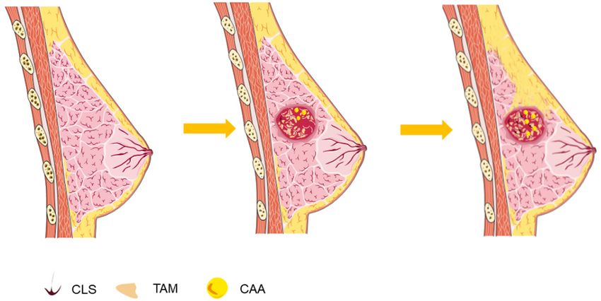
Yin-yang effect of tumour cells in breast cancer

matory cytokines in healthy hosts which is            complex vicious cycle orchestrated by cancer
defined as “Yang”, and ADs are usually induced        cells to promote tumour progression [12].
in case of disturbed homeostasis, which is            Experiments with adipocytes and tissues have
defined as “Yin” [9].                                 shown their correlation. Therefore, they are
                                                      considered to be a critical cell type and are
Therefore, we discussed unique functional             associated with the TME of BC.
characteristics of CAAs and TAMs in BC micro-
environment, and analysed the feasibility of          TAMs
crosstalk based on the relationship between
yin-yang as a potential therapeutic direction.        As an important subset of tumour infiltrating
                                                      immune cells, TAMs participate in the whole
Cancer-associated adipocytes (CAAs) and               process of tumour progression.
tumour-associated macrophages (TAMs) in
breast cancer                                         There are two main sources of TAMs: Monocyte-
                                                      Derived Macrophages (MDMs) derived from
CAAs                                                  bone marrow, and Tissue-Resident Macrophage
                                                      (TRMs) derived from the yolk sac and colonized
Adipose tissue is distributed throughout the          in specific tissues. In most cases, TAMs are
human body. In the breast, adipocytes account         derived primarily from circulating monocytes
for the largest proportion among the cells that       and aggregated around tumour tissue via the
comprise breast tissue [10]. There also exist         chemokine CCL2 [13].
adipocyte-precursor cells, preadipocytes in
breast tissue which have fibroblast-like mor-         Changes in surface markers usually occur when
phology and high proliferative activity.              circulating monocytes accumulate in tumour
                                                      tissue. Ly6C+ monocytes enter into inflamma-
The tumour genesis of breast tissue recapitu-         tion tissue, cell morphological changes, volu-
lates glandular epithelial cell proliferation, adi-   me increase, mononuclear cells continuously
pocyte differentiation, extracellular matrix          replace the yolk sac in macrophages, become
(ECM) remodeling, and reciprocal interactions         an MHC II+ macrophage, this process is also a
between epithelial cell and adipocytes are also       mature mononuclear cell differentiation pro-
observed in the process of carcinogenesis in          cess, called the “waterfall effect” (Figure 1)
BC [4].                                               [14]. This phenomenon originated from the
                                                      study of intestinal monocytes and may extend
Recent studies have shown that the graft of           to tumour tissues, providing a basic model for
adipose tissue could potentially promote or           describing the terminal differentiation of mono-
accelerate the development of a subclinical           cytes in tumour tissues. In tumour tissue, how-
tumour or support its locoregional recurrence,        ever, the distribution of different mature stage
which prompted the microenvironment sur-              TAMs is affected by tumour location, type,
rounding breast cancer cells and may stimulate        stage and the influence of size. The different
growth and promote progression of residual            stages of maturity TAMs play a certain role in
cancer cells when surgery is performed on the         the process of tumour development, such as
main tumour mass [11].                                MCH II+ TAMs presenting antigen, initiating an
                                                      immune response, effectively killing the tumour
The association of adipocytes and BC cells            cells.
have been demonstrated in vivo and in vitro evi-
dence that, (i) invasive cancer cells dramatical-     Although TAMs can inhibit tumour cells, it is
ly impact surrounding adipocytes; (ii) peritu-        believed that TAMs can promote tumor growth
moral adipocytes exhibit a modified phenotype         in other studies. This seemingly contradictory
and specific biological features sufficient to be     result is related to the phenotype of TAMs.
named CAAs; and (iii) CAAs modify the cancer          TAMs are considerably plastic and assume
cell characteristics/phenotype leading to a           opposing phenotypes and functions that can
more aggressive behavior.                             be either tumour-supportive (M2 macrophages)
                                                      or tumouricidal (M1 macrophages). In most
These results strongly support the innovative         tumours, the tumour-supportive M2 phenotype
concept that adipocytes participate in a highly       prevails [15].

384                                                           Am J Cancer Res 2020;10(2):383-392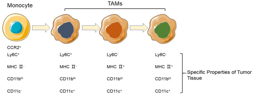
Yin-yang effect of tumour cells in breast cancer

Figure 1. Differentiation phenotype of TAMs in different stages.

M1 macrophages is regulated by cytokines                      sis and immunosuppression mediated by
secreted by Th1, such as interferon-γ (INF-γ),                macrophages.
lipopolysaccharide (LPS) and toll-like receptor
(TLR), and is characterized by the secretion of               Crosstalk of CAA with TAM in breast cancer
pro-inflammatory cytokines including IL-6, IL-
12, IL-23 and tumour necrosis factor-α (TNF-α).               Crown-like structures at the beginning of the
In addition, the tumour specific antigen pre-                 crosstalk
sented needs by M1 macrophages high ex-
                                                              Crown-like structures (CLS), composed of mac-
pression of major histocompatibility complex
                                                              rophages surrounding dead or dying adipo-
(major histocompatibility complex) I classes
                                                              cytes, are found in breast tissue regardless of
and II molecules. Therefore, M1 macrophages
                                                              the presence of breast cancer [19], and are a
are considered to be cells that kill bacteria and
                                                              histologic hallmark of the proinflammatory pro-
tumour cells and secrete a variety of pro-inflam-
                                                              cess which adipose tissue contributes to the
matory cytokines [16].
                                                              increased risk and worse prognosis of breast
M2 macrophages have the opposite function of                  cancer in obese, postmenopausal patients
M1 macrophages, which are recognized as                       [20]. The formation of CLS can be seen as a
anti-inflammatory and carcinogenic cells.                     sign of the beginning of crosstalk (Figure 2).

The generic use of M2 to define macrophage                    Firstly, the latest studies have shown that CLS
activation other than M1 is justified based on                formation is associated with breast cancer [21,
the sharing of selected functional properties                 22]. Secondly, the number and density of CLS
(e.g. low IL-12) and their general involvement in             increased in proportion to the size and number
type II responses, immunoregulation and tis-                  of adipocytes and the abundance of macro-
sue remodeling. We propose to refer to the                    phages in the parasitoid adipose tissue of
three well defined forms of M2 as: M2a (where                 breast cancer. Then, CLS formation, as a poten-
a also stands for alternative), induced by IL-4 or            tial mutagenic agent, triggers a series of vicious
IL-13; M2b, induced by exposure to the IC and                 cycles in the breast cancer tumour microenvi-
agonists of Toll-like receptors (TLRs) or IL-1R;              ronment. Macrophages in CLS engulf cell debris
and M2c, induced by IL-10 and glucocorticoid                  and lipid droplets, release fatty acids and tri-
hormones [17].                                                glycerides [23]. Fatty acids and triglycerides
                                                              are two of the most common metabolites of
Recent studies have suggested that teams                      tumour-associated fat cells. Studies have con-
tend to contribute to the environment of tumour               firmed that fatty acids can participate in the
formation and development, not only because                   glucose uptake process of various tumour cells,
macrophages can constitute up to 50% of                       such as prostate cancer cells, and promote the
tumour masses [18], but also because of the                   growth of malignant tumours by promoting the
occurrence of tumour cells vascular endosmo-                  release of inflammatory factors in malignant

385                                                                    Am J Cancer Res 2020;10(2):383-392
Yin-yang effect of tumour cells in breast cancer
Figure 2. Crown like structure (CLS) in adipose tissue nearby breast cancer.
tumours and mediating the recruitment of mac- abnormalities and special metabolic character-
rophages and lymphocytes to malignant tumour istics of tumours are caused. At the same time,
cells. Other studies have confirmed that triglyc- tumour microenvironment recruits and chang-
erides can reduce the apoptosis of malignant es the phenotype of TAMs, regulates the
tumour cells by inhibiting the release of immune response, including TAMs, and leads
ceramide in human body [23, 24]. to immune escape or tolerance.
The tumour microenvironment as a venue for Healthy cells rely on mitochondria to oxidize
crosstalk sugar molecules to release ATP, while the high
concentration of lactic acid in TME produces a
The so-called TME is being considered as a key special Warburg effect: infinitely increased
factor in the occurrence, progression and treat- energy metabolism of cancer cell changes,
ment of cancer lesions. In addition to genetic even in an aerobic environment, glycolysis is
mutation, the presence of malignant microenvi-
used to replace the aerobic cycle (aerobic gly-
ronments forms the basis of a new perspective
colysis). There is evidence that there is a large
in cancer biology, in which system-level linkag-
amount of TAMs aggregation in the hypoxic
es are the basis. From this point of view, all
region of progressive tumours. Casazza et al.
aspects of interaction between CAAs and TAMs
[25] confirmed that the recruitment of TAMs
can be considered from a unified perspective,
depends on the activation of the signal path-
thus forming a new research field and clinical
way of semaphores 3A/neuropilin-1 (Sema3A/
breakthrough point.
Nrp1). Sema3A can chemotactic macrophages
Hypoxia: Hypoxia in TME is particularly promi- by binding with homologous receptor complex
nent. The vascular system is highly disordered Nrp1/plexinA1 (pA1)/plexinA4 (pA4). Mean-
and constantly changing due to the increase while, Sema3A can promote the migration of
and decrease of blood vessels. The result of TAMs to hypoxic areas. When TAMs tend to
this change is the fluctuation of oxygen and glu- hypoxic regions, hypoxia can alter macrophage
cose levels, which leads to the coexistence of phenotypes and make them develop intotu-
hypoxia, anaerobic and aerobic glycolysis. In mourigenic phenotypes. Hypoxia-inducible fac-
this process, proliferating cells and dead cells tor 1α (HIF-1α) is a key transcription factor in
coexist in the hypoxic and acidic environments the phenotypic change of macrophages. The
and produce a large number of harmful cyto- expression of chemokine receptor 4 (C-X-C
kines. With the participation of CAAs, vascular motif) receptor 4, CXCR4-dependent HIF-1α in
386 Am J Cancer Res 2020;10(2):383-392Yin-yang effect of tumour cells in breast cancer
macrophages and the increase of its specific Chronic low-grade inflammation persists in
ligand CXCL12 also lead to the increase of tumour microenvironment, which is another
chemokine response in macrophages [26]. inducing condition for malignant transforma-
Increased expression of macrophage chemo- tion of TAMs. Under the induction of STAT3,
kines further led to more TAMs recruited into macrophages in the stroma of tumours tend to
hypoxic regions and become M2. M2 macro- differentiate into TAMs with M2 characteristics.
phages are highly localized in hypoxic tumours In addition, the high lactic acid state produced
and exhibit good angiogenic activity in vivo, and by glycolysis inhibited the migration ability of
the number of M2 macrophages increases with mononuclear macrophages and reduced the
the progression of tumours release of TNF and IL-6. At the same time,
under the action of lactic acid, macrophage
In hypoxic mammary adipose tissue, the prolif- phenotype transforms to M2 type, which inhib-
eration of oxygen decreases with the increase its its antigen presenting function, thus promot-
of cell volume, and the growth of blood vessels ing the occurrence of immune escape.
is impaired. Mitochondrial production of exces-
sive free fatty acids leads to an increase in Tumour factors act as crosstalk products
reactive oxygen species (ROS), which leads to
oxidative stress [27]. Therefore, adipokines When abnormal energy metabolism occurs in
secreted by adipose tissue (i.e. cell signalling malignant tumour cells and malignant tumour
proteins secreted by adipose tissue) is defec- microenvironment, the lipolysis of tumour-relat-
tive, leading to angiogenesis and inflammation ed adipocytes becomes very active, which
[28]. This chain reaction forms a malignant secretes tumour-related adipokines and inflam-
environment in the breast cancer region to pro- matory factors, including leptin, adiponectin,
mote the development of breast cancer cells. Visfatin, IL-6 and IL-8, and produces fatty acids
and other metabolites.
Inflammation: Chronic inflammation is consid-
ered a precursor to cancer development. Breast Leptin, spherical adiponectin, Resistin, insulin-
cancer patients are often accompanied by like growth factor binding protein-2 (IGFBP-2),
chronic low-grade inflammation, and the degree chemokine ligand 5 (CCL5) and other cytokines
of pathological changes increases with the derived from adipocytes in tumour microenvi-
accumulation of fat and inflammation of fat. At ronment can act as paracrine signals in breast
the same time, there is increasing evidence cancer cells and up-regulate the expression of
that adipose tissue inflammation is a key driver invasive related proteins or proteases, such as
of oestrogen production in obese postmeno- calcium binding protein S100A7, matrix metal-
pausal women, which plays an important role in loprotein-9 (MMP-9) and urokinase-type plas-
ER+ breast cancer. Chronic inflammation pro- minogen activator (UPA), or activate PI3K/AKT
duces malignant lesions by producing harmful signalling pathways, or media EMT to enhance
ROS and decays surrounding adipose tissue to the movement, migration or invasion of breast
form CAAs. With the release of CAAs pro-inflam- cancer cells [29-31]. On the other hand, recent
matory adipokines such as leptin, activating studies have confirmed that cancer cells can
transcription factors are activated, such as promote the release of FFA from adipocytes,
nuclear factor κB (NF-κB), transcription activa- and on the contrary, enhance the invasiveness
tor 3 (STAT3), HIF-1α, and various inflammatory of tumours by inducing metabolic remodeling
mediators such as cytokines, chemokines and [32].
prostaglandins are produced in large quanti-
ties. Various inflammatory cells such as TAM, The key point is that adipocytes can secrete
MDSC, mast cells and neutrophils are recruited leptin and IL-6 and activate the transcription
and further promoted. This process by influenc- factor Egr-1 in macrophages, thus up-regulate
ing the proliferation and survival of cells, pro- the expression of vascular endothelial growth
moting angiogenesis, inhibiting the anti-tumour factor-A (VEGF-A) in macrophages at the tran-
immune response, promoting the infiltration scriptional level and mediate the metastasis of
and metastasis of tumour cells, finally, medi- breast cancer cells. In the microenvironment of
ates the occurrence and development of tumours, Visfatin secreted by CAAs can pro-
tumours. mote the growth of malignant tumour cells
387 Am J Cancer Res 2020;10(2):383-392Yin-yang effect of tumour cells in breast cancer
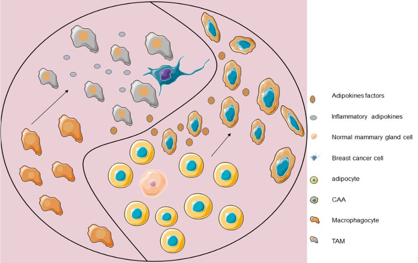
Figure 3. With the development of breast cancer, TAMs and CAAs play the role of yin-yang: in tumour microenviron-
ment, CAAs are derived from adipocytes and CAAs secreted adipokines to interfere with macrophages evolution in
tumour microenvironment. TAMs and CAAs secrete cytokines to interfere with the progress of cancer cells. At the
same time, they influence each other to form crosstalk through their own specific tumour factors.
by accelerating the glycolysis process of malig- cells and mediate the immune escape of
nant tumour cells, which can produce high tumours. TNF-α and IL-1 secreted by TAMs cells
lactic acid to induce polarization of M2 can promote the secretion of vascular endothe-
macrophages and promote the expression of lial growth factor, while the expression of
oncogene VEGF and Arg1 in macrophages [33]. Arg-1, cyclooxygenase-2 (COX-2) and inducible
VEGF expression in cancer tissues is usually nitric oxide synthase (iNOS) are significantly
regulated by HIF-1α, a tissue inducible factor increased, promoting the proliferation and dif-
that can play a stable role even in hypoxic envi- ferentiation of tumour cells [14]. TAMs also
ronments. It has also been reported that the secrete MMP, which can promote the metasta-
deletion of Arg1 in bone marrow cells can sig- sis of cancer cells by degrading extracellular
nificantly reduce the growth rate of subcutane- matrix components. The cytokines secreted by
ous transplanted tumours. It is speculated that TAMs also affect adipocytes.
the expression of Arg1 in TAMs can promote
the growth of tumours. Similarly, cell metabo- Aspirin can inhibit the differentiation and lipid
lites of tumour-related adipocytes, such as accumulation of adipocytes 3T3-L1, and in-
fatty acids, can participate in the glucose hibit the secretion of MCP-1. This effect is
uptake process of breast cancer cells and other more obvious when adipocytes are stimulated
cancer cells, and promote the growth of malig- by TNF-α from macrophages to produce in-
nant tumours by promoting the release of flammation.
inflammatory factors in malignant tumours and
mediating the recruitment of macrophages. TAMs and CAAs secrete cytokines to interfere
with the progress of cancer cells. At the same
When TAMs are recruited and activated, they time, they influence each other to form cross-
can directly release IL-10 or indirectly up-regu- talk through their own specific tumour factors
late Treg activity by releasing IL-23, thus inhib- (Figure 3). This characteristic can be used as a
ite the immune response of anti-tumor immune potential therapeutic target in the future, and it
388 Am J Cancer Res 2020;10(2):383-392Yin-yang effect of tumour cells in breast cancer
is also a difficult point to solve the progress of M2 phenotype to M1 phenotype [39]. In addi-
cancer. tion, macrophage transformation can effec-
tively inhibit the growth of breast tumours
Treatment based on crosstalk in breast cancer and inhibit the angiogenesis of tumours. For
example, histidine-rich_glycoprotein (HRG) can
Intervene TAMs down-regulate the expression of placental
growth factor (PIGF), induce macrophages to
There is growing evidence that TAMs are asso- transform into anti-cancer phenotype and vas-
ciated with poor prognosis. Tumour therapy tar- cular remodeling [40]. These results suggest
geting of TAMs may become a hot pot in the that the activation of TAMs is reversible, and a
future. At present, the main strategies of can- new therapeutic strategy for the redifferentia-
cer therapy targeting TAMs are: inhibiting the tion of TAMs is proposed.
recruitment of macrophages, transforming tu-
mourigenic M2 into anti-tumor M1, and inhibit- The strategy of improving the therapeutic
ing the survival of TAMs. effect of tumours by inhibiting the activity of
TAMs has been recognized and applied clini-
Chemokine (CK) derived from tumours and cally. For example, trabectidine (ET-743), a
stroma promotes the recruitment of macro- DNA damage drug for soft tissue sarcoma and
phages into tumours. Therefore, inhibition of recurrent platinum-sensitive ovarian cancer,
macrophage recruitment by regulating CK may can kill TAMs and exert anti-cancer effects [41].
be an effective cancer therapy. It specifically inhibits TAM by activating exoge-
nous apoptotic pathways through tumour
Current studies have shown that the phar-
necrosis factor-related apoptosis inducing
macological inhibition of CCL2 by Bindarit can
ligand (TRAIL) receptors. In mice model of drug-
significantly reduce macrophage recruitment
resistant transplanted tumours, trabectidine
and inhibit tumour growth [34]. The selective
can significantly inhibit the growth of tumours,
inhibition of vascular endothelial growth factor
and the density of TAMs in tumour microenvi-
receptor (VEGFR) 2 can effectively reduce ronment is significantly reduced. In addition,
the infiltration of macrophages and tumour docetaxel can also eliminate immunosuppres-
growth together with specific antibodies [35]. sive TAMs and exert anti-proliferation effect on
Meanwhile, the therapy targeting the colony myeloid-derived suppressor cells [42].
stimulating factor (CSF) 1 receptor may be
another new strategy for regulating the number Intervene CAAs
of macrophages in tumours. Studies showed
that in patients with diffuse giant cell tumours, CAAs are heterogeneous and closely related to
human monoclonal antibody RG7155 could the types, stages and grades of tumours. They
effectively inhibit the formation of CSF1-R are thus “inhibited” and “educated” by tumours
dimer, and then significantly reduce the infiltra- to varying degrees, which poses a challenge for
tion of CSF-1R+CD163+ macrophage subsets in targeting CAAs. Transforming CAAs into normal
tumours [36]. PLX3397, a tyrosine kinase inhib- adipocytes and inhibiting related biologically
itor of CSF1-R, can improve the effect of tumour active molecules are currently available means.
immunotherapy by reducing macrophage accu- Metformin has long been reported to have anti-
mulation and promoting lymphocyte infiltration tumor effects. It can not only directly target
in tumour tissues [37]. cancer cells, but also reverse dysfunctional adi-
pocytes and normalize them. Studies have
It has been found that activation of some TLRs shown that metformin not only regulates the
can transform M2 macrophages into M1 mac- expression of leptin, PAI-1 and other cytokines
rophages, which kill tumour cells [38]. In in adipocytes, but also participates in the regu-
tumour-bearing mice, activated TLR3/Toll-IL-1 lation of sugar uptake and lipid metabolism. In
rapidly induced the production of pro-inflamma- addition, metformin also regulates insulin sig-
tory cytokines by activating Poly (I:C) and then nalling pathway [43].
accelerated the production of M1 macro-
phages. Zoledronic acid is a clinical drug for In addition, PPAR γ agonists can not only
cancer. It has been found that zoledronic acid reverse the disorder of bioactive molecules,
can inhibit the occurrence of spontaneous such as up-regulation of adiponectin, but also
breast cancer by reducing macrophages from inhibit tumours. Factor, down-regulation of
389 Am J Cancer Res 2020;10(2):383-392Yin-yang effect of tumour cells in breast cancer
leptin, IL-6, TNF-α and other tumour factors can and functions of CAAs and TAMs, there are still
also promote adipogenic differentiation and many areas to be found. In the past, CAAs and
reduce the source of oestrogen. At present, TAMs may be a treatment barrier because they
TZDs such as rosiglitazone and pioglitazone participate in the mechanism of resistance to
have been used in the clinic [44, 45]. Similarly, various breast cancer therapies. Recent stud-
a large number of studies have found that a ies have proved that they interact in the TME,
variety of target genes specifically promote or and pointed out the possibility of targeting
inhibit adipogenic differentiation of microRNAs, therapy through intervention of interactive
such as microRNA143, microRNA21 and so on, products. In addition, the reversible transfor-
while microRNAs-27a/b and microRNAs-130 mation of tumour cells into adipocytes has
have inhibitory effects [46]. been confirmed. However, whether cancer cells
can reverse to macrophages and whether CAAs
The feasibility of research and clinical applica- and TAMs can be inhibited to form or transform
tion of bioactive molecules secreted by CAAs as into cell phenotypes with benign anti-cancer
targets has been reported. It has been report- effects need to be further clarified.
ed that polypeptide analogues located at the
binding site of leptin and leptin receptor (ObR) Acknowledgements
can inhibit the development and metastasis of
breast cancer by selectively inhibiting the inter- This work was supported by grants from the
action between leptin and leptin receptor [47]. National Natural Science Foundation of China
(Grant No. 81873311, No. 81573983 and No.
Promoting the benign transformation of tu- 81674028). Zhu Zhongbo thanks to the help
mour cells provided by School of Pharmacy, Health Science
Center, Xi’an Jiaotong University.
In the theory of yin-yang of Chinese philosophy,
yin-yang is mutually rooted and interdepen- Disclosure of conflict of interest
dent. In simple terms, yin-yang is homologous
and transformative. Interestingly, the occur- None.
rence and development of breast cancer accord
with the characteristics of yin-yang. Tumour cell Address correspondence to: Jianwei Dou, School of
plasticity and EMT are dynamic and can occur Pharmacy, Xi’an Jiaotong University, Xi’an 710061,
during different steps of cancer metastasis. P. R. China. Tel: +86-18093216671; Fax: +86-180-
93216671; E-mail: djw@mail.xjtu.edu.cn
Ronen et al. showed that the cellular plasticity
of cancer cells undergoing EMT can be exploit- References
ed to force transdifferentiation of breast can-
cer cells into post-mitotic and functional adipo- [1] Siegel RL, Miller KD and Jemal A. Cancer sta-
cytes, leading to the repression of primary tistics, 2019. CA Cancer J Clin 2019; 69: 7-34.
tumour invasion and metastasis formation [5]. [2] Burgos-Panadero R, Lucantoni F, Gamero-San-
demetrio E, Cruz-Merino L, Alvaro T and
Notably, adipogenic differentiation therapy with Noguera R. The tumour microenvironment as
a combination of Rosiglitazone and a MEK an integrated framework to understand cancer
inhibitor efficiently inhibits cancer cell invasion, biology. Cancer Lett 2019; 461: 112-122.
dissemination, and metastasis formation in [3] Frankenberger C, Rabe D, Bainer R, San-
various preclinical mouse models of breast karasharma D, Chada K, Krausz T, Gilad Y,
Becker L and Rosner MR. Metastasis suppres-
cancer. The results underscore the pivotal
sors regulate thetumour microenvironment by
role of cancer cell plasticity in malignant
blocking recruitment of prometastatictumour-
tumour progression and reveal the therapeutic associated macrophages. Cancer Res 2015;
potential that lies in the targeting of cellular 75: 4063-4073.
plasticity, for example by forcing post-mitotic [4] Choi J, Cha YJ and Koo JS. Adipocyte biology in
adipogenesis. breast cancer: from silent bystander to active
facilitator. Prog Lipid Res 2018; 69: 11-20.
Conclusions [5] Ishay-Ronen D, Diepenbruck M, Kalathur RKR,
Sugiyama N, Tiede S, Ivanek R, Bantug G, Mori-
Although remarkable progress has been made ni MF, Wang J, Hess C and Christofori G. Gain
in understanding the respective mechanisms fat-lose metastasis: converting invasive breast
390 Am J Cancer Res 2020;10(2):383-392Yin-yang effect of tumour cells in breast cancer
cancer cells into adipocytes inhibits cancer [19] Iyengar NM, Gucalp A, Dannenberg AJ and Hu-
metastasis. Cancer Cell 2019; 35: 17-32, e16. dis CA. Obesity and cancer mechanisms: tu-
[6] Zhang HJ and Wang ZX. Yin-yang and Zheng: mour microenvironment and inflammation. J
exported from Chinese medicine. Chin J Integr Clin Oncol 2016; 34: 4270-4276.
Med 2014; 20: 250-255. [20] Berger NA. Crown-like structures in breast adi-
[7] Zhang Z, Zhu Y, Wang Z, Zhang T, Wu P and pose tissue from normal weight women: impor-
Huang J. Yin-yang effect of tumour infiltrating B tant impact. Cancer Prev Res (Phila) 2017; 10:
cells in breast cancer: from mechanism to im- 223-225.
munotherapy. Cancer Lett 2017; 393: 1-7. [21] Bhardwaj P, Du B, Zhou XK, Sue E, Giri D, Har-
[8] Khachigian LM. The yin and yang of YY1 in tu- bus MD, Falcone DJ, Hudis CA, Subbaramaiah
mour growth and suppression. Int J Cancer K and Dannenberg AJ. Estrogen protects
2018; 143: 460-465. against obesity-induced mammary gland in-
[9] Qiao YC, Pan YH, Ling W, Tian F and Zhao HL. flammation in mice. Cancer Prev Res (Phila)
The Yin and Yang of regulatory T cell and ther- 2015; 8: 751-759.
apy progress in autoimmune disease. Autoim- [22] Morris PG, Hudis CA, Giri D, Morrow M, Falcone
mun Rev 2017; 16: 1058-1670. DJ, Zhou XK, Du B, Brogi E, Crawford CB, Ko-
[10] Vandeweyer E and Hertens D. Quantification of pelovich L, Subbaramaiah K and Dannenberg
glands and fat in breast tissue: an experimen- AJ. Inflammation and increased aromatase ex-
tal determination. Ann Anat 2002; 184: 181- pression occur in the breast tissue of obese
184. women with breast cancer. Cancer Prev Res
[11] Massa M, Gasparini S, Baldelli I, Scarabelli L (Phila) 2011; 4: 1021-1029.
and Repaci E. Interaction between breast can- [23] Jung UJ and Choi MS. Obesity and its metabol-
cer cells and adipose tissue cells derived from ic complications: the role of adipokines and
fat grafting. Aesthe Surg J 2015; 36: 358-63. the relationship between obesity, inflamma-
[12] Dirat B, Bochet L, Dabek M, Daviaud D, Dauvil- tion, insulin resistance, dyslipidemia and non-
lier S, Majed B, Wang YY, Meulle A, Salles B, Le alcoholic fatty liver disease. Int J Mol Sci 2014;
Gonidec S, Garrido I, Escourrou G, Valet P and 15: 6184-6223.
Muller C. Cancer-associated adipocytes exhibit [24] Glass CK and Olefsky JM. Inflammation and
an activated phenotype and contribute to lipid signaling in the etiology of insulin resis-
breast cancer invasion. Cancer Res 2011; 71: tance. Cell Metab 2012; 15: 635-645.
2455-2465. [25] Casazza A, Laoui D, Wenes M, Rizzolio S, Bas-
[13] Franklin RA, Liao W, Sarkar A, Kim MV, Bivona sani N, Mambretti M, Deschoemaeker S, Van
MR, Liu K, Pamer EG and Li MO. The cellular Ginderachter JA, Tamagnone L and Mazzone
and molecular origin of tumour-associated M. Impeding macrophage entry into hypoxictu-
macrophages. Science 2014; 344: 921-925. mour areas by Sema3A/Nrp1 signaling block-
[14] Bain CC, Bravo-Blas A, Scott CL, Perdiguero EG, ade inhibits angiogenesis and restores antitu-
Geissmann F, Henri S, Malissen B, Osborne LC, mor immunity. Cancer Cell 2013; 24: 695-709.
Artis D and Mowat AM. Constant replenish- [26] Schioppa T, Uranchimeg B, Saccani A, Biswas
ment from circulating monocytes maintains SK, Doni A, Rapisarda A, Bernasconi S, Sac-
the macrophage pool in the intestine of adult cani S, Nebuloni M, Vago L, Mantovani A, Mel-
mice. Nat Immunol 2014; 15: 929-937. illo G and Sica A. Regulation of the chemokine
[15] Bronte V and Murray PJ. Understanding local receptor CXCR4 by hypoxia. J Exp Med 2003;
macrophage phenotypes in disease: modulat- 198: 1391-1402.
ing macrophage function to treat cancer. Nat [27] Fridlyand LE and Philipson LH. Reactive spe-
Med 2015; 21: 117-119. cies and early manifestation of insulin resis-
[16] Gordon S and Martinez FO. Alternative activa- tance in type 2 diabetes. Diabetes Obes Metab
tion of macrophages: mechanism and func- 2010; 8: 136-145.
tions. Immunity 2010; 32: 593-604. [28] Hosogai N, Fukuhara A, Oshima K, Miyata Y,
[17] Mantovani A, Sica A, Sozzani S, Allavena P, Vec- Tanaka S, Segawa K, Furukawa S, Tochino Y,
chi A and Locati M. The chemokine system in Komuro R, Matsuda M and Shimomura I.
diverse forms of macrophage activation and Adipose tissue hypoxia in obesity and its im-
polarization. Trends Immunol 2004; 25: 677- pact on adipocytokine dysregulation. Diabetes
686. 2007; 56: 901-11.
[18] Bai J, Adriani G, Dang TM, Tu TY, Penny HX, [29] D’Esposito V, Liguoro D, Ambrosio MR, Collina
Wong SC, Kamm RD and Thiery JP. Contact- F, Cantile M, Spinelli R, Raciti GA, Miele C, Val-
dependent carcinoma aggregate dispersion entino R, Campiglia P, De Laurentiis M, Di Bo-
by M2a macrophages via ICAM-1 and β2 integ- nito M, Botti G, Franco R, Beguinot F and
rin interactions. Oncotarget 2015; 6: 25295- Formisano P. Adipose microenvironment pro-
25307. motes triple negative breast cancer cell inva-
391 Am J Cancer Res 2020;10(2):383-392Yin-yang effect of tumour cells in breast cancer
siveness and dissemination by producing [39] Coscia M, Quaglino E, Iezzi M, Curcio C, Panta-
CCL5. Oncotarget 2016; 7: 24495-24509. leoni F, Riganti C, Holen I, Mönkkönen H, Boc-
[30] Li K, Wei L, Huang Y, Wu Y, Su M, Pang X, Wang cadoro M, Forni G, Musiani P, Bosia A, Cavallo
N, Ji F, Zhong C and Chen T. Leptin promotes F and Massaia M. Zoledronic acid repolarizes
breast cancer cell migration and invasion via tumour-associated macrophages and inhibits
IL-18 expression and secretion. Int J Oncol mammary carcinogenesis by targeting the
2016; 48: 2479-87. mevalonate pathway. J Cell Mol Med 2010; 14:
[31] Sakurai M, Miki Y, Takagi K, Suzuki T, Ishida T, 2803-2815.
Ohuchi N and Sasano H. Interaction with adi- [40] Rolny C, Mazzone M, Tugues S, Laoui D, Jo-
pocyte stromal cells induces breast cancer hansson I, Coulon C, Squadrito ML, Segura I, Li
malignancy via S100A7 upregulation in breast X, Knevels E, Costa S, Vinckier S, Dresselaer T,
cancer microenvironment. Breast Cancer Res Åkerud P, De Mol M, Salomäki H, Phillipson M,
2017; 19: 70. Wyns S, Larsson E, Buysschaert I, Botling J,
[32] Wang YY, Attane C, Milhas D, Dirat B, Dauvillier Himmelreich U, Van Ginderachter JA, De Pal-
S, Guerard A, Gilhodes J, Lazar I, Alet N, Lau- ma M, Dewerchin M, Claesson-Welsh L and
rent V, Le Gonidec S, Biard D, Herve C, Bost F, Carmeliet P. HRG inhibitstumour growth and
Ren GS, Bono F, Escourrou G, Prentki M, Nieto metastasis by inducing macrophage polariza-
L, Valet P and Muller C. Mammary adipocytes tion and vessel normalization through down-
stimulate breast cancer invasion through met- regulation of PlGF. Cancer Cell 2011; 19: 31-
abolic remodeling of tumour cells. JCI Insight 44.
2017; 2: e87489. [41] Germano G, Frapolli R, Belgiovine C, Anselmo
[33] Merino M, Briones L, Palma V, Herlitz K and A, Pesce S, Liguori M, Erba E, Uboldi S, Zuc-
Escudero C. Role of adenosine receptors in the chetti M, Pasqualini F, Nebuloni M, van Rooijen
adipocyte-macrophage interaction during obe- N, Mortarini R, Beltrame L, Marchini S, Fuso
sity. Endocrinol Diabetes Nutr 2017; 64: 317-
Nerini I, Sanfilippo R, Casali PG, Pilotti S, Gal-
327.
marini CM, Anichini A, Mantovani A, D’Incalci
[34] Mizukami Y, Kono K, Kawaguchi Y, Akaike H,
M and Allavena P. Role of macrophage target-
Kamimura K, Sugai H and Fujii H. CCL17 and
ing in the antitumor activity of trabectedin.
CCL22 chemokines within tumour microenvi-
Cancer Cell 2013; 23: 249-262.
ronment are related to accumulation of Foxp3+
[42] Kodumudi KN, Karrune W, Gilvary DL, Eva S,
regulatory T cells in gastric cancer. Int J Cancer
Sheng W and Djeu JY. A novel chemoimmuno-
2008; 122: 2286-2293.
modulating property of docetaxel: suppression
[35] Gazzaniga S, Bravo AI, Guglielmotti A, van
of myeloid-derived suppressor cells in tumour
Rooijen N, Maschi F, Vecchi A, Mantovani A,
bearers. Clin Cancer Res 2010; 16: 4583-
Mordoh J and Wainstok R. Targetingtumour-
associated macrophages and inhibition of 4594.
MCP-1 reduce angiogenesis andtumour growth [43] Tebbe C, Chhina J, Dar SA, Sarigiannis K, Giri
in a human melanoma xenograft. J Invest Der- S, Munkarah AR and Rattan R. Metformin lim-
matol 2007; 127: 2031-2041. its the adipocytetumour-promoting effect on
[36] Ries CH, Cannarile MA, Hoves S, Benz J, War- ovarian cancer. Oncotarget 2014; 5: 4746-
tha K, Runza V, Rey-Giraud F, Pradel LP, Feuer- 4764.
hake F, Klaman I, Jones T, Jucknischke U, [44] Park J, Euhus DM and Scherer PE. Paracrine
Scheiblich S, Kaluza K, Gorr IH, Walz A, Abiraj and endocrine effects of adipose tissue on
K, Cassier PA, Sica A, Gomez-Roca C, de Visser cancer development and progression. Endocr
KE, Italiano A, Le Tourneau C, Delord JP, Lev- Rev 2011; 32: 550-570.
itsky H, Blay JY and Ruttinger D. Targetingtu- [45] Lee J, Imm JY and Lee SH. β‑catenin mediates
mour-associated macrophages with anti-CSF- anti-adipogenic and anticancer effects of arc-
1R antibody reveals a strategy for cancer tigenin in preadipocytes and breast cancer
therapy. Cancer Cell 2014; 25: 846-859. cells. J Agric Food Chem 2017; 65: 2513-
[37] Mok S, Koya RC, Tsui C, Xu J, Robert L, Wu L, 2520.
Graeber T, West BL, Bollag G and Ribas A. Inhi- [46] Arner P and Kulyte A. MicroRNA regulatory net-
bition of CSF-1 receptor improves the antitu- works in human adipose tissue and obesity.
mor efficacy of adoptive cell transfer immuno- Nat Rev Endocrinol 2015; 11: 276-288.
therapy. Cancer Res 2014; 74: 153-161. [47] Otvos L Jr, Kovalszky I, Scolaro L, Sztodola A,
[38] Shime H, Matsumoto M, Oshiumi H, Tanaka S, Olah J, Cassone M, Knappe D, Hoffmann R,
Nakane A, Iwakura Y, Tahara H, Inoue N and Lovas S, Hatfield MP, Beko G, Zhang S, Wade
Seya T. Toll-like receptor 3 signaling converts- JD and Surmacz E. Peptide-based leptin recep-
tumour-supporting myeloid cells totumouri- tor antagonists for cancer treatment and ap-
cidal effectors. Proc Natl Acad Sci U S A 2012; petite regulation. Biopolymers 2011; 96: 117-
109: 2066-2071. 125.
392 Am J Cancer Res 2020;10(2):383-392You can also read